
The Importance of Being Versatile: INFN-CHNet MA-XRF Scanner on Furniture at the CCR “La Venaria Reale”

 , , ,
, , ,  , ,
, ,  ,
,  and
and
Abstract
1. Introduction
2. Materials and Methods
3. Results and Discussion
3.1. Chinoiserie Lacquered Cabinet
3.2. Writing Desk by Pietro Piffetti
4. Conclusions
Author Contributions
Funding
Institutional Review Board Statement
Informed Consent Statement
Data Availability Statement
Acknowledgments
Conflicts of Interest
References
- Alfed, M.; Broekaert, J.A.C. Mobile Depth Profiling and Sub-Surface Imaging Techniques for Historical Paintings—A review. Spectrochiim. Acta Part B At. Spectrosc. 2013, 88, 211–230. [Google Scholar] [CrossRef]
- Angelici, D.; Borghi, A.; Chiarelli, F.; Cossio, R.; Gariani, G.; Lo Giudice, A.; Re, A.; Pratesi, G.; Vaggelli, G. µ-XRF analysis of trace elements in lapis lazuli-forming minerals for a provenance study. Microsc. Microanal. 2015, 21, 526–533. [Google Scholar] [CrossRef]
- Corsi, J.; Lo Giudice, A.; Re, A.; Agostino, A.; Barello, F. Potentialities of X-ray fluorescence analysis in numismatics: The case study of pre-Roman coins from Cisalpine Gaul. Archaeol. Anthropol. Sci. 2018, 10, 431–438. [Google Scholar] [CrossRef]
- Alfeld, M.; Siddons, D.P.; Janssens, K.; Dik, J.; Woll, A.; Kirkham, R.; van de Wetering, E. Visualizing the 17th century underpainting in Portrait of an Old Man by Rembrandt van Rijn using synchrotron-based scanning macro-XRF. Appl. Phys. A 2013, 111, 157–164. [Google Scholar] [CrossRef]
- Alfeld, M.; Van der Snickt, G.; Vanmeert, F.; Janssens, K.; Dik, J.; Appel, K.; van der Loeff, L.; Chavannes, M.; Meedendorp, T.; Hendriks, E. Scanning XRF investigation of a Flower Still Life and its underlying composition from the collection of the Kröller–Müller Museum. Appl. Phys. A 2013, 111, 165–175. [Google Scholar] [CrossRef]
- Romano, F.P.; Caliri, C.; Nicotra, P.; di Martino, S.; Pappalardo, L.; Rizzo, F.; Santos, H.C. Real-time elemental imaging of large dimension paintings with a novel mobile macro X-ray fluorescence (MA-XRF) scanning technique. J. Anal. At. Spectrom. 2017, 32, 773–781. [Google Scholar] [CrossRef]
- Alfeld, M.; Pedroso, J.V.; van EikemaHommes, M.; Van der Snickt, G.; Tauber, G.; Blaas, J.; Haschke, M.; Erler, K.; Dik, J.; Janssens, K. A mobile instrument for in situ scanning macro-XRF investigation of historical paintings. J. Anal. At. Spectrom. 2013, 28, 760–767. [Google Scholar] [CrossRef]
- Ravaud, E.; Pichon, L.; Laval, E.; Gonzalez, V.; Eveno, M.; Calligaro, T. Development of a versatile XRF scanner for the elemental imaging of paintworks. Appl. Physics. A. 2015, 122. [Google Scholar] [CrossRef]
- Alberti, R.; Frizzi, T.; Bombelli, L.; Gironda, M.; Aresi, N.; Rosi, F.; Miliani, C.; Tranquilli, G.; Talarico, F.; Cartechini, L. CRONO: A fast and reconfigurable macro X-ray fluorescence scanner for in-situ investigations of polychrome surfaces. X-ray Spectrom. 2017, 46. [Google Scholar] [CrossRef]
- Bergmann, U.; Bertrand, L.; Edwards, N.P.; Manning, P.L.; Wogelius, R.A. Chemical Mapping of Ancient Artifacts and Fossils with X-ray Spectroscopy; Jaeschke, E., Khan, S., Schneider, J., Hastings, J., Eds.; Synchrotron Light Sources and Free-Electron Lasers; Springer: Cham, Switzerland, 2019. [Google Scholar] [CrossRef]
- Debastiani, R.; Simon, R.; Batchelor, D.; Dellagustin, G.; Baumbach, T.; Fiederle, M. Synchrotron-based scanning macro-X-ray fluorescence applied to fragments of Roman mural paintings. Microchem. J. 2016, 126, 438–445. [Google Scholar] [CrossRef]
- Bergmann, U.; Manning, P.L.; Wogelius, R.A. Chemical mapping of paleontological and archeological artifacts with synchrotron X-rays. Annu. Rev. Anal. Chem. 2012, 5, 361–389. [Google Scholar] [CrossRef] [PubMed]
- Fernandez, J.E.; Taccetti, F.; Kenny, J.M.; Amendola, R. Conclusive editorial on non-destructive techniques for cultural heritage. Rend. Fis. Acc. Lincei 2020, 31, 819–820. [Google Scholar] [CrossRef]
- Castelli, L.; Felicetti, A.; Proietti, F. Heritage Science and Cultural Heritage: Standards and tools for establishing cross-domain data interoperability. Int. J. Digit Libr. 2019. [Google Scholar] [CrossRef]
- Sottili, L.; Guidorzi, L.; Mazzinghi, A.; Ruberto, C.; Castelli, L.; Czelusniak, C.; Giuntini, L.; Massi, M.; Taccetti, F.; Nervo, M.; et al. INFN-CHNet meets CCR La Venaria Reale: First results. In Proceedings of the 2020 IMEKO TC-4 International Conference on Metrology for Archaeology and Cultural Heritage 2020, Trento, Italy, 22–24October 2020; pp. 507–511. [Google Scholar]
- Re, A.; Zangirolami, M.; Angelici, D.; Borghi, A.; Costa, E.; Giustetto, R.; Gallo, L.M.; Castelli, L.; Mazzinghi, A.; Ruberto, C.; et al. Towards a portable X-ray Luminescence instrument for applications in the Cultural Heritage field. Eur. Phys. J. Plus 2018, 133, 362. [Google Scholar] [CrossRef]
- Czelusniak, C.; Palla, L.; Massi, M.; Carraresi, L.; Giuntini, L.; Re, A.; Lo Giudice, A.; Pratesi, G.; Mazzinghi, A.; Ruberto, C.; et al. Preliminary results on time-resolved ion beam induced luminescence applied to the provenance study of lapis lazuli. Nucl. Instrum. Methods Phys. Res. B 2016, 371, 336–339. [Google Scholar] [CrossRef]
- Palla, L.; Czelusniak, C.; Taccetti, F.; Carraresi, L.; Castelli, L.; Fedi, M.E.; Giuntini, L.; Maurenzig, P.R.; Sottili, L.; Taccetti, N. Accurate on line measurements of low fluences of charged particles. Eur. Phys. J. Plus 2015, 130. [Google Scholar] [CrossRef]
- Lo Giudice, A.; Corsi, J.; Cotto, G.; Mila, G.; Re, A.; Ricci, C.; Sacchi, R.; Visca, L.; Zamprotta, L.; Pastrone, N. A new digital radiography system for paintings on canvas and on wooden panels of large dimensions. In Proceedings of the 2017 IEEE International Instrumentation and Measurement Technology Conference (I2MTC 2017) Proceedings; IEEE: Piscataway, NJ, USA, 2017; pp. 1834–1839, FP17IMT-ART; ISBN 9781509035960. [Google Scholar]
- Ruberto, C.; Mazzinghi, A.; Massi, M.; Castelli, L.; Czelusniak, C.; Palla, L.; Gelli, N.; Bettuzzi, M.; Impallaria, A.; Brancaccio, R.; et al. Imaging study of Raffaello’s “La Muta” by a portable XRF spectrometer. Microchem. J. 2016, 126, 63–69. [Google Scholar] [CrossRef]
- Vadrucci, M.; Mazzinghi, A.; Sorrentino, B.; Falzone, S.; Gioia, C.; Gioia, P.; Loreti, E.M.; Chiari, M. Characterisation of ancient Roman wall-painting fragments using non-destructive IBA and MA-XRF techniques. X-ray Spectrom. 2020, 49, 668–678. [Google Scholar] [CrossRef]
- Dal Fovo, A.; Mazzinghi, A.; Omarini, S.; Pampaloni, E.; Ruberto, C.; Striova, J.; Fontana, R. Non-invasive mapping methods for pigments analysis of Roman mural paintings. J. Cult. Herit. 2020, 43, 311–318. [Google Scholar] [CrossRef]
- Mazzinghi, A.; Ruberto, C.; Castelli, L.; Ricciardi, P.; Czelusniak, C.; Giuntini, L.; Mandò, P.A.; Manetti, M.; Palla, L.; Taccetti, F. The importance of being little: MA-XRF on manuscripts on a Venetian island. X-ray Spectrom 2020, 1–7, in press. [Google Scholar] [CrossRef]
- Lazic, V.; Vadrucci, M.; Fantoni, F.; Chiari, M.; Mazzinghi, A.; Gorghinian, A. Applications of laser-induced breakdown spectroscopy for cultural heritage: A comparison with X-ray Fluorescence and Particle Induced X-ray Emission techniques. Spectrochim. Acta Part B At. Spectrosc. 2018, 149, 1–14. [Google Scholar] [CrossRef]
- Mazzinghi, A. Sviluppo di Strumentazione XRF a Scansione per Applicazioni ai Beni Culturali. Ph.D. Thesis, University of Florence, Firenze, Italy, 2016. [Google Scholar]
- Andersson, E.; Cattersel, V. A Dutch Seventeenth-Century European Lacquer Cabinet. Material-Technical Analysis to Gain Insight into the Deteriorated Surface. In Material Imitation and Imitation Materials in Furniture and Conservation; Vasques Dias, M., Ed.; Stichting Ebenist: Amsterdam, The Netherlands, 2017; pp. 190–206. [Google Scholar]
- Salvemini, F.; Grazzi, F.; Agostino, A.; Iannaccone, R.; Civita, F.; Hertmann, S.; Lehmann, E.; Zoppi, M. Non-invasive characterization through X-ray fluorescence and neutron radiography of an ancient Japanese lacquer. Archaeol. Anthropol. Sci. 2013, 5, 197–204. [Google Scholar] [CrossRef]
- Felix, V.S.; Mello, U.L.; Pereira, M.O.; Oliveira, A.L.; Ferreira, D.S.; Carvalho, C.S.; Silva, F.L.; Pimenta, A.R.; Diniz, M.G.; Freitas, R.P. Analysis of a European cupboard by XRF, Raman and FT-IR. Radiat. Phys. Chem. 2018, 151, 198–204. [Google Scholar] [CrossRef]
- Tagliante, S. Problematiche Conservative e Restauro di uno Stipo Settecentesco con Decorazioni in Lacca Orientale e “Alla China”. Master’s Thesis, University of Turin, Torino, Italy, 2018. [Google Scholar]
- Spantigati, C.; De Blasi, S. Il Restauro degli Arredi Lignei. L’ebanisteria Piemontese. Studi e Ricerche; Nardini Editore: Firenze, Italy, 2011. [Google Scholar]
- Re, A.; Albertin, F.; Avataneo, C.; Brancaccio, R.; Corsi, J.; Cotto, G.; De Blasi, S.; Dughera, G.; Durisi, E.; Ferrarese, W.; et al. X-ray tomography of large wooden artworks: The case study of ”Doppio corpo” by Pietro Piffetti. Herit. Sci. 2014, 2. [Google Scholar] [CrossRef]
- De Blasi, S.; Nervo, M.; Ravera, M.; Spantigati, C. Structural characters of Piedmontese eighteenth-century cabinetmaking: Historical documents, restorations and new technologies, in restoring joints, conserving structures. In Proceedings of the Tenth International Symposium on Wood and Furniture Conservation, Amsterdam, The Netherlands, 8–9 October 2010; pp. 98–107. [Google Scholar]
- Luciani, P.; De Blasi, S.; Nervo, M.; Piccirillo, A. Il Restauro del Mobile di Ebanisteria Piemontese del Settecento: Le Opere di Pietro Piffetti e Luigi Prinotto. Alcuni Casi Studio, Conservació-Restauració del Moble i la Fusta. L’experiència dels Experts; Costa Galobart, N., Ed.; Associació per a l’Estudi del Moble: Ajuntament de Barcelona, Institut de Cultura, Museu del Disseny de Barcelona: Barcelona, Spain, 2020; pp. 85–98. [Google Scholar]
- Taccetti, F.; Castelli, L.; Czelusniak, C.; Gelli, N.; Mazzinghi, A.; Palla, L.; Ruberto, C.; Censori, C.; Lo Giudice, A.; Re, A.; et al. A multipurpose X-ray fluorescence scanner developed for in situ analysis. Rend. Fis. Acc. Lincei 2019, 30, 307–322. [Google Scholar] [CrossRef]
- Fitzhugh, E. West, Orpiment and Realgar in Artists Pigments; West Fitzhugh, E., Ed.; National Gallery of Art: London, UK, 1997; Volume 3, pp. 45–81. [Google Scholar]
- Vermeulen, M.; Sanyova, J.; Janssens, K. Identification of artificial orpiment in the interior decorations of the Japanese tower in Laeken. Bruss. Belg. Herit. Sci. 2015, 3, 9. [Google Scholar] [CrossRef]
- Gettens, R.J.; Robert, L.F.; Chase, W.T. Vermilion and Cinnabar. Stud. Conserv. 1972, 17, 45–69. [Google Scholar] [CrossRef]
- Marika, S.; Grout, R.; White, R. ‘Black Earths’: A Study of Unusual Black and Dark Grey Pigments Used by Artists in the Sixteenth Century. Natl. Gallery Tech. Bull. 2003, 24, 96–114. [Google Scholar]
- Seccaroni, C.; Moioli, P. Fluorescenza X-Prontuario per l’ analisi XRF Portatile Applicata a Superfici Policrome; Nardini Editore: Firenze, Italy, 2002; pp. 60–89. [Google Scholar]
- Siddall, R. Mineral Pigments in Archaeology: Their Analysis and the Range of Available Materials. Minerals 2018, 8, 201. [Google Scholar] [CrossRef]
- Eastaugh, N. Pigment Compendium: A Dictionary and Optical Microscopy of Historical Pigments; Butterworth-Heinemann: Amsterdam, The Netherlands, 2008; pp. 239–241. [Google Scholar]
- Kriznar, A.; Muñoz, M.; Paz, F.; Respaldiza, M.; Vega, M. Non-destructive XRF analysis of pigments in a 15th century panel painting. In Proceedings of the 9th International Conference on NDT of Art, Jerusalem, Israel, 25–30 May 2008. [Google Scholar]
- Kirby, J.; Saunders, D. Fading and colour change of Prussian blue: Methods of manufacture and the influence of extenders. Natl. Gallery Tech. Bull. 2004, 25, 73–99. [Google Scholar]
- Vermeulen, M.; Sanyova, J.; Janssens, K.; Nuyts, G.; De Meyer, S.; De Wael, K. The Darkening of Copper- or Lead-Based Pigments Explained by a Structural Modification of Natural Orpiment: A Spectroscopic and Electrochemical Study. J. Anal. At. Spectrom. 2017, 32, 1331–1341. [Google Scholar] [CrossRef]


| Area | Dimension (mm2) | Source Voltage (kV) | Anode Current (μA) | Scanning Speed (mm/s) | Collimator Diameter (μm) | Step Size (mm) |
|---|---|---|---|---|---|---|
| Flowers | 145 × 105 | 28 | 30 | 3 | 800 | 1 |
| Flying bird | 175 × 135 | 28 | 30 | 3 | 800 | 1 |
| Area | Dimension (mm2) | Source Voltage (kV) | Anode Current (μA) | Scanning Speed (mm/s) | Collimator Diameter (μm) | Step Size (mm) |
|---|---|---|---|---|---|---|
| Drawer area 1 | 30 × 13 | 38 | 50 | 1 | 400 | 0.2 |
| Drawer area 2 | 20 × 10 | 28 | 50 | 1 | 400 | 0.2 |
| External side | 40 × 130 | 28 | 70 | 1 | 400 | 0.2 |
| Area Scanned | Colour | Elements Detected | Materials Hypothesised |
|---|---|---|---|
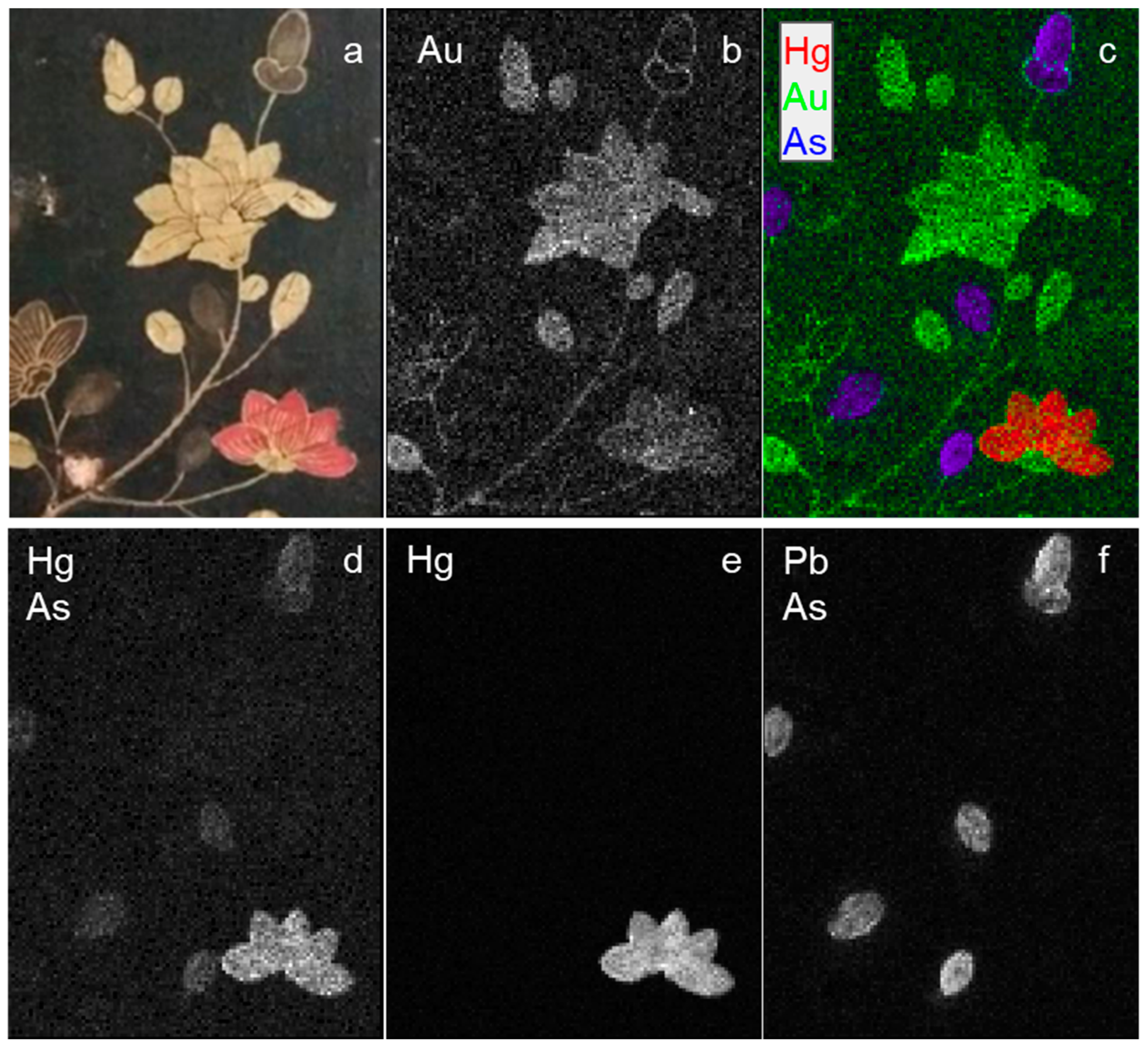
| Chinoiserie cabinet—flowers | red flower | Hg | cinnabar-vermilion |
| bright yellow flowers | Au | gold | |
| transparent yellow buds | As | arsenic-based compound | |
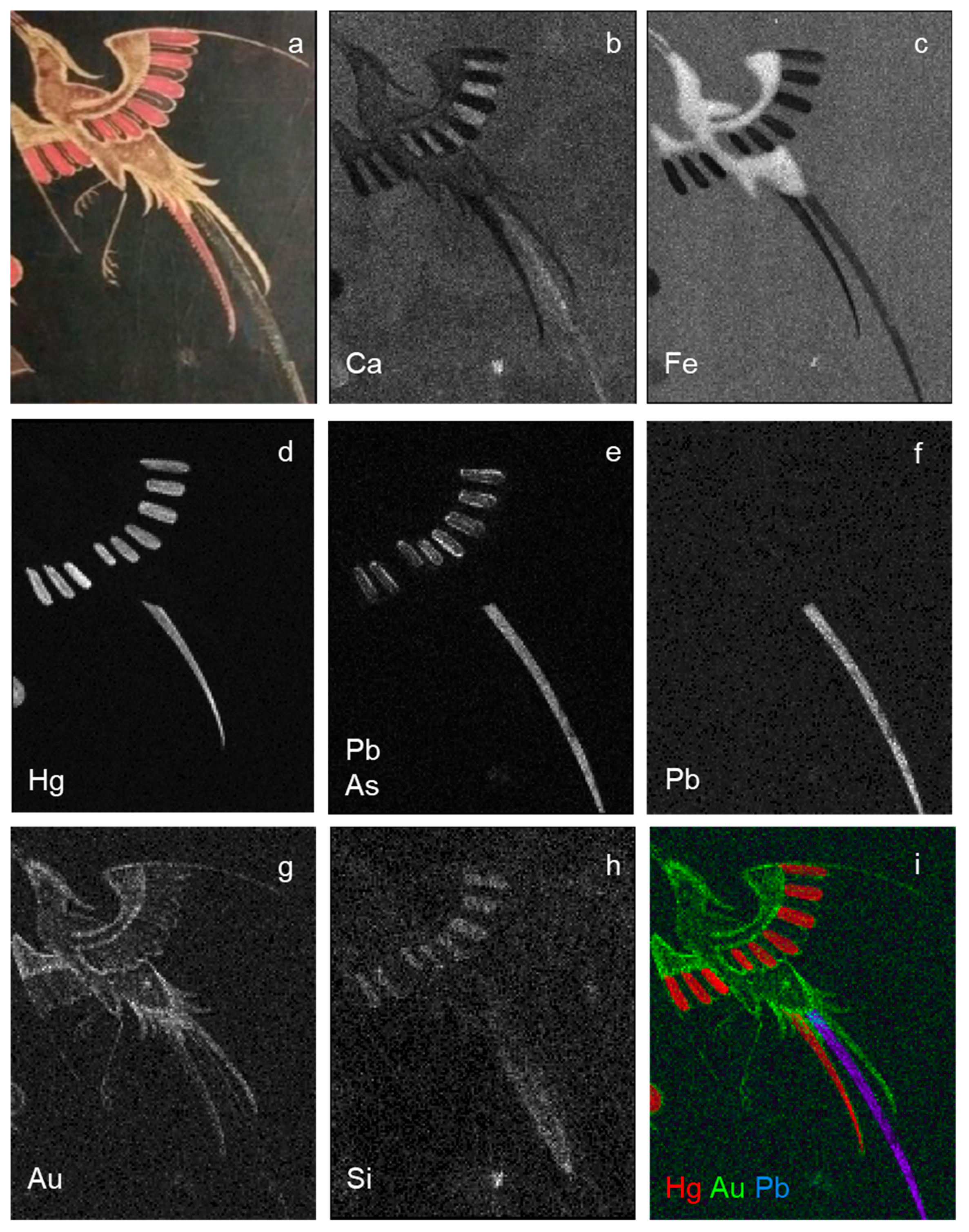
| Chinoiserie cabinet—flying bird | red tail and feathers | Hg | cinnabar-vermilion |
| bright yellow outlines | Au | gold | |
| buff body | Fe | ochres-earths | |
| black-blue feathers | As, Si, Ca | ? | |
| black tail | Pb | galena | |
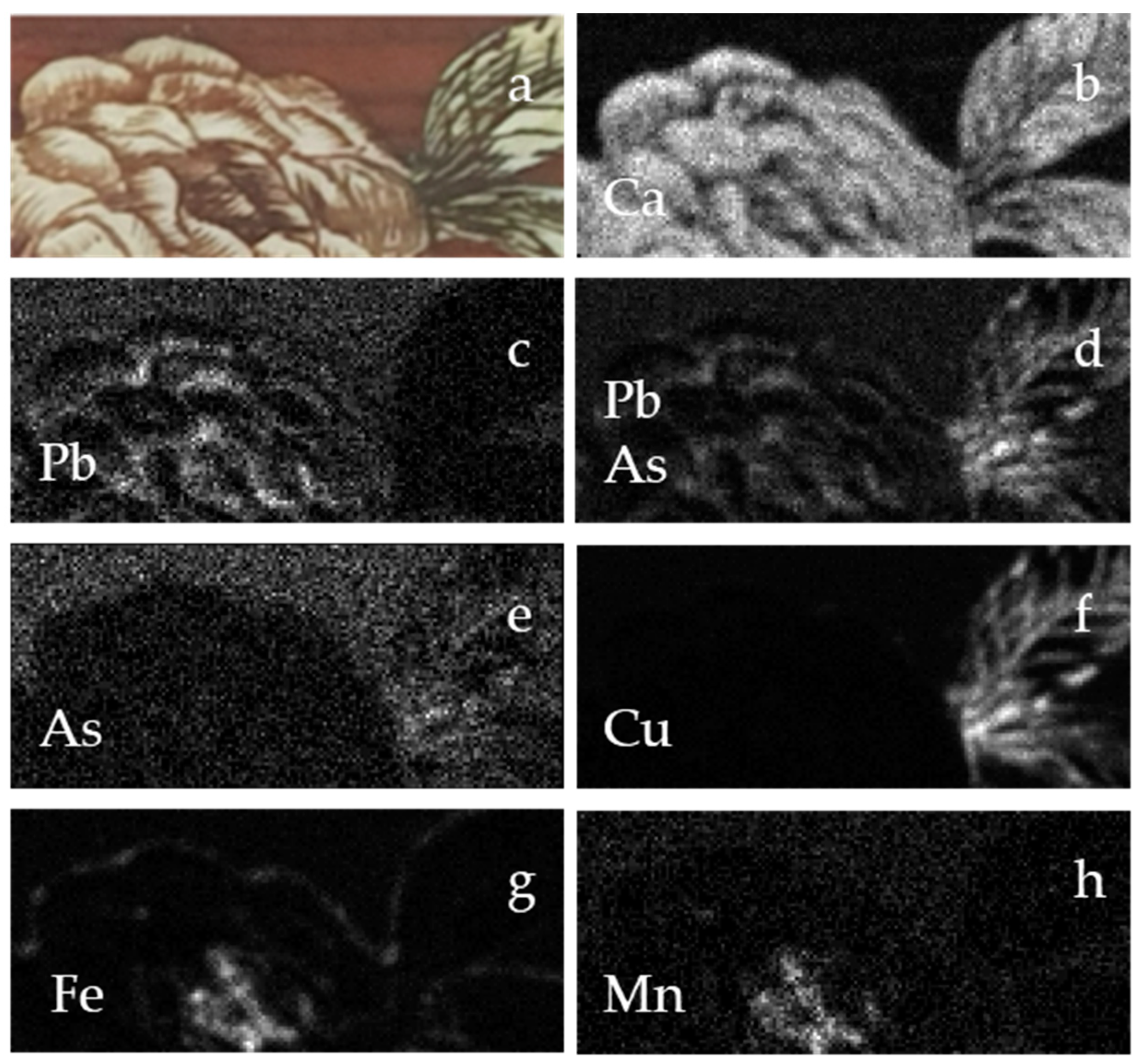
| Writing desk—drawer area 1 | green leaves | As, Cu | emerald green |
| dark rust tone of the flower | Fe, Mn | ochres-earths | |
| rust tone of the flower | - | organic compounds | |
| white highlights | Pb | lead white | |
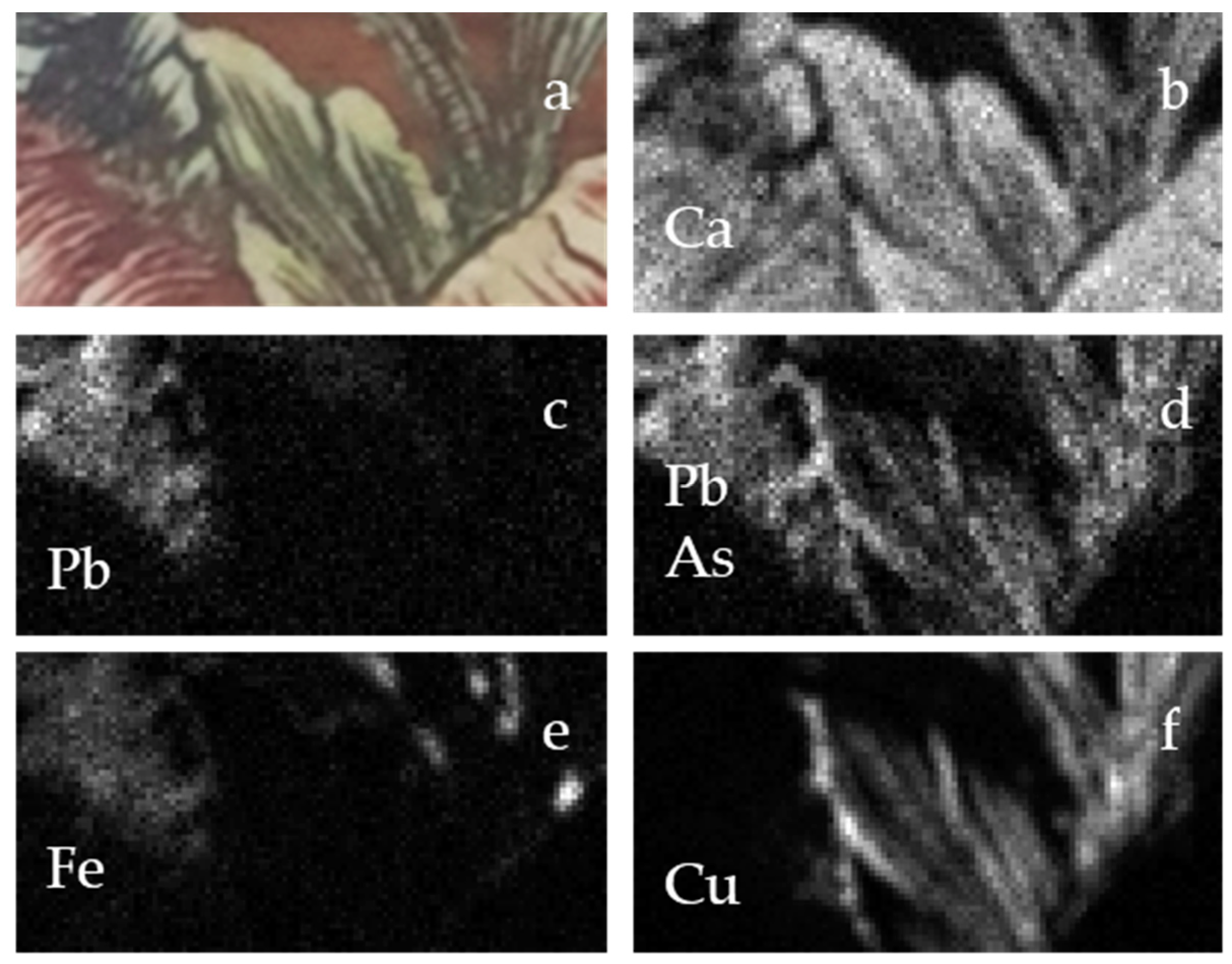
| Writing desk—drawer area 2 | black-blue flower | Pb, Fe | Prussian blue and lead white |
| green leaves | As, Cu | emerald green | |
| Writing desk—external side | green leaves | Cu, Cl | copper-based compound |
| black-blue flower | Pb, Fe | Prussian blue and lead white | |
| red flower | Hg | cinnabar-vermilion | |
| blue-black dark stem | Fe, Mn | ochres-earths | |
| dull yellow flower | - | organic compounds |
Publisher’s Note: MDPI stays neutral with regard to jurisdictional claims in published maps and institutional affiliations. |
© 2021 by the authors. Licensee MDPI, Basel, Switzerland. This article is an open access article distributed under the terms and conditions of the Creative Commons Attribution (CC BY) license (http://creativecommons.org/licenses/by/4.0/).
Share and Cite
Sottili, L.; Guidorzi, L.; Mazzinghi, A.; Ruberto, C.; Castelli, L.; Czelusniak, C.; Giuntini, L.; Massi, M.; Taccetti, F.; Nervo, M.; et al. The Importance of Being Versatile: INFN-CHNet MA-XRF Scanner on Furniture at the CCR “La Venaria Reale”. Appl. Sci. 2021, 11, 1197. https://doi.org/10.3390/app11031197
Sottili L, Guidorzi L, Mazzinghi A, Ruberto C, Castelli L, Czelusniak C, Giuntini L, Massi M, Taccetti F, Nervo M, et al. The Importance of Being Versatile: INFN-CHNet MA-XRF Scanner on Furniture at the CCR “La Venaria Reale”. Applied Sciences. 2021; 11(3):1197. https://doi.org/10.3390/app11031197
Chicago/Turabian StyleSottili, Leandro, Laura Guidorzi, Anna Mazzinghi, Chiara Ruberto, Lisa Castelli, Caroline Czelusniak, Lorenzo Giuntini, Mirko Massi, Francesco Taccetti, Marco Nervo, and et al. 2021. "The Importance of Being Versatile: INFN-CHNet MA-XRF Scanner on Furniture at the CCR “La Venaria Reale”" Applied Sciences 11, no. 3: 1197. https://doi.org/10.3390/app11031197
APA StyleSottili, L., Guidorzi, L., Mazzinghi, A., Ruberto, C., Castelli, L., Czelusniak, C., Giuntini, L., Massi, M., Taccetti, F., Nervo, M., De Blasi, S., Torres, R., Arneodo, F., Re, A., & Lo Giudice, A. (2021). The Importance of Being Versatile: INFN-CHNet MA-XRF Scanner on Furniture at the CCR “La Venaria Reale”. Applied Sciences, 11(3), 1197. https://doi.org/10.3390/app11031197