Abstract
To reveal the accumulation of the calcium oxalate crystals (COH Crystals) during the growth and development of the taproot of Panax ginseng, and develop a novel and rapid characterization method to evaluate the growth age of commercial ginseng, multiple methods in micro characterization techniques of SAXS, Micro-CT, FEG-ESEM and Micro-Raman were used to identify the COH Crystals and establish a quantitative counting method for growth age identification. In this study, a cross-analysis with multiple methods proved for the first time with a Raman and Energy spectrum that the high-density particles widely distributed in the parenchyma cells of the xylem and cortex are COH Crystals; we also first realized quantitative counting of the COH Crystals on the cross-section of fresh ginseng samples. Moreover, catering to the testing requirements of the modern trading of fresh ginseng products, we also specifically established an interesting and useful mathematical equation (Y = 2.3797X − 1.2404) for growth age identification. The technology and strategy in this study effectively compensated for the shortcomings of chemical testing and other methods in technical limitations; hence, the application of more ginseng varieties to perform the technical optimization is expected.
1. Introduction
Panax ginseng Meyer [1] is well known for its functional properties and has been used for health care and clinical use in East Asian countries for thousands of years. Due to the huge market demand every year, the taproot of P. ginseng (synonyms: ginseng) is one of the most popular functional food and herbal material in the world for domestic markets and the import–export trades [2]. Current quality standards of ginseng products are based on a strict control of chemical components. However, the classification of herbal ginseng is still dependent highly on traditional evaluative indicators or experiences of through word-of-mouth. For centuries, the number of the growth age of herbal ginseng in China has been the most important control index and evaluation parameter for quality control in the agricultural production and market circulation [3,4,5]. Recently, more and more evidences proved that the growth age of ginseng is virtually related to its chemical quality and efficacy [4,5]. So, it has been practically considered as an important criterion to determine the quality of ginseng.
In the past decade, we have been paying attention to and developing ginseng age identification technologies in different ways and several innovative methods have been developed gradually to accurately identify the growth age of ginseng. Liang [6] developed an exploratory method based on the telomere length and telomerase activity for ginseng age recognition. Unfortunately, this testing process involves multiple cumbersome molecular biology experiments, and its testing period is as long as 5–7 days, so it is difficult for the general testing institution to implement in their daily practice. Thus, we explored a rapid age identification based on a mathematical model in a strictly light of taproot radial development in vascular plants [7,8]. This method also provided an interesting exploration of a non-destructive identification by using X-ray tomography or synchrotron radiation 3D modeling, which initiated more extensive and practical strategies to establish identification methods of valuable medicinal plants based on X-ray tomography technology. Naturally, the radial growth of the taproot fully relies on the periodic activities of the vascular cambium, and the mathematical model of age identification developed based on this single index would be easily degraded to its detecting efficiency due to the different climatic conditions and agricultural technologies. Therefore, in order to evaluate ginseng growth age more accurately and develop more test methods related to ginseng quality characterization, we still need to vigorously explore new identifying characteristics or technical indicators to be applied for rapid detection, especially for the fresh herbal ginseng with frequent trading in the market.
In the past decade, we also tried to focus on the correlation analysis between the growth age and the number of calcium oxalate crystals (COH Crystals) inside the herbal ginseng sample by using the paraffin section method. Recently, it has also been reported that X-ray micro-tomography was used for the discovery and quantification of the COH Crystals in ginseng [9,10]. These studies first demonstrated the possibility of the COH Crystals to assist in the identification of the ginseng growth age. Thus, X-ray technologies, such as Small-angle X-ray scattering (SAXS) [7,8] and X-ray phase contrast micro-tomography [9,10], showed good application prospects in presenting and quantifying high-density calcium oxalate crystals in plants. However, due to technical limitations, X-ray tomography technologies are only suitable for a completely dry ginseng sample, and cannot actually conduct anything with a fresh plant sample. Moreover, previous studies did not use samples with consecutive ages for method verification and theoretical exploration. In this study, the research methodology of the study is showed in Figure 1, we not only established an age identification method for dried ginseng samples, but also focused on the development of growth age evaluation for the fresh ginseng sample by using another method of environmental scanning electron microscope (ESEM), which hinted at a clear and ultra-depth-of-field photographic ability on fresh plant samples [11,12]. Besides, under different instrument environments, this study also deliberately compared the technical advantages of using X-ray microscopic imaging to characterize the different micro-region features for growth age evaluation.
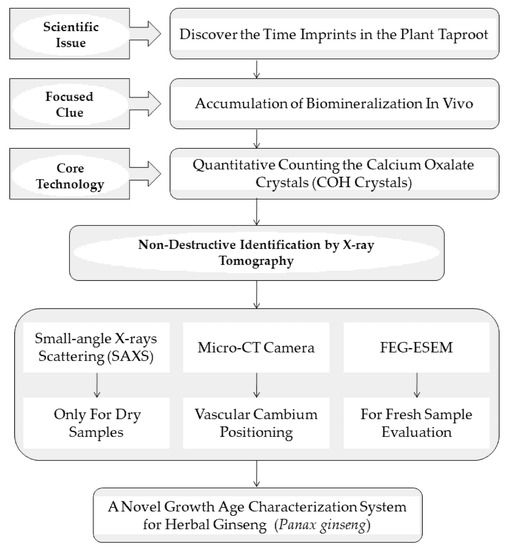
Figure 1.
Research methodology of the study.
2. Materials and Methods
2.1. Samples
All ginseng samples were collected with clear growth age from Dandong city, Liaoning Provence. The samples were divided into 14 groups according to their growth age, with 10 individuals in each group. All ginseng samples were morphological identified by Dr. Cheng Chun-Song as P. ginseng (Shizhu ginseng), an excellent farm-type cultivar of P. ginseng planted in Northeast China. Dried ginseng samples were produced by processing with a freeze vacuum dryer (YTLG-10A, produced in Shanghai Yetuo, China).
2.2. Tomography Imaging with Synchrotron Radiation of X-rays
According to the method described in Section 2.1, the taproot of Shizhu ginseng samples with different growth ages were first dried by using the freeze vacuum dryer, prepared into small segments or radial sectors and fixed in 1.5 or 2 mL EP tubes. Small-angle X-ray scattering (SAXS) (Institute of High Energy Physics Research (HEPR), Beijing, China) was used to obtain the projection and background images. Rotated 360° and produce projection data were obtained at the waist of samples in the Institute of High Energy Physics Research (HEPR), Chinese Academy of Sciences. Sections and three-dimensional images were reconstructed by DEI Reconstructor V3.6 which was developed by HEPR.
2.3. Tomography Imaging with Micro-CT Camera
Fresh ginseng samples growth—aged from 1 to14 were first stored at −80 °C and then dried under reduced pressure at −40 °C for more than 48 h to prepare completely dried ginseng samples. Tomography was performed on the completely dried samples to further explore the imaging of COH Crystals under normal micro-computed tomography conditions. Micro-CT camera (Bruker Inc., Kontich, Belgium) was used for tomography imaging and the image pixel size was 15 μm, the frame averaging was 4 and the rotating step was 0.2.
2.4. Cross-Sectional Imaging with FEG-ESEM
In accordance with reported papers [11,12], SEM stub with conductive carbon paint was used in this study and the fresh herbal ginseng sample slices were analyzed using a Quanta™ 250 FEG ESEM (FEI, Hillsboro, OR, USA) operating in low vacuum mode (pressure chamber set at 100 Pa) with the secondary electron signal range of 3, 5 or 15 kV.
2.5. Micro-Raman Spectroscopy Analysis
The taproots of fresh samples were sliced by hand and fixed on glass slides with deionizer water. Raman imaging was acquired with a LabRAM XploRA confocal Raman microscope equipped with a microscope (Olympus) and a motorized stage. The data were conducted with a high numerical aperture microscope objective from Olympus (40×) to achieve the high spatial resolution.
2.6. Analysis and Statistics
Image-pro Plus (Ipp software) was used to count the COH Crystals on the cross section of the fresh ginseng sample. R-studio (ggplot2, boxplot and lm function) was used to draw box-plots and perform the variance analysis and linear regression.
3. Results
3.1. Characterizing the Surface Structures of Fresh Sample by Using FEG-ESEM
Generally, fresh herbal ginseng is not easy to characterize with the imaging of calcium oxalate crystals under X-rays imaging conditions due to its high-water content. Therefore, a rare report can provide a practical method for the identification of fresh plant samples under micro imaging equipment with X-rays under different working conditions. However, it seems that FEG-ESEM can make a breakthrough for characterizing the surface micro structures of a fresh sample under X-rays conditions [13,14]. In this study, the surface characterization of COH Crystals was first achieved by using the FEG-ESEM system. As shown in the following Figure 2, the dim-white dots unevenly scattered on the surface of the slice (Figure 2A) were characterized by the energy spectrum as the Ca element (Figure 2B). Then, Micro Raman spectroscopy was used for further verification and characterization of the COH Crystals. The vibrational wave numbers (P480, P603, P891, P1406 cm−1, all shown in Figure 2C) were also consistent with the reported reference [15]. It was found that the locations where plant sieves and vessels were distributed presented uniform bright circles, which can easily be regarded as COH Crystals and challenge the counting of COH Crystals. As shown in Figure 2A and Figure S1 (see the Supplementary Materials), the three bright circles were initially considered as the K element distribution areas. In this way, we had to further study how to overcome the challenge of potassium distribution to the counting of COH Crystals. This result also indicates that it was necessary to critically review the reported X-ray tomography results on the counting or quantification of COH Crystals in the ginseng taproot. Whether it was necessary to design a systematic shielding method to improve the specificity of COH Crystals quantification had become another important scientific question that we needed to verify in the next stage.
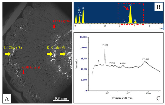
Figure 2.
Micro-scale surface analysis for counting of calcium oxalate crystals (COH Crystal) by using the FEG-ESEM system (A) and the physical and chemical characterization for COH Crystal through energy spectrum analysis (B) and Micro-Raman spectroscopy (C).
3.2. Tomography Used to Figure out More Specific Distribution of COH Crystals
In order to figure out a more specific distribution of COH Crystals inside the ginseng taproot, the completely dried ginseng samples prepared with a vacuum freeze drier, were also tested with tomography and 3D reconstruction by using the synchrotron radiation of X-rays. As shown in the following Figure 3, the high-density solid particles were clearly presented in three dimensions by computer algorithms. According to our statistics, more than 70% of the particles were distributed in the root cortex, and the remaining part was almost entirely distributed in the xylem among the parenchyma cells. In this X-ray working environment, the bright bands of potassium were not present in those images at all. Therefore, these results showed that the high energy X-ray measurement based on synchrotron radiation and corresponding computer reconstruction program specifically presented COH Crystals. Moreover, this result provided an important suggestion that the COH Crystals were also distributed partly among in the parenchyma cells surrounding the phloem fibers (Figure 3A,B). Obviously, the metal ion band would not interfere with the counting of COH Crystals under higher energy X-ray imaging conditions.
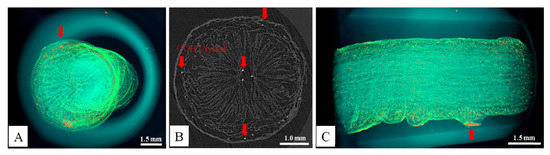
Figure 3.
Small-angle X-ray scattering (SAXS) was used for the tomographic 3D reconstruction of dried ginseng samples (the red arrow points to the preferred position of COH Crystals aggregation in the taproot of ginseng). (A) One of the 3D rendering angles of ginseng taproot for counting COH Crystals; (B) the original cross section of tomography; (C) the other one of the 3D rendering angles of ginseng taproot for counting COH Crystals.
3.3. Mathematical Modeling to Realize the Ginseng Age Identification
The specific identification method for fresh ginseng, especially the intelligent recognition technology of the growth age of herbal ginseng, was still worthy of development. Hence, we further used the cambium as the key recognition center to focus on the specific microscopic vision and quickly counted the COH Crystals to identify the growth age. The confirmation of cambium was first verified by micro CT results in this study (Figure S2, see the Supplementary Materials). As shown in Figure 4, the fresh taproots were cut crosswise as shown in Figure 4A, the two bright potassium circles (Figure 4B, K+ Circle (S) and K+ Circle (V)) were used to locate and identify the vascular cambium, phloem and cortex. More than 100 individual growths aged from 1 to 14 were tested with this counting method (Figure 4C). Available X-ray images came from taking photos of randomly selected microscopic fields covering a quarter of the fresh taproot section, all the bright granular COH Crystals between the two metal ion bands should have been counted for the construction of the mathematical model. The results showed that the counts of COH Crystals were significantly different in fresh taproot sections of different growth ages (N = 8, ANOVA testing, F = 362.11, p = 1.28 × 10−76 < 0.001). Moreover, the linear regression analysis showed a good increasing trend that the number of COH Crystals increased positively with the growth age (Figure 4D, age identification). The modeled equation is:
Y = 2.3797X − 1.2404
R2 = 0.9765
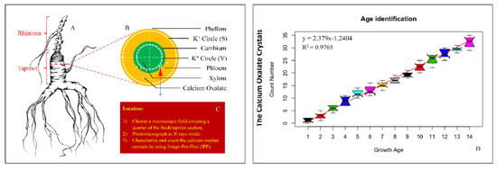
Figure 4.
Quantitative counting of the calcium oxalate crystals (COH Crystals) with special micro-positioning and testing the ginseng samples with gradient growth age for acquiring age identification equation. (A): Chinese Shizhu ginseng (A farm variety of Panax ginseng), the ginseng figure was drawn by Dr. Chunsong Cheng (程春松); (B): schematic diagram of positioning for microscopic field observation; (C): important implementation procedures for counting COH Crystals; (D): this was an independent result of linear regression, the unary regression equation is the mathematical model used for age identification.
While Y is the count of COH Crystals, X is the fixed number of growth age.
R is the correlation coefficient of the linear fitting.
4. Discussions
In this study, a multi-environment platform of X-ray practices was of such great significance, that it first cross-answered the question of the specificity and resolution of different X-ray tomography imaging methods for COH Crystals. In view of this, Ye L.L.’s method [9,10] has sufficient basis to continue to be widely used in the quality identification of herbal medicines.
We also found that the section structure features imaged by the method of FEG-ESEM had a super depth of field level, so that the COH Crystals we observed actually came from the multiple superimposed planes. Compared with traditional paraffin sections [16], the technology we provided here was more stable and reproducible.
Additionally, we have been engaged in the growth age recognition of ginseng for more than 10 years. In recent testing practices, the judgment of ginseng age based on taproot growth and development models [7,8] was easily affected by many meteorological and agricultural factors, such as precipitation, swelling agents, etc., which indeed created testing challenges. Therefore, auxiliary identification measures were very important to the discovery of evaluating the growth age of herbal ginseng. As the direct trade of fresh ginseng has taken place frequently in recent years, more and more purchasers are eager to quickly evaluate the growth time of ginseng in the farmland or market. Therefore, we are pleased to report this result quickly in the hope of obtaining more cooperation and promoting the establishment of different identification parameters and systems for more farm varieties widely planted in Northeast China, South Korea and Japan.
At present, we verified that the number of COH Crystals increased with the growth age in the specific parts of the ginseng taproot. Simultaneously, we observed and confirmed the bright bands of metal ions in the FEG-ESEM mode for the first time. Our research also provided detailed information for the first time to confirm that the metal ions could not interfere with the counting of COH Crystals under other X-ray tomography scanning modes. In terms of the considerable practical significance and market prospects of the growth age identification method developed in this study, in detail, this result provided another brand-new scientific evaluation program that can be made available to support our general estimated model for ginseng growth age identification [8]. In the next implementation scenarios of quality control of herbal ginseng, the COH Crystals accumulation of more types of ginseng landraces will be observed by using FEG-ESEM. More COH Crystal-related biological issues need further attention and in-depth study, including the differences in the accumulation of COH Crystals’ biomass between different landraces, the closely related biological mechanisms of growth and development, biological defense characteristics of ginseng root, as well as the correlation to the secondary metabolism.
5. Conclusions
With the FEG-ESEM imaging system, a novel quantitative counting of COH Crystals by using X-ray microscopic imaging was first realized for growth age identification in a fresh ginseng cross-section. We used the specific microscopic field of vision for focus positioning, which masterly avoided the confusion of the result caused by the optical imaging of other metal ions. So far, a possibility effective inspection and linear mathematical model were also developed for the first time specifically for Chinese Shizhu ginseng, a kind of landrace of P. ginseng which retains the traits of wild ginseng to the greatest extent. Therefore, it will be a meaningful and interesting work and this study provides a new age identification strategy by quantitatively counting the COH Crystals. There are many cultivars of ginseng in the current medicinal material markets, and our method needs more verification and practice in the main types of ginseng cultivars, such as “Damaya”, “Biantiao”, “Ermaya”, etc. Biomineralization represented by COH Crystals is a very interesting research direction. The relationship between COH Crystals’ bioaccumulation and biological age involves the cross-interaction among ginseng physiology, cultural techniques, as well as the ecological environments. Therefore, we will use the biological age as a scientific clue, and continue to be concerned about the biological issues of herbal ginseng at different growth and development stages, and the quality control technologies contributing to the industrial norms.
Supplementary Materials
The following are available online at https://www.mdpi.com/article/10.3390/app11188389/s1, Figure S1: The radial distribution of six common metal ions in ginseng cross section, Figure S2: The positions of cambium and phloem fiber were further confirmed by microstructure analysis using Micro-CT.
Author Contributions
C.C. conducted the strategy and experimental design, data analysis and wrote and reviewed the main manuscript text; S.F., P.G. and Y.H. conducted the data analysis. All authors have read and agreed to the published version of the manuscript.
Funding
This research was funded by Special Project in Lushan Botanical Garden, CAS: 2021ZWZX08.
Institutional Review Board Statement
Not applicable.
Informed Consent Statement
Not applicable.
Data Availability Statement
Not applicable.
Acknowledgments
This research was funded by the Science and Technology Development Fund of Lushan Botanical Garden, Chinese Academy of Science (2021ZWZZX08). The authors are also grateful for the advice of Jie Liang from the Macau University of Science and Technology, and Qinxi Yuan from the Institute of High Energy Physics, Chinese Academy of Science.
Conflicts of Interest
The authors declare no conflict of interest.
References
- Park, J.D.; Rhee, D.K.; Lee, Y.H. Biological activities and chemistry of saponins from Panax ginseng CA Meyer. Phytochem. Rev. 2005, 4, 159–175. [Google Scholar] [CrossRef]
- Chen, C.F.; Chiou, W.F.; Zhang, J.T. Comparison of the pharmacological effects of Panax ginseng and Panax quinquefolium. Acta Pharmacol. Sin. 2008, 29, 1103–1108. [Google Scholar] [CrossRef] [PubMed]
- Kim, N.; Kim, K.; Choi, B.Y.; Lee, D.; Shin, Y.S.; Bang, K.H.; Lee, D. Metabolomic approach for age discrimination of Panax ginseng using UPLC-Q-Tof MS. J. Agric. Food Chem. 2011, 59, 10435–10441. [Google Scholar] [CrossRef] [PubMed]
- Shan, S.M.; Luo, J.G.; Huang, F.; Kong, L.Y. Chemical characteristics combined with bioactivity for comprehensive evaluation of Panax ginseng CA Meyer in different ages and seasons based on HPLC-DAD and chemometric methods. J. Pharm. Biomed. Anal. 2014, 89, 76–82. [Google Scholar] [CrossRef] [PubMed]
- Huang, B.M.; Zha, Q.L.; Chen, T.B.; Xiao, S.Y.; Xie, Y.; Luo, P.; Zhou, H. Discovery of markers for discriminating the age of cultivated ginseng by using UHPLC-QTOF/MS coupled with OPLS-DA. Phytomedicine 2018, 45, 8–17. [Google Scholar] [CrossRef] [PubMed]
- Liang, J.; Jiang, C.; Peng, H.; Shi, Q.; Guo, X.; Yuan, Y.; Huang, L. Analysis of the age of Panax ginseng based on telomere length and telomerase activity. Sci. Rep. 2015, 5, 7985. [Google Scholar] [CrossRef] [PubMed]
- Cheng, C.; Peng, D.; Huang, L.; Ma, X. Years-identification mathematical model of paeonialactiflora pall. based on the allometric-scaling. Microsc. Res. Tech. 2013, 76, 201–208. [Google Scholar] [CrossRef] [PubMed]
- Cheng, C.; Yuan, Q.; Zhou, H.; Huang, L. Nondestructive estimation of growth year in ginseng cultivars using the means of mathematical modeling on the basis of allometry. Microsc. Res. Tech. 2016, 79, 98–105. [Google Scholar] [CrossRef] [PubMed]
- Ye, L.L.; Xue, Y.L.; Tan, H.; Chen, R.C.; Qi, J.C.; Xiao, T.Q. X-ray phase contrast microtomography and its application in quantitative 3D imaging study of wild ginseng characteristic microstructures. Acta Opt. Sin. 2013, 33, 1234002–1234006. [Google Scholar]
- Ye, L.; Xue, Y.; Wang, Y.; Qi, J.; Xiao, T. Identification of ginseng root using quantitative X-ray microtomography. J. Ginseng Res. 2017, 41, 290–297. [Google Scholar] [CrossRef] [PubMed][Green Version]
- Dal Cortivo, C.; Barion, G.; Visioli, G.; Mattarozzi, M.; Mosca, G.; Vamerali, T. Increased root growth and nitrogen accumulation in common wheat following PGPR inoculation: Assessment of plant-microbe interactions by ESEM. Agric. Ecosyst. Environ. 2017, 247, 396–408. [Google Scholar] [CrossRef]
- Gan, L.; Zhang, W.; Fang, F.; Yang, L. Evidence of a trade-off between root aeration and architecture in Vallisnerianatans in the presence of Pseudomonas putida KT2440. Aquat. Bot. 2020, 162, 103189. [Google Scholar] [CrossRef]
- Stabentheiner, E.; Zankel, A.; Pölt, P. Environmental scanning electron microscopy (ESEM)—A versatile tool in studying plants. Protoplasma 2010, 246, 89–99. [Google Scholar] [CrossRef] [PubMed]
- Tihlaříková, E.; Neděla, V.; Đorđević, B. In-situ preparation of plant samples in ESEM for energy dispersive x-ray microanalysis and repetitive observation in SEM and ESEM. Sci. Rep. 2019, 9, 2300. [Google Scholar] [CrossRef] [PubMed]
- Edwards, H.G.M.; Farwell, D.W.; Jenkins, R.; Seaward, M.R.D. Vibrational Raman spectroscopic studies of calcium oxalate monohydrate and dihydrate in lichen encrustations on Renaissance frescoes. J. Raman Spectrosc. 1992, 23, 185–189. [Google Scholar] [CrossRef]
- Wang, M.; Jin, W.; Guo, H.; Wang, H.; Sun, L. Study on the relevance between ginseng root tissue morphology and biologically active ingredients. J. Cent. China Norm. Univ. (Nat. Sci.) 2015, 3. [Google Scholar]
Publisher’s Note: MDPI stays neutral with regard to jurisdictional claims in published maps and institutional affiliations. |
© 2021 by the authors. Licensee MDPI, Basel, Switzerland. This article is an open access article distributed under the terms and conditions of the Creative Commons Attribution (CC BY) license (https://creativecommons.org/licenses/by/4.0/).