Abstract
This study aims to evaluate the functional variations of the electromyographic response and clinical symptomatology in TMD patients. This study has been performed and compared before and after the application of a therapeutic protocol based on the use of an oral device working on the proper mandibular repositioning through the proprioceptive-based lingual re-education called “Lingual Ring”. Between January to December 2016, 32 TMD patients have been recruited out of a series of 321 individuals recruited at the Neurosensorial and Motorial Surgery Department, in Palermo (Italy). All the patients underwent the Surface Electromyography (sEMG) of Masseter and Temporal muscles, with different registrations at T0, T1, T2, and T3; to evaluate the variations of the Electromyographic values, it was assigned the application of the Lingual Ring as the only therapy. The study demonstrated a neat rebalancing of the EMG tracks and important improvements with the TMD related issues. The clinical responses, due to the treatment at the end of the therapeutic protocol, were: absence or reduction of muscular or articular pain; absence or reduction of articular noises; absence or reduction of the cephalalgia. Hence, significant results, both clinical and in terms of instrumental EMG, were assessed. We can affirm that the adopted methodology allowed the monitoring of the Electromyographic variation and clinical symptoms. Moreover, the usage of the “Lingual Ring device” allowed to carry out a simple and immediate therapeutic protocol that is minimally invasive, ensuring a clear rebalancing of the EMG tracks as well as the TMD related pain.
1. Introduction
Temporomandibular disorders (TMD) belong to a series of diseases affecting the oral cavity, involving several co-factors in their etiology. TMD are characterized by a neuromuscular imbalance that particularly affects the masticatory muscles: it often involves also the postural muscles, resulting in a trigger that may alter the entire body structure, falling within the large group of the so-called “cranio-cervico-mandibular disorders” (CCMD) [1].
The causes of CCMD are pretty heterogeneous. Nevertheless, dental malocclusion and muscular impairments are the main factors involved in the pathogenesis, able to induce the alteration of masticatory function, leading to unfavorable biomechanical alterations impacting the TMJ (temporomandibular joints). Trauma of cervical region (whiplash syndrome) frequently represents a trigger for CCMD onset. Other disorders should also be carefully evaluated, such as functional alterations of the oculomotor muscles and dystonia of the neck muscles [2,3,4].
Surface electromyography (sEMG) is the most commonly used research tool, mainly used for evaluating the muscle activity through the use of surface sensors. The advantage of sEMG is its total non-invasiveness: moreover, it is a painless, simple to perform, and easy to apply test that allows to simultaneously record the potential of several muscles. The electrical activity recorded is directly related to the activity of the muscles [5,6,7].
This specific functional examination is also suitable for evaluating the efficacy of physiotherapeutic, surgical, or pharmacological treatments [8]. The numerous advantages of this technique have allowed the creation of low-cost sEMG devices, some of which have been widely validated by several scientific research groups [9,10,11].
The Surface Electromyography (sEMG) analysis is useful to investigate the main functional activities of the masticatory muscles, especially in subjects affected by temporomandibular disorders (TMD), where the muscular balance is frequently altered [12,13,14]. Interestingly, the Surface Electromyography can clearly describe the muscular activity to better understand specific alterations between different muscular groups over time [15,16,17,18,19].
In this study, the sEMG has been used as a reliable and repeatable method to investigate the functional alterations between different muscular groups over time. More specifically, sEMG has recorded the activity of Masseter and Temporal muscles at three different times (before, during, and after therapy), to assess the efficacy of a novel therapeutic protocol. The therapeutic protocol was based on a new preformed occlusal device called “Lingual Ring” and commercially identified with the name “Ripara” [20,21,22]: we must highlight that this device has been created by the same authors of this paper. This device has been created with the aim of obtaining the repositioning of the articular discs through the induction of proper movements of the masticatory muscles and the tongue. Ripara differs from other “Bites” or “Splints” [23], as it is based on the re-education of the tongue through a lingual ring: basically, the tongue is the only support where the device is applied, triggering the muscular balancing among Masseter and Temporal muscles [24,25,26]. Previous studies have been carried out by the authors on this device [20,21,22]: the working group has highlighted the most impacting clinical outcomes obtained with this device; in this study, the authors aim to provide more data on the preliminary results achieved in previous studies.
This “Lingual Ring” has been conceived as a novel and innovative device, able to actively involve the patients in the therapeutic protocols [20,21,22,27,28,29,30,31]. The main outcome is the rehabilitation of a proper lingual posture: this result can play a strategic role to obtain a stable and good balancing of the extrinsic tongue muscles [32]. Of course, the main requirement for this therapy it that it needs the fully active collaboration of the patient, who has to learn how to handle the new device; therefore, this oral device offers different management strategies compared to a traditional bite that works mainly as a passive appliance [20,21,22,23,27]. In fact, the universal occlusal splints used in TMD therapy are designed to be passive “pads” that basically counteract the load produced by the masticatory muscles. The new device aims to work on the vertical dimension, stimulating the muscles elongation, and guiding at the same time the tongue position to remain higher and more forward: these actions are potentially able to modify the position of the hyoid bone and the paravertebral muscles of rachis in an active and homogeneous way.
This study aims to evaluate the functional variations of the electromyographic response and clinical symptomatology in TMD patients. This study has been performed and compared before and after the application of a therapeutic protocol based on the use of an oral device working on the proper mandibular repositioning through the proprioceptive-based lingual re-education called “Lingual Ring”.
2. Materials and Methods
A total of 32 patients affected by Temporo-Mandibular Disorders (TMDs) (26 women and 6 men), aged from 18 to 58 years (average: 38 years old; SD = 6), have been recruited. Patients were selected from a list of 321 patients recruited at the Neurosensorial and Motor Surgery Department of the Complex Operative Unit of Odontostomatology, AOU Policlinico “Paolo Giaccone” in Palermo (Italy), between January 2016 and December 2016. All the patients were found to have masticatory dysfunctions and (or) temporomandibular joint anomalies, according to the international criteria to assess the presence of Temporomandibular Disorders [33,34].
The study was conducted according to the guidelines of the Declaration of Helsinki and approved by the Institutional Review Board of the Neurosensorial and Motor Surgery Department of the Complex Operative Unit of Odontostomatology, AOU Policlinico “Paolo Giaccone” in Palermo (Italy). (Ref. n. RIPARA project–20/01/2016).
All the participants expressed their informed consent on a dedicated agreement.
Inclusion criteria [33,34]:
- temporomandibular joint (TMJ) pain ≥ 30 on the numerical visual scale (NVS);
- muscular pain (Masseters and Temporalis) ≥ 30 on the NVS;
- tensive cephalgia and (or) migraine ≥ 30 on the NVS;
- cervical pain and (or) backaches of tensive origin ≥ 30 on the NVS.
Painful symptomatology has been divided into 4 categories: mild (0–30); moderate (30–50); strong (50–80); severe (80–100).
Exclusion criteria [33,34]:
trauma, fractures, or malformations affecting TMJ;
- recent surgery of TMJ;
- systemic pathologies (such as, rheumatoid and psoriatic arthritis, Ehlers–Danlos syndrome);
- neuropathic cephalgia;
- edentulism, involving eight or more missing teeth;
Therapeutic indications:
The therapeutic protocol with the lingual-ring has been articulated in the following manner: the standardized application of the device ranged from a minimum of night-only use (6–8 h) to a maximum of 12 h per day, including night, with the indication to put the tongue at the top of the spot. The maximum duration established for an entire cycle of treatment was 3 months.
The recruited patients underwent the sEMG examination, with the aid of a computer-controlled technology to process the data. A dedicated software was able to digitalize the sEMG analogic signals. The sEMG recordings and signal detection were carried out following the Seniam’s recommendations; briefly, the operators should apply single-use, bipolar, and superficial skin-electrodes (21 × 41 mm), placed in parallel with the muscular fibers on the Masseter and Temporal muscles (anterior fascia), on both the right and left sides. Inter-electrodes’ distance should be 21 ± 1 mm. The reference (mass) electrode is placed on the forehead (glabella) (Figure 1). Before placing the electrodes, the skin must be cleaned with 90% ethanol, in order to reduce the superficial electric impedance. Patients should sit in a natural position, with the head free from any support, and eyes looking to the horizon [35,36].
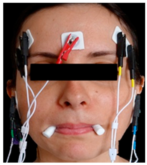
Figure 1.
First recording (T0), with cotton rolls between the second premolars.
The electromyographic evaluation at time zero (T0) was first performed with the aid of two 10 mm-thick cotton rolls placed between the second premolars of the mandible, with maximum voluntary tightening (MVC) lasting no less than 5 s (Figure 1).
Then, the electromyographic evaluation was newly performed at time zero (T0) with the aid of the intraoral device called “Lingual Ring” (Figure 2).
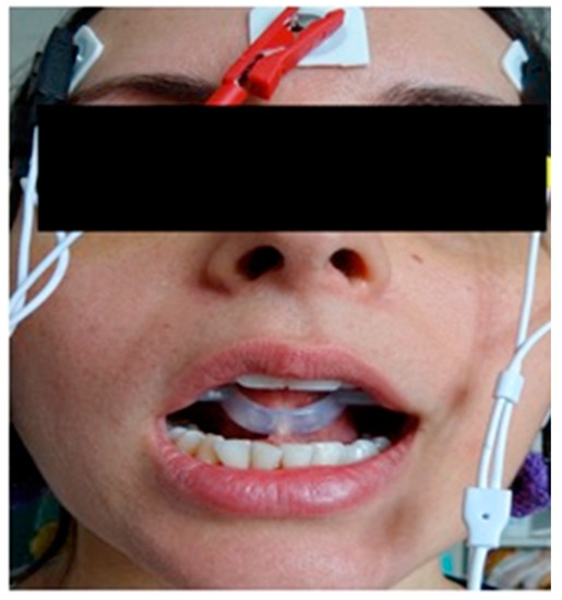
Figure 2.
Second recording (T0): with the intra-oral Lingual Ring application.
The “Lingual Ring” has been previously described in the scientific literature [20,21,22]. Briefly, it is a medical platinic silicone, in compliance with international regulation (UNI EN ISO 10993 1:2010) and European directives number 93-42 EC (Class 1 medical devices). This device is made of three different parts: (1) The Central part, representing the Lingual ring; (2) the reinforcement, anchoring, balancing, fitting, and stabilization systems; and (3) the functional part working to promote the rehabilitation of masticatory function.
The procedures just described were replicated with the same conditions after 15 days (T1), one month (T2), and two months (T3).
No other drugs or specific therapies were prescribed during the treatment period.
The electromyography (EMG) was performed following a specific protocol, to ensure a bias-free recording. The “normalization” of EMG results was made before the experimental tests, mainly reducing the biological and technical alterations that may have impact on the data recorded.
The resulting EMG standardized potentials have been, therefore, considered only as the result of the muscular contraction correlated to the dental occlusal surfaces and the Lingual Ring interactions.
Finally, the Masseter POC (Percentage of Overlapping Coefficient) was used to indicate the impact of the teeth touching the muscles, compared to the standard calibration records obtained with only the cotton rolls.
3. Results
The first table (Table 1) shows the number of patients involved in the study, the minimum and maximum values, the values’ average, and the standard deviation at each time. In Table 1, we can observe the statistical analysis of the values of Masseter POC (MPOC) obtained at different time points, with or without the use of the Ripara device.

Table 1.
Masseter POC (MPOC) values at different time points, with (MPOCW) and without (MPOCWO) Ripara device.
Figure 3 shows the minimum and maximum values relating to the Masseter POC (MPOC) value, obtained at different time points (T0–T3) without using the Ripara device. On the other hand, Figure 4 shows the minimum and maximum values relating to the Masseter POC (MPOC) value obtained at different time points (To–T3) with the use of the Ripara device. From the comparison of these two figures, we can observe that the Ripara device does not affect the reported parameters in any of the reported time points.
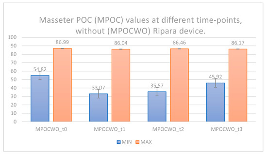
Figure 3.
Masseter POC (MPOC) values at different time points, without (MPOCWO) Ripara device.
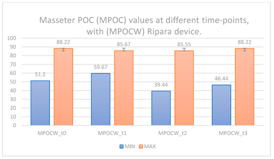
Figure 4.
Masseter POC (MPOC) values at different time points, with (MPOCW) Ripara device.
Table 2 shows the difference between two average values obtained at different time points, with or without the use of the Ripara device. From the analysis of this table, we can observe quite similar average values; therefore, there are no statistically relevant differences in the MPOC.
Table 2.
Paired difference between the 2-time-point averages of the MPOC, with and without Ripara device.
Table 3 shows the differences of pain (DOP) reported on a Visual Analogue Scale (VAS) from T0 to T3: these data were recorded for both the right and left Masseters.
Table 3.
Differences of pain from T0 to T3: these data were recorded for both the right and left Masseters.
After careful evaluation, we can point out that patients reported a reduction of pain on both sides from T0 to T3, suggesting a normalization of the electromyographic values, with a better masticatory efficiency with the use of the Ripara device.
Therefore, based on Table 3, we can argue that we have several promising results in pain control with the use of the lingual ring tested here. The statistical data reported here have clearly demonstrated how, at the end of the therapy with the lingual ring (T3), the pain at palpation progressively decreases in all patients.
The results thereby suggest that therapy with the innovative device decreases the muscular hypertonus in TMD patients; moreover, this device increases the muscular hypotonus for the patients who present an opposite characteristic, bringing back isometry and symmetry to the muscular apparatus.
Normalization of the electromyographic values also resulted in a better masticatory efficiency, considered as a better contraction strength, symmetry, and balance during mastication. These results refer to the electromyographic values at the end of the therapy, indicating that the patient finds a new neuromuscular scheme equipped with more harmony and syntony between the right and left antimeres, and also muscular isotonicity. Thus, the therapy carried out with the Lingual Ring improves the instrumental parameters even without being in position after the therapy.
Table 4 reports the analysis of the bivariate correlation between the differences of pain (DOP) reported on a Visual Analogue Scale (VAS) from T0 to T3: these data were recorded for both the right and left Masseters. The difference between right and left Masseter is correlated with the right and left Masseter POC; in particular, the difference observed in the timespan T0–T3 related to the right Masseter pain is directly linked with the difference related to the left Masseter pain (0.035 significance).
Table 4.
Analysis of the bivariate correlation between the differences of pain (DOP) reported on a Visual Analogue Scale (VAS) from T0 to T3 with (DOP_W) and without (DOP_WO) Ripara device.
Table 4 also reports the “Pearson Correlation” analysis, which describes if two variables are dependent or independent with a specific condition: it seems that the clinical parameter of pain and the instrumental parameter of Masseter POC are closely linked. This means that the instrumental values can be related to the clinic values: in this context, our study has assessed that the pain values found in the right and left Masseters are associated with a high significance, suggesting that the masticatory cycle should be considered as a mathematical set that should be treated with our lingual ring as a single variable.
Finally, Table 4 reports a significance of 0.037 referred to the direct correlation between the pain to palpation at the right Masseter, and the electromyography values: this statistical association is highly significant, making the use of such methods for pain detection highly reliable. Moreover, patients have reported, in a specific point of a questionnaire that we distributed at the end of the study, an overall reduction of headache onset.
In this trial, the correlation between clinical and instrumental values allows to describe that Masseter muscles are constantly adapting during therapy with the lingual ring, ensuring that, at the end of the therapeutic protocol, there is a stable improvement in the muscular stability, and also without the further use of the lingual ring in the months following the therapy.
4. Discussion
This trial has highlighted a clear rebalancing of the EMG tracks. The clinical improvement after the therapy with the lingual ring was relevant: basically, we found a significant reduction of muscular pain at T3 in all the patients investigated. Moreover, we found a reduction of headache, also linked to a significant reduction of temporo-mandibular articulation clicks, which occurred right after our functional therapy. Interestingly, despite the fact that this study has investigated the outcomes after two months of this therapy, the clinical and gnathological improvement of the signs and symptoms has also been reported six months after the removal of the Ripara device, maintaining the EMG values normalized (new study, in press).
These results confirmed that the functional therapy with the lingual ring is able to reset the masticatory muscles’ impairment, re-educating both the correct movements and structure. Furthermore, the combination of clinical and electromyographic parameters are a valid combination for the reliable control and monitoring of a proper functional therapy.
Our study has investigated a novel and useful method for treating patients affected by Temporo-Mandibular Disorders. Our data were compared in a small group of patients, and our main outcomes basically correlate to findings already reported in the international literature with other functional devices [37,38]. The vast majority of research groups strongly state that the occlusal appliances are essential devices to intercept and reduce TMD associated with muscular pain. To date, however, it remains unclear how occlusal appliance/splint is able to work on such pain. A limitation to be considered in this study is related to the lack of measurement of the Maximum voluntary bite force (MVBF): MVBF is an important parameter to assess the functional state of the masticatory organ, and it is greatly considered during the planning of implant surgery and prosthetic restorations [39]. Despite the fact that MVBF measurements remain difficult to perform, and depending on many factors, it has been established that in the Western population, the average maximum occlusal force usually occurs between molars and is in the range of 600–750 N.
We have demonstrated that the lingual ring works through the re-education of the neuromuscular mechanisms, directly influencing the masticatory muscles and their behavior.
5. Conclusions
In conclusion, we may say that the methodology reported and investigated here could allow a safe and reliable monitoring of the masticatory muscles from both a clinical and electromyographic point of view. Moreover, from our study, it appears clear that there is a correlation between the electromyographic and clinical parameters: more specifically, we found that the balancing of the Masseter POC values is closely followed by a pain reduction. Thus, the use of the Lingual Ring can be considered as a simple and safe therapeutic appliance to rebalance the EMG tracks, thereby obtaining an improvement of the symptomatology in TMD patients.
This study certainly needs further investigations and a long-lasting clinical monitoring of a larger study sample to better validate our preliminary results.
Author Contributions
G.C., M.F., A.R. (Alessandro Rampello), A.R. (Alessio Rampello) and G.A. were involved in the conceptualization, methodology, and validation of this clinical trial. All authors have read and agreed to the published version of the manuscript.
Funding
This research received no external funding.
Institutional Review Board Statement
The study was conducted according to the guidelines of the Declaration of Helsinki, and approved by the Institutional Review Board of the Neurosensorial and Motor Surgery Department of the Complex Operative Unit of Odontostomatology, AOU Policlinico “Paolo Giaccone” in Palermo (Italy). (RIPARA project–20 January 2016).
Informed Consent Statement
Informed consent was obtained from all subjects involved in the study. Written informed consent has been obtained from the patients, to publish this paper.
Data Availability Statement
Raw data are available if required.
Conflicts of Interest
The authors declare that they have no conflict of interest.
References
- Peck, C.; Goulet, J.P.; Lobbezoo, F. Expanding the taxonomy of the diagnostic criteria for temporomandibular disorders (DC/TMD). J. Oral Rehabil. 2014, 41, 2–23. [Google Scholar] [CrossRef] [PubMed]
- De Barros Pascoal, A.L.; de Freitas, R.F.C.P.; Silva, L.F.G.D.; Oliveira, A.G.R.C.; Calderon, P.D.S. Effectiveness of Counseling on Chronic Pain Management in Patients with Temporomandibular Disorders. J. Oral Facial Pain Headache 2020, 34, 77–82. [Google Scholar] [CrossRef] [PubMed]
- Tatullo, M.; Marrelli, M.; Amantea, M.; Paduano, F.; Santacroce, L.; Gentile, S.; Scacco, S. Bioimpedance Detection of Oral Lichen Planus Used as Preneoplastic Model. J. Cancer 2015, 6, 976–983. [Google Scholar] [CrossRef] [PubMed]
- Marrelli, M.; Codispoti, B.; Shelton, R.M.; Scheven, B.A.; Cooper, P.R.; Tatullo, M.; Paduano, F. Dental Pulp Stem Cell Mechanoresponsiveness: Effects of Mechanical Stimuli on Dental Pulp Stem Cell Behavior. Front. Physiol. 2018, 9, 1685. [Google Scholar] [CrossRef] [PubMed]
- Mortka, K.; Wiertel-Krawczuk, A.; Lisiński, P. Muscle Activity Detectors-Surface Electromyography in the Evaluation of Abductor Hallucis Muscle. Sensors 2020, 20, 2162. [Google Scholar] [CrossRef] [PubMed]
- Marrelli, M.; Pacifici, A.; Di Giorgio, G.; Cassetta, M.; Stefanelli, L.V.; Gargari, M.; Promenzio, L.; Annibali, S.; Cristalli, M.P.; Chiaravalloti, E.; et al. Diagnosis and treatment of a rare case of adenomatoid odontogenic tumor in a young patient affected by attenuated familial adenomatosis polyposis (aFAP): Case report and 5-year follow-up. Eur. Rev. Med. Pharmacol. Sci. 2014, 18, 265–269. [Google Scholar] [PubMed]
- Tartaglia, G.M.; Moreira Rodrigues da Silva, M.A.; Bottini, S.; Sforza, C.; Ferrario, V.F. Masticatory muscle activity during maximum voluntary clench in different research diagnostic criteria for temporomandibular disorders. (RDC/TMD) groups. Man Ther. 2008, 13, 434–440. [Google Scholar] [CrossRef] [PubMed]
- Mohammadi, S.; Roostayi, M.M.; Naimi, S.S.; Baghban, A.A. The effects of cupping therapy as a new approach in the physiotherapeutic management of carpal tunnel syndrome. Physiother. Res. Int. 2019, 24, 1770. [Google Scholar] [CrossRef]
- Fuentes Del Toro, S.; Wei, Y.; Olmeda, E.; Ren, L.; Guowu, W.; Díaz, V. Validation of a low-cost electromyography (EMG) system via a commercial and accurate EMG device: Pilot study. Sensors 2019, 19, 5214. [Google Scholar] [CrossRef] [PubMed]
- Jang, M.H.; Ahn, S.J.; Lee, J.W.; Rhee, M.H.; Chae, D.; Kim, J.; Shin, M.J. Validity and Reliability of the Newly Developed Surface Electromyography Device for Measuring Muscle Activity during Voluntary Isometric Contraction. Comput. Math. Methods Med. 2018, 2018, 4068493. [Google Scholar] [CrossRef]
- Heywood, S.; Pua, Y.H.; McClelland, J.; Geigle, P.; Rahmann, A.; Bower, K.; Clark, R. Low-cost electromyography—Validation against a commercial system using both manual and automated activation timing thresholds. J. Electromyogr. Kinesiol. 2018, 42, 74–80. [Google Scholar] [CrossRef] [PubMed]
- Szyszka-Sommerfeld, L.; Machoy, M.; Lipski, M.; Woźniak, K. The Diagnostic Value of Electromyography in Identifying Patients with Pain-Related Temporomandibular Disorders. Front. Neurol. 2019, 10, 180. [Google Scholar] [CrossRef] [PubMed]
- Smaglyuk, L.V.; Liakhovska, A.V. EMG-characteristic of masticatory muscles in patients with class II malocclusion and temporomandibular disorders. Wiad. Lek. 2019, 72, 1043–1047. [Google Scholar] [CrossRef] [PubMed]
- Valentino, R.; Cioffi, I.; Vollaro, S.; Cimino, R.; Baiano, R.; Michelotti, A. Jaw muscle activity patterns in women with chronic TMD myalgia during standardized clenching and chewing tasks. Cranio 2019, 21, 1–7. [Google Scholar] [CrossRef]
- Ferrario, V.F.; Sforza, C.; Zanotti, G.; Tartaglia, G.M. Maximal bite forces in healthy young adults as predicted by surface electromyography. J. Dent. 2004, 32, 451–457. [Google Scholar] [CrossRef] [PubMed]
- Ferrario, V.F.; Piancino, M.G.; Dellavia, C.; Castroflorio, T.; Sforza, C.; Bracco, P. Quantitative analysis of the variability of unilateral chewing movements in young adults. Cranio 2006, 24, 274–282. [Google Scholar] [CrossRef] [PubMed]
- Ferrario, V.F.; Sforza, C.; Tartaglia, G.M.; Dellavia, C. Immediate effect of a stabilization splint on masticatory muscle activity in temporomandibular disorder patients. J. Oral Rehabil. 2002, 29, 810–815. [Google Scholar] [CrossRef] [PubMed]
- Ferrario, V.F.; Sforza, C.; D’Addona, A.; Miani, A., Jr. Reproducibility of electromyographic measures: A statistical analysis. J. Oral Rehabil. 1991, 18, 513–521. [Google Scholar] [CrossRef] [PubMed]
- Ferrario, V.F.; Dellavia, C.; Turci, M.; Sforza, C. Fatigue in the sternocleidomastoid muscle and hip dysplasia: A surface electromyographic assessment in adult women. J. Manip. Physiol. Ther. 2006, 29, 275–278. [Google Scholar] [CrossRef]
- Rampello, A.; Falisi, G.; Panti, F.; DI Paolo, C. A new aid in TMD Therapy: The Universal Neuromuscular Immediate Relaxing appliance “UNIRA”. Oral Implantol. 2010, 3, 20–32. [Google Scholar]
- Rampello, A.; Saccucci, M.; Falisi, G.; Panti, F.; Polimeni, A.; Di Paolo, C. A new aid in temporomandibular joint disorders’ therapy: The universal neuromuscular immediate relaxing appliance. J. Biol. Regul. Homeost. Agents 2013, 27, 1011–1019. [Google Scholar] [PubMed]
- Rampello, A.; Papi, P.; Pompa, G.; Rampello, A.; Polimeni, A.; Di Paolo, C. A novel universal device “LINGUAL RING Ri.P.A.Ra.” for TMDs and cranio-cervico-mandibular pains: Preliminary results of a randomized control clinical trial. Eur. Rev. Med. Pharmacol. Sci. 2018, 22, 1180–1190. [Google Scholar] [PubMed]
- Boeckx, P.; Essig, H.; Kokemuller, H.; Tavassol, F.; Gellrich, N.C.; Swennen, G.R. Presentation and Evaluation of a Modified Wax-Bite Dental Splint for Surgical Navigation in Craniomaxillofacial Surgery. J. Oral Maxillofac. Surg. 2015, 73, 2189–2195. [Google Scholar] [CrossRef] [PubMed]
- Inchingolo, F.; Tatullo, M.; Abenavoli, F.M.; Marrelli, M.; Inchingolo, A.D.; Inchingolo, A.M.; Dipalma, G. Non-Hodgkin lymphoma affecting the tongue: Unusual intra-oral location. Head Neck Oncol. 2011, 3, 1. [Google Scholar] [CrossRef] [PubMed]
- Marrazzo, P.; Paduano, F.; Palmieri, F.; Marrelli, M.; Tatullo, M. Highly Efficient in Vitro Reparative Behaviour of Dental Pulp Stem Cells Cultured with Standardised Platelet Lysate Supplementation. Stem Cells Int. 2016, 2016, 7230987. [Google Scholar] [CrossRef] [PubMed]
- Tatullo, M.; Spagnuolo, G.; Codispoti, B.; Zamparini, F.; Zhang, A.; Esposti, M.D.; Aparicio, C.; Rengo, C.; Nuzzolese, M.; Manzoli, L.; et al. PLA-Based Mineral-Doped Scaffolds Seeded with Human Periapical Cyst-Derived MSCs: A Promising Tool for Regenerative Healing in Dentistry. Materials 2019, 12, 597. [Google Scholar] [CrossRef]
- Tuncer, A.B.; Ergun, N.; Tuncer, A.H.; Karahan, S. Effectiveness of manual therapy and home physical therapy in patients with temporomandibular disorders: A randomized controlled trial. J. Bodyw. Mov. Ther. 2013, 17, 302–308. [Google Scholar] [CrossRef]
- Gadotti, I.C.; Hulse, C.; Vlassov, J.; Sanders, D.; Biasotto-Gonzalez, D.A. Dentists’ Awareness of Physical Therapy in the Treatment of Temporomandibular Disorders: A Preliminary Study. Pain Res. Manag. 2018, 2018, 1563716. [Google Scholar] [CrossRef] [PubMed]
- De Freitas, R.F.; Ferreira, M.Â.; Barbosa, G.A.; Calderon, P.S. Counselling and self-management therapies for temporomandibular disorders: A systematic review. J. Oral Rehabil. 2013, 40, 864–874. [Google Scholar] [CrossRef]
- Segura-Pérez, M.; Hernández-Criado, M.T.; Calvo-Lobo, C.; Vega-Piris, L.; Fernández-Martín, R.; Rodríguez-Sanz, D. A multimodal approach for myofascial pain syndrome: A prospective study. J. Manip. Physiol. Ther. 2017, 40, 397–403. [Google Scholar] [CrossRef]
- Michelotti, A.; Iodice, G.; Vollaro, S.; Steenks, M.H.; Farella, M. Evaluation of the short-term effectiveness of education versus an occlusal splint for the treatment of myofascial pain of the jaw muscles. J. Am. Dent. Assoc. 2012, 143, 47–53. [Google Scholar] [CrossRef] [PubMed]
- Kakizaki, Y.; Uchida, K.; Yamamura, K.; Yamada, Y. Coordination between the masticatory and tongue muscles as seen with different foods in consistency and in reflex activities during natural chewing. Brain Res. 2002, 929, 210–217. [Google Scholar] [CrossRef]
- Messina, G. The tongue, mandible, hyoid system. Eur. J. Transl. Myol. 2017, 27, 6363. [Google Scholar] [CrossRef] [PubMed]
- Dworkin, S.F.; Leresche, L. Research diagnostic criteria for temporomandibular disorders: Review, criteria, examinations and specifications, critique. J. Craniomandib. Disord. 1992, 6, 301–355. [Google Scholar] [PubMed]
- Ohrbach, R.; Gonzalez, Y.; List, T.; Michelotti, A.; Schiffman, E. Diagnostic Criteria for Temporomandibular Disorders (DC/TMD) Clinical Examination Protocol: Version 2013. Available online: www.rdc-tmdinternational.org (accessed on 13 February 2021).
- Steenks, M.H.; Türp, J.C.; De WiJer, A. Reliability and validity of the diagnostic criteria for temporomandibular disorders Axis I in clinical and research settings: A critical appraisal. J. Oral Facial Pain Headache 2018, 32, 7–18. [Google Scholar] [CrossRef] [PubMed]
- Abraham, J.; Pierce, C.; Rinchuse, D.; Zullo, T. Assessment of buccal separators in the relief of bruxism activity associated with myofasciaI Pain dysfunction. Angle Orthod. 1992, 62, 177–184. [Google Scholar]
- Alajbeg, I.; Živković, K.; Gikić, M. The role of stabilization splint in the treatment of temporomandibular disorders. Acta Med. Croat. 2015, 69, 33–43. [Google Scholar]
- Dobrzański, L.A.; Dobrzański, L.B.; Achtelik-Franczak, A.; Dobrzańska, J. Application Solid Laser-Sintered or Machined Ti6Al4V Alloy in Manufacturing of Dental Implants and Dental Prosthetic Restorations According to Dentistry 4.0 Concept. Processes 2020, 8, 664. [Google Scholar] [CrossRef]
Publisher’s Note: MDPI stays neutral with regard to jurisdictional claims in published maps and institutional affiliations. |
© 2021 by the authors. Licensee MDPI, Basel, Switzerland. This article is an open access article distributed under the terms and conditions of the Creative Commons Attribution (CC BY) license (https://creativecommons.org/licenses/by/4.0/).