Article

Preparation and Characterization of Electrosprayed Nanocapsules Containing Coconut-Oil-Based Alkyd Resin for the Fabrication of Self-Healing Epoxy Coatings

by
Roya Malekkhouyan
1,
Saied Nouri Khorasani
1,*,
Rasoul Esmaeely Neisiany
2,
Reza Torkaman
3,
Mohammad Sadegh Koochaki
1 and
Oisik Das
4,*
1
Department of Chemical Engineering, Isfahan University of Technology, Isfahan 84156-83111, Iran
2
Department of Materials and Polymer Engineering, Faculty of Engineering, Hakim Sabzevari University, Sabzevar 9617976487, Iran
3
Department of Materials Engineering, Isfahan University of Technology, Isfahan 84156-83111, Iran
4
Department of Engineering Sciences and Mathematics, Luleå University of Technology, 97187 Luleå, Sweden
*
Authors to whom correspondence should be addressed.
Appl. Sci. 2020, 10(9), 3171; https://doi.org/10.3390/app10093171
Submission received: 18 March 2020 / Revised: 22 April 2020 / Accepted: 29 April 2020 / Published: 1 May 2020

Abstract

:
In the present study, the preparation of nanocapsules using the coaxial electrospraying method was investigated. Poly(styrene-co-acrylonitrile) (SAN) was used as a shell material and coconut-oil-based alkyd resin (CAR) as a core. Chemical structure, thermal stability, and morphology of nanocapsules were characterized by Fourier transform infrared (FTIR) spectroscopy, thermal gravimetric analysis (TGA), and field emission scanning electron microscopy (FE-SEM), respectively. In addition, the formation of the core–shell structure was approved by transmission electron microscopy (TEM) and FE-SEM micrographs of the fractured nanocapsules. Furthermore, differential scanning calorimetry tests (DSC) were carried out to investigate the reactivity of released healing agents from the nanocapsules. The prepared nanocapsules were then incorporated into the epoxy resins and applied on the surfaces of the steel panels. The effect of capsule incorporation on the properties of the coating was evaluated. The self-healing performance of the coatings in the salty and acidic media was also assessed. The FTIR results revealed the presence of both shell and core in the prepared nanocapsules and proved that no reaction occurred between them. The morphological studies confirmed that the electrosprayed nanocapsules’ mean diameter was 708 ± 252 nm with an average shell thickness of 82 nm. The TGA test demonstrated the thermal stability of nanocapsules to be up to 270 °C while the DSC results reveal a successful reaction between CAR and epoxy resin, especially in the acidic media. The electrochemical impedance spectroscopy (EIS) test results demonstrate that the best self-healing performance was achieved for the 2 and 1 wt.% nanocapsules incorporation in the NaCl, and HCl solution, respectively.

1. Introduction

Metals are extensively utilized in numerous industries due to their superior physical and mechanical properties [1]. However, metallic structures are usually susceptible to corrosion, wear, and erosion causes financial damages. According to the World Corrosion Organization, the global annual cost of corrosion is approximately 1.3 trillion Euros, or 3.1% to 3.5% of a country’s Gross National Product [2]. Therefore, the corrosion process of metallic structures is considered as a global economic issue. Polymeric coatings are considered as one of the most facile andeconomical strategies to protect metals from corrosion. The polymeric coatings provide barrier action as well as active corrosion inhibition due to their low permeability to corrosive chemicals. Polymeric coatings act as an obstacle between the metallic substrates and the corrosive environment around them and extend the lifetime of the metallic structures [3,4]. However, the polymeric coatings are susceptible to damages, once they encounter temperature, UV rays, and external mechanical shocks. The created damages consequently deteriorate the barrier performance of the coatings. Therefore, self-healing coatings, as smart coatings, have been developed to enhance the lifespan of the polymeric coating and consequently the metallic structures underneath.
In recent years, several strategies for the development of self-healing have been employed, such as the release of healing agents, reversible bonds and also nanoparticles with the ability to migrate [5,6]. It has been proved that the release of the healing agent is the most well-studied method among the others [7,8]. When a crack initiates, the release of the healing agent protects the crack from propagation and decreases the penetration of oxygen, water, and ions into the substrate. Three strategies for storing the healing agent are as follows: the usage of micro/nanocapsules, micro/nanofibers [9], and micro/nano vascular [10] based systems. In recent years, many attempts have been made to produce an optimized self-healing coating using the capsules-based system. It was reported that the nanocapsules showed better self-healing performance in comparison with microcapsules [11,12].
Among the several employed healing agents, the alkyd resins have recently gained considerable attention. Alkyd resins are special types of polyesters with distinctive properties that are made by the polycondensation of three kinds of monomers, including polyols, polybasic acids, or anhydrides, fatty acids obtained from triglyceride oils (or vegetable oils). [13,14]. There are two types of fatty acids: drying alkyds with enough unsaturated fatty acids that cure with oxygen, and non-drying alkyds with a lower amount of unsaturated fatty acids that cannot be cured with oxygen. Non-drying alkyd can be used as a healing agent due to its crosslinking sites (the presence of carboxyl and hydroxyl groups) [15], while they are stable against oxidation reactions due to the lack of unsaturation bonds [16]. Considering some properties, including low cost, safe to work with, a renewable source, and eco-friendly, these types of oils can be extensively used in anticorrosive coatings for different industries. Figure 1 shows a plausible structure of the alkyd resin. According to Figure 1, the hydroxyl and carboxyl groups can possibly react with other functional groups, i.e., residual epoxy groups, or oxygen to form a solid binder, making it an eco-friendly healing agent for the epoxy coatings [17]. In addition, in the real condition, the reaction is catalyzed by the protons and possibly chloride ions, which are available in the environment [18].
Since the introduction of self-healing coatings, several methods, i.e., in-situ and interfacial polymerization, multi-stage emulsion polymerization, and solvent evaporation, have been developed to encapsulate healing agents within micro/nanocapsules [19,20]. These methods are expensive, time consuming, and need purification. To address the aforementioned shortcomings, the electrospraying method has been used for the encapsulation of materials, while most researches were carried out for other applications, such as drug delivery. Electrospray, known as electrohydrodynamic atomization (EHDA), has attracted attention recently due to its high encapsulation yield and loading efficiency [21]. This method consists of liquid acceleration and cone–jet formation, which is the consequence of force balances [22]. The parameters that affect electrospraying are high voltage, flow rate and distance of particle collector in addition to the characteristics of polymer solution, namely viscosity and concentration [23].
In the present study, CAR was encapsulated in SAN nanocapsules. SAN (as the shell of capsules) is used due to its superior encapsulation yield, which is an important factor for shell materials [24]. The coaxial electrospraying method was employed for the preparation of the capsules. The prepared nanocapsules were then incorporated into epoxy resins and applied on the steel panels to develop a self-healing coating. The effect of nanocapsule incorporation on the properties of the coating was evaluated and the self-healing performance of the coating was investigated using EIS tests in the 3.5 wt.% NaCl and 0.5 M HCl media.

2. Materials and Methods

2.1. Materials

CAR with the viscosity of 5.6 stocks (5.6 × 10⁻⁴ m2/s), acid value of 10 mg KOH g−1 and oil length of 62% was purchased from Aria Resin Co., Tehran, Iran. SAN (Mw = 185 kDa, acrylonitrile 30 wt.%) and N,N-dimethylformamide (DMF, 99.8%) were obtained from Sigma-Aldrich (St. Louis, MO, USA). The dichloromethane (DCM) was provided by Merck (Darmstadt, Germany). All the materials and chemicals were used as received without any further purification. For the preparation of the coatings, EPON 828, from Hexion (Columbus, OH, USA) with the viscosity of 11–15 Pa.s was used as epoxy resin. Diglycidyl ether of 1,6-hexanediol (ED 180) with the viscosity of 0.015–0.025 Pa.s was purchased from Inchem Ltd. (Budapest, Hungary) and used as the reactive diluent. Polyaminoamide (Merginamide A280) with amine value of 0.25–0.29 g KOH/g and viscosity of 1–2 Pa.s was provided by Hobum Oleochemicals (Hamburg, Germany) and utilized as the curing agent of epoxy resin.

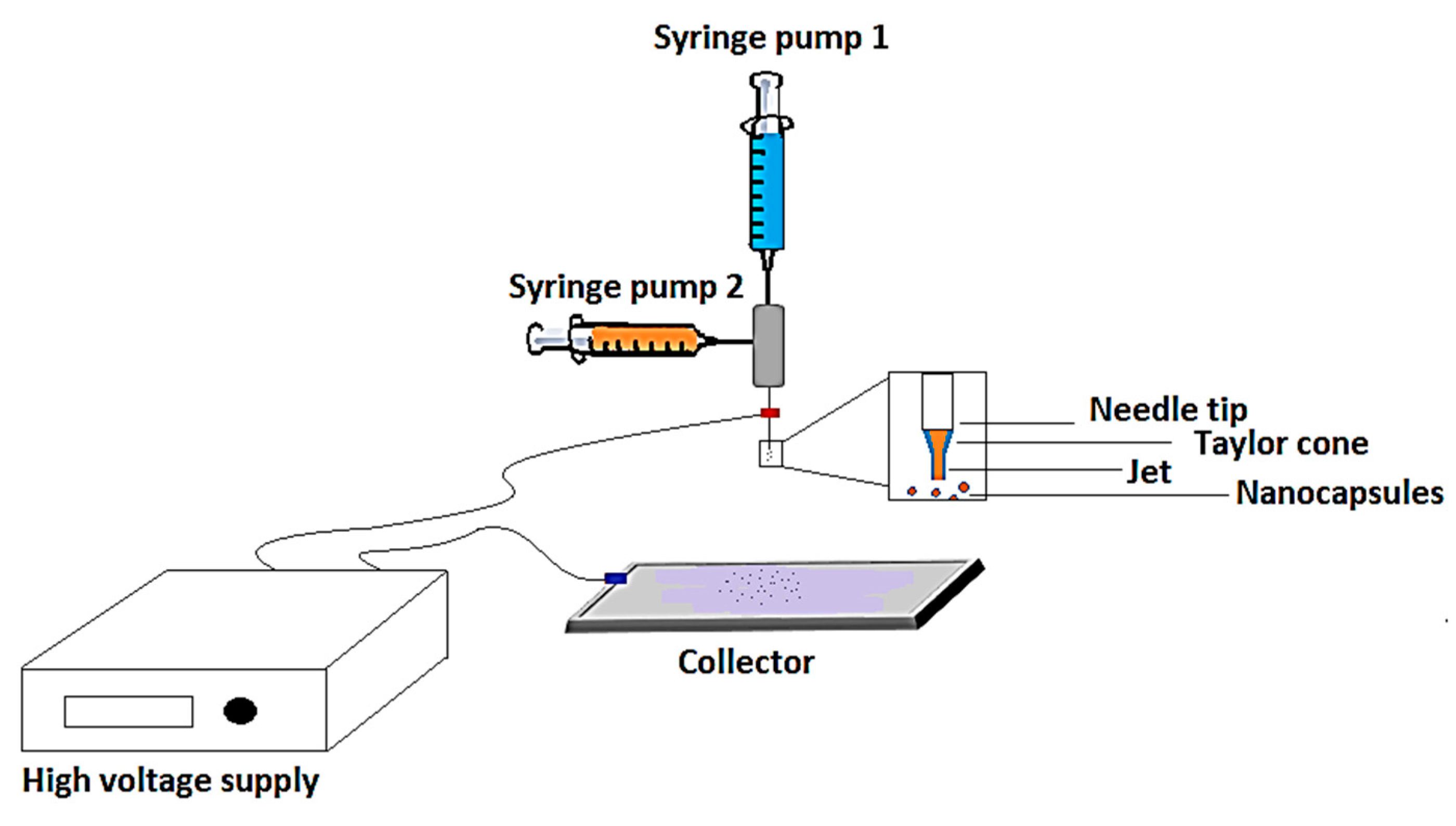
2.2. Encapsulation Process

Figure 2 schematically represents the employed coaxial electrospray setup for the encapsulation process. The spraying home-made nozzle consists of two concentric stainless steel needles: a core–shell needle with gauge 16 and an inner needle with gauge 24. The needle was charged to the cathode of high voltage supply and the anode was connected to the plate covered with aluminum foil (acting as a collector of the nanocapsules).
Coaxial electrospraying was carried out using SAN solution (5% w/v) at the outer needle (as a shell), while the CAR was filled in the inner needle. The blend of DCM:DMF with a ratio of 50:50 was used for the preparation of the SAN solution. The flow rate of the shell and the core solutions were changed from 0.5 to 0.7 mL/h and 0.04 to 0.06 mL/h, respectively. The applied high voltage was varied between 18 and 26 kV, and the distance between the tip of the needle and the collector was set at 15 cm.

2.3. Preparation of Self-Healing Coatings for Corrosion and Mechanical Tests

Epoxy resin was mixed with reactive diluent with a 3:1 weight ratio to reduce its viscosity. The produced nanocapsules were dispersed into diluted epoxy resin using mechanical stirring with a speed of 200 rpm for 5 min. Then, the curing agent was added with a proportion of 100:50 (mass ratio of epoxy to polyaminoamide). The nanocapsules’ content inside the prepared mixture was set at 1, 2, and 4 wt.%. Before applying the coating on the steel panels, their surface was polished with sandpaper and washed by acetone to eliminate any contaminants on the surfaces. The panels were coated by epoxy mixtures using a film applicator (Zehtner Zau 2000.80, Zurich, Switzerland) and the thickness of the coatings was controlled to 100 µm. The coatings were allowed to cure for seven days at room temperature. For the self-healing evaluations, the cured coatings were scratched using a scalpel blade. The scratched coatings were then kept at room temperature for seven days to enable the healing process of the coating. The control panels were prepared by the same procedure but without embedding any nanocapsules into the resin.

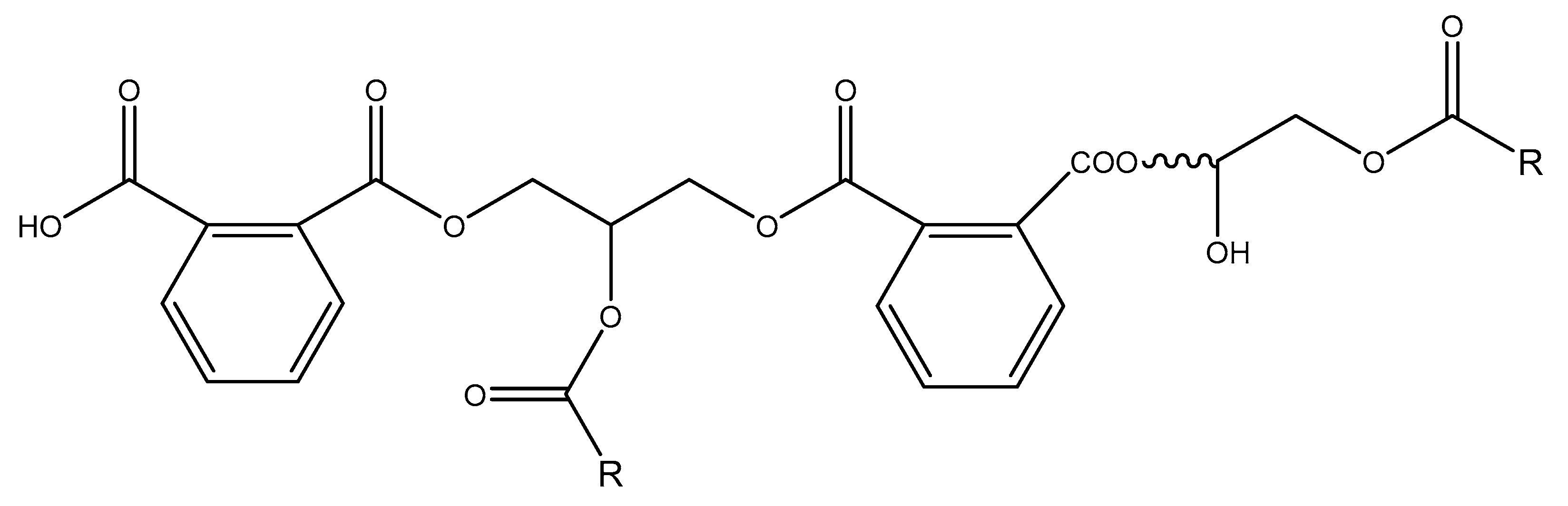
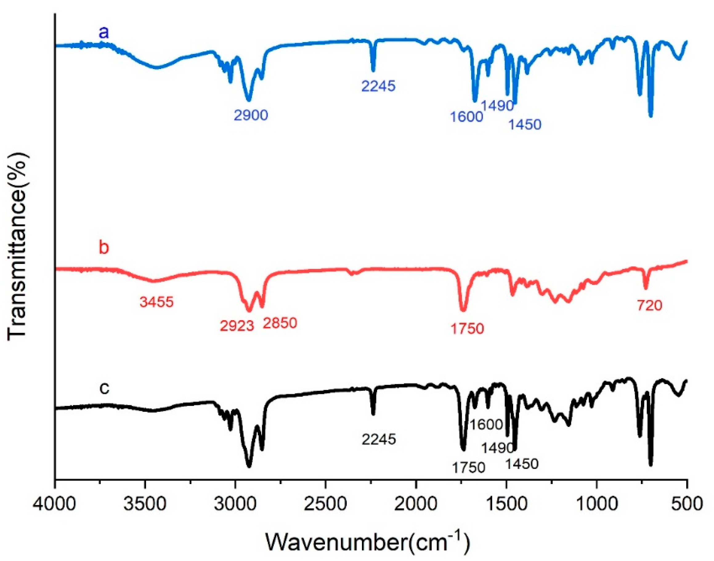
2.4. Chemical Structure Evaluation

The chemical structure of SAN, CAR, and nanocapsules were investigated separately using a FTIR spectrometer (WQF-510A, China), with 32 scans from 4000 to 500 cm−1 at the resolution of 4 cm−1. The samples of empty nanocapsules (only shell material) and those containing CAR were prepared by grinding with KBr, while, for the CAR sample, a thin layer of the CAR was spread onto the KBr pellet.

2.5. Evaluation of Encapsulation Yield and Core Content

To investigate the amount of core content in the prepared nanocapsules, the extraction of CAR from nanocapsules was performed according to previous research [15]. Therefore, a known weight of the nanocapsules was crushed with mortar and pestle. As a result of crushing, the shells of nanocapsules were broken and the core was subsequently washed by ethanol. After dissolving the core in the solvent, the shell was filtered, washed with ethanol several times and dried at 40 °C for 24 h in an oven. The practical percentage of the core-content (Wpractical) was calculated according to Equation (1).
% Core   content   ( W practical ) =   W ca W sh W ca × 100
where Wca refers to the weight of nanocapsules and Wsh refers to the weight of the shell [15].
The theoretical core content (Wtheoretical) can be also assessed by using the flow rates and densities of electrosprayed solutions according to [25]. Consequently, the yield of encapsulation can be obtained through Equation (2).
% Encapsulation   yield = W practical W theoretical × 100

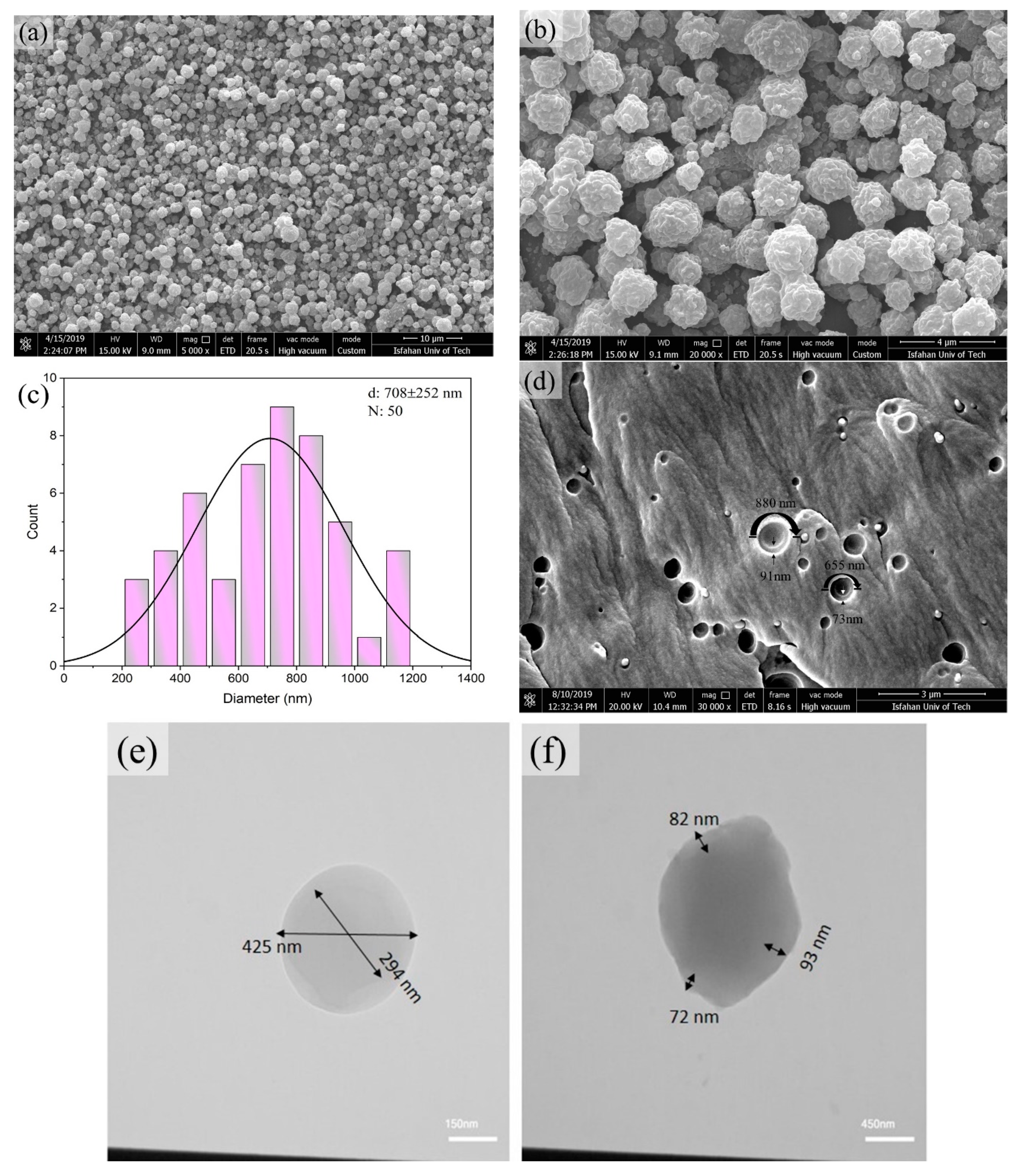
2.6. Morphological Studies

The surface morphology, shape, and size of the prepared nanocapsules were investigated by a FE-SEM (QUANTA FEG 450, Graz, Austria).
To confirm the successful encapsulation of the core by the shell, the thermodynamic behavior of polymer solution, shell, and CAR (core) should be assessed. In general, the tendency of a polymer phase to spread on a liquid or solid substrate for the formation of a shell layer can be explained by the spreading coefficient λij (Harkin’s equation [26]):
λij = γj − γi − γij
where γi, γj, and γij are the surface tensions of polymer phase and CAR, and the interfacial surface tension between the two phases, respectively. The Harkin’s equation is for describing the propagation of a liquid on a solid, and then the equation was extended for two immiscible solutions in a third immiscible phase [27]. Based on Harkin’s equation, the spreading of phase i on phase j will occur when the spreading coefficient is positive and when negative, the spreading is reversed [28]. To perform the calculation of the spreading coefficient in Equation (3), the interfacial surface tension can be approximated by employing the surface tension components directly (Equation (4)) [29]:
γ ij = ( γ i γ j ) 2
These evaluations are an approximation in equilibrium condition, which is not easily approachable in electrospraying, so there may be differences between experimental and theoretical results [30]. The surface tensions are measured using tensiometer (DCAT 11-Dataphysics, Filderstadt, Germany).
The transmission electron microscopy (DS-960A DSS, Zeiss, Germany), operating at 120 kV was employed to practically confirm the core–shell structure of the prepared nanocapsules. Therefore, the nanocapsules were electrosprayed onto a Lacey Formvar/carbon-coated copper grids.
Furthermore, the core–shell structure of capsules was studied through the investigation of the fractured capsules in the composite using FE-SEM. Therefore, the capsules were electrosprayed onto the epoxy resin and the composite was prepared by the addition of epoxy resin curing agent. The mixture was poured into a mold and cured at room temperature for three days. After three days, the composite was fractured in liquid nitrogen and was investigated by FE-SEM.

2.7. Thermal Stability Evaluations

The thermal stability of the prepared nanocapsules, CAR, and neat SAN, were analyzed using a thermogravimetric analyzer (Perkin Elmer STA 6000 TGA system, Waltham, MA, USA) under an argon atmosphere. For the evaluation of the thermal behavior of the prepared nanocapsules, they were firstly washed with methanol three times and then dried in a vacuum oven for 24 h at 40 °C. For all TGA tests, the heating rate was adjusted at 10 °C min−1 in the temperature range of 25 to 700 °C.

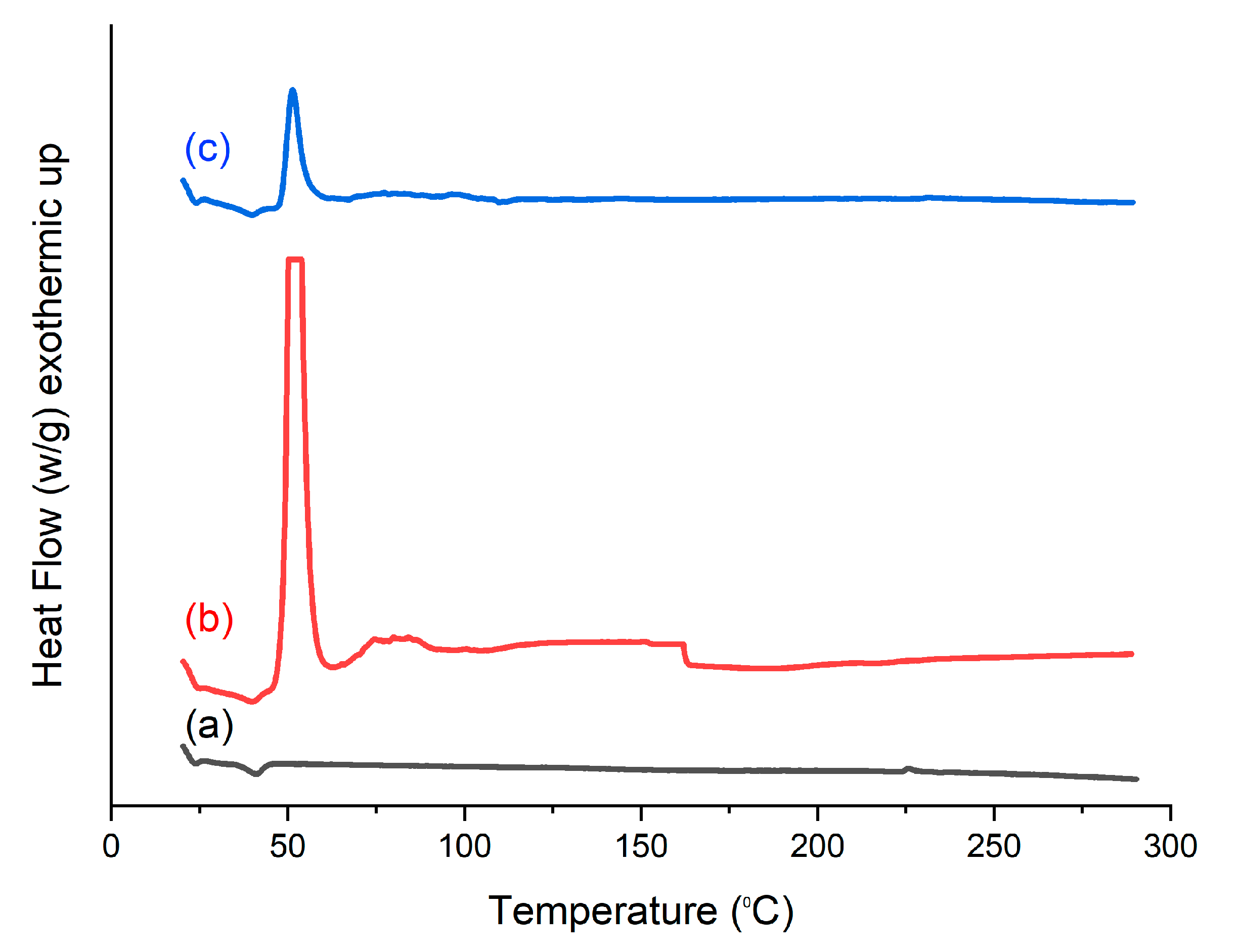
2.8. Evaluation of Healing Reaction Heat

In order to investigate the reactivity of the embedded core material (the healing agent) inside the nanocapsules with epoxy resin during any damages, DSC tests were evaluated using a BÄHR-DSC302 (Hüllhorst, Germany) from room temperature to 300 °C. The DSC tests were carried out under nitrogen atmosphere.

2.9. Evaluation of the Coating Properties

For investigating the effect of embedding nanocapsules on the adhesion strength of the coatings, pull-off adhesion tests were carried out according to ASTM D 4541 using a PosiTest AT-M from DeFelsko (Ogdensburg, NY, USA). Each sample was tested three times and the average of measurements was calculated and reported. In addition, the elongation at break of the coatings containing nanocapsules was evaluated through the bending tests according to ASTM D 522 test method A (conical mandrel bending tester SHEEN SH801-UK).

2.10. Evaluation of the Self-Healing Performance of the Coatings

To assess the anticorrosive and self-healing ability of the coatings, salt spray testes were accomplished on the scratched control coating and coatings containing nanocapsules according to ASTM B117. The total exposure time of the coatings was about 72 h. It should be noted that the scratched coatings were kept at room temperature for seven days before testing to provide enough time for the release of the healing agent and healing reaction. In addition, the self-healing ability of the coatings containing nanocapsules was evaluated in the NaCl and HCl solutions through the electrochemical measurements. The corrosion resistance of the coated substrates was determined by EIS tests using an Ivium–potentiostat (Ivium Technologies, Eindhoven, Netherland). A conventional three-electrode arrangement was utilized with a KCl saturated calomel electrode (SCE) as a reference electrode, a platinum sheet as a counter electrode and scratched coatings as the working electrode. Before electrochemical evaluation, the scratched coatings were kept in 3.5%wt. NaCl solution for 8 days separately to be stabilized at open circuit potential (OCP). The EIS measurements were carried out in a frequency range between 100 and 10 mHz with an amplitude of 10 mV rms−1. The results of the corrosion tests were estimated by extrapolating the polarization curve based on ASTM-G102-89. The obtained EIS data were analyzed in terms of electrical equivalents circuits utilizing the Z-View software. In addition, the EIS tests were also conducted on the scratched coatings immersed in a 0.5 M HCl solution, to evaluate the self-healing performance of the coatings in the acidic media.

3. Results and Discussion

3.1. The Electrospraying Process Observation

To achieve a stable cone-jet, the effective parameters in electrospinning or electrospraying process, namely, the concentration of the shell solution, the solvent characteristics, applied voltage, flow rates, and the distance between the needle tip and collector, should be optimized [31,32].
The concentration of the shell solution is important not only to control the viscosity, electrical conductivity, and interfacial tension of the solution but it also affects the stability of the tailor cone. It is difficult to obtain an intact shell with a low concentration of polymer and is challenging to achieve a stable cone-jet mode with high polymer concentration. It may also cause the formation of fibers instead of capsules, as shown in Figure S1 in the supplementary file. Therefore, the shell concentrations of 4, 5, and 6 wt.% were examined. The applied voltage also plays a critical role in the formation of core–shell capsules. The stable cone–jet can be obtained by changing the applied voltage. The dripping mode occurs when low voltage is applied, and, by applying high voltage, multi-jet occurs in the electrospray process [31]. Therefore, the applied voltages of 18 to 24 kV were examined to obtain the optimum applied voltage. Figure S2a, of the supplementary file, shows the FE-SEM image of the collector in dripping mode (low voltage), and Figure S2b shows the FE-SEM images of the prepared capsules in the multi-jet mode (high voltage). In addition, the inner and the outer flow rates in coaxial electrospray are the other effective factors for cone–jet stability and droplet size. A stable core–shell structure can be formed for a small range of inner/outer liquid flow rates [31]. Therefore, in this research, several feed rates were investigated, 0.5 to 1 mL.h−1 as a shell feed rate and 0.01 to 0.06 mL.h−1 as a core feed rate. Some of the unsuccessful encapsulations are shown through the FE-SEM images of Figure S3. Another important process parameter is the distance between the tip and the collector. At a constant voltage, a shorter distance is advantageous for generating a higher electrical field strength and consequently smaller particles are prepared. However, very short distance results in low solvent evaporation and, consequently, the coalescence and aggregation of wet particles at the collector. On the other hand, the long collector to tip distance would need higher applied voltage to compensate for the reduced electrical field strength. In general, one could expect to obtain larger particles compared to particles produced at a shorter collector to tip distance setup [33]. In this research, 13, 15, and 17 cm were examined as the distance of the needle to the collector. Figure S4 shows the FE-SEM images of the prepared capsules in two needle-to-collector distances. Using DMF as the solvent of shell material keeps the collector wet due to its slow evaporation rate, while DCM solvent leads to the formation of bigger particles (micro-scale) that is not appropriate for coating applications. Therefore, using a mixture of these solvents with equal amounts addressed the issue by optimizing solvent evaporation rate and electrical conductivity of the solution, which is reported in previous studies [34,35]. Figure S5 depicts the FE-SEM images of the prepared capsules when the neat DCM and DMF were employed as shell solvent.
Other effective parameters were optimized by changing a parameter and keeping the other constant. The parameters were chosen as optimum at those values where the electrospraying process is stable and led to the preparation of the nanocapsules in a spherical shape with the minimum average of the capsule’s diameter. The optimized parameters obtained were 22 kV, 0.6 and 0.05 mL/h, 15 cm, and 5 w/v % for applied voltage, flow rates of the shell and core solutions, the distance of needle and collector, and the concentration of the shell solution, respectively.

3.2. The Chemical Structure of the Prepared Capsules

Figure 3 shows the FTIR spectra of the neat shell material, the neat CAR, and crushed nanocapsules. The FTIR spectrum of neat SAN shows the characteristic peaks at 2237 and 1600 cm−1 corresponding to C≡N and C=C in styrene ring bending, respectively [24,36]. All the absorption of SAN can be observed in spectrum, such as 700, 2900, 3025 cm−1 for CH, 2852 cm−1 for CH2 and 540, 760, 1490 cm−1 for CH aromatic ring [37].
Characteristic bands of CAR are observed at 1740 cm−1 for ester groups and small twin peaks at 1604, and 1584 cm−1 corresponding to C=C stretching vibration of the aromatic ring originated from phthalate groups that formed the alkyd resin (as shown in the plausible structure of the alkyd resin in Figure 2). In addition, the aromatic C-H bending arising from this aromatic functional group appears at 720 cm−1 as a sharp peak. The broad stretching bands at 3455 cm−1 confirm the presence of free hydroxyl and carboxyl groups, while the peaks at 2850 and 2923 cm−1 are attributed to C-H aliphatic stretching. Peaks are also observed at 1008–1240 cm−1 for the C-O-C stretching of ester that supports the structure of CAR [15].
The FTIR spectrum of crushed nanocapsules shows all the characteristic peaks of neat SAN and CAR, while no more peak can be discerned. It can be concluded that both SAN and CAR are available in the prepared nanocapsules, considering no reaction occurred between them during the encapsulation process.
The practical core content of the prepared nanocapsules was measured to be 32 wt.% using the extraction method, while the theoretical core content was calculated to be 46 wt.% according to the feed rate of the shell and core solutions. Therefore, the encapsulation yield can be obtained from Equation (2) at 69%, confirming the high efficiency of the electrospray method in comparison to other researches with an encapsulation yield of 10–20% [38].

3.3. Morphological Studies

Figure 4a,b represent FE-SEM micrographs of the nanocapsules at two different magnifications. Spherical nanocapsules are clearly seen, which guarantee easy dispersion into the resins before applying coating [15]. In addition, the rough outer surface of the capsules provides enhanced adhesion to the resin and metallic substrate. The rough surface of capsules can be attributed to the phase change of CAR to solid during the encapsulation process. To investigate the mean diameter and size distribution of synthesized nanocapsules, ImageJ software was used and the average of 50 particles’ diameter was determined. Nanocapsules with a mean diameter of 708 ± 252 nm were found. Figure 4c shows the size distribution of the prepared nanocapsules. It can be seen that the coaxial electrospraying method led to the formation of nanocapsules with a wide range of size distribution. The capsules with a wide range in the size distribution showed great potential in self-healing performance due to covering and healing all sizes of cracks, from nano to microscale [39].
The fractured surface of the epoxy composite is shown in Figure 4d. According to Figure 4d, the core–sheath structure, along with a smooth inner layer of the nanocapsules, was formed, which confirms the FTIR results regarding the presence of CAR in nanocapsules without any reaction with SAN shell materials. Moreover, Figure 4d reveals an increased bonding of nanocapsules and the epoxy matrix, which is attributed to the high roughness of nanocapsules and the chemical reaction of unencapsulated CAR on the surfaces of the nanocapsules and epoxy matrix.
Figure 4e shows the TEM images of nanocapsules, where the dark and bright fields are observed in these images. It proves the formation of the core–shell structure of nanocapsules while the single-core structure of nanocapsules can be observed. According to the TEM and FE-SEM images of the fractured capsules, the thickness of shells is in the range of 35–95 nanometers, and, consequently, a scratch longer than 95 nm has the ability to break the capsules’ shell [40]. From these measurements, the volume fraction of the core was calculated to be in the range of 27–33%. Knowing the density of CAR and SAN solution (1.027 g/mL and 1.08 g/mL, respectively) led to the weight percentage of the core content in the range of 27% to 32% [40]. These values are in good agreement with the measured core content from the extraction method (32 wt.%).

Thermodynamic of the Encapsulation Process

In the coaxial electrospraying method, the surface tensions of the components are important factors for the successful encapsulation process [30]. The surface tensions of the core and shell materials were measured and listed in Table 1. According to Table 1 and Equations (3) and (4), the spreading coefficient was calculated to be 3.02 mN/m. The positive spreading coefficient proves the spreading of the shell solution on CAR and confirms the successful encapsulation of CAR within the SAN shell [30].

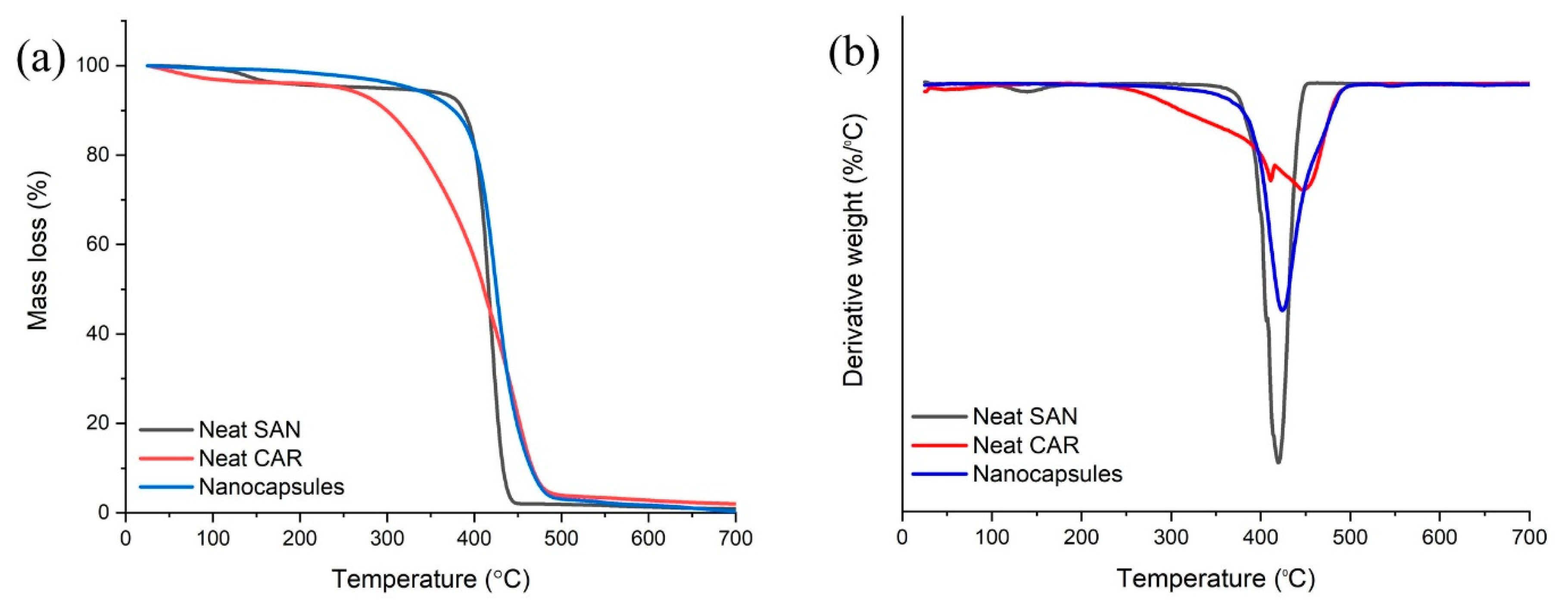
3.4. Thermal Stability of the Nanocapsules

TGA tests were carried out to investigate the thermal stability of the prepared capsules. Figure 5 represents the TGA and derivative thermogravimetric analysis (DTGA) diagrams of capsules containing the healing agent, neat SAN capsules (without core), and neat CAR. The TGA curve of neat SAN nanocapsules shows that the first weight loss (around 100 °C) of the neat SAN nanocapsules can be attributed to the evaporation of adsorbed moisture and residual solvent on the surface of the nanocapsules. There was a significant thermal decomposition event that initiated at 303 °C and continued till 452 °C. The thermal decomposition at 303 °C is associated with nitrile oligomerization, leading to the production of volatile products, e.g., NH3, HCN, CH3CN, etc., which are the components of acrylonitrile sections of the copolymer [41]. The CAR thermal decomposition occurs between the temperature range of 230–500 °C. The thermal decomposition of the core–shell nanocapsules started at 270 °C, contributed to the decomposition of the shell and continued up to 500 °C due to the decomposition of encapsulated CAR. It can be seen that the thermal decomposition curve of core–shell nanocapsules is contiguous with the mass loss curve of the shell (SAN) in the beginning and is adjacent to the mass loss curve of the core (CAR) at the end of the analysis. This proves the presence of both the components in the prepared capsules (Figure 5a). In addition, the temperature decomposition range reveals the high thermal stability of the produced nanocapsules.

3.5. Evaluation of the Reactivity of the Encapsulated Healing Agent

The heat flow and existence of an exothermic peak in the DSC diagram of epoxy and the healing agent was reported as evidence for the healing reaction [24,42]. Figure 6a represents the DSC diagram of epoxy and CAR mixture without any catalyst. The endothermic peak around 40 °C is attributed to the melting of CAR (this peak is also observed in two other diagrams). Figure 6b demonstrates that the DSC curve of epoxy and CAR resin, with the presence of HCl as a catalyst, shows a considerable exothermic peak at 54 °C, confirming the reaction between epoxy and CAR. In addition, Figure 6c represents the DSC diagram of the crushed epoxy composite containing 2 wt.% of nanocapsules in the presence of HCl. It can be discerned that this curve also shows an exothermic peak at 54 °C, which proves the healing process and the reactivity of the released healing agent from crushed nanocapsules.

3.6. Evaluation of the Coating’s Properties

The results of pull-off adhesion strength and elongation at break of the control sample and self-healing coatings are summarized in Table 2. Pull-off adhesion tests were carried out to evaluate the adhesion strength of coatings. According to the results, increasing the capsule content in the matrix led to a decrease in the adhesion strength of the coatings that can be attributed to the reduction in contact areas between the coating and its substrate [11,43]. Furthermore, elongation at break was measured for all of the samples thorough a conical mandrel bending test. The results revealed that the bending elongation at break decreased by increasing the nanocapsule content. This result can be due to the negative effect of adding nanocapsules on the adhesion strength of the coatings as well as their agglomeration at higher contents, which act as defects in the coating matrix [15].

3.7. Evaluation of the Self-Healing Ability of the Coatings

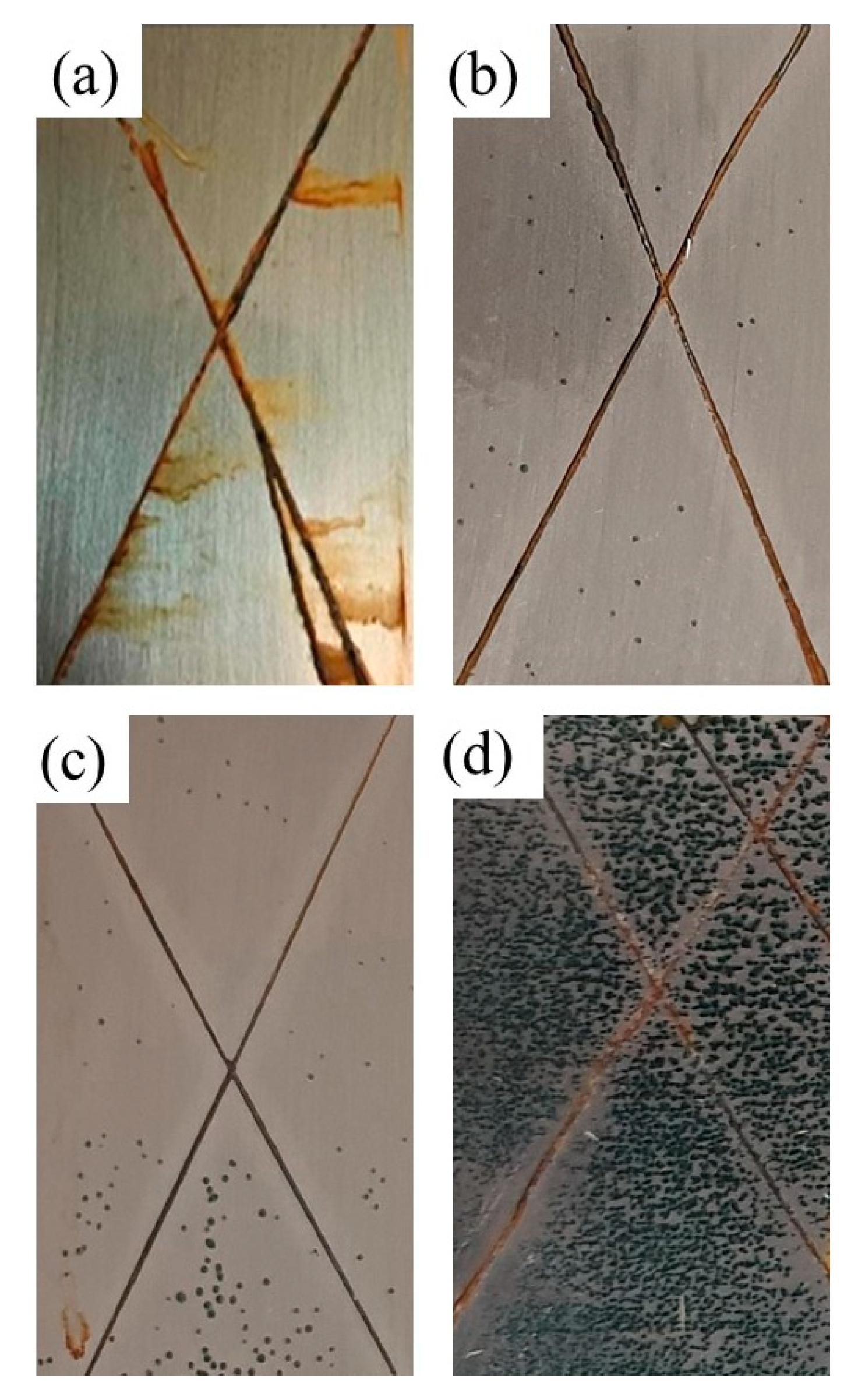
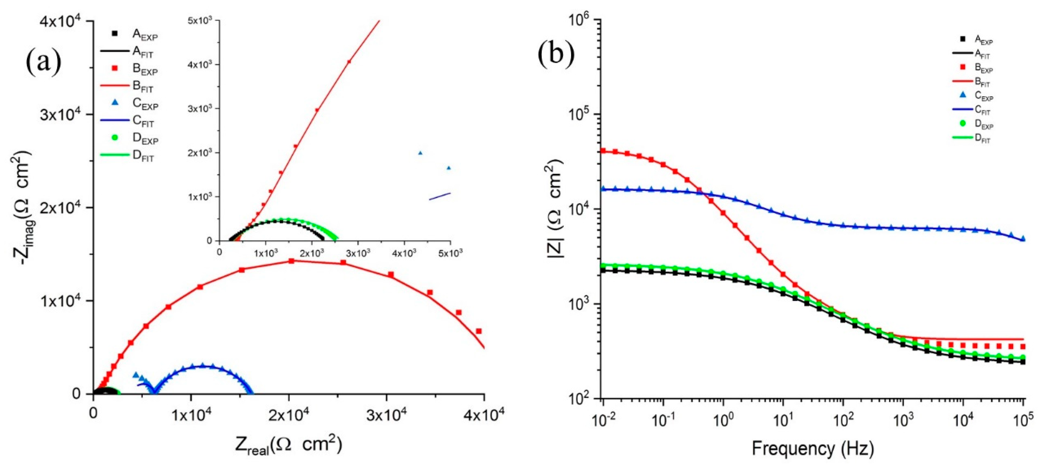
3.7.1. Salt Spray Tests

The results of the scratched coatings after 72 h of salt spray corrosion tests are presented in Figure 7. The control panel and the panel without capsules showed severe corrosion after exposure to the salt solution (Figure 7a). It can be seen in Figure 7 that with increasing the capsule content in the coatings, the corrosion in the scratch area was considerably decreased. This can be due to the healing reaction occurring when capsules rapture in the crack area. However, it can be seen that by increasing capsule content to 4 wt.%, the number of dark spots representing the porosity has increased in the matrix, due to capsule agglomeration [12]. According to Figure 7, the coating containing 2 wt.% of capsules showed the most efficient corrosion protection of the coating after scratch and exposure in the corrosive environment (5 wt.% NaCl solution). The anticorrosive property of this sample is because of the efficient healing reaction between epoxy and released CAR from capsules and the formation of a new film on the substrate [44].

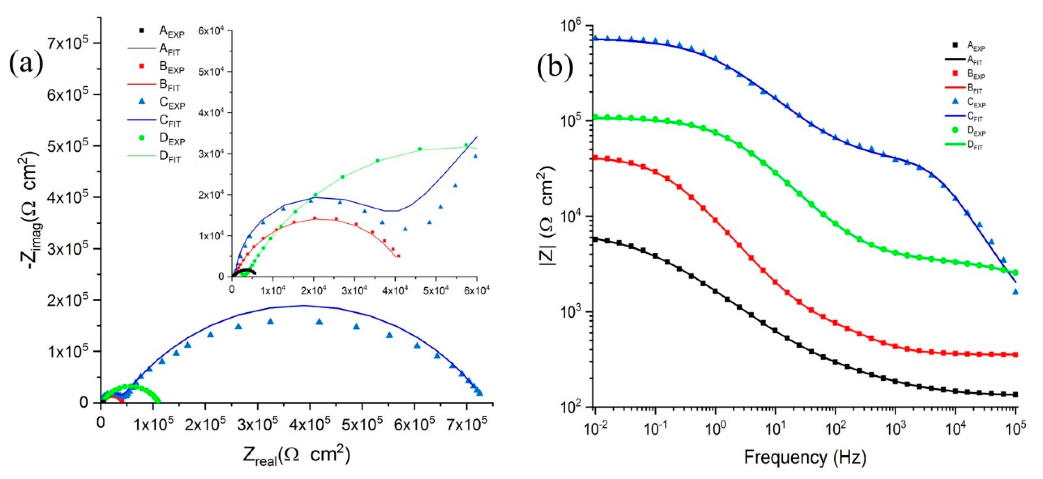
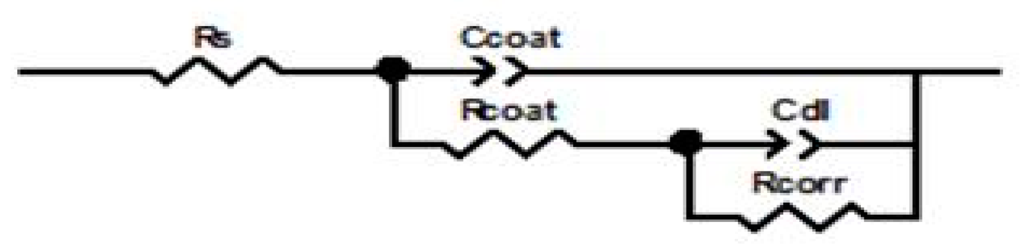
3.7.2. Evaluation of the Self-Healing Ability by EIS Tests in NaCl and HCl Solutions

The EIS measurements were carried out to assess the self-healing performance of the coatings. Figure 8 shows the Nyquist and bode diagrams for the scratched samples, which were immersed in 3.5 wt.% NaCl solution for 8 days. The corresponding fitted curves according to the proposed electrical equivalent circuit are also presented. Similarly, the same results for the coatings that were immersed in 0.5 M HCl solution are presented in Figure 9. Comparing the results for the samples immersed in the NaCl solution reveals that Sample C, the coating containing 2 wt.% of the nanocapsules, has the best corrosion resistance, while acidic media sample B showed the best self-healing performance. According to the EIS results, adding the capsules increased the corrosion resistance of scratched coatings for both the immersion solutions. However, increasing the nanocapsule content more than 2 and 1 wt.%, reduced the Nyquist semi-circle radii for the coatings immersed in NaCl and HCl solutions, respectively, indicating the reduction in the corrosion resistance of the samples.
Figure 10 shows the equivalent electrical circuit utilized to fit the EIS data. In this circuit, Rs, Rcoat and Rcorr are the solution resistance, the coating resistance, and charge transfer resistance, respectively. Table 3 represents the impedance data extracted from the equivalent electrical circuit of the samples immersed in the 3.5 wt.% NaCl solution. In addition, Table 4 summarizes those acquired for the samples immersed in the 0.5 M HCl solution. In Nyquist plots, the high-frequency time constant is attributed to the capacitive behavior of the epoxy coating containing nanocapsules. Whereas the low-frequency one can be related to the charge transfer at the interface of the metallic substrate and self-healing coating. According to Figure 8a, in the Nyquist data of sample C, there are two capacitive loops that are attributed to the adequately released healing agent for the self-healing process. In fact, there is a significant difference between the semi-circle radii of sample C and the other samples, indicating the effectiveness of the corrosion protection of the self-healing coating. When nanocapsules in the epoxy coating increased from 2 to 4 wt.%, the corrosion resistance clearly decreased due to smaller dimensions of the capacitive loop. This is due to the agglomeration of the nanocapsules in the epoxy coating leading to localized corrosion and penetration of the corrosive NaCl solution into the surface of the self-healing coated substrates. Generally, the agglomerated area provides a localized form of corrosion through some defects, such as holes, cavities, and cracks [45]. It can be seen that the radius of the capacitive loop increased when the self-healing nanocapsule was incorporated in the epoxy coating, proving the self-healing agent’s role in the protection of the metallic surface. Comparing the corrosion resistance, the coatings with various amounts of nanocapsules show that the corrosion resistance of the samples in the NaCl solution was significantly higher than those acquired for the samples immersed in the HCl solution. For example, Rcorr for the sample C in NaCl solution was 697,000 Ω cm2, which reduced to 10,100 Ω cm2 when it was immersed in the HCl solution.
In addition, Figure 8b and Figure 9b demonstrate the bode plots of the coatings immersed in NaCl, and HCl solution, respectively. It should be noted in bode plots that the impedance modulus |Z| at lower frequencies shows the total corrosion resistance of the utilized system and consequently the efficiency of the self-healing performance. These results are in good agreement with the Nyquist plots, according to the fact that samples C and B have the highest total impedance for NaCl and HCl immersion solutions, respectively. According to these results, it can be concluded that the released CAR was not enough to protect the scratched area for sample B in NaCl solution. However, for the HCl media, the corrosion resistance and barrier property of the coating are more sensitive to particle agglomeration and the adhesion of the coatings. In fact, the increase in the nanocapsules content decreased the adhesion of the coating and made it more susceptible to the penetration of the corrosive ions to the metallic substrate in the acidic media. On the other hand, the hydrogen ion possibly catalyzed the healing reaction between epoxy and CAR functional groups at the initial release of CAR. Since the healing reaction and healing solidification is accelerated, fewer healing agents were allowed to release. Consequently, the healing reaction in the acidic media is less sensitive to the available healing agent and is mostly controlled by the adhesion of the coatings to the metallic substrate. Therefore, for the scratched samples immersed in the HCl solution, the highest self-healing performance was observed for sample B. The healing reaction efficiency was measured to be 99% and 95% for the samples C (in NaCl solution) and B (in HCl solution), respectively, according to Equation (5) [46,47]:
%HE = (1 − Rcorr0/Rcorr) × 100
where Rcorr0 and Rcorr are the charge transfer resistances of the control and self-healing coatings, respectively.

4. Conclusions

In the present study, a CAR was encapsulated in SAN nanocapsules through the coaxial electrospraying for self-healing purposes. Morphological studies revealed that the uniform core–shell nanocapsules with a mean diameter of 708 ± 252 were prepared, while the core content and encapsulation yield were measured to be 32% and 69%, respectively. In addition, morphological studies revealed the formation of the rough outer surface, which made them suitable for better dispersion and enhanced self-healing performance, and a soft inner surface of the nanocapsules, confirming the successful encapsulation process without any reaction between shell and core materials. The FTIR and TGA test results confirmed the presence of both SAN and CAR in the prepared nanocapsules and the thermal stability of the capsules up to 270 °C. Furthermore, the DSC test results confirmed the reactivity of released healing agent from nanocapsules. The evaluation of the mechanical properties of the coatings showed that the incorporation of the nanocapsules decreased both the adhesion of the coating to the substrate and bending elongation of the coatings. The evaluation of the self-healing performance revealed that the coatings containing nanocapsules have the self-healing ability during the salt spray test. In addition, the EIS test results showed that the coating has the autonomous ability to heal in both NaCl and HCl media. The best self-healing performance occurred for the coating containing 2 wt.% nanocapsules in the NaCl solution with 99% healing efficiency. In contrast, for HCl media, the best self-healing performance was observed for the coating containing 1 wt.% of the nanocapsules with 95% healing efficiency.

Supplementary Materials

The following are available online at https://www.mdpi.com/2076-3417/10/9/3171/s1, Figure S1: FESEM images of the prepared capsules at 22 kV, 0.6 and 0.05 mL/h, and 15 cm of applied voltage, flow rates of the shell and core solutions, and the distance of needle to the collector, respectively at the shell solution concentration of (a) 4 w/v %; (b) 6 w/v %. Figure S2: FESEM images of the prepared capsules at 0.6 and 0.05 mL/h, 15 cm, and 5 w/v % of flow rates of the shell and core solutions, the distance of needle to the collector, and concentration of the shell solution, respectively at the applied voltage of (a) 20 kV (dripping mode) and (b) 24 kV (multi-jet mode). Figure S3: FESEM images of the prepared capsules at 22 kV, 15 cm, and 5 w/v % of the applied voltage, the distance of needle to the collector, and concentration of the shell solution, respectively. The flow rate of the core solution is 0.05 mL/h while the flow rate of shell solution is; (a) 0.5 mL/h; (b) 0.7 mL/h and. The flow rate of shell solution is 0.6 mL/h and the flow rate of core solution is; (c) 0.04 mL/h; (d) 0.06 mL/h. Figure S4: FESEM images of the prepared capsules at 22 kV, 0.6 and 0.05 mL/h, and 5 w/v % of the applied voltage, flow rates of the shell and core solutions, and concentration of the shell solution, respectively at the distance between needle to the collector of (a) 13 cm, and (b) 17 cm. Figure S5: FESEM images of the prepared nanocapsules at 22 kV, 0.6 and 0.05 mL/h, 15 cm, and 5 w/v % of the applied voltage, flow rates of the shell and core solutions, the distance of needle and collector, and concentration of the shell solution, respectively while the solvent was (a) DCM, and (b) DMF (wet collector and incapability of the process for encapsulating the healing agent).

Author Contributions

Conceptualization, R.M., S.N.K. and R.E.N.; methodology R.M., S.N.K., R.E.N., and M.S.K.; software, R.M., R.E.N., M.S.K.; validation, R.E.N., M.S.K., and O.D.; formal analysis, R.M., R.T.; investigation, R.M., and R.T.; resources, S.N.K.; data curation, R.E.N., M.S.K., R.T., and O.D.; writing—original draft preparation, R.M.; R.E.N., R.T. M.S.K., writing—review and editing, S.N.K., R.E.N., M.S.K., and O.D.; supervision, S.N.K., and R.E.N.; project administration, S.N.K.; funding acquisition, S.N.K. All authors have read and agreed to the published version of the manuscript.

Funding

This research received no external funding.

Conflicts of Interest

The authors declare no conflict of interest. The funders had no role in the design of the study; in the collection, analyses, or interpretation of data; in the writing of the manuscript, or in the decision to publish the results.

References

  1. Borisova, D.; Möhwald, H.; Shchukin, D.G. Mesoporous silica nanoparticles for active corrosion protection. ACS Nano 2011, 5, 1939–1946. [Google Scholar] [CrossRef] [PubMed]
  2. Ataei, S.; Khorasani, S.N.; Neisiany, R.E. Biofriendly vegetable oil healing agents used for developing self-healing coatings: A review. Prog. Org. Coat. 2019, 129, 77–95. [Google Scholar] [CrossRef]
  3. Zheludkevich, M. Self-Healing anticorrosion coatings. Self Heal. Mater. 2009. [Google Scholar] [CrossRef]
  4. D’Elia, F.M.; Magni, M.; Trasatti, P.M.S.; Schweizer, B.T.; Niederberger, M.; Caseri, W. Poly(phenylene methylene)-Based Coatings for Corrosion Protection: Replacement of Additives by Use of Copolymers. Appl. Sci. 2019, 9, 3551. [Google Scholar] [CrossRef] [Green Version]
  5. Esmaeely Neisiany, R.; Enayati, M.S.; Sajkiewicz, P.; Pahlevanneshan, Z.; Ramakrishna, S. Insight Into the Current Directions in Functionalized Nanocomposite Hydrogels. Front. Mater. 2020, 7, 25. [Google Scholar] [CrossRef] [Green Version]
  6. Sanka, R.V.S.P.; Krishnakumar, B.; Leterrier, Y.; Pandey, S.; Rana, S.; Michaud, V. Soft Self-Healing Nanocomposites. Front. Mater. 2019, 6, 137. [Google Scholar] [CrossRef]
  7. Guadagno, L.; Raimondo, M.; Naddeo, C.; Longo, P.; Mariconda, A.; Binder, W.H. Healing efficiency and dynamic mechanical properties of self-healing epoxy systems. Smart Mater. Struct. 2014, 23, 045001. [Google Scholar] [CrossRef]
  8. Wang, Y.; Li, Y.; Zhang, Z.; Zhao, H.; Zhang, Y. Repair Performance of Self-Healing Microcapsule/Epoxy Resin Insulating Composite to Physical Damage. Appl. Sci. 2019, 9, 4098. [Google Scholar] [CrossRef] [Green Version]
  9. Mirmohammad Sadeghi, S.A.; Borhani, S.; Zadhoush, A.; Dinari, M. Single nozzle electrospinning of encapsulated epoxy and mercaptan in PAN for self-Healing application. Polymer 2020, 186, 122007. [Google Scholar] [CrossRef]
  10. Cuvellier, A.; Torre-Muruzabal, A.; Van Assche, G.; De Clerck, K.; Rahier, H. Selection of healing agents for a vascular self-Healing application. Polym. Test. 2017, 62, 302–310. [Google Scholar] [CrossRef] [Green Version]
  11. Hatami Boura, S.; Peikari, M.; Ashrafi, A.; Samadzadeh, M. Self-Healing ability and adhesion strength of capsule embedded coatings—Micro and nano sized capsules containing linseed oil. Prog. Org. Coat. 2012, 75, 292–300. [Google Scholar] [CrossRef]
  12. Sun, J.; Wang, Y.; Li, N.; Tian, L. Tribological and anticorrosion behavior of self-Healing coating containing nanocapsules. Tribol. Int. 2019, 136, 332–341. [Google Scholar] [CrossRef]
  13. Nabuurs, T.; Baijards, R.; German, A. Alkyd-Acrylic hybrid systems for use as binders in waterborne paints. Prog. Org. Coat. 1996, 27, 163–172. [Google Scholar] [CrossRef]
  14. Azimi, A.; Yahya, R.; Gan, S.-N. Investigating effect of conventional and nano zinc pigments on air-drying property of palm-stearin-based alkyd resin paints. Int. J. Polym. Mater. Polym. Biomater. 2013, 62, 199–202. [Google Scholar] [CrossRef]
  15. Khorasani, S.N.; Ataei, S.; Neisiany, R.E. Microencapsulation of a coconut oil-based alkyd resin into poly (melamine–urea–formaldehyde) as shell for self-healing purposes. Prog. Org. Coat. 2017, 111, 99–106. [Google Scholar] [CrossRef]
  16. Shahabudin, N.; Yahya, R.; Gan, S. Microcapsules filled with a palm oil-based alkyd as healing agent for epoxy matrix. Polymers 2016, 8, 125. [Google Scholar] [CrossRef] [Green Version]
  17. Ataei, S.; Khorasani, S.N.; Torkaman, R.; Neisiany, R.E.; Koochaki, M.S. Self-healing performance of an epoxy coating containing microencapsulated alkyd resin based on coconut oil. Prog. Org. Coat. 2018, 120, 160–166. [Google Scholar] [CrossRef]
  18. Whalen, D.L.; Ross, A.M. Specific effects of chloride ion on epoxide hydrolysis. The pH-dependence of the rates and mechanisms for the hydrolysis of indene oxide. J. Am. Chem. Soc. 1976, 98, 7859–7861. [Google Scholar] [CrossRef]
  19. Choi, H.; Kim, K.Y.; Park, J.M. Encapsulation of aliphatic amines into nanoparticles for self-Healing corrosion protection of steel sheets. Prog. Org. Coat. 2013, 76, 1316–1324. [Google Scholar] [CrossRef]
  20. Li, Q.; Mishra, A.K.; Kim, N.H.; Kuila, T.; Lau, K.-T.; Lee, J.H. Effects of processing conditions of poly (methylmethacrylate) encapsulated liquid curing agent on the properties of self-Healing composites. Compos. Part B Eng. 2013, 49, 6–15. [Google Scholar] [CrossRef]
  21. Zamani, M.; Prabhakaran, M.P.; San Thian, E.; Ramakrishna, S. Protein encapsulated core–Shell structured particles prepared by coaxial electrospraying: Investigation on material and processing variables. Int. J. Pharm. 2014, 473, 134–143. [Google Scholar] [CrossRef] [PubMed]
  22. Xu, Q.; Qin, H.; Yin, Z.; Hua, J.; Pack, D.W.; Wang, C.-H. Coaxial electrohydrodynamic atomization process for production of polymeric composite microspheres. Chem. Eng. Sci. 2013, 104, 330–346. [Google Scholar] [CrossRef] [PubMed] [Green Version]
  23. Tapia-Hernández, J.A.; Rodríguez-Félix, F.; Katouzian, I. Nanocapsule formation by electrospraying. In Nanoencapsulation Technologies for the Food and Nutraceutical Industries; Elsevier: Amsterdam, The Netherlands, 2017; pp. 320–345. [Google Scholar]
  24. Neisiany, R.E.; Lee, J.K.Y.; Khorasani, S.N.; Ramakrishna, S. Towards the development of self-Healing carbon/epoxy composites with improved potential provided by efficient encapsulation of healing agents in core-shell nanofibers. Polym. Test. 2017, 62, 79–87. [Google Scholar] [CrossRef]
  25. Neisiany, R.E.; Khorasani, S.N.; Lee, J.K.Y.; Ramakrishna, S. Encapsulation of epoxy and amine curing agent in PAN nanofibers by coaxial electrospinning for self-Healing purposes. RSC Adv. 2016, 6, 70056–70063. [Google Scholar] [CrossRef]
  26. Sundberg, E.J.; Sundberg, D.C. Morphology development for three-component emulsion polymers: Theory and experiments. J. Appl. Polym. Sci. 1993, 47, 1277–1294. [Google Scholar] [CrossRef]
  27. Torza, S.; Mason, S. Three-Phase interactions in shear and electrical fields. J. Colloid Interface Sci. 1970, 33, 67–83. [Google Scholar] [CrossRef]
  28. Hobbs, S.; Dekkers, M.; Watkins, V. Effect of interfacial forces on polymer blend morphologies. Polymer 1988, 29, 1598–1602. [Google Scholar] [CrossRef]
  29. Pollauf, E.J.; Pack, D.W. Use of thermodynamic parameters for design of double-Walled microsphere fabrication methods. Biomaterials 2006, 27, 2898–2906. [Google Scholar] [CrossRef]
  30. Davoodi, P.; Feng, F.; Xu, Q.; Yan, W.-C.; Tong, Y.W.; Srinivasan, M.; Sharma, V.K.; Wang, C.-H. Coaxial electrohydrodynamic atomization: microparticles for drug delivery applications. J. Control. Release 2015, 205, 70–82. [Google Scholar] [CrossRef]
  31. Zhang, L.; Huang, J.; Si, T.; Xu, R.X. Coaxial electrospray of microparticles and nanoparticles for biomedical applications. Expert Rev. Med. Devices 2012, 9, 595–612. [Google Scholar] [CrossRef] [Green Version]
  32. Neisiany, R.E.; Enayati, M.S.; Kazemi-Beydokhti, A.; Das, O.; Ramakrishna, S. Multilayered Bio-Based Electrospun Membranes: A Potential Porous Media for Filtration Applications. Front. Mater. 2020, 7, 67. [Google Scholar] [CrossRef]
  33. Nguyen, D.N.; Clasen, C.; Van den Mooter, G. Pharmaceutical Applications of Electrospraying. J. Pharm. Sci. 2016, 105, 2601–2620. [Google Scholar] [CrossRef] [PubMed] [Green Version]
  34. Ho, H.; Lee, J. PEG/PLA core/shell particles from coaxial electrohydrodynamic spray drying. Macromol. Res. 2011, 19, 815–821. [Google Scholar] [CrossRef]
  35. Jing, Y.; Zhu, Y.; Yang, X.; Shen, J.; Li, C. Ultrasound-Triggered smart drug release from multifunctional core−Shell capsules one-Step fabricated by coaxial electrospray method. Langmuir 2010, 27, 1175–1180. [Google Scholar] [CrossRef]
  36. Mai, J.; Wang, L. Reaction mechanism of suspension graft copolymerization of styrene and acrylonitrile in the presence of ethylene propylene diene terpolymer. Polym. Chem. 2014, 5, 2118–2129. [Google Scholar] [CrossRef]
  37. Tan, W.; Radhi, M.; Ab Rahman, M.; Kassim, A. Synthesis and characterization of grafted polystyrene with acrylonitrile using gamma-Irradiation. J. Appl. Sci. 2010, 10, 139–144. [Google Scholar] [CrossRef] [Green Version]
  38. Huang, M.; Zhang, H.; Yang, J. Synthesis of organic silane microcapsules for self-Healing corrosion resistant polymer coatings. Corros. Sci. 2012, 65, 561–566. [Google Scholar] [CrossRef]
  39. Mirmohseni, A.; Akbari, M.; Najjar, R.; Hosseini, M. Self-Healing waterborne polyurethane coating by pH-Dependenat triggered-Release mechanism. J. Appl. Polym. Sci. 2019, 136, 47082. [Google Scholar] [CrossRef]
  40. Li, J.; Hu, Y.; Qiu, H.; Yang, G.; Zheng, S.; Yang, J. Coaxial electrospun fibres with graphene oxide/PAN shells for self-Healing waterborne polyurethane coatings. Prog. Org. Coat. 2019, 131, 227–231. [Google Scholar] [CrossRef]
  41. Igarashi, S.; Kambe, H. Thermogravimetric analysis of styrene-Acrylonitrile copolymer. Die Makromol. Chem. Macromol. Chem. Phys. 1964, 79, 180–188. [Google Scholar] [CrossRef]
  42. Zhang, X.-L.; Qiu, L.-F.; Ding, M.-Z.; Hu, N.; Zhang, F.; Zhou, R.-F.; Chen, X.-S.; Kita, H. Preparation of Zeolite T Membranes by a Two-Step Temperature Process for CO2 Separation. Ind. Eng. Chem. Res. 2013, 52, 16364–16374. [Google Scholar] [CrossRef]
  43. Samadzadeh, M.; Boura, S.H.; Peikari, M.; Ashrafi, A.; Kasiriha, M. Tung oil: An autonomous repairing agent for self-healing epoxy coatings. Prog. Org. Coat. 2011, 70, 383–387. [Google Scholar] [CrossRef]
  44. Tatiya, P.D.; Hedaoo, R.K.; Mahulikar, P.P.; Gite, V.V. Novel polyurea microcapsules using dendritic functional monomer: synthesis, characterization, and its use in self-Healing and anticorrosive polyurethane coatings. Ind. Eng. Chem. Res. 2013, 52, 1562–1570. [Google Scholar] [CrossRef]
  45. Torkaman, R.; Darvishi, S.; Jokar, M.; Kharaziha, M.; Karbasi, M. Electrochemical and in vitro bioactivity of nanocomposite gelatin-forsterite coatings on AISI 316 L stainless steel. Prog. Org. Coat. 2017, 103, 40–47. [Google Scholar] [CrossRef]
  46. Atta, A.M.; El-Azabawy, O.E.; Ismail, H.; Hegazy, M. Novel dispersed magnetite core–shell nanogel polymers as corrosion inhibitors for carbon steel in acidic medium. Corros. Sci. 2011, 53, 1680–1689. [Google Scholar] [CrossRef]
  47. Koochaki, M.S.; Khorasani, S.N.; Neisiany, R.E.; Ashrafi, A.; Magni, M.; Trasatti, S.P. Facile strategy toward the development of a self-Healing coating by electrospray method. Mater. Res. Express 2019, 6, 116444. [Google Scholar] [CrossRef]
Figure 1. Probable structure of alkyd resin (fatty acid in triglyceride oil is shown by R).
Applsci 10 03171 g001
Figure 2. Schematic illustration of the employed coaxial electrospraying setup.
Applsci 10 03171 g002
Figure 3. FTIR spectra of (a) neat SAN nanocapsules; (b) neat CAR; (c) and crushed nanocapsules obtained from coaxial electrospraying.
Applsci 10 03171 g003
Figure 4. FE-SEM micrographs of the prepared nanocapsules at: (a) 5000× and (b) 20,000× magnification; (c) the size distribution of nanocapsules; (d) FE-SEM image of the fractured capsules in the epoxy composites; and (e,f) TEM images of the prepared nanocapsules.
Applsci 10 03171 g004
Figure 5. (a) TGA and (b) DTGA curves of neat SAN, neat CAR, and the prepared nanocapsules.
Applsci 10 03171 g005
Figure 6. DSC diagrams of (a) epoxy and CAR in the absence of any catalyst; (b) a mixture of epoxy resin and CAR in the presence of HCl; and (c) a crushed epoxy composite containing nanocapsules with HCl.
Applsci 10 03171 g006
Figure 7. Images of the scratched coatings after salt spray test: (a) Sample A; (b) Sample B; (c) Sample C; (d) Sample D.
Applsci 10 03171 g007
Figure 8. (a) Nyquist and (b) bode diagrams of the scratched samples immersed in the NaCl solution for 8 days.
Applsci 10 03171 g008
Figure 9. (a) Nyquist and (b) bode diagrams of the scratched samples immersed in the HCl solution for 8 days.
Applsci 10 03171 g009
Figure 10. Employed electrical equivalent circuit for the fitting of the Nyquist and bode curves.
Applsci 10 03171 g010
Table 1. Surface tension of core and shell materials.
MaterialSurface Tension (mN/m)
5 wt.% SAN solution in an equal ratio of DMF and DCM (shell)30.8
CAR (core)33.9
The interfacial tension between CAR and the 5% SAN solution (calculated from Equation (4))0.074
Spreading coefficient3.02 > 0
Table 2. Impact of nanocapsules content on the properties of the coatings.
Sample CodeNanocapsules Content (wt.%)Adhesion Strength (MPa)Total Bending Elongation (%)
A02.5924.8
B12.1119.8
C22.1019.6
D41.4318.8
Table 3. Electrochemical impedance spectroscopy (EIS) parameters for the scratched coatings and immersed in the 3.5 wt.% NaCl.
Sample CodeRs (Ω cm2)CPEcoat-T (sn Ω−1 cm−2)CPEcoat-pRcoat (Ω cm2)CPEdl-T (sn Ω−1 cm−2)CPEdl-PRcorr (Ω cm2)
A129.601.05×10−45.44×10−1276.21.03×10−45.64×10−16.62×103
B353.606.89×10−67.92×10−16111.94×10−57.38×10−14.19×104
C847.301.20×10−99.77×10−13.77×1044.44×10−76.32×10−16.97×105
D875.401.03×10−75.92×10−12.69×1031.75×10−66.97×10−11.05×105
Table 4. The EIS parameters for the scratched coatings and immersed in the 0.5 M HCl.
Sample CodeRs (Ω cm2)CPEcoat-T (sn Ω−1 cm−2)CPEcoat-pRcoat (Ω cm2)CPEdl-T (sn Ω−1 cm−2)CPEdl-PRcorr (Ω cm2)
A2.43×1021.14×10−99.04×10−1234.106.38×10−55.15×10−12.05×103
B421.201.63×10−99.85×10−1476.602.00×10−57.51×10−14.07×104
C4.00×1031.45×10−99.90×10−12.21×1031.51×10−56.90×10−11.01×104
D25.612.95×10−109.90×10−1227.806.41×10−54.97×10−12.38×103

