1. Introduction
Executive function (EF) refers to a set of cognitive skills necessary for individuals to consciously regulate their thoughts, behaviors, and emotions to achieve specific goals (
Zelazo et al., 2016). It comprises three subcomponents: inhibitory control (IC), working memory (WM), and cognitive flexibility (CF) (
Lehto et al., 2003;
Miyake et al., 2000). IC refers to an individual’s ability to demonstrate self-control, engage in more adaptive behaviors, and avoid impulsive responses (
Tominey & McClelland, 2016). WM denotes an individual’s capacity to retain and select relevant information while manipulating it (
Baddeley, 2010). CF signifies an individual’s ability to shift perspectives or consider multiple viewpoints when necessary (
Dajani & Uddin, 2015). These psychological skills promote the organization and control of goal-directed behavior (
Best, 2010), jointly influencing an individual’s attention, emotional, and physiological responses to stimuli (
Blair & Raver, 2014), thereby supporting more effective learning (
Diamond, 2013). Research indicates that the development of EFs during the preschool years correlates with language and mathematical abilities (
Blair & Razza, 2007;
Bull et al., 2008;
Diamond & Lee, 2011;
Verdine et al., 2014), long-term physical and mental health (
Diamond & Lee, 2011), and positive social relationships (
Diamond, 2012). Early EF development provides children with significant advantages at school entry (
Blair & Razza, 2007), promotes subsequent academic achievement (
Raver et al., 2011), and yields more pronounced effects among disadvantaged groups with socio-economic disparities (
Fitzpatrick et al., 2014).
Physical activity, as an economical, accessible, and child-friendly educational intervention, has been widely applied in the field of cognitive development (
Carson et al., 2016;
J.-Y. Zhang et al., 2022). Numerous studies reveal that physical activity positively influences cognitive abilities, including executive functions (
Pindus et al., 2016). However, not all physical activities yield equivalent cognitive benefits (
Tomporowski et al., 2015). Differences in exercise intensity, intervention duration, and activity type result in varying effects on executive function improvement (
J. Li et al., 2025;
O’Callaghan et al., 2024). Regarding exercise intensity, moderate-intensity acute physical activity significantly enhances children’s executive function more effectively than other intensities (
Ji et al., 2019). Moreover, the relationship between cognitive performance and exercise intensity after acute physical activity follows an inverted U-shaped curve (
Kamijo et al., 2004). Regarding intervention duration, both acute and long-term physical activity effectively enhance EF in children and adolescents (
Liu et al., 2020). With respect to potential mechanisms, acute physical activity may enhance EF by increasing physiological arousal and optimizing the allocation of cognitive resources (
Tomporowski, 2003), by transiently elevating catecholaminergic neurotransmitters such as dopamine and epinephrine (
McMorris, 2016), and by upregulating brain-derived neurotrophic factor in central and peripheral circulation (
Loprinzi et al., 2013). Furthermore, a study has demonstrated that acute physical activity lasting 20 min yields significantly greater cognitive benefits than 10 min or 45 min sessions (
Chang et al., 2015). Concerning exercise modality, the level of cognitive engagement incorporated into physical activity has garnered extensive attention (
Tomporowski et al., 2015). Appropriate cognitive stimulation can enhance EF by modulating physiological and psychological arousal levels (
Pesce, 2012). Physical activities incorporating cognitive engagement elements offer greater advantages than simple repetitive activities, yielding additional cognitive benefits (
Preston et al., 2025). Cognitively engaging physical activity (CEPA) incorporates conditions such as situational interference, mental control, and exploration/discovery into physical activities. It requires participants to allocate attention resources and cognitive effort appropriately within challenging and complex environments (
Kolovelonis et al., 2022b). As a form of physical activity intervention, it is characterized by its operationalizability of cognitive engagement and high interest in physical activity contexts, making it particularly suitable for preschool children. Multiple studies indicate that CEPA effectively promotes cognitive development, attention, and EF in children and adolescents (
Jia et al., 2021;
Kolovelonis & Goudas, 2023;
Vazou & Mavilidi, 2021). Existing research confirms that acute physical activity interventions involving cognitive demands can produce immediate positive effects on young children’s performance in motor inhibition tasks (
Stein et al., 2017). Further studies indicate that short-duration physical activities with moderate cognitive engagement yield more significant improvements in EF and self-regulation compared to activities with high or low cognitive demands (
Ureña et al., 2020). Although existing research confirms the effectiveness of acute physical activity and cognitive engagement in enhancing EF, evidence regarding the impact of cognitively engaged physical activity on preschool children’s EF remains limited and inconclusive. Compared to conventional physical activity, the intervention effects and underlying mechanisms of such activities require further clarification.
The prefrontal cortex (PFC) plays a central role in cognitive control and EF. Particularly during the cognitive development of children and adolescents, the maturation of its structure and function is closely associated with improvements in cognitive abilities (
Casey et al., 2018;
Friedman & Robbins, 2022;
Luna et al., 2015). Preschool children exhibit significant activation in prefrontal regions during executive function tasks (
Moriguchi & Hiraki, 2013). As a higher-order association cortex within the frontal lobe, the dorsolateral prefrontal cortex (DLPFC) occupies the anterior portion of the middle and superior frontal gyri, adjacent to the posterior part of the precentral gyrus. It corresponds roughly to Brodmann areas 9 and 46 and is mainly responsible for maintaining and updating WM, controlling attention, monitoring conflicts, and carrying out goal-directed behaviors. It is recognized as one of the core structures within the cognitive control system (
Miller & Cohen, 2001;
Niendam et al., 2012). The frontopolar cortex (FPC) is the most anterior region of the PFC, located at the foremost position of the frontal lobe within Brodmann area 10. It plays a pivotal role in regulating higher-order cognitive processes (
Friedman & Robbins, 2022).
Currently, functional near-infrared spectroscopy (fNIRS) serves as a non-invasive brain imaging technique. By attaching optical probes to the tissue surface to emit and receive near-infrared light (650–1000 nm), it measures concentration changes in oxyhemoglobin (HbO
2) and deoxyhemoglobin (HHb), thereby reflecting hemodynamic alterations in the tissue. This method is commonly employed to examine cortical activation during cognitive tasks and infer underlying neural activity in the brain (
Herold et al., 2018;
Huppert et al., 2006). Its portability and low sensitivity to motion artifacts make it suitable for preschool children studies (
Zhan et al., 2024). Research has confirmed that acute physical activity activates physiological arousal systems, enhancing prefrontal cortical activation associated with cognitive tasks and thereby improving EF (
Byun et al., 2014). Different levels of cognitive engagement also exert distinct effects on cortical neural activity. Compared to monotonous movements, physical activities involving moderate physical or cognitive demands more effectively elevate oxygenated hemoglobin concentration in the PFC (
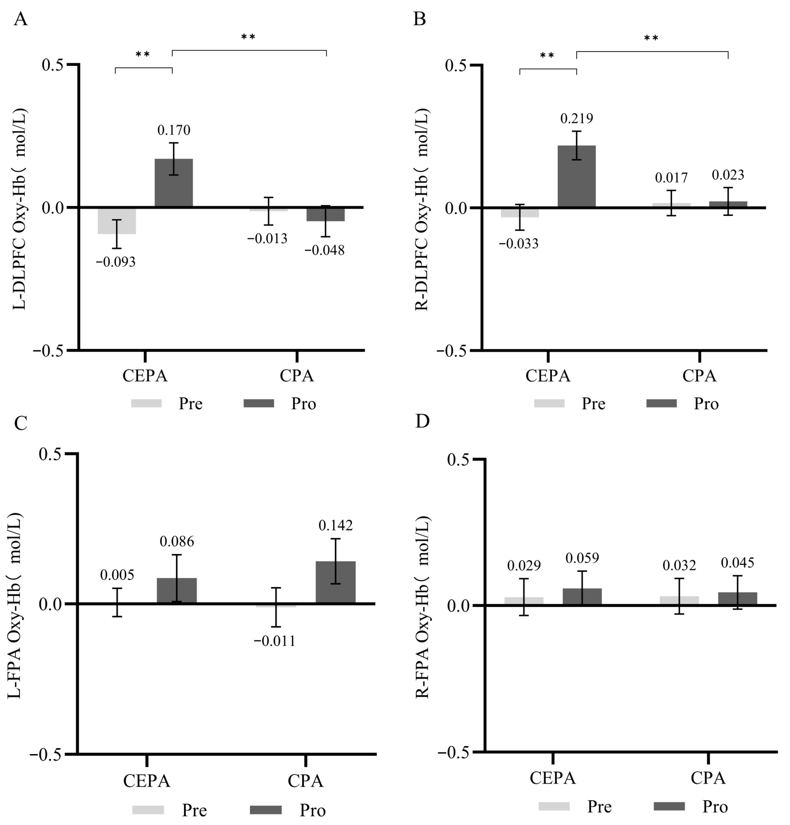
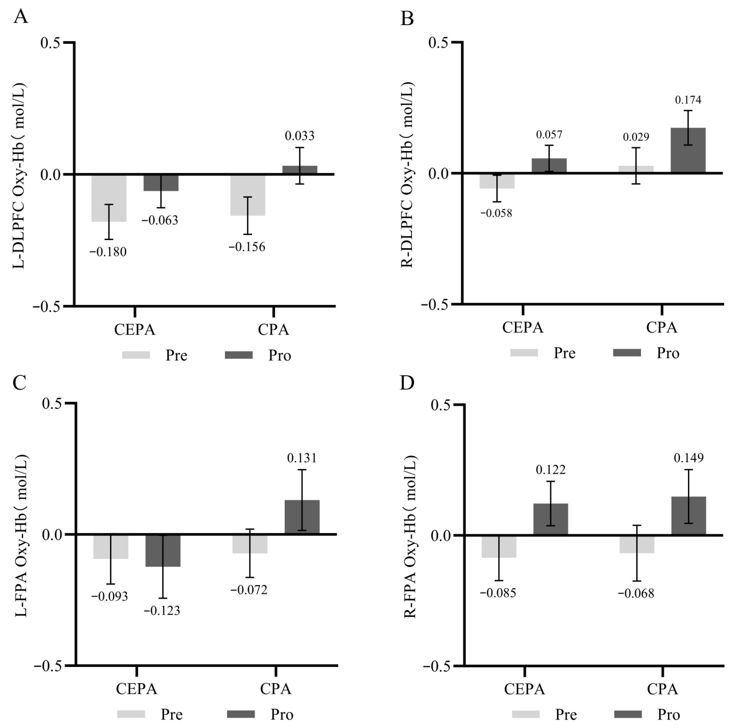
Naito et al., 2024). Currently, fNIRS technology is widely applied to monitor prefrontal activation states and analyze the mechanisms underlying effects before and after interventions, effectively elucidating the benefits of different intervention approaches on cognitive performance enhancement and their corresponding neural activity characteristics. However, the mechanisms underlying changes in cerebral blood flow within the PFC during executive function tasks in preschool children following acute cognitively engaged physical activity interventions remain unclear. Therefore, this study employs fNIRS to compare pre- and post-intervention activation patterns in the PFC. It aims to deepen understanding of the neural mechanisms underlying acute cognitive engagement during physical activity interventions that enhance preschoolers’ EF performance. This research will enrich the theoretical foundation in this field, broaden perspectives on improving preschoolers’ cognitive performance, and provide scientific evidence and practical pathways for designing and implementing cognitively engaging physical activity intervention programs.
In conclusion, cognitively engaged physical activity has surfaced as a promising intervention for preserving and augmenting individual cognitive function. However, the relationship between such activities and EF benefits, along with their underlying mechanisms, remains unclear. This study employs fNIRS technology to investigate the behavioral benefits of EF improvement before and after physical activity interventions with varying levels of cognitive engagement, as well as the characteristic changes in oxygenated hemoglobin concentration in the PFC. Specifically, this study examines (1) whether cognitively engaged physical activity yields greater improvements in EF compared to conventional physical activity among preschool children; (2) the patterns and differences in changes in PFC HbO2 concentration in preschool children before and after conventional and cognitively engaged physical activity interventions; and (3) the relationship between changes in behavioral measures of EF and cerebral blood flow in preschool children before and after physical activity interventions.
2. Materials and Methods
2.1. Trial Design
This study employed a parallel group design using a 2 (group) × 2 (test time) mixed-design experimental setup to investigate the acute effects of cognitive engagement factors on EF. All participating children were required to visit the experimental site twice: the first visit involved basic information collection, testing, and learning the intervention content; the second visit involved completing the physical activity intervention and data collection.
2.2. Procedures
2.2.1. Testing Sites
The subjects for this experiment were drawn from two kindergartens under the same educational group in Xi’an City, Shaanxi Province, China. Both kindergartens were comparable in terms of geographical location, facility scale, teacher qualifications, and curriculum design. The experimental intervention sites were established in open physical activity areas within two kindergartens, featuring similar environmental conditions in terms of size and layout. To meet fNIRS data collection requirements as closely as possible, the data acquisition site was set up in a quiet, dimly lit, independent room within the kindergarten.
2.2.2. Eligibility Criteria
A total of 64 preschool children were selected based on inclusion criteria: children in the senior kindergarten class, right-handedness, normal vision or corrected vision, normal intellectual development, no mental disorders, and no health conditions affecting participation in physical activities. All intervention implementers were master’s degree candidates from the School of Physical Education at Shaanxi Normal University. They received at least one day of formal training provided by the experiment designers and conducted one simulated intervention prior to the formal experiment.
2.2.3. Randomization
The randomization list was prepared by a researcher who was not involved in data collection or intervention implementation, and the group assignments were implemented after the first visit. Stratified random sampling with proportional allocation was conducted at the kindergarten level to recruit participants, ensuring that the number of children from each kindergarten was proportional to its size. Then, all 64 eligible participants were randomly assigned to either the cognitively engaging physical activity group (n = 32) or the conventional physical activity group (n = 32) using the RANDBETWEEN(1,2) function in Microsoft Excel to generate random allocation codes.
2.2.4. Ethical Considerations
The Academic Committee of Shaanxi Normal University has authorized this study (Scientific Ethics Review Number: 202416060). All parents or legal guardians comprehended the research protocols and provided written informed consent prior to their children’s involvement. Moreover, the testing procedure meticulously adhered to the personal choices of the participating children, who retained the autonomy to resign from the study at any moment.
2.3. Intervention and Comparator
The physical activity intervention in this study was of moderate intensity, defined as 64% to 95% of maximum heart rate (HRmax). The formula for calculating maximum heart rate is 220 minus age. During the experiment, children’s heart rates were monitored using the Domor portable heart rate monitor (
American College of Sports Medicine, 2013). Each child received the intervention individually and completed the tasks at each station in sequence for three cycles, with each intervention session lasting 20 min. To control for teaching style effects, the intervention was delivered in a rotating stations format, with teachers providing only necessary prompts based on game rules. The balance beams, mats, toddler soccer balls, sandbags, horizontal bars, and other materials required for the intervention are all standard physical education equipment commonly used in kindergartens. To control for potential interference from the kindergarten curriculum on intervention effectiveness, all participants completed the tests between 9:30 AM and 11:30 AM.
Cognitively engaging physical activity involves elements such as situational interference, mental control, and discovery. Participating children were required to complete games based on fundamental motor skills and engage in physical activity within the game’s rules. The balance and stability motor skills segment required children to mimic static poses of different animals, locate and sequentially walk across balance beams of varying colors, and perform forward rolls toward directions randomly indicated by the instructor. The operational motor skills segment required children to dribble a ball with their feet while navigating around obstacles and throw soft beanbags toward a grid 3 m away, aiming to form a straight line. Mobility motor skills involved sideways crawling and stepping over bars of varying heights to reach the finish line; displaying red, yellow, and green signs during running to indicate stop, slow down, and normal running, respectively; and performing frog jumps in randomly indicated directions. Additional rules during the intervention required children to innovate movements and methods during play, respond promptly to unpredictable situations, and continuously adjust behavioral strategies.
Conventional physical activity similarly employed moderate-intensity activities incorporating basic motor skills, but communication with children involved only simple encouragement and instructions, excluding game rules with cognitive engagement elements. The balance and stability motor skills section required children to perform single- and double-leg static stands, balance beam walking, and forward rolls on triangular mats. The operational motor skills section required children to dribble in a straight line and throw soft beanbags at distant targets. The locomotor motor skills segment required children to perform lateral walking, running at a constant speed around the field, and forward frog jumps. Compared to cognitively engaged physical activity, rules involving cognitive participation were reduced, but identical equipment specifications and consistent station sequences were maintained. The intervention content is detailed in
Table 1.
2.4. Outcomes
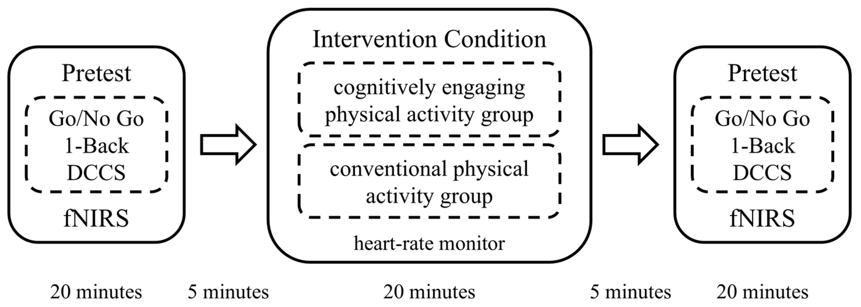
Prior to the intervention, each participant completed three EF cognitive tasks in a consistent order while concurrently undergoing fNIRS data collection. Subsequently, participants individually engaged in either a 20 min cognitively engaging physical activity intervention or a conventional physical activity intervention, with real-time heart rate monitored during the activity using a Domor portable heart rate monitor. After the exercise session, participants repeated the EF cognitive task tests and fNIRS data collection in the same order as the pre-test. Interveners are responsible only for their assigned group’s testing, with no interference between groups. Test administrators were unaware of participants’ intervention group assignments. The experimental procedure is illustrated in
Figure 1.
2.4.1. Executive Function
All EF tests were programmed using the PsychToolBox (PTB) toolkit in MATLAB. IC was assessed using a Go/No-Go task paradigm; WM was evaluated using a 1-back task paradigm; and CF was measured using a dimensional shift card sorting task. Prior to each task, participants received training to ensure comprehension of the task rules. During the testing phase, participants were instructed to remain seated and fixate on a “+” displayed on the screen during rest periods. Responses were recorded via keyboard operation when stimuli appeared on the computer screen, including accuracy and reaction time. Task presentation and behavioral data collection were performed using E-Prime 3.0 software.
The Go/No-Go cognitive task, adapted from a classic research paradigm, is widely used to measure IC in children (
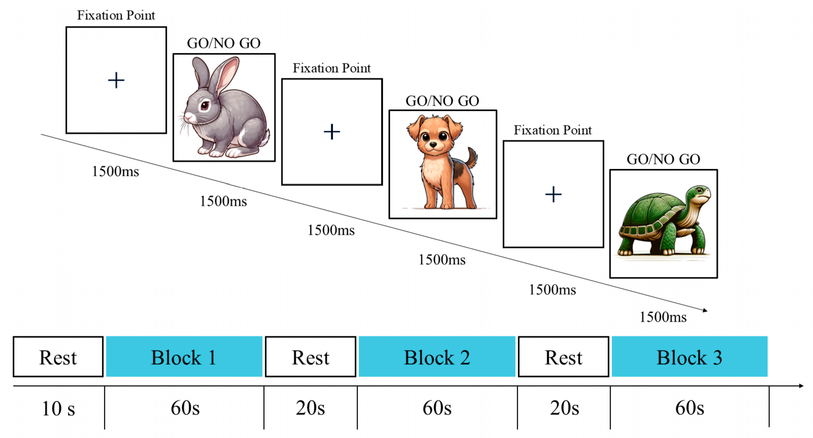
Howard & Melhuish, 2017). The Go/No-Go cognitive task paradigm in this study comprised three block sets. To minimize interference from cognitive processes on response inhibition, each block set randomly distributed 10 Go trails and 10 No-Go trails (
Figure 2) to assess non-selective response inhibition (
Masharipov et al., 2022). Stimuli comprised animal images familiar to preschoolers. During each trial, an animal stimulus (turtle, rabbit, sheep, or dog) randomly appeared at the screen center for 1500 ms. Participants were instructed to press the spacebar immediately upon seeing any animal except the puppy (the no-go stimulus). No response was required when the puppy image appeared. To minimize potential practice effects, all subjects learned the test rules and completed 8 practice trials before the formal task. The testing procedure is illustrated in
Figure 2.
The 1-back cognitive task was adapted from the classic paradigm and modified to suit the cognitive levels of children aged 3–6 years (
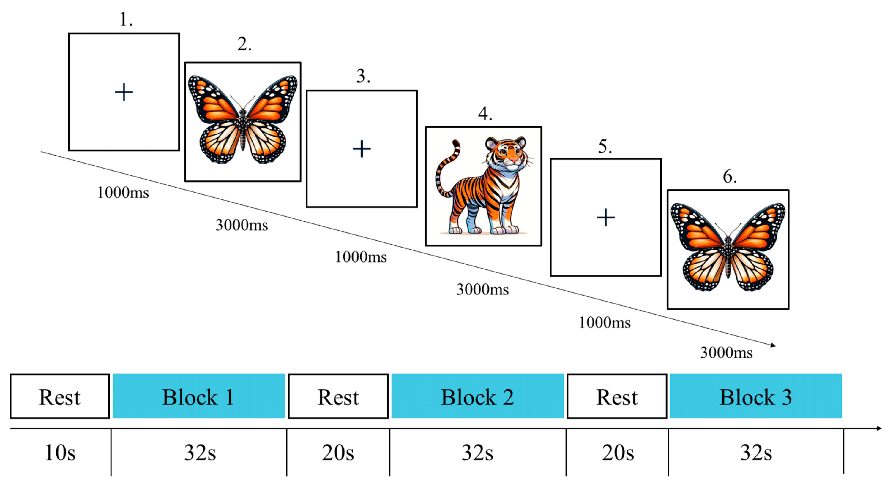
Chatham et al., 2011). The 1-back cognitive task paradigm in this study comprised three block sets, each containing eight trials (
Figure 3). Stimuli featured five animal images familiar to children—elephant, butterfly, tiger, rabbit, and turtle—ensuring task relevance to their everyday experiences. During each trial, each animal image appeared centrally on the screen for 3000 ms, followed by a 1000-millisecond fixation point between images. Children were instructed to press the corresponding key to indicate whether the current image matched the previously displayed one, using the “F” key for match and the “J” key for no match. All trials were presented in randomized order to prevent sequence effects from influencing results. Prior to the formal task, all participants learned the test rules and completed 6 practice trials. The testing procedure is illustrated in
Figure 3.
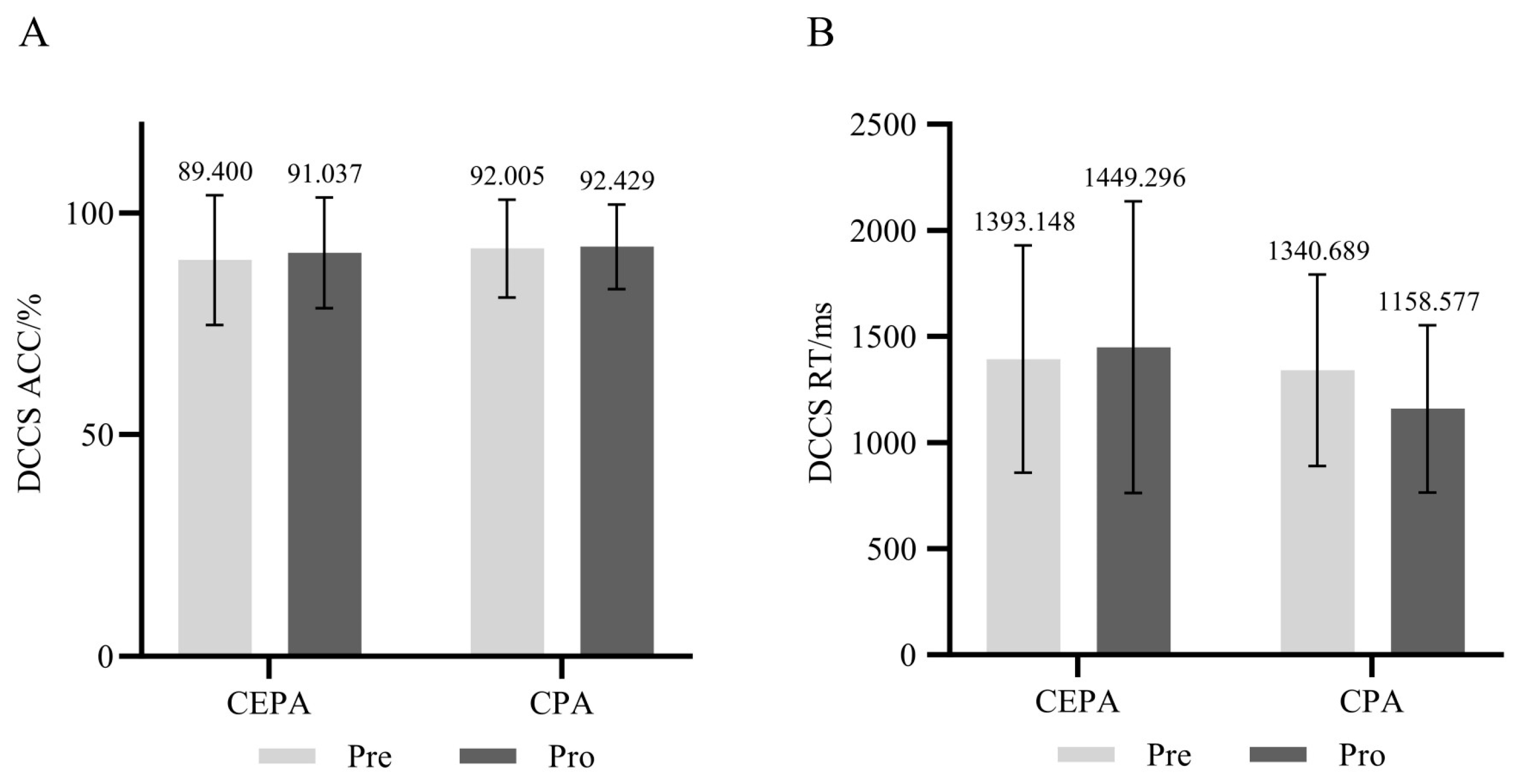
The Dimension Change Card Sorting task has been widely used in previous studies to measure CF in preschool children (
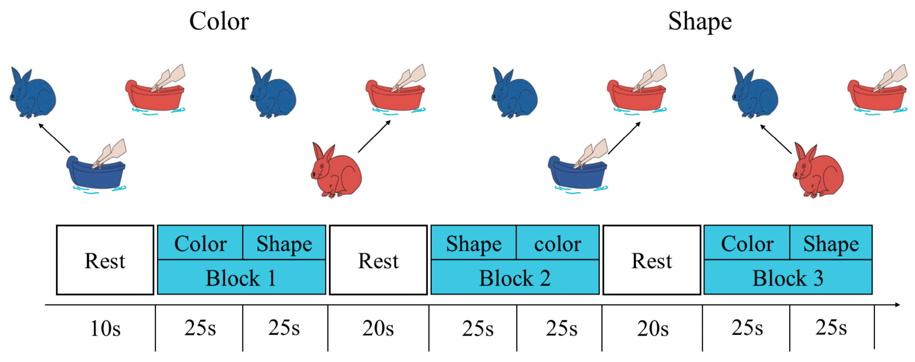
H. Li et al., 2021). The DCCS cognitive task paradigm in this study comprised three block sets, each containing 12 trials. Each block set consisted of 6 shape-matching trials and 6 color-matching trials (
Figure 4). During testing, two target images (rabbit and boat) served as stimuli and were displayed in the upper half of the screen. A test image (rabbit or boat) appeared at the center bottom of the screen for 3000 ms. Participants matched the test image to the target image according to the rule: pressing the “F” key matched it to the left image, while pressing the “J” key matched it to the right image. The rule sequence for the three block sets was fixed and applied uniformly across all participants to control for learning effects. Prior to the formal task, all subjects learned the test rules and completed 6 practice trials. The testing procedure is illustrated in
Figure 4.
2.4.2. fNIRS
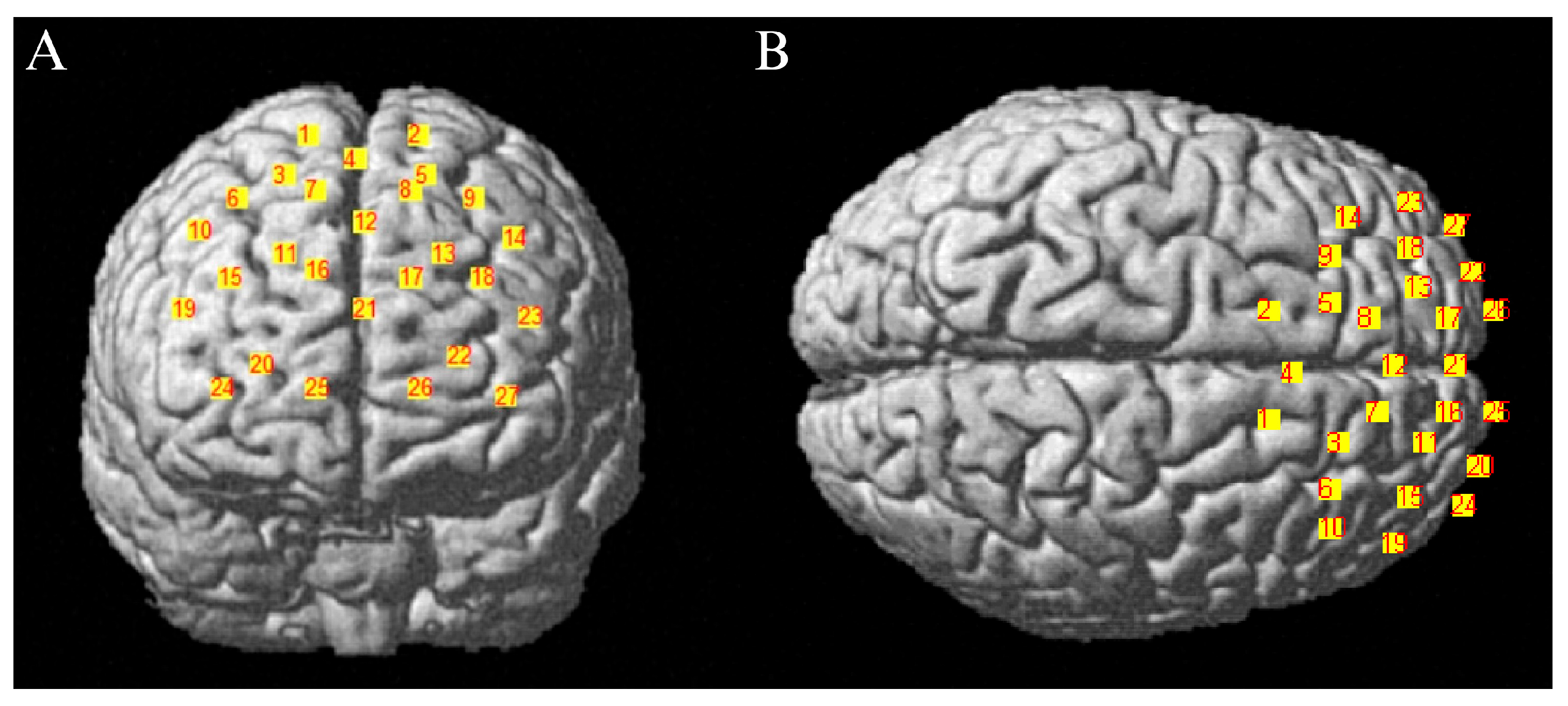
This study employed the Artinis Corporation (Holland) brite MKIII portable near-infrared brain imaging system to record hemodynamic changes in the PFC during subjects’ performance of WM tasks. The hardware configuration comprised 10 emitters and 8 receiver probes. Based on the distribution of brain regions associated with EF and the arrangement scheme of regions of interest (ROIs) from prior studies (
Xiang et al., 2022), a 2 × 13 channel matrix layout was employed, yielding a total of 27 acquisition channels covering the PFC. Emitter and receiver probes were spaced approximately 3 cm apart, with light sources centered at wavelengths of 760/850 nm. The sampling rate was set to 25 Hz. Precise scalp coordinates for each channel were determined using the Polhemus Patriot 3D head tracking system (USA). Three-dimensional channel coordinates were acquired and spatially registered within the Matlab environment using the “NIRS_SPM” software package (
https://www.nitrc.org/projects/nirs_spm/ (accessed on 15 July 2025)). Channel distribution is shown in
Figure 5. Based on the spatial layout of the 27 channels, four regions of interest (ROIs) were further delineated. The correspondence between each channel and the respective brain regions is detailed in
Table 2.
2.5. Sample Size
Sample size was determined using G*Power 3.1 to pre-set statistical power based on repeated measures ANOVA (interaction) (
Faul et al., 2009). The effect size was established at a medium value of 0.25 (effect size f = 0.25), accompanied by a two-tailed α level of 0.05 and a target power of 0.95 (
Bedard et al., 2021). Inputting the number of groups and measurements (n = 2), the calculated sample size for this study should exceed 54, with each group requiring at least 27 participants. After inputting the number of groups and measurements (n = 2), the calculated sample size required for this study was determined to be 54 or more. No significant differences were observed between the two groups in baseline characteristics (
Table 3). During formal testing, some children were unable to complete assessments due to excessive activity and inability to remain still. Ultimately, 59 preschoolers completed all tests. During near-infrared data preprocessing, 3 children were excluded due to excessive artifacts in cerebral oxygenation signals. The final sample included 56 children: 29 males and 27 females. The groups were a cognitively engaging physical activity group (n = 27) and a conventional physical activity group (n = 29).
2.6. Data Processing
Using the E-Merge3 feature in E-Prime 3.0, behavioral records for all subjects were consolidated and subsequently imported into Excel for preliminary organization. Data points exceeding ±3 standard deviations from the mean were excluded. fNIRS data preprocessing was performed in the MATLAB environment (R2013b, MathWorks, Natic, MA, USA). The Oxysoft2matlab plugin provided by Artinis was used to convert raw “.oxy5” and “.oxyproj” files into “.nirs” format. Subsequently, the Homer2 toolkit was employed to convert light intensity data into hemoglobin concentration. Motion artifacts were corrected across all channels for every subject, and poor channels were flagged. A bandpass filter of 0.01–0.1 Hz was applied to suppress physiological noise such as respiration and heartbeat. Data from the two seconds preceding each block stimulus served as the baseline for correction. Blood oxygen signals from all blocks of the same stimulus type were averaged to obtain the mean values for HbO2, deoxygenated hemoglobin (HHb), and total hemoglobin (t-Hb). HbO2 exhibits a higher signal-to-noise ratio and greater sensitivity to blood oxygen changes compared to HHb. Therefore, this study employs HbO2 as the primary blood oxygen concentration metric for subsequent statistical analysis.
2.7. Statistical Methods
All data were statistically analyzed using SPSS 22.0. Data distribution characteristics were assessed using Shapiro–Wilk normality tests and Levene’s tests for homogeneity of variance. For normally distributed continuous variables, data were described as mean ± standard deviation (M ± SD). Independent samples t-tests were used to compare heart rate between the cognitively engaging physical activity group and the conventional physical activity group during the intervention period. Behavioral metrics and oxygenated hemoglobin concentration were analyzed by a 2 (group) × 2 (time) repeated-measures ANOVA, with time as the within-subjects variable and group as the between-subjects variable. The main analysis assessed the group × time interaction, and post hoc multiple comparisons were adjusted using Bonferroni correction for p-values. Pearson correlation analyses examined the linear relationship between changes in HbO2 concentrations across four regions of interest and changes in behavioral measures.