You are currently viewing a new version of our website. To view the old version click .
Animals
  • Article
  • Open Access

17 November 2025

Cloning and Expression of Col10a1 Gene and Its Response to Wnt/TGF-β Signaling Inhibitors in the Chinese Three-Keeled Pond Turtle (Mauremys reevesii)

,
,
,
,
,
,
,
,
and
1
School of Biological Sciences and Engineering, Shaanxi University of Technology, Hanzhong 723001, China
2
Key Laboratory of Tropical & Subtropical Fishery Resource Application & Cultivation of Ministry of Agriculture and Rural Affairs, Pearl River Fisheries Research Institute, Chinese Academy of Fishery Sciences, Guangzhou 510380, China
3
South China Sea Fisheries Research Institute, Chinese Academy of Fishery Sciences, Guangzhou 510300, China
4
Science and Technology Research Center of China Customs, Beijing 100026, China
Animals2025, 15(22), 3315;https://doi.org/10.3390/ani15223315 
(registering DOI)
This article belongs to the Section Aquatic Animals

Simple Summary

The formation of Mauremys reevesii carapaces requires precisely regulated collagen deposition and ossification. The Col10a1 gene may play a key role in this process, but its specific function remains unclear. In this study, we aim to investigate the role of Col10a1 in M. reevesii carapace formation through molecular cloning, expression profiling, and functional characterization. We successfully cloned the Col10a1 gene and characterized its molecular properties using bioinformatics tools. Furthermore, we sought through spatiotemporal expression analysis at key embryonic developmental stages and in multiple adult tissues, combined with in vitro experiments using M. reevesii carapace-derived cells, to investigate the potential regulatory mechanisms of the Col10a1. Our findings provide important insights into the molecular mechanisms underlying M. reevesii carapace development and lay a foundation for further investigations into reptilian carapace formation mechanisms.

Abstract

The formation of the Chinese three-keeled pond turtle (Mauremys reevesii) carapace is a complex biological event involving developmental processes such as collagen deposition and ossification. As a key regulator of collagen deposition and ossification, Col10a1 may play a crucial role in carapace development, though its specific mechanism remains unclear. To investigate the role of Col10a1 during carapace development and its regulatory mechanisms, we cloned its cDNA sequence and performed bioinformatic analysis. This revealed that Col10a1 encodes a stable, hydrophilic, and basic protein, with phylogenetic analysis showing closest evolutionary relationships to other reptiles and the greatest divergence from fish. Further RT-qPCR analysis examined Col10a1 expression patterns in M. reevesii embryos at stages 14, 18, and 22, as well as in various tissues of adult males and females. Results indicated that during embryonic development, Col10a1 expression levels progressively increased alongside the progression of carapace ossification and collagen deposition, suggesting its involvement in regulating this process. In adult tissues, Col10a1 exhibited widespread expression, with particularly high levels in the brain, kidneys, and liver, suggesting potential specialized functions in these organs. Finally, in vitro experiments demonstrated that inhibition of the Wnt/β-catenin pathway with salinomycin sodium salt downregulated the expression of both its target genes (Sp5, Myc, Ccnd1) and Col10a1. In contrast, inhibition of the TGF-β/Smad pathway with oxymatrine suppressed its target genes (Serpine1, Cdkn1a) but concomitantly upregulated Col10a1. These results suggest that Col10a1 expression may be positively regulated by the Wnt/β-catenin pathway and negatively regulated by the TGF-β/Smad pathway. Our findings provide novel insights into the molecular mechanisms governing carapace development and collagen deposition in M. reevesii, laying a crucial foundation for further investigations into the regulatory networks involving Col10a1 during carapace formation.

1. Introduction

As a key structural component of the extracellular matrix (ECM), collagen is the most abundant and widely distributed functional protein in animals [,]. It plays crucial roles in maintaining the mechanical properties and physiological functions of various tissues []. At present, mammalian tissues are the primary sources for collagen extraction []. However, their use is limited by zoonotic disease safety concerns and regional religious restrictions [,,], driving the search for safer alternatives. Among these, aquatic collagen has garnered increased attention, with sources including fish [], starfish [], jellyfish [], and turtle/tortoise []. In particular, turtle-and-tortoise-derived collagen has emerged as a high-quality alternative with considerable potential. The Chinese three-keeled pond turtle (Mauremys reevesii) is a commercially important aquaculture species in China, valued for culinary and medicinal purposes [,]. Its carapace, a traditional Chinese medicinal ingredient rich in collagen and other bioactive compounds [], is a key raw material for producing turtle shell gelatin, turtle shell peptides, and the traditional Chinese medicinal dessert turtle jelly [,]. However, the mechanisms of collagen biosynthesis and deposition in the M. reevesii carapace remain poorly investigated.
The Col10a1 is a pivotal member of the collagen gene family, playing an essential role in collagen biosynthesis []. It encodes the alpha-1 chain of type X collagen, a short-chain secreted protein initially identified in the tibial growth plate cartilage of chicken embryos [,]. As a specific marker of the ECM in hypertrophic chondrocytes, Col10a1 is integral to endochondral ossification, where it facilitates matrix mineralization and compartmentalization of matrix components, thereby critically supporting the ossification process within skeletal growth plate cartilage [,]. Furthermore, Col10a1 mediates the remodeling of the growth plate cartilage ECM, constructing a microenvironment conducive to angiogenesis and bone replacement. It forms a directed scaffold on the surface of degraded cartilage templates, thereby promoting the directed migration of osteoprogenitor cells and the ordered deposition of type I collagen [,]. The role of Col10a1 has been primarily characterized in mammals and birds, whereas its function in reptiles—particularly in unique skeletal structures like the turtle carapace—remains poorly understood. This knowledge gap hinders a comprehensive understanding of both its evolutionary conservation and its potential lineage-specific functions in reptilian skeletal development. Previous studies from our group revealed significant upregulation of Col10a1 during M. reevesii carapace collagen synthesis, suggesting its potential involvement in carapace collagen matrix construction.
Collagen metabolism and skeletal development are closely linked to the Wnt/β-catenin and TGF-β/Smad signaling pathways. The Wnt/β-catenin pathway regulates the differentiation of chondrocyte precursors into hypertrophic chondrocytes by maintaining their proliferative state, thereby providing the cellular foundation for subsequent endochondral ossification []. Concurrently, the TGF-β/Smad signaling pathway contributes to early cartilage template establishment by promoting mesenchymal cell aggregation and cartilage nodule formation. It also influences the stability and remodeling of the collagen fiber network by regulating the dynamic balance between matrix metalloproteinases (MMPs) and their inhibitors (TIMPs) []. Thus, the spatiotemporal coordination of these two pathways during M. reevesii carapace formation may provide a unique regulatory model for Col10a1-mediated collagen deposition and endochondral ossification. However, systematic investigations into the expression characteristics and potential biological functions of Col10a1 in M. reevesii carapace collagen formation remain scarce. To address this knowledge gap, the present study cloned the cDNA sequence of Col10a1 from M. reevesii, performed bioinformatics analysis on its sequence, explored its spatial-temporal expression patterns in carapace tissues at different embryonic stages and adult male/female tissues, and investigated the gene’s response to Wnt/β-catenin and TGF-β/Smad inhibitor treatments. These findings will provide foundational data for further investigation into the molecular mechanisms of Col10a1 in M. reevesii carapace collagen deposition and endochondral ossification.

2. Materials and Methods

2.1. Experimental Animals and Ethical Declaration

The M. reevesii individuals used in this experiment were three-winter-old adults obtained from the Turtle and Soft-shelled Turtle Breeding and Conservation Base of the Pearl River Fisheries Research Institute, Chinese Academy of Fishery Sciences (Guangzhou, China). A total of six healthy individuals were selected, comprising three females and three males. Following anesthesia and blood collection, tissues including liver, kidney, spleen, heart, lung, muscle, brain, testis, and ovaries were harvested. These tissues were placed in enzyme-free cryopreservation tubes and snap-frozen in liquid nitrogen.
Additionally, 200 fertilized eggs of M. reevesii were collected. The eggs were buried in vermiculite (vermiculite-to-water ratio, 1:1) with the fertilization spots facing upward, and incubated in a constant-temperature incubator (Tai Hong Zhu Jiang, model FHX-250, 250 L) maintained at 32 ± 1 °C []. Embryonic developmental stages of M. reevesii were primarily determined according to the morphological criteria established by Tokita and Kuratani [], supplemented by the criteria proposed by Greenbaum []. Three fertilized eggs were dissected daily for developmental stage identification. At stage 14 (formation of the primitive carapace margin), stage 18 (appearance of pigment deposition and keel initiation), and stage 22 (fully developed morphology with defined structures), carapace samples were collected from 30 fertilized eggs per stage. Freshly collected carapace samples were rapidly frozen in liquid nitrogen and stored at −80 °C.
All animal experiments were conducted in accordance with protocols approved by the Laboratory Animal Ethics Committee of the Pearl River Fisheries Research Institute, Chinese Academy of Fishery Sciences.

2.2. Total RNA Extraction and cDNA Preparation

Total RNA was isolated from various adult M. reevesii tissues and from embryonic carapaces at stages 14, 18, and 22 using Trizol reagent (Ambion, Austin, TX, USA). RNA concentration was measured using the NanoQTM Nucleic Acid Detector (Thermo Scientific, Madison, WI, USA), and its integrity was assessed by 1% agarose gel electrophoresis. Subsequently, the first-strand cDNA was synthesized with the HiScript® III RT SuperMix for qPCR (+gDNA wiper) reverse transcription kit (Vazyme, Nanjing, China) and stored at −20 °C for later use.

2.3. Cloning of the Col10a1 Gene

Gene-specific primers (Table 1) were designed based on the M. reevesii Col10a1 cDNA sequence (GenBank accession no. XM_039529706.1) and synthesized by Guangzhou Tianyi Huiyuan Gene Technology Co., Ltd. (Guangzhou, China). Primer specificity, verified by NCBI BLAST, was further confirmed by single-peak melting curves (Supplementary Figure S1). The qRT-PCR standard curves showed high linearity (R2 ≥ 0.99), with slopes ranging from −3.45 to −3.21, corresponding to amplification efficiencies of 94% to 104% (Supplementary Figure S2). Polymerase chain reaction (PCR) was performed using M. reevesii carapace-derived cDNA to amplify the coding sequence (CDS) of Col10a1 with the specific primer pair Col10a1-F/Col10a1-R (Table 1). The reaction system consisted of a 20 μL volume: 10 μL 2× SanTaq PCR Master Mix, 7 μL ddH2O, 1.0 μL cDNA template, and 1.0 μL each of Col10a1-F and Col10a1-R primers. Amplification program: 95 °C pre-denaturation for 5 min; 35 cycles of 95 °C for 30 s, 58 °C for 30 s, 72 °C for 2 min; 72 °C extension for 10 min. Amplified products were analyzed by 1% agarose gel electrophoresis, and the target band was excised and purified using a Gel Extraction Kit (Omega, Norcross, GA, USA). Subsequently, the purified product was ligated into the pMD19-T vector (TaKaRa, Beijing, China), and transformed into DH5α competent cells (Vazyme, Nanjing, China). Transformed cells were plated on LB agar, and single colonies were selected and screened by colony PCR. Positive clones were cultured, and plasmid DNA was submitted to Guangzhou Tianyi Huiyuan Gene Technology Co., Ltd. for Sanger sequencing.
Table 1. Primers used for cDNA cloning and expression analysis of M. reevesii.

2.4. Bioinformatics Analysis of the Col10a1

The cDNA sequences obtained from sequencing were translated into amino acid sequences using DNAMAN 8 []. Multiple sequence alignment was performed with ClustalW (BioEdit) software, and amino acid sequence homology analysis was conducted using BLAST []. Physicochemical properties, hydrophilicity and hydrophobicity profiles, signal peptides, and potential phosphorylation sites were predicted with ProtParam, ProtScale, SignalP-6.0, and NetPhos 3.1, respectively []. The secondary and tertiary structures of the Col10a1 protein were predicted using SOPMA [] and SWISS-MODEL []. Protein domains were predicted using SMART (https://smart.embl.de (accessed on 30 June 2025)). A phylogenetic tree was constructed using the maximum likelihood (ML) method in MEGA 11 [], with 1000 bootstrap replicates. Using the MEME Suite, conserved motifs within the Col10a1 protein were predicted. Domain annotation was then performed with the National Center for Biotechnology Information (NCBI) Conserved Domain Database (CDD). All sequences used for the analyses, including amino acid homology comparisons, phylogenetic construction, conserved motifs and structural domain analysis, were obtained from the NCBI. The corresponding accession numbers are provided in Table 2.
Table 2. NCBI accession numbers of the Col10a1 protein sequences across different species.

2.5. Expression Analysis of the Col10a1 Gene in Different Tissues of M. reevesii

The Gapdh gene was used as the housekeeping gene for normalization []. Both the reference and target gene primers for RT-qPCR are listed in Table 1. Using cDNA templates from various adult M. reevesii tissues and embryonic carapaces, the relative expression levels of the Col10a1 were detected using the Applied Biosystems StepOnePlus Real-Time PCR Systems (Applied Biosystems, Singapore). The reaction system consisted of 20.0 μL: 10 μL iTaq Universal SYBR® Green Supermix (Bio-Rad, Hercules, CA, USA), 7.0 μL ddH2O, 1.0 μL each of Col10a1-qF and Col10a1-qR (Table 1) primers, and 1.0 μL cDNA template. The qPCR conditions for all target genes were as follows: 95 °C for 5 min; 40 cycles of 95 °C for 10 s, 60 °C for 20 s, and 72 °C for 20 s. The melting curve analysis was performed at 95 °C for 15 s, 60 °C for 60 s, and 95 °C for 15 s.

2.6. Treatment of M. reevesii Carapace Cells with Salinomycin Sodium Salt and Oxymatrine

The M. reevesii carapace cells (MRCCs) line used in this experiment was previously established by our research group. This cell line was established by isolating carapace tissue during M. reevesii embryonic development and performing primary culture using the tissue block adhesion method, ultimately yielding a stably proliferating cell population. Well-growing MRCCs were seeded at appropriate concentrations into six-well plates. Upon reaching 60–70% confluence, salinomycin sodium salt (Wnt/β-catenin pathway inhibitor) [] and oxymatrine (TGF-β/Smad pathway inhibitor) [] stock solutions using DMSO according to manufacturer instructions. The prepared drug solutions were added to the cell culture system, with an equal volume of DMSO solvent added as a negative control group. After 24 h of drug treatment, total RNA was isolated from collected cell samples and reverse-transcribed into cDNA. The expression levels of Col10a1 and key genes in the Wnt (Sp5, Myc, Ccnd1) and TGF-β (Serpine1, Cdkn1a) signaling pathways were quantified by RT-qPCR. Primer information is shown in Table 1.

2.7. Data Analysis

Data are presented as the mean of three independent biological replicates. Gene expression levels were determined using the 2−ΔΔCt method []. For statistical comparison, one-way analysis of variance (ANOVA) coupled with least significant difference (LSD) test was applied in SPSS 21.0, and differences with p < 0.05 were deemed statistically significant.

3. Results

3.1. Cloning and Sequence Analysis of the M. reevesii Col10a1

The coding sequence (CDS) of the Col10a1 gene was successfully cloned from M. reevesii carapace-derived cDNA. Colony PCR yielded a single band of approximately 2000 bp (Supplementary Figure S3), and sequencing confirmed a synonymous single-base mutation (T to C) compared to the NCBI reference sequence (Supplementary Figure S4).
The cloned cDNA is 2194 bp in length and contains a 2034 bp CDS encoding a protein of 677 amino acid (Supplementary Figure S5a). Bioinformatic analysis predicted the encoded protein to be alkaline and stable, with a theoretical isoelectric point (pI) of 9.39, an instability index of 25.91, and an aliphatic index of 58.26. Hydrophobicity analysis identified the most hydrophobic residue at position 8 (score: 2.744) and the most hydrophilic at position 577 (score: −3.233) (Supplementary Figure S5b). Furthermore, the M. reevesii Col10a1 was predicted to contain 28 phosphorylation sites and an N-terminal 18-amino-acid signal peptide (Supplementary Figure S5c,d). The secondary structure of M. reevesii Col10a1 was predicted to be predominantly composed of random coils (73.71%), along with extended strands (11.08%), β-turns (9.31%), and α-helices (5.91%) (Supplementary Figure S5e), and the predicted tertiary structure was consistent with these secondary structure elements (Supplementary Figure S5f). Domain analysis identified two low-complexity regions, five collagen-specific domains with Gly-X-Y repeats, and a conserved C-terminal globular C1q domain (NC1) (Supplementary Figure S5g) [].

3.2. Amino Acid Similarity Analysis of Col10a1 Protein and Construction of Phylogenetic Tree

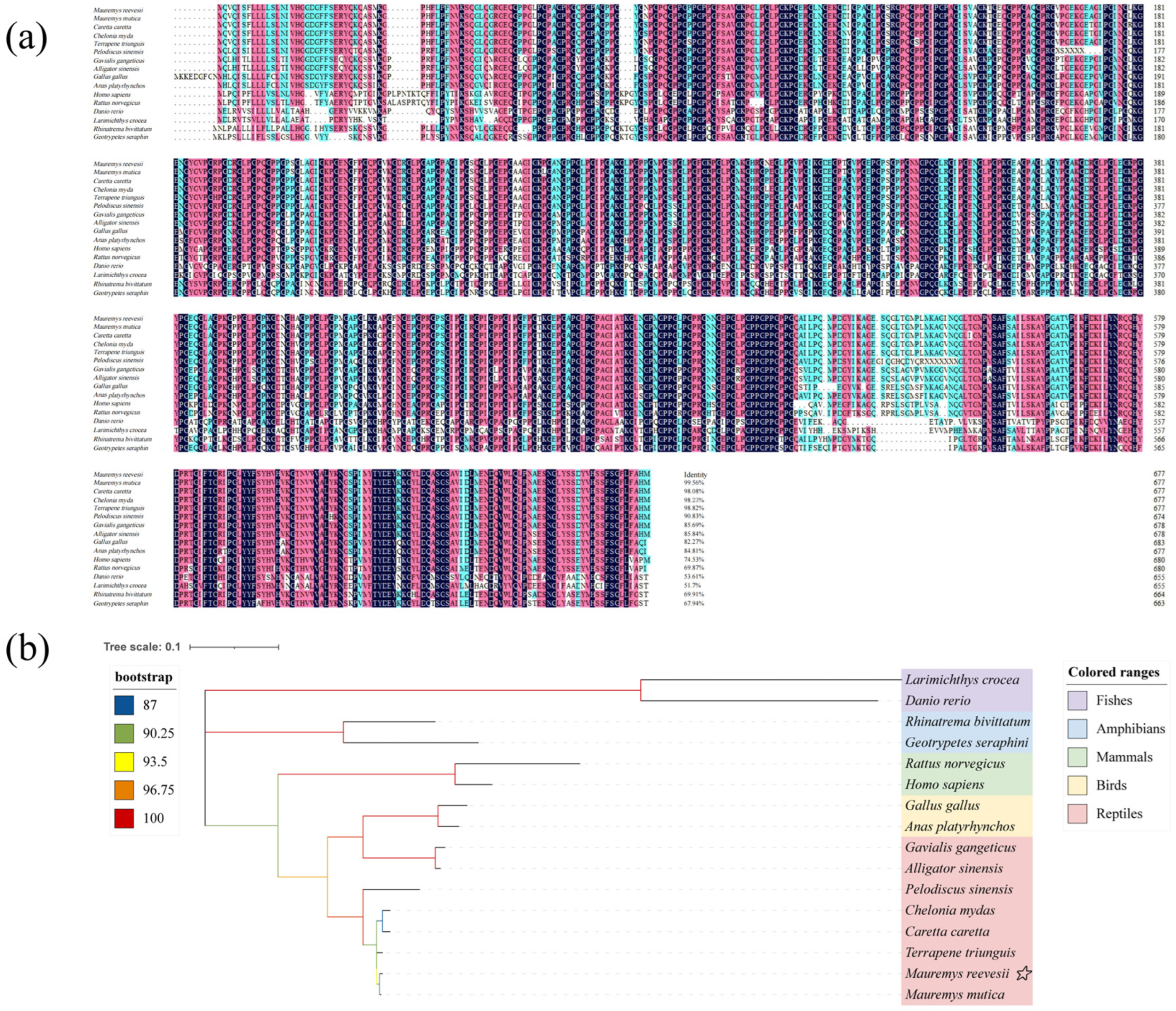
Amino acid sequence homology alignment results indicate that the M. reevesii Col10a1 exhibits high identity with reptiles, showing the highest identity with Mauremys mutica at 99.56%. It also demonstrates significant identity with other turtle and tortoise species, including Caretta caretta (98.08%), Chelonia mydas (98.23%), Terrapene triunguis (98.82%), and Pelodiscus sinensis (90.83%) (Figure 1a). However, the Col10a1 exhibits low conservation in fish, with only 51.70% identity with Larimichthys crocea. Phylogenetic tree results (Figure 1b) indicate that M. reevesii Col10a1 clusters with reptilian species, forming a clade most closely related to M. mutica and showing the greatest evolutionary distance from fish species. This finding fully aligns with amino acid sequence homology analysis, together confirming the conservation of Col10a1 across turtle and soft-shelled turtle species.
Figure 1. The comparison of amino acid sequence homology similarity and phylogenetic tree of Col10a1 between M. reevesii and other species. (a) amino acid sequence homology similarity; (b) phylogenetic tree. Notation: (b). The yellow star symbol indicates M. reevesii, the target species of this study, which is marked as a key node in this phylogenetic tree.

3.3. Conserved Motifs and Structural Domain Analysis of Col10a1 Proteins Across Different Species

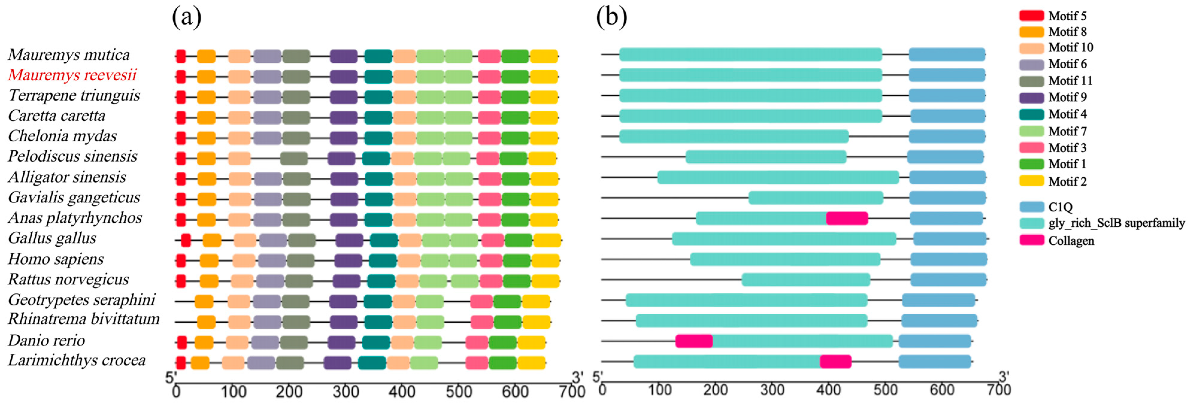
Through prediction of conserved amino acid regions, 11 highly conserved motifs were identified in Col10a1 proteins across diverse vertebrates (Figure 2a). Furthermore, Col10a1 proteins across these vertebrates contain identical structural domains, namely the gly_rich_SclB superfamily and C1q domain (Figure 2b). Among these, the gly_rich_SclB superfamily is most conserved in turtle species, while the C1q domain is highly conserved across various vertebrate lineages.
Figure 2. Conserved motifs and structural domain analysis of Col10a1 across different species. (a) Conserved motifs; (b) structural domain.

3.4. Expression Changes in Col10a1 in the Carapace of M. reevesii Embryos During Development

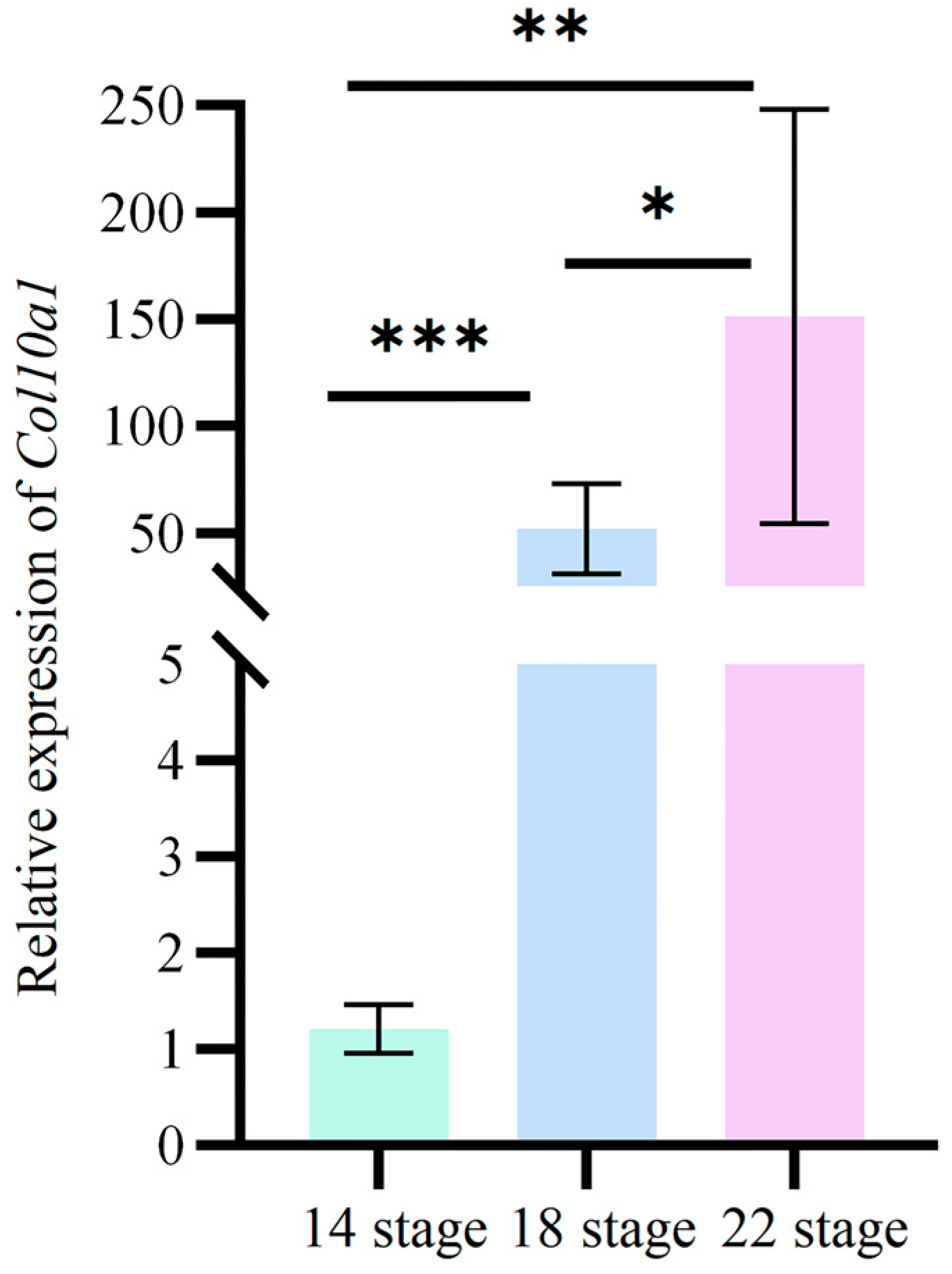
RT-qPCR analysis was conducted to examine the expression dynamics of the Col10a1 in carapace tissues of M. reevesii embryos at developmental stages 14, 18, and 22. The results (Figure 3) demonstrate that Col10a1 expression increased gradually throughout embryonic development, reaching its highest level at stage 22. Expression at stage 22 is significantly higher than at stages 14 and 18.
Figure 3. Relative expression of Col10a1 in the carapace of M. reevesii embryos at developmental stages 14, 18, and 22. Significant differences are indicated by * (p < 0.05), ** (p < 0.01) and *** (p < 0.001). Gapdh was used as the internal reference gene for the 2−ΔΔCt method. The original data are available in Supplementary Table S1.

3.5. Expression Profile of the Col10a1 in Adult M. reevesii Tissues

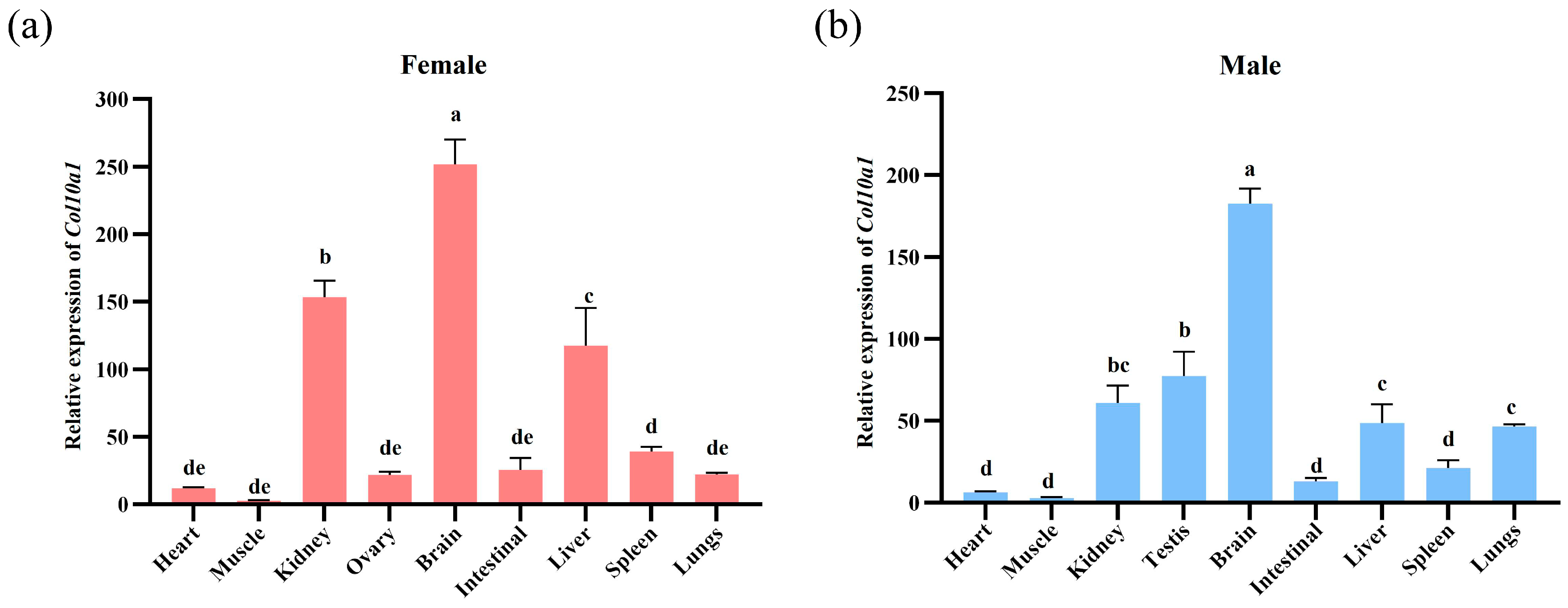
The expression profile of the Col10a1 in three-winter-old male and female M. reevesii tissues is shown below (Figure 4). The Col10a1 was expressed in all examined tissues in both male and female adults, with highly similar expression patterns between sexes. Relatively high expression levels were observed in brain, kidney, and liver, whereas muscle showed the lowest expression. Among these, brain tissue demonstrates the most abundant expression, with significantly higher levels than other tissues (p < 0.05).
Figure 4. Relative expression of Col10a1 in various tissues of three-winter-old M. reevesii. (a) expression of Col10a1 in female individuals; (b) expression of Col10a1 in male individuals. Different lowercase letters indicate significant differences between groups (p < 0.05). Gapdh was used as the internal reference gene for the 2−ΔΔCt method. The original data are available in Supplementary Table S2.

3.6. Effects of Salinomycin Sodium Salt and Oxymatrine on Gene Expression in MRCCs

MRCCs were treated with different concentrations (1 mM and 10 mM) of salinomycin sodium salt and oxymatrine, as shown in Figure 5a,b, respectively. Compared to the negative control group (DMSO group), Col10a1 expression in MRCCs decreased significantly in response to salinomycin sodium salt in a dose-dependent manner, with the 10 mM treatment group showing a highly significant reduction (p < 0.001). In contrast, both 1 mM and 10 mM doses of oxymatrine significantly upregulated Col10a1 expression (p < 0.001). Furthermore, in MRCCs, treatment with salinomycin sodium salt downregulated the expression of Wnt/β-catenin signaling pathway target genes (Sp5, Myc, Ccnd1), whereas oxymatrine treatment reduced the expression of TGF-β/Smad signaling pathway target genes (Serpine1, Cdkn1a).
Figure 5. Relative expression of Col10a1 in MRCCs after treatment with salinomycin sodium salt (a) and oxymatrine (b). Significant differences are indicated by * (p < 0.05), ** (p < 0.01) and *** (p < 0.001). Gapdh was used as the internal reference gene for the 2−ΔΔCt method. The original data are available in Supplementary Table S3.

4. Discussion

This study successfully cloned the Col10a1 gene from M. reevesii, which spans 2194 bp, including a 2034 bp coding sequence (CDS) that encodes a protein of 677 amino acids. The encoded Col10a1 protein is an alkaline-stable hydrophilic protein. Amino acid sequence alignment and ML phylogenetic tree analysis indicate that M. reevesii Col10a1 is most closely related to turtle and tortoise species within the reptilian clade. Protein structure determines function. The M. reevesii Col10a1 protein is predicted to be secreted and contains an N-terminal signal peptide that serves as a molecular localization signal. This guides the newly synthesized Col10a1 precursor protein into the endoplasmic reticulum (ER) for post-translational modification. Subsequently, the signal peptide is specifically cleaved by signal peptidase within the ER lumen, releasing the mature Col10a1 protein into the ECM []. The C-terminal NC1 domain of Col10a1 plays a pivotal role in collagen X assembly by driving the homotrimerization of three α1(X) chains, making it essential for Col10a1 secretion [,]. Furthermore, the NC1 domain contains a specific calcium ion-binding site; binding of calcium ions is known to promote the initiation of mineralization in growth plate cartilage [].
RT-qPCR analysis revealed that Col10a1 gene expression in the carapace of M. reevesii embryos exhibited distinct temporal characteristics, with expression levels progressively increasing throughout carapace development. Notably, Col10a1 gene expression was significantly upregulated (p < 0.05) in carapace tissue during stage 22 of M. reevesii embryonic development. This expression pattern coincides with the biological processes of carapace ossification and collagen deposition during M. reevesii embryonic development. Previous studies indicate that during vertebrate skeletal development, the X-type collagen gene (Col10a1) is specifically expressed in the hypertrophic chondrocyte zone during endochondral ossification [,], and its upregulation is closely associated with cartilage matrix mineralization and the formation of ossification centers []. Furthermore, Col10a1 facilitates ECM remodeling in the growth plate, providing a scaffold for vascular invasion and subsequent ossification, as well as guiding osteoblast migration and facilitating Type I collagen deposition on the mineralized cartilage surface [,]. Thus, our results suggest that during M. reevesii carapace development, elevated Col10a1 expression may contribute to carapace bone plate formation by promoting collagen synthesis and mineralized matrix deposition.
Additionally, in studies of tissue expression profiles in adult M. reevesii, Col10a1 was expressed in all tissues, with higher expression levels in the brain, kidneys, and liver. Its expression level in brain tissue was particularly significant (p < 0.05). Col10a1 encodes the α1 chain of type X collagen, typically expressed in cartilage growth plates and involved in skeletal development []. However, its prominent expression in non-skeletal tissues such as the brain, kidney, and liver, however, suggests potential tissue-specific functions beyond osteogenesis. Sustained Col10a1 expression in the adult brain may indicate a role in neural tissue homeostasis. Although the precise function of type X collagen in the brain remains incompletely understood, other collagen family members (such as type IV collagen) have been demonstrated to be critical for the nervous system. Studies have shown that type IV collagen is a core component of the blood–brain barrier basement membrane [,], which plays a key role in maintaining blood–brain barrier function []. Moreover, Type IV collagen plays a central role in arterial stiffness and dementia pathogenesis, with Col4a1 mutations and dysregulation of its synthesis and degradation closely associated with vascular dysfunction and cognitive decline []. These findings suggest that Col10a1 may participate in physiological or pathological processes of the nervous system by influencing brain ECM dynamics or vascular stability, though the precise mechanisms require further investigation. Furthermore, in the kidney, collagen serves as a critical structural component. Type IV collagen and type V collagen constitute the primary constituents of the glomerular basement membrane (GBM) and glomerular mesangial ECM, respectively [,], while the tubulointerstitial ECM contains types I, III, IV, V, VI, VII, and VIII collagen []. Recent studies reveal that type X collagen is upregulated during renal fibrosis, and Col10a1 overexpression has been shown to promote aberrant ECM deposition following tubulointerstitial injury, thereby exacerbating renal fibrosis []. In the liver, Col10a1 expression is associated with fibrosis regulation. It is induced during hepatic stellate cell activation, subsequently promoting collagen deposition and driving the progression of liver fibrosis [].
Collagen metabolism is jointly regulated by a series of signaling pathways. Among these, the Wnt/β-catenin and TGF-β/Smad signaling pathways are classical pathways regulating collagen metabolism, primarily involved in the deposition of ECM [,,,]. Previous studies have established that salinomycin sodium salt is a potent Wnt/β-catenin pathway inhibitor, as it acts by blocking the phosphorylation of the Wnt coreceptor lipoprotein receptor related protein 6 (LRP6) and subsequently inducing its degradation [,,]. In contrast, oxymatrine inhibits the TGF-β/Smad pathway through a dual mechanism, which involves suppressing the expression of Smad3 and its coactivator CBP while promoting the expression of the inhibitory Smad7 [,,]. In this study, pathway inhibitor treatment of MRCCs revealed that salinomycin sodium salt downregulated Col10a1 expression, whereas oxymatrine upregulated it. This opposing effect suggests that the signaling pathways targeted by the two drugs may have opposing regulatory effects on Col10a1 expression. To further verify the pathway inhibition effects of salinomycin sodium salt and oxymatrine in MRCCs, we analyzed the expression of key target genes in the relevant pathways. Sp5, Myc and Ccnd1 are established direct target genes of the Wnt/β-catenin pathway, such that pathway activation enables nuclear β-catenin to complex with TCF/LEF and initiate their transcription, whereas pathway inhibition impairs the formation or function of this complex and diminishes transcriptional output [,,]. In contrast, Serpine1 and Cdkn1a are classic direct transcriptional targets of the TGF-β/Smad signaling pathway, and as key markers of the pathway’s activity, their expression is precisely regulated through the interaction of Smad proteins with specific promoter elements [,]. The results of this study indicate that in MRCCs, treatment with salinomycin sodium salt significantly reduced the expression of Wnt/β-catenin target genes (Sp5, Myc, Ccnd1), while oxymatrine decreased that of TGF-β/Smad target genes (Serpine1, Cdkn1a), confirming that each drug effectively inhibited its respective target pathway. Therefore, it can be inferred that within MRCCs, salinomycin sodium salt and oxymatrine likely inhibit the Wnt/β-catenin and TGF-β/Smad pathways, respectively, mediating the suppression and enhancement of Col10a1 expression, and revealing that the Wnt/β-catenin and TGF-β/Smad pathways function as positive and negative regulators of Col10a1, respectively. Evidence suggests that Wnt/β-catenin signaling promotes Col10a1 expression by activating the transcription factor Runx2, mediating chondrocyte hypertrophy, accelerating chondrocyte maturation, and advancing endochondral ossification [,]. In contrast, within the TGF-β/Smad signaling pathway, TGF-β/Smads activate signaling that suppresses Runx2 and Col10a1 expression, reducing cartilage cell ECM degradation and thereby delaying cartilage cell hypertrophy [,,]. Therefore, the deposition of collagen in M. reevesii carapace and the ossification process may be regulated by the antagonistic control of Col10a1 expression through the Wnt/β-catenin and TGF-β/Smad signaling pathways. This precise regulatory mechanism ensures the timely deposition of collagen and orderly ossification within carapace tissue during the transition of chondrocytes toward hypertrophy.
However, this study has certain limitations. The precise mechanisms by which salinomycin and oxymatrine inhibit the Wnt/β-catenin and TGF-β/Smad pathways in MRCCs, respectively, are not fully elucidated, and their potential off-target effects remain to be evaluated. Consequently, elucidating their precise mechanisms of action will be an important future direction.

5. Conclusions

This study successfully cloned and characterized Col10a1 from M. reevesii. Bioinformatics analysis revealed that M. reevesii Col10a1 is most closely related to other reptilian species. The M. reevesii Col10a1 is a basic, hydrophilic secretory molecule, whose secondary structure is dominated by α-helices and random coils and features a C-terminal NC1 domain. Expression analysis revealed Col10a1 expression increases gradually during embryonic carapace development and is highly expressed in brain, kidneys, and liver of adult M. reevesii. Pharmacological inhibition experiments suggest that Col10a1 expression is potentially positively regulated by the Wnt/β-catenin pathway and negatively regulated by the TGF-β/Smad pathway. These findings confirm the key role of Col10a1 in collagen deposition and carapace ossification in M. reevesii, offer new insights into the underlying mechanisms, and lay a foundation for exploring Wnt/TGF-β crosstalk in carapace development and hardening.

Supplementary Materials

The following supporting information can be downloaded at: https://www.mdpi.com/article/10.3390/ani15223315/s1, Figure S1: Melting curve analysis of Col10a1 (a), Gadph (b), Sp5 (c), Myc (d), Ccnd1 (e), Serpine1 (f), Cdkn1a (g); Figure S2: Amplification efficiency analysis of Gadph (a), Col10a1 (b), Col10a1 (a), Gadph (b), Sp5 (c), Myc (d), Ccnd1 (e), Serpine1 (f), Cdkn1a (g); Figure S3: Agarose gel electrophoresis image of PCR from Col10a1 gene cloned colonies; Figure S4: Alignment of the cloned Col10a1 sequence from M. reevesii with the NCBI reference sequence (XM_039529706.1); Figure S5: (a) Nucleotide of M. reevesii Col10a1 and its encoded protein sequence; (b) Prediction of hydrophilicity/hydrophobicity of M. reevesii Col10a1; (c) Phosphorylation prediction results of M. reevesii Col10a1; (d) Signal peptide prediction results of M. reevesii Col10a1; (e) M. reevesii Col10a1 secondary structure prediction; (f) M. reevesii Col10a1 tertiary structure prediction; (g) M. reevesii Col10a1 domains prediction; Table S1: Figure 3-Relative Expression Calculation Data for Col10a1; Table S2: Figure 4-Relative Expression Calculation Data for Col10a1; Table S3: Figure 5-Relative Expression Calculation Data for Col10a1.

Author Contributions

Conceptualization, L.D., X.Z. and W.L.; methodology, X.L., C.C., L.J., J.Z. and W.L.; validation, Y.L.; formal analysis, Y.L., T.R.,C.C., L.J., J.Z. and W.L.; investigation, Y.L. and J.Z.; resources, T.R., X.L., X.H., C.W. and H.C.; data curation, Y.L.; writing—original draft preparation, Y.L.; writing—review and editing, Y.L., X.Z. and J.Z.; visualization, Y.L.; supervision, J.Z., T.R., C.C., L.J., X.H., X.L., C.W. and H.C.; project administration, L.D. and W.L.; funding acquisition, L.D., X.Z. and W.L. All authors have read and agreed to the published version of the manuscript.

Funding

This research was funded by the special funding for basic scientific research business expenses from the Central-level Public Welfare Scientific Research Institutes of the Chinese Academy of Fishery Sciences (2025XK01), the National Natural Science Foundation of China (32470482), the Shaanxi Provincial Department of Science and Technology project of China (2025NC-YBXM-066), the Shaanxi Provincial Department of Education Project of China (24JP035), the Selection Breeding of the high-quality fast-growing Chinese Soft-Shelled Turtle and Studies on Efficient and Healthy Farming Technology (2024-SPY-00-014), the Central Public-interest Scientific Institution Basal Research Fund, CAFS (2023TD38), the China-ASEAN Maritime Cooperation Fund (CAMC-2018F).

Institutional Review Board Statement

The animal research protocol was approved by the Institutional Review Board of the Pearl River Fisheries Research Institute, Chinese Academy of Fishery Sciences (approval number: LAEC-PRFRI-2024-02-20; approval date: 20 February 2024).

Data Availability Statement

The raw data analyzed in this study can be downloaded from the National Center for Biotechnology Information (NCBI) databases.

Conflicts of Interest

The authors declare no conflicts of interest.

References

  1. Shen, Z.; Zhang, Q.; Li, L.; Li, D.; Takagi, Y.; Zhang, X. Properties of Grass Carp (Ctenopharyngodon idella) Collagen and Gel for Application in Biomaterials. Gels 2022, 8, 699. [Google Scholar] [CrossRef]
  2. Ampitiya, A.G.D.M.; Gonapinuwala, S.T.; Fernando, C.A.N.; De Croos, M.D.S.T. Extraction and Characterisation of Type I Collagen from the Skin Offcuts Generated at the Commercial Fish Processing Centres. J. Food Sci. Technol. 2023, 60, 484–493. [Google Scholar] [CrossRef]
  3. Gelse, K. Collagens—Structure, Function, and Biosynthesis. Adv. Drug Deliv. Rev. 2003, 55, 1531–1546. [Google Scholar] [CrossRef] [PubMed]
  4. Zhang, X.; Ookawa, M.; Tan, Y.; Ura, K.; Adachi, S.; Takagi, Y. Biochemical Characterisation and Assessment of Fibril-Forming Ability of Collagens Extracted from Bester Sturgeon Huso huso × Acipenser ruthenus. Food Chem. 2014, 160, 305–312. [Google Scholar] [CrossRef] [PubMed]
  5. Sionkowska, A.; Lewandowska, K.; Adamiak, K. The Influence of UV Light on Rheological Properties of Collagen Extracted from Silver Carp Skin. Materials 2020, 13, 4453. [Google Scholar] [CrossRef]
  6. Atef, M.; Ojagh, S.M.; Latifi, A.M.; Esmaeili, M.; Udenigwe, C.C. Biochemical and Structural Characterization of Sturgeon Fish Skin Collagen (Huso huso). J. Food Biochem. 2020, 44, e13256. [Google Scholar] [CrossRef]
  7. Li, L.; Yu, Y.; Wu, W.; Wang, P. Extraction, Characterization and Osteogenic Activity of a Type I Collagen from Starfish (Asterias amurensis). Mar. Drugs 2023, 21, 274. [Google Scholar] [CrossRef]
  8. Khong, N.M.H.; Yusoff, F.M.; Jamilah, B.; Basri, M.; Maznah, I.; Chan, K.W.; Armania, N.; Nishikawa, J. Improved Collagen Extraction from Jellyfish (Acromitus hardenbergi) with Increased Physical-Induced Solubilization Processes. Food Chem. 2018, 251, 41–50. [Google Scholar] [CrossRef]
  9. Xu, R.; Li, D.; Peng, J.; Fang, J.; Zhang, L.; Liu, L. Cloning, Expression and Antioxidant Activity of a Novel Collagen from Pelodiscus sinensis. World J. Microbiol. Biotechnol. 2016, 32, 100. [Google Scholar] [CrossRef]
  10. Peng, M. Research on Industrialized Farming and All-Female Seed Breeding Technology of Mauremys reevesii. Master’s Thesis, Zhejiang Ocean University, Zhoushan, China, 2025. [Google Scholar] [CrossRef]
  11. Du, W.; Hu, L.; Lu, J.; Zhu, L. Effects of Incubation Temperature on Embryonic Development Rate, Sex Ratio and Post-Hatching Growth in the Chinese Three-Keeled Pond Turtle, Chinemys reevesii. Aquaculture 2007, 272, 747–753. [Google Scholar] [CrossRef]
  12. Ren, M.; Lu, J.; Song, Y.; Zhu, J.; Lin, L.; Lu, H.; Wan, Q.; Jiang, S. Comparison between High-Pressure Steam and Vinegar Stir-Frying on Grinding Characteristics and Nutrient Components of Turtle Shells: Difference between Carapace and Plastron. J. Food Compos. Anal. 2024, 132, 106391. [Google Scholar] [CrossRef]
  13. Li, Q.; Zhu, X.; Shi, Y.; Zhu, J.; Tong, Q. Analysis of the Research and Development of Tortoise Shell Glue. Henan Tradit. Chin. Med. 2023, 43, 968–972. [Google Scholar] [CrossRef]
  14. Li, H.; Gao, Y.; Song, L.; Shi, W.; Li, L.; Chen, T.; Hou, H.; Zhang, G.; Ye, Z. Effect of Guiling Jelly in Treating Excess Fire from Yin Deficiencyin Model Rats by Nourishing Yin and Purging Fire. J. Guangxi Univ. Chin. Med. 2022, 25, 4–9. [Google Scholar] [CrossRef]
  15. Liu, X.; Li, K.; Zhang, J. Research Progress on Collagen. Life Sci. Res. 2016, 20, 542–547. [Google Scholar] [CrossRef]
  16. Schmid, T.M.; Linsenmayer, T.F. Immunohistochemical Localization of Short Chain Cartilage Collagen (Type X) in Avian Tissues. J. Cell Biol. 1985, 100, 598–605. [Google Scholar] [CrossRef]
  17. Schmid, T.M.; Linsenmayer, T.F. Developmental Acquisition of Type X Collagen in the Embryonic Chick Tibiotarsus. Dev. Biol. 1985, 1072, 373–381. [Google Scholar] [CrossRef]
  18. Gu, J.; Lu, Y.; Li, F.; Qiao, L.; Wang, Q.; Li, N.; Borgia, J.A.; Deng, Y.; Lei, G.; Zheng, Q. Identification and Characterization of the Novel Col10a1 Regulatory Mechanism during Chondrocyte Hypertrophic Differentiation. Cell Death Dis. 2014, 5, e1469. [Google Scholar] [CrossRef]
  19. Shen, G. The Role of Type X Collagen in Facilitating and Regulating Endochondral Ossification of Articular Cartilage. Orthod. Craniofac. Res. 2005, 8, 11–17. [Google Scholar] [CrossRef] [PubMed]
  20. Knuth, C.; Andres Sastre, E.; Fahy, N.; Witte-Bouma, J.; Ridwan, Y.; Strabbing, E.; Koudstaal, M.; Van De Peppel, J.; Wolvius, E. Collagen Type X Is Essential for Successful Mesenchymal Stem Cell-Mediated Cartilage Formation and Subsequent Endochondral Ossification. Eur. Cells Mater. 2019, 38, 106–122. [Google Scholar] [CrossRef] [PubMed]
  21. Chan, D.; Jacenko, O. Phenotypic and Biochemical Consequences of Collagen X Mutations in Mice and Humans. Matrix Biol. 1998, 17, 169–184. [Google Scholar] [CrossRef] [PubMed]
  22. Baron, R.; Kneissel, M. WNT Signaling in Bone Homeostasis and Disease: From Human Mutations to Treatments. Nat. Med. 2013, 19, 179–192. [Google Scholar] [CrossRef]
  23. Janssens, K.; Ten Dijke, P.; Janssens, S.; Van Hul, W. Transforming Growth Factor-Β1 to the Bone. Endocr. Rev. 2005, 26, 743–774. [Google Scholar] [CrossRef] [PubMed]
  24. Xiong, L. Research on the Epigenetics of Mauremys reevesii Sex Determination System and Early Gonadal Development and Adult Function Maintenance. Ph.D. Thesis, Anhui Normal University, Wuhu, China, 2020. [Google Scholar] [CrossRef]
  25. Tokita, M.; Kuratani, S. Normal Embryonic Stages of the Chinese Softshelled Turtle Pelodiscus sinensis (Trionychidae). Zool. Sci. 2001, 18, 705–715. [Google Scholar] [CrossRef]
  26. Greenbaum, E. A Standardized Series of Embryonic Stages for the Emydid Turtle Trachemys scripta. Can. J. Zool. 2002, 80, 1350–1370. [Google Scholar] [CrossRef]
  27. Zhang, Y.; Zhang, Y.; Xu, L.; Wang, H.; Shao, F.; Yu, J.; Gilbert, E.; Gu, Z. Molecular Cloning, Tissue Expression and Polymorphism Analysis of the Caveolin-3 Gene in Ducks. Br. Poult. Sci. 2021, 62, 17–24. [Google Scholar] [CrossRef]
  28. Kohli, D.K. CLOURE: Clustal Output Reformatter, a Program for Reformatting ClustalX/ClustalW Outputs for SNP Analysis and Molecular Systematics. Nucleic Acids Res. 2003, 31, 3501–3502. [Google Scholar] [CrossRef] [PubMed]
  29. Liu, C.; Lian, C.; Lyu, Z.; Zhang, Y.; Li, D.; Zhang, X. Molecular Cloning, Tissue Expression and Bioinformatics Analysis of socs1 Gene in Zhongshan Ma Duck. Chin. J. Anim. Sci. 2025, 61, 199–207. [Google Scholar] [CrossRef]
  30. Geourjon, C.; Deléage, G. SOPMA: Significant Improvements in Protein Secondary Structure Prediction by Consensus Prediction from Multiple Alignments. Bioinformatics 1995, 11, 681–684. [Google Scholar] [CrossRef]
  31. Waterhouse, A.; Bertoni, M.; Bienert, S.; Studer, G.; Tauriello, G.; Gumienny, R.; Heer, F.T.; de Beer, T.A.P.; Rempfer, C.; Bordoli, L.; et al. SWISS-MODEL: Homology Modelling of Protein Structures and Complexes. Nucleic Acids Res. 2018, 46, W296–W303. [Google Scholar] [CrossRef]
  32. Tamura, K.; Stecher, G.; Kumar, S. MEGA11: Molecular Evolutionary Genetics Analysis Version 11. Mol. Biol. Evol. 2021, 38, 3022–3027. [Google Scholar] [CrossRef]
  33. Yang, J.; Xia, Y.; Li, S.; Chen, T.; Zhang, J.; Weng, Z.; Zheng, H.; Jin, M.; Bao, C.; Su, S.; et al. Gene Regulation during Carapacial Ridge Development of Mauremys reevesii: The Development of Carapacial Ridge, Ribs and Scutes. Genes 2022, 13, 1676. [Google Scholar] [CrossRef]
  34. Lu, D.; Choi, M.Y.; Yu, J.; Castro, J.E.; Kipps, T.J.; Carson, D.A. Salinomycin Inhibits Wnt Signaling and Selectively Induces Apoptosis in Chronic Lymphocytic Leukemia Cells. Proc. Natl. Acad. Sci. USA 2011, 108, 13253–13257. [Google Scholar] [CrossRef]
  35. Wu, X.; Zeng, W.; Jiang, M.; Qin, J.; Xu, H. Effect of Oxymatrine on the TGFbeta-Smad Signaling Pathway in Rats with CCl4-Induced Hepatic Fibrosis. World J. Gastroenterol. 2008, 14, 2100. [Google Scholar] [CrossRef]
  36. Wang, Y.; Zhu, J.; Li, J.; Chen, C.; Ji, L.; Hong, X.; Liu, X.; Wang, Y.; Wu, C.; Yu, W. Molecular Characteristics of Dkkl1 Gene in Chinese Soft-Shelled Turtle (Pelodiscus sinensis) and Its Response to Exogenous Hormone Treatment. South China Fish. Sci. 2023, 19, 166–172. [Google Scholar] [CrossRef]
  37. Brass, A.; Kadler, K.E.; Thomas, J.T. The Fibrillar Collagens, Collagen VIII, Collagen X and the Clq Complement Proteins Share a Similar Domain in Their C-Terminal Non-Collagenous Regions. FEBS Lett. 1992, 303, 126–128. [Google Scholar] [CrossRef] [PubMed]
  38. Chan, D.; Ho, M.P.; Cheah, K.E. Aberrant Signal Peptide Cleavage of Collagen X in Schmid Metaphyseal Chondrodysplasia. J. Biol. Chem. 2001, 276, 7992–7997. [Google Scholar] [CrossRef] [PubMed]
  39. Chan, D.; Weng, Y.M.; Hocking, A.M.; Golub, S.; McQuillan, D.J.; Bateman, J.F. Site-Directed Mutagenesis of Human Type X Collagen. J. Biol. Chem. 1996, 271, 13566–13572. [Google Scholar] [CrossRef]
  40. Frischholz, S.; Beier, F.; Girkontaite, I.; Wagner, K.; Pöschl, E.; Turnay, J.; Mayer, U.; Von Der Mark, K. Characterization of Human Type X Procollagen and Its NC-1 Domain Expressed as Recombinant Proteins in HEK293 Cells. J. Biol. Chem. 1998, 273, 4547–4555. [Google Scholar] [CrossRef]
  41. Lefebvre, V.; Bhattaram, P. Vertebrate Skeletogenesis. Curr. Top. Dev. Biol. 2010, 90, 291–317. [Google Scholar]
  42. Kwan, K.M.; Pang, M.K.M.; Zhou, S.; Cowan, S.K.; Kong, R.Y.C.; Pfordte, T.; Olsen, B.R.; Sillence, D.O.; Tam, P.P.L.; Cheah, K.S.E. Abnormal Compartmentalization of Cartilage Matrix Components in Mice Lacking Collagen X: Implications for Function. J. Cell Biol. 1997, 136, 459–471. [Google Scholar] [CrossRef]
  43. Kamakura, T.; Jin, Y.; Nishio, M.; Nagata, S.; Fukuda, M.; Sun, L.; Kawai, S.; Toguchida, J. Collagen X Is Dispensable for Hypertrophic Differentiation and Endochondral Ossification of Human iPSC-derived Chondrocytes. JBMR Plus 2023, 7, e10737. [Google Scholar] [CrossRef] [PubMed]
  44. Mayne, R. Collagenous Proteins of Blood Vessels. Arterioscler. Off. J. Am. Heart Assoc. Inc. 1986, 6, 585–593. [Google Scholar] [CrossRef] [PubMed]
  45. Tilling, T.; Korte, D.; Hoheisel, D.; Galla, H. Basement Membrane Proteins Influence Brain Capillary Endothelial Barrier Function In Vitro. J. Neurochem. 1998, 71, 1151–1157. [Google Scholar] [CrossRef]
  46. Xu, L.; Nirwane, A.; Yao, Y. Basement Membrane and Blood–Brain Barrier. Stroke Vasc. Neurol. 2019, 4, 78–82. [Google Scholar] [CrossRef]
  47. Rabkin, S.W. Collagen Type IV as the Link between Arterial Stiffness and Dementia. Am. J. Transl. Res. 2023, 15, 5961–5971. [Google Scholar]
  48. Yuan, X.; Su, Q.; Wang, H.; Shi, S.; Liu, L.; Lv, J.; Wang, S.; Zhu, L.; Zhang, H. Genetic Variants of the COL4A3, COL4A4, and COL4A5 Genes Contribute to Thinned Glomerular Basement Membrane Lesions in Sporadic IgA Nephropathy Patients. J. Am. Soc. Nephrol. 2023, 34, 132–144. [Google Scholar] [CrossRef]
  49. Bülow, R.D.; Boor, P. Extracellular Matrix in Kidney Fibrosis: More than Just a Scaffold. J. Histochem. Cytochem. 2019, 67, 643–661. [Google Scholar] [CrossRef]
  50. Zhao, J.; Wang, X.; Wu, Y.; Zhao, C. Krüppel-like Factor 4 Modulates the miR-101/COL10A1 Axis to Inhibit Renal Fibrosis after AKI by Regulating Epithelial–Mesenchymal Transition. Ren. Fail. 2024, 46, 2316259. [Google Scholar] [CrossRef]
  51. Sauer, J.; Steixner-Kumar, A.A.; Gabler, S.; Motyka, M.; Rippmann, J.F.; Brosa, S.; Boettner, D.; Schönberger, T.; Lempp, C.; Frodermann, V.; et al. Diverse Potential of Secretome from Natural Killer Cells and Monocyte-Derived Macrophages in Activating Stellate Cells. Front. Immunol. 2024, 15, 1232070. [Google Scholar] [CrossRef]
  52. Li, Q.; Hong, L. Research Progress on Signaling Pathways Related to Collagen Metabolism. China Med. Her. 2017, 14, 56–59. [Google Scholar]
  53. Hamburg-Shields, E.; DiNuoscio, G.J.; Mullin, N.K.; Lafyatis, R.; Atit, R.P. Sustained Β-catenin Activity in Dermal Fibroblasts Promotes Fibrosis by Up-regulating Expression of Extracellular Matrix Protein-coding Genes. J. Pathol. 2015, 235, 686–697. [Google Scholar] [CrossRef]
  54. Ma, Y.; Zou, H.; Zhu, X.; Pang, J.; Xu, Q.; Jin, Q.; Ding, Y.; Zhou, B.; Huang, D. Transforming Growth Factor β: A Potential Biomarker and Therapeutic Target of Ventricular Remodeling. Oncotarget 2017, 8, 53780–53790. [Google Scholar] [CrossRef]
  55. Hirose, T.; Nakazato, K.; Song, H.; Ishii, N. TGF-β1 and TNF-α Are Involved in the Transcription of Type I Collagen α2 Gene in Soleus Muscle Atrophied by Mechanical Unloading. J. Appl. Physiol. 2008, 104, 170–177. [Google Scholar] [CrossRef] [PubMed]
  56. Liu, P.; Wang, J.; Du, W.; Chen, L. LncRNA SNHG12 Promotes Proliferation and Migration of Hepatic Progenitor Cells via the Wnt/β-Catenin Pathway. Adv. Clin. Exp. Med. 2023, 32, 1017–1027. [Google Scholar] [CrossRef] [PubMed]
  57. Chen, J.; Liu, J.; Chen, S.; Lai, R.; Zheng, C.; Lu, J.; Jiang, X.; He, F.; Yang, C.; Li, K. Salinomycin Alleviates Osteoarthritis Progression via Inhibiting Wnt/β-Catenin Signaling. Int. Immunopharmacol. 2022, 112, 109225. [Google Scholar] [CrossRef]
  58. Wang, H.W.; Shi, L.; Xu, Y.P.; Qin, X.Y.; Wang, Q.Z. Oxymatrine Inhibits Renal Fibrosis of Obstructive Nephropathy by Downregulating the TGF-Β1-Smad3 Pathway. Ren. Fail. 2016, 38, 945–951. [Google Scholar] [CrossRef]
  59. Wang, D.; Lou, X.; Jiang, X.; Yang, C.; Liu, X.; Zhang, N. Oxymatrine Protects against the Effects of Cardiopulmonary Resuscitation via Modulation of the TGF-Β1/Smad3 Signaling Pathway. Mol. Med. Rep. 2018, 17, 4747–4752. [Google Scholar] [CrossRef] [PubMed]
  60. Mohanty, S.; Lekven, A.C. Divergent Functions of the Evolutionarily Conserved, yet Seemingly Dispensable, Wnt Target, Sp5. Differentiation 2025, 141, 100829. [Google Scholar] [CrossRef]
  61. He, T.C.; Sparks, A.B.; Rago, C.; Hermeking, H.; Zawel, L.; Da Costa, L.T.; Morin, P.J.; Vogelstein, B.; Kinzler, K.W. Identification of C-MYC as a Target of the APC Pathway. Science 1998, 281, 1509–1512. [Google Scholar] [CrossRef]
  62. Tetsu, O.; McCormick, F. B-Catenin Regulates Expression of Cyclin D1 in Colon Carcinoma Cells. Nature 1999, 398, 422–426. [Google Scholar] [CrossRef]
  63. Koinuma, D.; Tsutsumi, S.; Kamimura, N.; Taniguchi, H.; Miyazawa, K.; Sunamura, M.; Imamura, T.; Miyazono, K.; Aburatani, H. Chromatin Immunoprecipitation on Microarray Analysis of Smad2/3 Binding Sites Reveals Roles of ETS1 and TFAP2A in Transforming Growth Factor β Signaling. Mol. Cell. Biol. 2009, 29, 172–186. [Google Scholar] [CrossRef]
  64. Datta, P.K.; Blake, M.C.; Moses, H.L. Regulation of Plasminogen Activator Inhibitor-1 Expression by Transforming Growth Factor-β-Induced Physical and Functional Interactions between Smads and Sp1. J. Biol. Chem. 2000, 275, 40014–40019. [Google Scholar] [CrossRef]
  65. Chen, H.; Tan, X.; Hu, S.; Liu, R.; Peng, L.; Li, Y.; Wu, P. Molecular Mechanisms of Chondrocyte Proliferation and Differentiation. Front. Cell Dev. Biol. 2021, 9, 664168. [Google Scholar] [CrossRef]
  66. Dong, Y.; Soung, D.Y.; Schwarz, E.M.; O’Keefe, R.J.; Drissi, H. Wnt Induction of Chondrocyte Hypertrophy through the Runx2 Transcription Factor. J. Cell. Physiol. 2006, 208, 77–86. [Google Scholar] [CrossRef] [PubMed]
  67. Xiao, L.; Xu, S.; Xu, Y.; Liu, C.; Yang, B.; Wang, J.; Xu, H. TGF-β/SMAD Signaling Inhibits Intermittent Cyclic Mechanical Tension-induced Degeneration of Endplate Chondrocytes by Regulating the miR-455-5p/RUNX2 Axis. J. Cell. Biochem. 2018, 119, 10415–10425. [Google Scholar] [CrossRef] [PubMed]
  68. Janssen, J.N.; Batschkus, S.; Schimmel, S.; Bode, C.; Schminke, B.; Miosge, N. The Influence of TGF-Β3, EGF, and BGN on SOX9 and RUNX2 Expression in Human Chondrogenic Progenitor Cells. J. Histochem. Cytochem. 2019, 67, 117–127. [Google Scholar] [CrossRef]
  69. Hu, S.; Zhao, X.; Mao, G.; Zhang, Z.; Wen, X.; Zhang, C.; Liao, W.; Zhang, Z. MicroRNA-455-3p Promotes TGF-β Signaling and Inhibits Osteoarthritis Development by Directly Targeting PAK2. Exp. Mol. Med. 2019, 51, 1–13. [Google Scholar] [CrossRef] [PubMed]
Disclaimer/Publisher’s Note: The statements, opinions and data contained in all publications are solely those of the individual author(s) and contributor(s) and not of MDPI and/or the editor(s). MDPI and/or the editor(s) disclaim responsibility for any injury to people or property resulting from any ideas, methods, instructions or products referred to in the content.

Article Metrics

Citations

Article Access Statistics

Article metric data becomes available approximately 24 hours after publication online.