Error in Figure
In the original publication [1], there was a mistake in “Figure 2C. The morphological characteristics of Umbilical Cord, and Figure 3D. The migration of HUVEC cells in vitro after 24 h of treatment with Exo-MS, Exo-OS, or PBS” as published. “This picture was improperly used due to a mistake in organizing pictures during the data handover process during the COVID-19 pandemic, but it does not affect the results and conclusions of the paper”. The corrected “Figure 2. Angiogenesis of OS pig was repressed, and Figure 3. The pro-angiogenesis role of Exo-OS was repressed.” appear below. The authors state that the scientific conclusions are unaffected. This correction was approved by the Academic Editor. The original publication has also been updated.
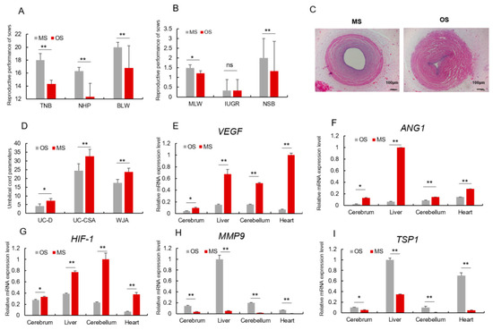
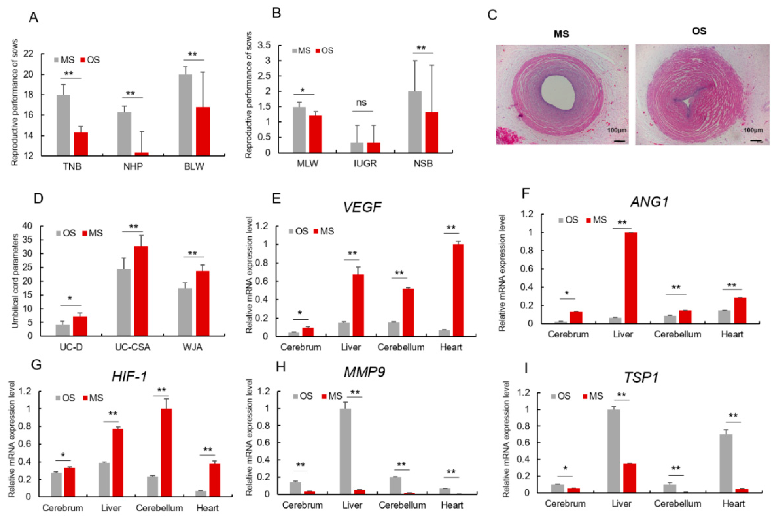
Figure 2.
Angiogenesis of OS pig was repressed. (A,B) Reproductive performance of OS and MS sows. (C) The morphological characteristics of Umbilical Cord, 40×, scale = 100 μm, umbilical vein of MS; umbilical vein of OS. (D) Umbilical vein diameter (UV-D), umbilical vein cross-sectional area (UV-CSA), Wharton’s jelly (WJA). (E–G) mRNA relative expression level of pro-angiogenesis genes (VEGF, ANG1, HIF-1) in both MS and OS piglet’s tissues. (H,I) mRNA relative expression level of angiogenesis-inhibiting genes (MMP9, TSP1) in both MS and OS piglet’s tissues. * or ** represents significance at the 0.05 or 0.01 level, respectively, and ns represents no significance.
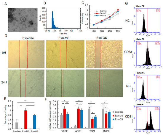
Figure 3.
The pro-angiogenesis role of Exo-OS was repressed. (A) The 3D morphology of isolated pig UCB-EXO observed in electron microscope. (B) The line profile of electron microscope image size for UCB-EXO. X- and Y-axes are the size and events, respectively. (C) Cell viability was measured following Exo-OS, Exo-MS, or PBS treatment for 24 h, using the CCK8 analysis. (D) The migration of HUVEC cells in vitro after 24 h of treatment with Exo-MS, Exo-OS, or PBS was evaluated using scratch assay (n = 3). (E) Percentage of cells healed. (F) The expression level of VEGF, ANG1, TSP1, and MMP9 mRNA after treating Exo-MS, Exo-OS, or PBS. (G) Positive rate of exosome marker proteins CD63 and CD81 detected by flow cytometry. * or ** represents significance at the 0.05 or 0.01 level, respectively.
Reference
- Pu, Q.; Chai, J.; Chen, L.; Liu, C.; Yang, C.; Huang, Y.; Luo, J. Exosome miRNA Expression in Umbilical Cord Blood of High-Parity Sows Regulates Their Reproductive Potential. Animals 2022, 12, 2456. [Google Scholar] [CrossRef] [PubMed]

Disclaimer/Publisher’s Note: The statements, opinions and data contained in all publications are solely those of the individual author(s) and contributor(s) and not of MDPI and/or the editor(s). MDPI and/or the editor(s) disclaim responsibility for any injury to people or property resulting from any ideas, methods, instructions or products referred to in the content. |
© 2024 by the authors. Licensee MDPI, Basel, Switzerland. This article is an open access article distributed under the terms and conditions of the Creative Commons Attribution (CC BY) license (https://creativecommons.org/licenses/by/4.0/).