Article

A Description of Echinochasmus pseudobeleocephalus n. sp. (Echinochasmidae) Based on Morphological and Molecular Data

by
Kristina Andreevna Kalinina
1,*,
Vladimir Vladimirovich Besprozvannykh
1,
Yulia Viktorovna Tatonova
1 and
Mikhail Yurievich Shchelkanov
1,2
1
Federal Scientific Center of the East Asia Terrestrial Biodiversity, Far Eastern Branch, Russian Academy of Sciences, pr-t 100-letiya Vladivostoka 159a, Vladivostok 690022, Russia
2
G.P. Somov Research Institute of Epidemiology and Microbiology, Russian Federal Service for Surveillance on Consumer Rights Protection and Human Wellbeing, Selskaya St. 1, Vladivostok 690022, Russia
*
Author to whom correspondence should be addressed.
Animals 2023, 13(20), 3236; https://doi.org/10.3390/ani13203236
Submission received: 18 August 2023 / Revised: 21 September 2023 / Accepted: 7 October 2023 / Published: 17 October 2023
(This article belongs to the Section Animal Genetics and Genomics)

Abstract

:

Simple Summary

Far Eastern trematodes of the genus Echinochasmus were studied. As the analysis of the nuclear 28S rRNA gene sequence showed, the examined Far Eastern individuals did not belong to the species E. beleocephalus despite their morphological similarities and represented a new species, Echinochasmus pseudobeleocephalus n. sp. An analysis of phylogenetic relationships in Echinochasmidae supported their status as an independent species. The subdivision of individuals of the genus Echinochasmus into two groups was also confirmed on the basis of the number of head-collar spines and the tail length in cercariae.

Abstract

Adult individuals of Echinochasmus pseudobeleocephalus n. sp. were obtained during an experimental study on trematodes’ life cycle. An analysis of the morphometric characteristics of the developmental stages and involvement of first intermediate hosts, snails of the genus Boreoelona, in their life cycle, revealed the identity of the obtained trematodes to the European species Echinochasmus beleocephalus previously discovered in the south of the Russian Far East. However, an analysis of molecular data, in particular sequences of the 28S rRNA gene, showed that the Far Eastern trematodes examined do not belong to European E. beleocephalus despite their morphological similarities. An analysis of phylogenetic relationships within the family Echinochasmidae supported the status of E. pseudobeleocephalus n. sp. as an independent species. Our new data confirmed that the individuals attributed to Echinochasmus can be subdivided into two groups on the basis of the number of head-collar spines and the tail length in cercariae on an intergeneric level.

1. Introduction

The trematode family Echinochasmidae Odhner 1910 comprises numerous species that in their mature stage parasitize mammals including humans, birds, and, less commonly, reptiles [1,2]. It is also known that members of this family use only prosobranch snails as first intermediate hosts and mostly fish as second intermediate hosts. In some cases, in addition to fish, mollusks and tadpoles can be involved in their life cycle as second intermediate hosts [3,4].
On both specific and higher levels, the taxonomy of Echinochasmidae is mainly based on the morphological characters of mature individuals. Molecular data have been obtained for a relatively small number of species in this family. However, these data allowed Tkach et al. (2016) [5] to place the species combined in the subfamily Echinochasminae Odhner, 1910 into a separate family, Echinochasmidae Odhner, 1910 [2]. In addition, differences in molecular characteristics were found between individuals of Echinochasmus Dietz, 1909 on the generic level. Some of them had 24 head-collar spines and cercariae with a tail length comparable to the body length, while in others cercariae had a longer tail and adult worms had 20–22 spines. The latter clustered with species of the genus Stephanoprora Odhner 1902 [5,6,7].
During a parasitological study of freshwater prosobranch snails of the family Bithyniidae Gray, 1857, collected in a lake of the Arsenyevka River basin (Primorsky Krai, Russia), we found snails emitting cercariae that were morphologically similar to cercariae of the family Echinochasmidae. The subsequent experimental completion of their life cycles, a study of their developmental stages, and an analysis of molecular markers have shown that the trematodes belong to a species of the genus Echinochasmus. The results of the study are presented below.

2. Materials and Methods

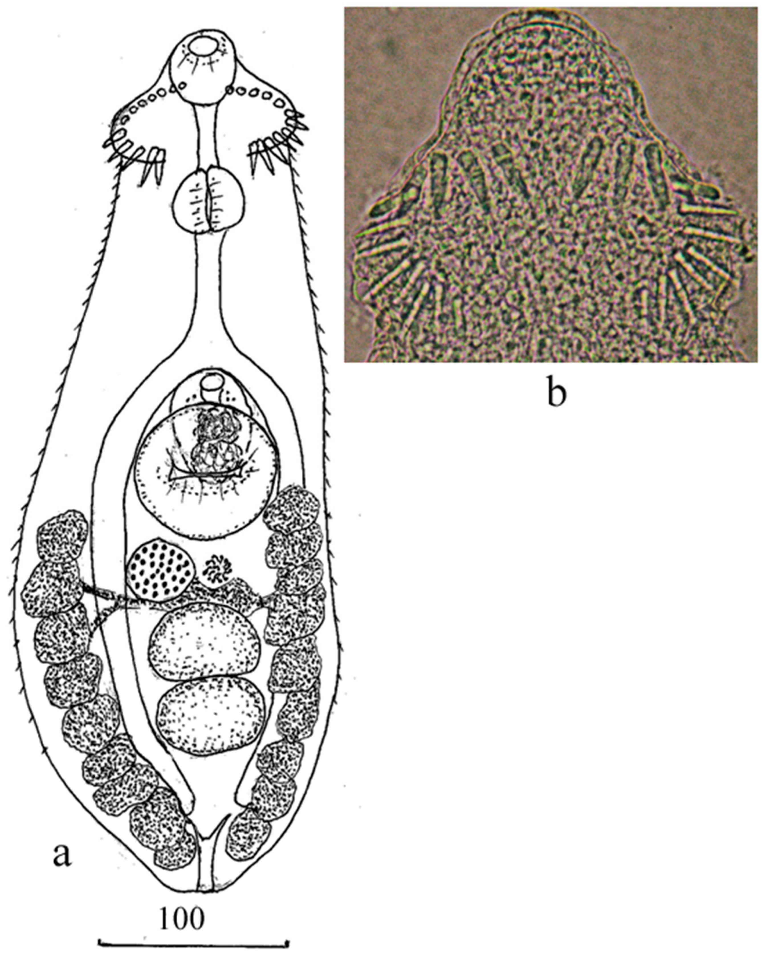
2.1. Life Cycle and Morphology of Adult Individuals

One snail Boreoelona ussuriensis (Ehrmann in Büttner and Ehrmann, 1927) that emitted cercariae similar in morphological parameters to short-tailed cercariae of Echinochasmidae was found among the examined prosobranch snails of the family Bithyniidae collected in a lake of the Arsenyevka River basin. A map of sampling location is provided in the Supplementary Material. According to the available information about the life cycles of Echinochasmidae in the south of the Russian Far East [4], tadpoles are one of the second intermediate hosts for echinochasmids with short-tailed cercariae. To obtain metacercariae from emitted cercariae, tadpoles of Rana dybowskii Günther, 1876 caught in an artificial pond were used. First, 50 tadpoles from this pond were dissected to confirm the absence of trematode metacercariae. Three tadpoles were placed in a Petri dish with cercariae being emitted from the snail into the water. One day later, encysted cercariae were found in tadpoles’ visceral tissues. After that, the snail emitting cercariae was placed with 10 tadpoles in a container filled with 500 mL water. After a 4-h exposure to cercariae, the tadpoles were transferred into another container. On day 24 post-exposure, one of the infected tadpoles was dissected, and 12 metacercariae were obtained from its visceral tissue, which were morphologically similar to those of the genus Echinochasmus. Other tadpoles were fed to a duckling and a chicken: four tadpoles to each bird. After 8 days, 23 mature flukes were found in the small intestine of the duckling, while there were no flukes in the chicken.
The obtained adult individuals were washed, fixed in 70% ethanol, and then some of them were transferred to 96% ethanol for further DNA isolation. Whole mounts of the adult flukes were prepared by staining with carmine alum, dehydrating in a graded ethanol series, clearing in clove oil, and embedding in Canada balsam. All measurements are in micrometers (µm).

2.2. DNA Extraction, Amplification, and Sequencing

In the genetic analysis, one cercaria and two adult specimens representing the genus Echinochasmus were used. The adult specimens were obtained from cercariae through the experimental completion of the life cycle. DNA was extracted by the HotSHOT method [8]. Partial sequences of the 28S rRNA gene (28S) were amplified using the specific primers Digl2 (5′-AAG-CAT-ATC-ACT-AAG-CGG-3′, forward) and 1500R (5′-GCT-ATC-CTG-AGG-GAA-ACT-TCG-3′, reverse) [9]. For sequencing, internal primers were used: 900F (5′-CCG-TCT-TGA-AAC-ACG-GAC-CAA-G-3′, forward) [10] and 1200R (5′-GAA-GGA-CGA-ATC-GCT-TCG-TG-3′, reverse) [11]. To amplify the ITS2 spacer region, the following primers were used: forward 1/F [12] and reverse BD2 (5′-ATC-TAG-ACC-GGA-CTA-GGC-TGT-G-3′) [13]. The ITS2 spacer region was sequenced using external primers.
The resulting nucleotide sequences were visually checked using the FinchTV ver. 1.4.0 and aligned manually in MEGA ver. 5.03 [14]. Sequences of other members of the family Echinochasmidae were accessed from GenBank (Table 1).
The length of the sequences used in the phylogenetic analysis was 731 bp taking into account alignment. Phylogenetic relationships were reconstructed using the Bayesian Inference (BI) algorithm in the MrBayes (BI) program [20] and the Maximum Likelihood (ML) algorithm in the PhyML 3.1 program. The optimal model GTR + I + G based on the Akaike information criterion was obtained in the jModeltest 2.1.5 program [21]. For the former (BI) phylogenetic reconstruction, 500,000 generations were performed; in the ML analysis, 100 repetitions were used. Both phylogenetic reconstructions had a similar topology, and, therefore, a consensus tree is presented here. Genetic distances (p-distances) between individual sequences and clusters were calculated using MEGA ver. 5.03.

3. Results

Echinochasmus pseudobeleocephalus n. sp.
Syn. Echinochasmus beleocephalus [4].
Host: Anas platyrhynchos dom. (experimental host).
Localization: small intestine.
Intensity of infection: 23 specimens.
First intermediate host: Boreoelona ussuriensis.
Second intermediate host: tadpoles of Rana dybowskii (experimental host).
Localization: visceral tissue.
Type locality: lake in the Arsenyevka River basin, south of the Russian Far East (44°44′ N, 133°57′ E).
Type-deposition: holotype No. 222-Tr; paratype Nos. 223-228-Tr.
This material was deposited in the parasitological collection of the Zoological Museum (Federal Scientific Center of the East Asia Terrestrial Biodiversity, Far Eastern Branch, Russian Academy of Sciences, Vladivostok, Russia) on 22 November 2022; e-mail: [email protected].
Etymology: The species epithet indicates a coincidental visual similarity to Echinochasmus beleocephalus.
Adult worm (based on seven specimens; Figure 1; Table 2). The body was elongated, covered by spines from the anterior end to the level of the posterior testis, with the most densely concentrated spines in the anterior third of the body. Oral sucker subterminal. Head-collar with 24 spines, arranged into single row interrupted dorsally. Prepharynx long; pharynx oval or rounded; esophagus longer than prepharynx. Intestinal bifurcation immediately anteriorly of cirrus-sac. Intestinal branches terminate, slightly separated from the posterior end of the body. Ventral sucker oval or rounded, in the middle third of the body. Testes two, tandem or slightly oblique, transversely-oval, adjacent to each other, in the posterior third of the body. Cirrus-sac oval, at median line of body and partly covered by ventral sucker. Internal seminal vesicle bipartite. Genital pore between intestinal bifurcation and anterior margin of ventral sucker. The ovary was rounded or transversely oval, sinistrally to the median line of the body, adjacent to the anterior testis, or at some distance anteriorly of the testis. Uterine seminal receptacle present. Mehlis’ gland left to the ovary, between the ventral sucker and anterior testis. Uterus short, located between the caeca, posterior margin of ventral sucker and anterior margin of the anterior testis, usually containing one or two large eggs. Vitelline fields lateral, extending from the level of the middle of the ventral sucker to the posterior end of the body. The vitelline reservoir on the median line of the body at the level of the anterior end of the anterior testis. Excretory vesicle Y-shaped. Stem of excretory vesicle short.
Molecular data. The nucleotide sequences of 28S with a length of 1020 bp were identical between the three Echinochasmus specimens (one cercaria and two adult specimens).
The complete nucleotide sequences of the ITS2 region with a length of 695 bp were obtained. The resulting sequences were identical to that of the species Echinochasmus japonicus (MT268119). Thus, further analysis for this marker was not performed.

4. Remark

4.1. Morphological Identification

In their morphological characteristics, the experimentally obtained adult trematodes were similar both to Echinochasmus japonicus Tanabe, 1926 and to E. beleocephalus (Linstow, 1873). The former species was first discovered in Japan; subsequently, individuals identified as E. japonicus were found in Western Siberia, in the south of the Russian Far East, and in Vietnam [3,4,15,24]. Data on the life cycle and morphology of developmental stages were obtained for trematodes from each of the above-listed regions. The specimens of E. japonicus found in these regions differed slightly from each other in morphology, both at the stage of cercariae and at the adult stage. At the former stage, the main morphological differences between them were the presence of cuticular formations on the oral and ventral suckers in the specimens from Vietnam and Western Siberia and the absence of these formations in the Japanese and Russian Far Eastern specimens [3,4,15,25]. At the adult stage, there were differences in the number of head-collar spines: the Far Eastern individuals had 22 spines vs. 24 spines in individuals from other regions. The first intermediate hosts for Japanese, Vietnamese, and Russian Far Eastern trematodes were snails of the genus Parafossarulus; for West Siberian trematodes, snails of the genus Codiella Locard, 1894, also belonging to Bithyniidae.
Another representative of Echinochasmus from the Far Eastern region of Russia was attributed to the European species E. beleocephalus. It was identified on the basis of the life cycle and morphology of developmental stages [4,26,27]. However, these trematodes were also identical to the E. japonicus specimens from Vietnam and Western Siberia in the morphology of cercariae and adult individuals and had similarities with E. japonicus from Japan [24] and the Far East of Russia. They differed from the Japanese E. japonicus by the presence of cuticular formations on the suckers of cercariae and had a different number of spines on the head collar compared to those in E. japonicus from the Russian Far East (24 vs. 22, respectively).
As for the metric characteristics, adult trematodes identified as E. japonicus and E. beleocephalus had significant differences in the sizes of the body, oral and ventral suckers, pharynx, and cirrus sac (Table 2). For example, the E. japonicus individuals from Japan had lower values of most of these parameters than E. japonicus from the Russian Far East; the Vietnamese individuals of this species had smaller sizes of body length and cirrus sacs than the trematodes from both Japan and Russia. These sizes in the European E. beleocephalus were larger than in the Russian Far Eastern E. beleocephalus. However, in most of the parameters, the former individuals had the strongest metric similarity with E. japonicus from the Russian Far East (and differed only in the number of head-collar spines, as mentioned above), and the latter were similar to the Vietnamese E. japonicus (Table 2).
On the basis of the morphological characteristics of the developmental stages, the metric characteristics of adult trematodes (Table 2), and the features of their life cycles, the individuals obtained in the present study were identical to the Far Eastern flukes previously referred by Besprozvannykh (2009) [4] to as E. beleocephalus. Based on the above-listed morphological characteristics, the trematodes in our material, as well as those from the European and East Asia regions identified as E. japonicus and E. beleocephalus, are most probably representatives of a group of cryptic species. Among them, the individuals from the Far East of Russia identified as E. japonicus and having 22 head-collar spines (unlike other E. japonicus individuals), as well as E. beleocephalus individuals with 24 spines, most likely represented a separate species. For the definitive determination of species affiliation of the East Asian trematodes under study, molecular data for E. japonicus from the type locality are required. However, to date, nucleotide sequences have been obtained only for the specimens identified as E. japonicus from Vietnam and for those identified as E. beleocephalus from Europe.

4.2. Molecular Identification

The reconstruction of phylogenetic relationships among Echinochasmidae using 28S sequences showed a subdivision of members of this family into two clusters with an intergeneric distance between them being 6.3% (without a member of the genus Microparyphium, removed from the analysis (Figure 2)). The level of genetic distances between Microparyphium facetum and representatives of Echinochasmus from Cluster 1 were in a range from 5 to 6%. The distances between Echinochasmus species grouped into Cluster 1 ranged from 0.3 to 2%. In Cluster 2, where members of the family attributed to Echinochasmus were grouped with Stephanoprora, the differences ranged from 0.2 to 1.9% (Table 3).
In Cluster 1, the individuals obtained in this study were grouped with four other species of Echinochasmus and a representative of Microparyphium. However, they formed a single branch with E. japonicus from Vietnam with a genetic distance between them being 0.3%, which was minimal within the Cluster. The European E. beleocephalus and Echinochasmus sp. 1, not identified as species, formed a separate branch that occupied an external position relative to our specimens and E. japonicus from Vietnam with 0.7% differences from these two species. The rest of the species from Cluster 1 held an external position relative to all the above-mentioned representatives of Echinochasmus with a range of distances from 0.7 to 2%. Judging by the values of interspecies differences within the family Echinochasmidae, previously estimated by analyses of 28S in the studies of Schwelm et al. (2020, 0.2–1.5%) [16] and Tatonova et al. (2020, 0.2–2.6%) [7], the specimens of E. japonicus from Vietnam and those in our material were representatives of a different Echinochasmus species. Therefore, we attributed them to a new species, Echinochasmus pseudobeleocephalus n. sp.

5. Discussion

The results of the phylogenetic reconstruction of relationships in the family Echinochasmidae including new data for representatives of Echinochasmus were consistent with those presented in the publications by Tkach et al. (2016) [5] and Tatonova et al. (2020) [7] and confirmed the identification of the clusters that comprised representatives of Echinochasmus and Stephanoprora with similar numbers of head-collar spines and morphologies of cercariae. However, as in the studies by Tkach et al. (2016) [5] and Tatonova et al. (2020) [7], it was found that the genetic distances within the clusters in the reconstruction based on 28S were in the range of intrageneric level, while the difference between the clusters reached the intergeneric level. In Cluster 1, which included the Echinochasmus species, the adoral disk of mature individuals was equipped with 24 head-collar spines, and cercariae had short tails, equal to or slightly longer than the body length. The species affiliation of one of the specimens included in this cluster, identified as Echinochasmus bursicola, remains unresolved. The trematodes with 20, 22, and 24 head-collar spines were described as this species [1,23,27,28,29,30]. Tkach et al. (2016) [5] obtained molecular data for individuals with 24 spines from Ukraine and attributed them to E. bursicola. Nucleotide sequences of 28S identical to those obtained by Tkach et al. (2016) [5] were also found in short-tailed cercariae of Echinochasmus from snails Bithynia tentaculata collected in Germany [16]. On the basis of this marker, the trematodes found in Germany at the cercarial stage were also attributed to E. bursicola. The vast majority of studies on echinochasmids showed that different species are characterized by a different number of head-collar spines. If we assume that the adult trematodes in the material of Tkach et al. (2016) [5] were, indeed, E. bursicola, then the rest of the individuals with 20 and 22 spines should belong to other species. This also applies to the Russian Far Eastern trematodes identified as E. japonicus but having 22 head-collar spines, in contrast to the individuals of this species with 24 spines described by Tanabe (1926) [31]. As for 28S, this marker does not always provide reliable species differentiation for echinochasmids. There is a report about the identity of this gene sequence between specimens belonging to different Echinochasmidae species [7]. In view of the considerations above, the question as to which individuals belong to E. bursicola and which do not, in our opinion, still remains open.
Cluster 2 includes representatives of Echinochasmus and Stephanoprora with 20–22 head-collar spines and long-tailed cercariae. In this group of echinochasmids, seven species were identified. However, only for five of them, the determination of species affiliation was based on both the morphology of developmental stages and nucleotide sequences, which were obtained in the same studies. The set of such data provides sufficient accuracy in the attribution of genetic characteristics to the species identified. Unfortunately, for a number of specimens in Cluster 2, including those with identified species, there were no morphological data. This greatly complicates the resolution of their taxonomy and the determination of the level of relationships with other individuals of Echinochasmidae. For example, the morphology and photographs of the cercariae of Echinochasmus sp. 1 (MN726946 and MN726947) [16] indicate their similarity with cercariae of Stephanoprora, to which they may probably belong. Furthermore, there is still no complete clarity as regards the trematodes identified as E. donaldsoni Beaver, 1941, for which partial 28S have been sequenced but no morphological characteristics have been provided for the studied individuals. If these specimens actually belong to E. donaldsoni, then the question remains as to whether the morphology of their cercariae matches that of the specimens grouped in Cluster 2. Thus, according to Beaver (1941) [32], the tail length in cercariae of E. donaldsoni is equivalent to the body length, while Yamaguti (1975) [22] described the tail of E. donaldsoni cercariae as “tail powerful with annular ridges throughout its length when contracted”, which is typical of Stephanoprora cercariae. However, judging by the sizes that the author reported [22] for these cercariae, the lengths of their tails are equivalent to the body length. Therefore, in our opinion, additional information including both morphological and molecular data is required to clarify the taxonomic affiliation of the specimen identified as E. donaldsoni (KT956930).
It should also be noted that the principle of differentiation between Echinochasmidae on the basis of morphological criteria is also followed in reconstructions using other nuclear markers [7]. Moreover, if relationships between individuals of the family in the clusters based on 28S have an intrageneric level, and an intergeneric level is observed between individuals of different clusters, then echinochasmids are divided into groups of a higher taxonomic rank on the basis of another nuclear marker, ITS2 rDNA region [7]. However, in view of the limited amount of morphological and molecular data available to date for members of Echinochasmidae, claiming these taxonomic problems as resolved is premature. The taxonomy of this large group of trematodes will only be reliable if both morphological and molecular studies cover a sufficiently large number of species of this family.

6. Conclusions

In this study, a life cycle experiment was set up and the morphology of the developmental stages of an allegedly new trematode species from the Russian Far East was examined. As the new molecular data have shown, the obtained individuals from the genus Echinochasmus represent a new species despite its morphological characteristics similar to those of the European E. beleocephalus. Thus, the Far Eastern trematode has been given a new scientific name, Echinochasmus pseudobeleocephalus. Apparently, Echinochasmus pseudobeleocephalus n. sp. and E. japonicus and E. beleocephalus from different regions are a group of cryptic species whose successful differentiation requires a combination of data on the morphology of developmental stages and the molecular characteristics of individual species obtained in a single study.

Supplementary Materials

The following supporting information can be downloaded at: https://www.mdpi.com/article/10.3390/ani13203236/s1, Figure S1. Sampling location (the Arsenyevka river) of Echinochasmus pseudobeleocephalus n.sp. in this study.

Author Contributions

K.A.K. conducted the study, wrote the first, draft version of the manuscript, and took part in the writing of the final version. V.V.B. and Y.V.T. supervised the study and made substantial contributions to the analysis and all versions. M.Y.S. made a significant contribution to the versions of the manuscript. All authors have read and agreed to the published version of the manuscript.

Funding

The research was carried out within the framework of the state assignment from the Ministry of Science and Higher Education of the Russian Federation (topic No. 121031000154-4) and government assignment no. 123022200035-0 “The structure of natural foci of parasitic diseases in the south of the Russian Far East”.

Institutional Review Board Statement

Euthanasia of animals was carried out in accordance with the resolution of the Committee on the Ethics of Animal Experiments, Federal Scientific Center of the East Asia Terrestrial Biodiversity, FEB RAS, Russia, and with the ethical standards of the national guides. Permit Number: 1 of 25.04.2022.

Informed Consent Statement

Not applicable.

Data Availability Statement

The obtained sequences of the 28S gene for the new species were deposited in the National Center for Biotechnology Information database (NCBI, https://www.ncbi.nlm.nih.gov, accessed on 18 August 2023).

Acknowledgments

The experiments described in this study were set up using the equipment at the Instrumental Centre for Biotechnology and Gene Engineering, Federal Scientific Center of the East Asia Terrestrial Biodiversity, Far Eastern Branch, Russian Academy of Sciences (Vladivostok, Russia).

Conflicts of Interest

The authors declare no conflict of interest.

References

  1. Skrjabin, K.I.; Bashkirova, E.Y. Family Echinostomatidae Diestz, 1909. Trematodes Animals and Humans. Fundamentals of Trematodology; Publishing House of the Academy of Sciences of the USSR: Moscow, Russia, 1956; Volume 21, pp. 51–915. (In Russian) [Google Scholar]
  2. Kostadinova, A.; Jones, A.; Bray, R.A.; Gibson, D.I. Family Psilostomidae Looss, 1900. Keys to the Trematoda; Jones, A., Bray, R.A., Gibson, D.I., Eds.; CABI Publishing and the Natural History Museum: Sofia, Bulgaria, 2005; Volume 2, pp. 99–118. [Google Scholar]
  3. Filimonova, L.V. On the phenomenon of amphixia in trematodes Echinochasmus japonicus (Echinostomatidae). J. Zool. 1979, 58, 1103–1110. (In Russian) [Google Scholar]
  4. Besprozvannykh, V.V. Life cycles of trematodes Echinochasmus japonicus and E. beleocephalus (Echinostomatidae) in conditions of Primorsky Region. Parazitologia 2009, 43, 248–258. (In Russian) [Google Scholar]
  5. Tkach, V.V.; Kudlai, O.; Kostadinova, A. Molecular phylogeny and systematics of the Echinostomatoidea Looss, 1899 (Platyhelminthes: Digenea). Int. J. Parasitol. 2016, 46, 171–185. [Google Scholar] [CrossRef]
  6. Besprozvannykh, V.V.; Ermolenko, A.V. Stephanoprora chasanensis n. sp. (Digenea: Echinochasmidae): Morphology, life cycle, and molecular data. Int. J. Parasitol. 2017, 66, 863–870. [Google Scholar] [CrossRef]
  7. Tatonova, Y.V.; Izrailskaia, A.V.; Besprozvannykh, V.V. Stephanoprora amurensis sp. nov., Echinochasmus milvi Yamaguti, 1939 and E. suifunensis Besprozvannykh, 1991 from the Russian southern Far East and their phylogenetic relationships within the Echinochasmidae Odhner 1910. Parasitology 2020, 147, 1469–1479. [Google Scholar] [CrossRef]
  8. Truett, G.E.; Heege, P.; Mynatt, R.L.; Truett, A.A.; Walker, J.A.; Warman, M.L. Preparation of PCR–quality mouse genomic DNA with hot sodium hydroxide and tris (HotSHOT). Biotechniques 2000, 29, 52–54. [Google Scholar] [CrossRef]
  9. Tkach, V.V.; Timothy, D.; Littlewood, J.; Olson, P.D.; Kinsella, J.M.; Swiderski, Z. Molecular phylogenetic analysis of the Microphalloidea Ward, 1901 (Trematoda: Digenea). Syst. Parasitol. 2003, 56, 1–15. [Google Scholar] [CrossRef]
  10. Lockyer, A.E.; Olson, P.D.; Ostergaard, P.; Rollinson, D. The phylogeny of the Schistosomatidae based on three genes with emphasis on the interrelationships of Schistosoma Weinland, 1858. Parasitology 2003, 126, 203–224. [Google Scholar] [CrossRef]
  11. Sawabe, T.; Kita–Tsukamoto, K.; Thompson, F.L. Inferring the evolutionary history of vibrios by means of Multilocus Sequence Analysis. J. Bacteriol. 2007, 21, 7932–7936. [Google Scholar] [CrossRef]
  12. Krieger, J.; Fuerst, P.A. Evidence of multiple alleles of the nuclear 18S ribosomal RNA gene in sturgeon (Family: Acipenseridae). J. Appl. Ichthyol. 2002, 18, 290–297. [Google Scholar] [CrossRef]
  13. Bowles, J.; Blair, D.; McManus, D.P. A molecular phylogeny of the human schistosomes. Mol. Phylogenet. Evol. 1995, 4, 103–109. [Google Scholar] [CrossRef]
  14. Tamura, K.; Peterson, D.; Peterson, N.; Stecher, G.; Nei, M.; Kumar, S. MEGA5: Molecular evolutionary genetics analysis using maximum likelihood, evolutionary distance, and maximum parsimony methods. J. Mol. Struct. 2011, 28, 2731–2739. [Google Scholar] [CrossRef]
  15. Besprozvannykh, V.V.; Ermolenko, A.V.; Ngo, H.D.; Ha, N.V.; Hung, N.M.; Rozhkovan, K.V. Descriptions of digenean parasites from three snail species, Bithynia fuchsiana (Morelet), Parafossarulus striatulus Benson and Melanoides tuberculata Müller, in North Vietnam. Helmintologia 2013, 50, 190–204. [Google Scholar] [CrossRef]
  16. Schwelm, J.; Kudlai, O.; Smit, N.J.; Selbach, C.; Sures, B. High parasite diversity in a neglected host: Larval trematodes of Bithynia tentaculata in Central Europe. J. Helminthol. 2020, 94, e120. [Google Scholar] [CrossRef]
  17. Staneviciute, S.; Virmantas, S.; Romualda, P. Phylogenetic relationships of some species of the family Echinostomatidae Odner, 1910 (Trematoda), inferred from nuclear rDNA sequences and karyological analysis. Comp. Cytogenet. 2015, 9, 257–270. [Google Scholar] [CrossRef] [PubMed]
  18. Tkach, V.V.; Pawlowski, J.; Mariaux, J. Phylogenetic analysis of the suborder Plagiorchiata (Platyhelminthes, Digenea) based on partial lsrDNA sequences. Int. J. Parasitol. 2000, 30, 83–93. [Google Scholar] [CrossRef] [PubMed]
  19. Tkach, V.V.; Mariaux, J.; Swiderski, Z. Molecular phylogeny of the suborder Plagiorchiata and its position in the system of Digenea. In Interrelationships of the Platyhelminthes. In Interrelationships of the Platyhelminthes; CRC Press: Boca Raton, FL, USA, 2001; Volume 17, pp. 186–193. [Google Scholar]
  20. Ronquist, F.; Huelsenbeck, J.P. MrBayes 3: Bayesian phylogenetic inference under mixed models. Bioinformatics 2003, 19, 1572–1574. [Google Scholar] [CrossRef] [PubMed]
  21. Darriba, D.; Taboada, G.L.; Doallo, R.; Posada, D. jModelTest 2: More models, new heuristics and parallel computing. Nat. Methods 2012, 9, 772. [Google Scholar] [CrossRef]
  22. Yamaguti, S. A Synoptical Review of Life Histories of Digenetic Trematodes of Vertebrates: With Special Reference to the Morphology of Their Larval Forms; Kaigaku Pub Co.: Tokyo, Japan, 1975; pp. 1–590. [Google Scholar]
  23. Bashkirova, V.Y. Echinostomatids of birds of the USSR and a review of the cycles of their development. Bashkir NIVS 1941, 3, 243–300. (In Russian) [Google Scholar]
  24. Yamaguti, S. Studies on the helminth fauna of Japan. Part 25. Trematodes of birds IV. Biology 1939, 8, 130–210. [Google Scholar]
  25. Ito, J. A monograph of cercaria in Japan and adjacent territories. Prog. Med. Parasitol. Jpn. 1964, 1, 395–550. [Google Scholar]
  26. Karmanova, E.M. Knowledge of the life cycle of the trematodes Echinochasmus coaxatus Dietz, 1909 and E. beleocephalus (Linstow, 1873) (Echinostomatidae). Tr. GELAN 1974, 24, 46–53. (In Russian) [Google Scholar]
  27. Oshmarin, P.G.; Dotsenko, T.K. About Epizootology of Helminthosises among DOMESTIC Birds in Lowlying Land Near Khanka Lake; Soobchenia DVF AN SSSR: Moscow, Russian, 1951; Volume 3, pp. 8–11. (In Russian) [Google Scholar]
  28. Braun, M. The so–called “free–swimming sporocyst”. Centralbl. Bakt. Parasit. 1891, 10, 215–219. (In Germany) [Google Scholar]
  29. Dietz, E. Die Echinostomiden der Vögel. Zoologische Jahrbücher 1910, 12, 265–512. [Google Scholar]
  30. Odhner, T. Nordostafrikanische Trematoden, grösstenteils vom Weissen Nile. I. Fascioliden. Results of the Swedish Zoological Expedition to Egypt and the White Nile, 1901, under the Direction of LA Jägerskiöld, No. 23A (1910) 1-170. Die Homologien der weiblichen Genitalwege beiden Trematoden und Cestoden. Zool. Anz. 1912, 23, 1–170. [Google Scholar]
  31. Tanabe, H. A new species Echinochasmus, E. japonicus n. sp. Nippon Byor. Gakk. Shi. 1926, 16, 295–296. [Google Scholar]
  32. Beaver, P.C. Studies on the life history of Echinochasmus donaldsoni n. sp., a trematode (Echinochasmidae from the Pied—Billed grebe. J. Parasitol. 1941, 27, 347–355. [Google Scholar] [CrossRef]
Figure 1. Echinochasmus pseudobeleocephalus n. sp. (a). Adult worm, (b). Head-collar.
Animals 13 03236 g001
Figure 2. The phylogeny of the family Echinochasmidae based on the 28S rRNA gene sequences (1020 bp) using the Bayesian Inference (BI) algorithm. Values of a posterior probability ≥ 60 for BI and nodal support of ≥0.60 for Maximum Likelihood (ML) are shown at the nodes, respectively. The scale bar indicates the number of substitutions per site. The sequences obtained in the present study are highlighted in bold. The outgroup species are listed in Table 2.
Animals 13 03236 g002
Table 1. List of species used in the analysis with sequences of the 28S rRNA gene and other associated information.
SpeciesDevelopmental StageAccession Numbers
(GenBank)
ReferenceCountrySequence Length, bp
family Echinochasmidae
Echinochasmus pseudobeleocephalus n. sp.AdultOR076694, OR076695this studyRussia701
CercariaOR076696
Echinochasmus japonicusAdultJQ890579, JQ890580[15]Russia1370
Echinochasmus beleocephalusAdultKT956929[5]Ukraine1178
Echinochasmus sp. 2CercariaMN726948[16]Germany1199
Echinochasmus coaxatusAdultKJ542643[17]Ukraine;969
AdultKT956928[5]Ukraine1200
CercariaMN726944[16]Germany1200
Echinochasmus bursicolaAdultKT956938[5]Ukraine1173
Microparyphium facetumAdultKT956933[5]USA1291
Echinochasmus sp. VS-2012CercariaJQ088098[17]Lithuania1258
Echinochasmus mordaxAdultKT956931[5]Ukraine1175
Stephanoprora chasanensisAdultKT873320, KT873321[6]Russia1369
Echinochasmus sp. AdultKT956932[5]USA1161
Echinochasmus donaldsoniAdultKT956930[5]USA1240
Stephanoprora amurensisAdultMT447050, MT447051[7]Russia1145
Echinochasmus sp. 1CercariaMN726946, MN726947[16]Germany1199
Echinochasmus milviAdultKT873317, KT873318 [6]Russia
Russia
1369
MT447054, MT447055[7]1145
Echinochasmus suifunensisAdultMT447056, MT447057[7]Russia1145
Stephanoprora sp. 1AdultKT956936[5]USA1181
Stephanoprora sp. 2KT956937
Stephanoprora pseudoechinataAdultKT956934, KT956935[5]Ukraine1249
Outgroup
Psilostomum brevicolleAdultKT956950[5]Ukraine1292
Psilochasmus oxyurusAdultAF151940[18]Ukraine1239
Sphaeridiotrema pseudoglobulusAdultKT956957[5]USA1206
Notocotylus attenuatusAdultAF184259[19]Ukraine1261
Table 2. Morphometric parameters of adult Echinochasmus individuals (µm).
CharactersE. pseudobeleocephalus n. sp.E. japonicusE. beleocephalus
This Study[22][3][4][15][23][4]
HolotypeRange (n = 7)Mean
Body length (Bl)454454–554502600–900540–620780–810520–580715–924550–620
Body width (Bw)173139–173152160–180150–180220–250162–235253–330130–170
Bw/Bl (%) *38.127.2–38.130.3
Forebody length (Fo)204204–258234
Fo/Bl (%) **44.944.4–48.646.6
Oral sucker length3931–393638–4240–5145–5023–3547–5134–39
Oral sucker width3535–393738–4240–575627–4247–5139–42
Ventral sucker length6962–817070–9668–9195–11050–65132–14359–70
Ventral sucker width7365–857270–9674–8667–8954–77132–15462–73
Ratio of suckers’ lengths1.781.59–2.101.94
Ratio of suckers’ widths2.091.67–2.091.95
Head-collar width9285–11696
Prepharynx length3531–504030–6046–6828–3415–4233–3848–50
Pharynx length3535–423835–3934–4667–8419–2751–5628–40
Pharynx width3931–393427–3234–5145–5023–3151–5634–39
Oesophagus length5858–10078110–21097–12095–13092–96132–19867–130
Ovary length35–3931–393436–4840–5141–60 35–4238–4334–42
Ovary width31–3527–353222–3046–6738–4935–424731–50
Anterior testis length4235–624860–75 40–6883–13558–8043–8850–70
Anterior testis width5853–776154–8034–46100–13265–9234–8859–80
Posterior testis length4239–6247
Posterior testis width5846–6259
Cirrus sac length5858–696175–9091–13050–6586–13262–81
Cirrus sac width4639–464136–4860–7442–5866–7734–48
Post-testicular field length6958–6965
Egg length deformed 77–9063–8684–898073–8184
Egg width 51–5746–57505334–4361
Number of head-collar spines242424242422242424
* Bw/Bl, body width as percentage of body length; ** Fo/Bl, forebody length as percentage of body length.
Table 3. Genetic distances (below the diagonal) and standard error estimate (above the diagonal) between species of family Echinochasmidae e based on the 28S rRNA gene sequences.
NoSpecies123456789101112131415161718
Cluster 1 *1Microparyphium facetum 0.0080.0080.0090.0090.0090.0090.0080.0070.0080.0070.0080.0080.0070.0080.0080.0070.008
2Echinochasmus bursicola0.047 0.0040.0030.0030.0040.0040.0090.0090.0090.0090.0090.0090.0090.0090.0090.0080.008
3E. coaxatus0.0590.011 0.0040.0040.0050.0050.0100.0090.0090.0100.0100.0100.0100.0100.0100.0090.009
4Echinochasmus. sp. 20.0540.0070.016 0.0000.0030.0030.0100.0090.0090.0100.0100.0100.0090.0100.0100.0090.009
5E. beleocephalus0.0540.0070.0160.000 0.0030.0030.0100.0090.0090.0100.0100.0100.0090.0100.0100.0090.009
6E. japonicus0.0590.0110.0200.0070.007 0.0020.0090.0090.0090.0090.0090.0090.0090.0100.0100.0090.009
7E. peudobeleocephalus0.0590.0110.0200.0070.0070.003 0.0090.0090.0090.0090.0090.0090.0100.0100.0100.0090.009
Cluster 2 *8Echinochasmus sp. VS-20120.0620.0590.0690.0660.0660.0590.062 0.0040.0030.0040.0040.0040.0040.0050.0050.0030.003
9E. mordax0.0590.0560.0660.0630.0630.0560.0590.011 0.0030.0040.0040.0030.0050.0050.0050.0040.004
10Stephanoprora chasanensis0.0600.0540.0640.0620.0620.0540.0570.0070.007 0.0030.0030.0020.0040.0040.0040.0030.003
11Echinochasmus sp.0.0670.0620.0720.0690.0690.0620.0640.0110.0110.007 0.0030.0030.0050.0050.0050.0040.004
12E. donaldsoni0.0640.0590.0690.0660.0660.0590.0620.0110.0110.0070.006 0.0030.0050.0050.0050.0040.004
13S. amurensis0.0600.0570.0670.0640.0640.0570.0600.0100.0070.0030.0070.007 0.0040.0040.0040.0030.003
14Echinochasmus sp. 10.0590.0560.0660.0630.0630.0590.0620.0140.0140.0100.0170.0170.013 0.0050.0050.0040.004
15E. milvi0.0670.0620.0720.0630.0630.0620.0640.0140.0170.0100.0170.0170.0130.017 0.0000.0040.004
16E. suifunensis0.0670.0620.0720.0630.0630.0620.0640.0140.0170.0100.0170.0170.0130.0170.000 0.0040.004
17Stephanoprora sp.0.0540.0540.0640.0620.0620.0570.0600.0070.0100.0060.0130.0130.0060.0100.0130.013 0.002
18S. pseudoechinata0.0560.0560.0660.0630.0630.0590.0620.0090.0110.0070.0140.0140.0070.0110.0140.0140.001
* Representatives of Clusters 1 and 2 and the distances within the clusters are indicated in green and violet, respectively.
Disclaimer/Publisher’s Note: The statements, opinions and data contained in all publications are solely those of the individual author(s) and contributor(s) and not of MDPI and/or the editor(s). MDPI and/or the editor(s) disclaim responsibility for any injury to people or property resulting from any ideas, methods, instructions or products referred to in the content.

