Communication

First Description of Sarcoptic Mange in a Free-Ranging European Wildcat (Felis silvestris silvestris) from Spain

by
Fernando Nájera
1,2,*,
Elena Crespo
2,3,
Amalia García-Talens
2,4,
Rebeca Grande-Gómez
2,
Francisco Javier Herrera-Sánchez
2,
Michaela Gentil
5,
Carmen Cortés-García
5,
Elisabeth Müller
5,
Rafael Calero-Bernal
6,* and
Luis Revuelta
1
1
Department of Animal Physiology, Faculty of Veterinary Medicine, Complutense University of Madrid, 28040 Madrid, Spain
2
Asistencia Técnica de la Dirección General del Medio Natural y Desarrollo Sostenible de la Junta de Comunidades de Castilla-La Mancha, Plaza del Cardenal Siliceo s/n, 45071 Toledo, Spain
3
“El Chaparrillo” Wildlife Rehabilitation Center, Ctra de Porzuna s/n, 13071 Ciudad Real, Spain
4
“La Alfranca” Wildlife Rehabilitation Center Finca de la Alfranca s/n, Pastriz, 50195 Zaragoza, Spain
5
Laboklin GmbH & Co. KG, Steubenstrasse 4, 97688 Bad Kissingen, Germany
6
SALUVET, Animal Health Department, Faculty of Veterinary Medicine, Complutense University of Madrid, Avenida Puerta de Hierro s/n, 28040 Madrid, Spain
*
Authors to whom correspondence should be addressed.
Animals 2021, 11(9), 2494; https://doi.org/10.3390/ani11092494
Submission received: 2 August 2021 / Revised: 15 August 2021 / Accepted: 23 August 2021 / Published: 25 August 2021
(This article belongs to the Special Issue Wildlife Disease Threats)

Abstract

:

Simple Summary

Sarcoptic mange caused by the mite Sarcoptes scabiei is a worldwide-distributed skin infestation with a wide range of hosts, among them several species within the Felidae family. Sarcoptes scabiei was diagnosed in a dead adult female European wildcat (Felis silvestris silvestris) from Spain. This is the first description of Sarcoptes scabiei in a European wildcat. Since this is a species of conservation concern due to its critical demography in the southernmost population of the Iberian Peninsula, the impacts of infectious diseases, including sarcoptic mange, should be considered during disease surveillance programs of the species’ populations.

Abstract

Sarcoptic mange caused by the mite Sarcoptes scabiei is a worldwide-distributed skin infestation with a wide range of hosts, among them several species within the Felidae family. Sarcoptes scabiei was diagnosed in a dead adult female European wildcat (Felis silvestris silvestris) from Spain, based on histological evaluation of skin biopsies and identification of the arthropod from skin scrapings and molecular methods. This is the first description of Sarcoptes scabiei in a European wildcat. Due to its critical demography in the southernmost population of the Iberian Peninsula, the impacts of infectious diseases, including sarcoptic mange, as a new potential threat should be considered during disease surveillance programs of the species’ populations.

1. Introduction

Sarcoptic mange caused by arthropods of the genus Sarcoptes is a highly contagious skin disease that has been observed to affect nearly 150 species of mammals, leading to acute or chronic forms depending on factors such as host immunity and mite lineage [1,2]. Its relevance relies on an economic, zoonotic and ecological basis [3,4,5]. Additionally, sarcoptic mange could represent a potential threat to be considered within the Carnivore guild, which is already of conservation concern, since 14% (15/108) of the species under suboptimal conservation status within the order Carnivora are considered susceptible to this disease [2]. Sarcoptic mange has been described in several species of wild felids, including the African lion (Panthera leo), the cheetah (Acinonyx jubatus), the serval (Felis serval), the leopard (Panthera pardus), the Eurasian lynx (Lynx lynx) and the Iberian lynx (Lynx pardinus) [6,7].
The European wildcat (Felis silvestris silvestris) remains a widely distributed felid species in Europe, although it is assumed to be an endangered taxa in most of the countries in which it lives [8]. The demographic situation of the southernmost population of the species in the Iberian Peninsula is somewhat critical, primarily due to low density and habitat fragmentation [9]. Under these circumstances, the species may benefit from health surveillance to detect disease threats for the populations. The present study reports the first observation of a case of sarcoptic mange in a European wildcat.

2. Materials and Methods
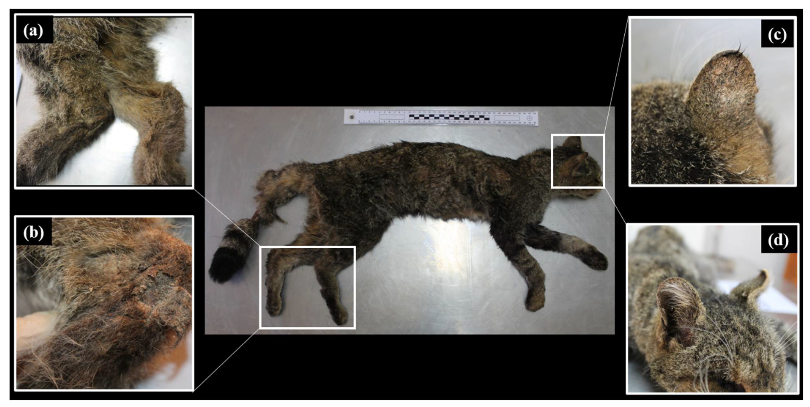
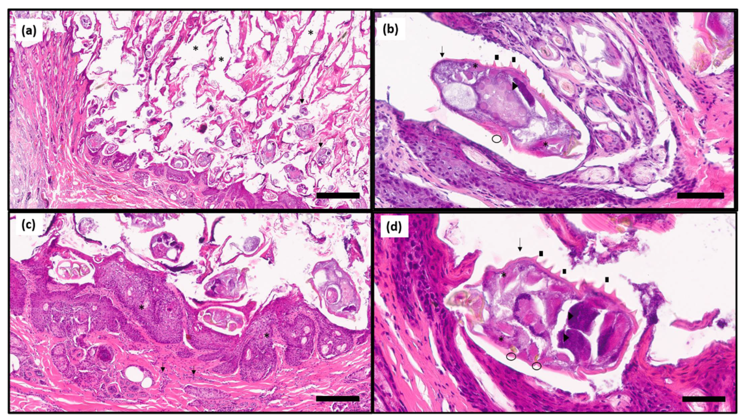
On 1 November 2020, during a routine field surveillance performed under the Iberian lynx conservation and reintroduction program in Castilla-La Mancha (South Central Spain), an adult (>2-year-old) female wildcat was found dead in a private estate that consisted of Mediterranean scrubland in Las Virtudes (Ciudad Real province, 859857X 340946Y). The dead wildcat was found in right lateral recumbency with no signs of struggle and no injuries. The animal was brought to El Chaparrillo Wildlife Rehabilitation Center (Ciudad Real), kept in refrigeration and necropsied within 12 h after harvesting.
On physical examination, we discarded a putative individual based on coat patterns from dorsal, lateral, ventral, head and tail, showing a pelage characteristic typical of true wildcats [10,11,12]. The carcass suffered from an initial stage of decay and was calculated between 3–4 days of postmortem interval. It weighed 2.4 kg and it was given a 1/5 body condition score. Skin lesions were clearly visible on the head, and both tarsus and skin scrapings were taken from both pinnae and tarsus for microscopic examination and direct detection under light microscopy (x40–100). Two 5 × 5 cm patches of skin were submitted to the laboratory to perform molecular testing for S. scabiei via Taqman real-time PCR (qPCR), targeting the internal transcribed spacer (ITS)-2 region (primer Sarco S: 5′-GCT AAA GAA TCC AAG TGC CA-3′, Sarco R: 5′-TCT TTT CCT CCG CTT ATT TAT ATG-3′, probe Sarco TM: 5′-6FAM-CGG GTA TTC TCG CTT GAT CTG AGG TC-BBQ-3′). Prior to nucleic acid extraction, skin samples were incubated in a lysis buffer (containing proteinase K) in “MagNA Lyser Green Bead” tubes (Roche Diagnostics, Mannheim, Germany), which contain ceramic beads and crush the tissue through mechanical disruption (MagNA Lyser, Roche Diagnostics, Mannheim, Germany). Afterwards, automated total nucleic acid extraction was carried out using a commercially available kit (“MagNA Pure 96 DNA and Viral NA Small Volume Kit”, Roche Diagnostics, Mannheim, Germany). Each PCR run included a negative and a positive control, as well as an extraction control in each sample, to check for nucleic acid extraction and PCR inhibition.
Additionally, portions of skin samples were fixed in 10% buffered formalin and submitted to histopathological analyses by standard hematoxylin and eosin staining.
In addition, aiming for ancillary analyses, several tissue samples from mesenteric ganglia, spleen, bone marrow, clot, kidney and lung were taken to perform PCR analysis and/or culture for selected pathogens; blood (1 mL) was also taken to perform feline leukemia virus p27 antigen and feline immunodeficiency antibodies point-of-care enzyme-linked immunosorbent assays, and liver for anticoagulant rodenticide analysis (Table 1).

3. Results

On gross examination, skin revealed thickening of the edge of both pinnae, slight crusting of both pinnae, loss of hair density in nasal and rostral planes and thickening and crusting of the skin of both tibia–tarsal joints. The only alopecic area found in the carcass corresponded to the central area of the tail, but it was considered a post-mortem sign (Figure 1). On examination of the subcutaneous tissue, congestion was observed in the rostral plane, both tarsus and proximal area of the tail. In addition, severe muscle mass loss and minimal body fatty stores were observed. The lungs and liver presented several 1 × 1 mm white foci scattered in their serosa.
By means of light microscopy examination, mites detected in the skin scrapes were identified as the adult stage of Sarcoptes scabiei according to morphological identification keys [21]. Nevertheless, skin patches submitted to the laboratory were processed to confirm mite identification by molecular methods; an early amplification was shown by the qPCR (Ct value: 19.3) indicating a high parasite load in the samples (Figure 2).
Histological examination revealed moderate hyperplastic epidermis, stratum corneum expanded by a thick layer of parakeratotic hyperkeratosis embedding numerous mite tunnels and superficial dermis diffusely expanded by moderate numbers of perivascular to interstitial neutrophils, macrophages, eosinophils, fewer lymphocytes and plasma cells (Figure 3). The high number of mites observed per section is in agreement with the low Ct value observed in the qPCR.
Results from the ancillary tests performed in the European wildcat are expressed in Table 1.

4. Discussion

According to arthropod identification, clinical signs, histological examination and parasite DNA amplification, a case of sarcoptic mange by Sarcoptes scabiei was confirmed for the first time in a European wildcat. In this case, we used reliable diagnostic tools to identify the ectoparasite. As a limitation, carcass preservation prevented enough blood extraction to obtain serum to carry out an indirect ELISA, used for the immunodiagnosis of S. scabiei in various animal species [7,22,23,24,25,26].
Although specific molecular diagnosis was performed for S. scabiei, its lineage identity discrimination was not achieved. Host specificity could be relevant to predict sarcoptic mange virulence in novel hosts and explore cross-species transmission dynamics [2]. The area where the carcass was retrieved represents a fair example of an anthropogenic landscape, a suitable scenario for cross-species disease transmission. Sarcoptes scabiei cross-species transmission has been reported for 38% (n = 56) of the known host species [2]. In Spain, red foxes are endemic hosts for sarcoptic mange [27], and in this region wildcats are sympatric to this canid, making fox–wildcat transmission the most plausible route of infestation. However, the role of domestic dogs, cats or even rabbits (a keystone prey species in the Mediterranean ecosystem) as a mange source for this individual cannot be ruled out [28,29]. Within a cross-species transmission network, domestic species present higher connectivity and may have a key role in S. scabiei transmission pathways to wildlife hosts [2].
As for transmission dynamics, direct or indirect exposure may be considered in our case. Direct contact via predator–prey species, intraguild predation or carrion consumption have been proposed as non-social transmission mechanisms in solitary species [30,31,32,33,34,35]. Indirect transmission (i.e., environmental transmission) may be enabled by adequate microclimate of resting areas, dens and burrows used by foxes or badgers, both sympatric to European wildcats [34,36,37].
The development of clinical signs in our case may be due to a reduction in resistance to infestation, and environmental stress of this individual, but we cannot rule out the role of a concomitant disease, body condition and/or stressing environmental conditions on the development of immune response, as observed in other wildlife species [2,4]. We cannot determine the relevance of the anticoagulant (brodifacoum) exposure in this wildcat but we hypothesized that, even at the low levels detected (0.04 ppm), it might play a role due to the sublethal effects of these compounds. Evidence of both inflammatory response and immune suppression associated with anticoagulant rodenticide exposure could influence susceptibility to opportunistic infections as observed in Southern California bobcats (Lynx rufus) [38]. This finding also raises concern in relation to the potential exposure to rodenticides of another sympatric endangered felid in this region, the Iberian lynx.
We could not define the ultimate cause of death in this case, since some ancillary tests are lacking in this study necessary to aid in the diagnosis (e.g., bacterial culture/histopathology of liver and lung). Due to its poor body condition, and gross necropsy findings, sepsis could have played a role as it has been frequently found to be related to sarcoptic-mange-affected wildlife, as a consequence of immunosuppression [39,40,41,42]. A secondary bacterial infection could have developed by immunosuppression due to sarcoptic mange, or due to the immune suppression derived from anticoagulant rodenticide exposure [38].

5. Conclusions

The European wildcat population across its range faces several threats to its long-term survival due to habitat loss, roadkill, hybridization with its domestic counterpart and disease transmission [43,44,45]. In the Mediterranean Iberian Peninsula, other threats include the loss of its main prey, the European wild rabbit, Oryctolagus cuniculus, and the low density and fragmentation of its populations [9,46,47]. Under this scenario of silent extinction, understanding the impact of diseases such as sarcoptic mange is paramount, since the introduction of parasites to naïve host populations can result in drastic population declines and localized extinctions [48,49]. Further research is warranted to monitor this disease in European wildcats in order to recognize the potential risk that sarcoptic mange could represent for the conservation of the species.

Author Contributions

Conceptualization, F.N., E.C. and R.C.-B.; methodology, C.C.-G., M.G. and E.M.; validation, F.N., L.R. and R.C.-B.; formal analysis, C.C.-G., M.G. and E.M.; investigation, F.N., E.C., A.G.-T., R.G.-G. and F.J.H.-S.; resources, L.R.; writing—original draft preparation, F.N.; writing—review and editing, F.N., M.G., R.C.-B., E.C. and F.J.H.-S.; supervision, F.N., E.C. and F.J.H.-S.; project administration, F.N. and E.C.; funding acquisition, L.R. All authors have read and agreed to the published version of the manuscript.

Funding

This research received no external funding.

Institutional Review Board Statement

Not applicable.

Data Availability Statement

Data supporting reported results are available upon request to authors.

Acknowledgments

This work is included within the Rural Development Plan of Castilla-La Mancha (media 4.4: Plan de Desarrollo Rural), supported by the FEDER fund. We thank the Dirección General de Medio Natural y Biodiversidad (especially Antonio Aranda, Marino López de Carrión and Rafael Cubero) and Sección de Vida Silvestre de Ciudad Real (especially Victor Diez). We are indebted to all the wardens (Agentes de Medio Ambiente) of Castilla-La Mancha for their field work, especially to all the wildlife wardens/rangers in charge of the surveillance and tracking of Iberian lynxes and sympatric carnivores in Ciudad Real province. We are also indebted to LABOKLIN GmbH & Co.KG, particularly the Departments of Molecular Biology, Pathology and Haematology, and technicians from the Laboratorio Regional Agrario (LARAGA) of Castilla-La Mancha.

Conflicts of Interest

The authors declare no conflict of interest.

References

  1. Bornstein, S.; Mörner, T.; Samuel, W.M. Parasitic diseases of wild mammals. Sarcoptes scabiei and sarcoptic mange. In Parasitic Diseases of Wild Mammals, 2nd ed.; Samuel, W.M., Pybus, M.J., Kocan, A.A., Eds.; Iowa State University Press: Ames, IO, USA, 2001; pp. 107–119. [Google Scholar]
  2. Escobar, L.E.; Carver, S.; Cross, P.C.; Rossi, L.; Almberg, E.S.; Yabsley, M.J.; Niedringhaus, K.D.; Van Wick, P.; Dominguez-Villegas, E.; Gakuya, F.; et al. Sarcoptic mange: An emerging panzootic in wildlife. Transbound. Emerg. Dis. 2021, 1–16. [Google Scholar] [CrossRef]
  3. Mörner, T. Sarcoptic mange in Swedish wildlife. Rev. Sci. Tech. 1992, 11, 1115–1121. [Google Scholar] [CrossRef] [Green Version]
  4. Martin, R.W.; Handasyde, K.A.; Skerratt, L.F. Current distribution of sarcoptic mange in wombats. Aust. Vet. J. 1998, 76, 411–414. [Google Scholar] [CrossRef]
  5. Kalema-Zikusoka, G.; Koch, R.A.; Macfie, E.J. Scabies in freeranging mountain gorillas (Gorilla berengei berengei) in Bwindi impenetrable National Park, Uganda. Vet. Rec. 2002, 150, 12. [Google Scholar] [CrossRef]
  6. Munson, L.; Terio, K.A.; Ryser-Degiorgis, M.P.; Lane, E.P.; Courchamp, F. Wild felid diseases: Conservation implications and management strategies. In Biology and Conservation of Wild Felids, 1st ed.; Macdonald, D.W., Loveridge, A.J., Eds.; Oxford University Press: New York, NY, USA, 2010; pp. 237–259. [Google Scholar]
  7. Oleaga, A.; García, A.; Balseiro, A.; Casais, R.; Mata, E.; Crespo, E. First description of sarcoptic mange in the endangered Iberian lynx (Lynx pardinus): Clinical and epidemiological features. Eur. J. Wildl. Res. 2019, 65, 1–12. [Google Scholar] [CrossRef]
  8. Lozano, J.; Malo, A.F. Conservation of the European wildcat (Felis silvestris) in Mediterranean environments: A reassessment of current threats. In Mediterranean Ecosystems: Dynamics, Management and Conservation, 1st ed.; Williams, G.S., Ed.; Nova Science Publishers: Hauppauge, NY, USA, 2012; pp. 1–31. [Google Scholar]
  9. Gil-Sánchez, J.M.; Barea-Azcón, J.M.; Jaramillo, J.; Herrera-Sánchez, F.J.; Jiménez, J.; Virgós, E. Fragmentation and low density as major conservation challenges for the southernmost populations of the European wildcat. PLoS ONE 2020, 15, e0227708. [Google Scholar] [CrossRef] [PubMed]
  10. Ballesteros-Duperón, E.; Virgós, E.; Moleón, M.; Barea-Azcón, J.M.; Gil-Sánchez, J.M. How accurate are coat traits for discriminating wild and hybrid forms of Felis silvestris? Mammalia 2014, 79, 101–110. [Google Scholar] [CrossRef]
  11. Krüger, M.; Hertwig, S.T.; Jetschke, G.; Fischer, M.S. Evaluation of anatomical characters and the question of hybridization with domestic cats in the wildcat population of thuringia, Germany. J. Zoolog. Syst. Evol. Res. 2009, 47, 268–282. [Google Scholar] [CrossRef]
  12. Kitchener, A.C.; Yamaguchi, N.; Ward, J.M.; Macdonald, D.W. A diagnosis for the Scottish wildcat (Felis silvestris): A tool for conservation action for a critically-endangered felid. Anim. Conserv. 2005, 8, 223–237. [Google Scholar] [CrossRef] [Green Version]
  13. Gentil, M.; Gruber, A.D.; Müller, E. Prevalence of Dog Circovirus in healthy and diarrhoeic dogs. Tierärztl. Prax 2017, 45, 89–94. [Google Scholar]
  14. Gut, M.; Leutenegger, C.M.; Huder, J.B.; Pedersen, N.C.; Lutz, H. One-Tube fluorogenic reverse transcription- polymerase chain reaction for the quantitation of feline coronaviruses. J. Virol. Meth. 1999, 77, 37–46. [Google Scholar] [CrossRef]
  15. Brunner, C.; Kanellos, T.; Meli, M.L.; Sutton, D.J.; Gisler, R.; Gomes-Keller, M.A.; Hofmann-Lehmann, R.; Lutz, H. Antibody induction after combined application of an adjuvanted recombinant FeLV vaccine and a multivalent modified live virus vaccine with a chlamydial component. Vaccine 2006, 24, 1838–1846. [Google Scholar] [CrossRef] [PubMed]
  16. Langhammer, S.; Hübner, J.; Kurth, R.; Denner, J. Antibodies neutralizing feline leukaemia virus (FeLV) in cats immunized with the transmembrane envelope protein p15E. Immunology 2006, 117, 229–237. [Google Scholar] [CrossRef]
  17. Klein, D.; Musil, C.; Hirt, R.; Gold, P.; Thalhammer, J.G.; Günzburg, W.H. Möglichkeiten und Grenzen neuer molekularer Untersuchungsmethoden in der klinischen Mikrobiologie: Dargestellt am Beispiel des Felinen Immundefizienzvirus. Wien. Tierärztl. Mschr. 2000, 87, 269–277. [Google Scholar]
  18. Elia, G.; Decaro, N.; Martella, V.; Cirone, F.; Lucente, M.S.; Lorusso, E.; Di Trani, L.; Buonavoglia, C. Detection of canine distemper virus in dogs by real-time RT-PCR. J. Virol. Methods 2006, 136, 171–176. [Google Scholar] [CrossRef] [PubMed]
  19. Francino, O.; Altet, L.; Sánchez-Robert, E.; Rodriguez, A.; Solano-Gallego, L.; Alberola, J.; Ferrer, L.; Sánchez, A.; Roura, X. Advantages of real-time PCR assay for diagnosis and monitoring of canine leishmaniosis. Vet. Parasitol. 2006, 137, 214–221. [Google Scholar] [CrossRef]
  20. Stoddard, R.A.; Gee, J.E.; Wilkins, P.P.; McCaustland, K.; Hoffmaster, A.R. Detection of pathogenic Leptospira spp. through TaqMan polymerase chain reaction targeting the LipL32 gene. Diagn. Micr. Infec. Dis. 2009, 64, 247–255. [Google Scholar] [CrossRef]
  21. Wall, R.; Shearer, D. Mite (Acari). In Veterinary Entomology: Arthropod Ectoparasites of Veterinary Importance, 1st ed.; Wall, R., Shearer, D., Eds.; Chapman and Hall: London, UK, 1997; pp. 59–64. [Google Scholar]
  22. Casais, R.; Prieto, M.; Balseiro, A.; Solano, P.; Parra, F.; Martín Alonso, J.M. Identification and heterologous expression of a Sarcoptes scabiei cDNA encoding a structural antigen with immunodiagnostic potential. Vet. Res. 2007, 38, 435–450. [Google Scholar] [CrossRef] [Green Version]
  23. Oleaga, A.; Casais, R.; González-Quirós, P.; Prieto, M.; Gortázar, C. Sarcoptic mange in red deer from Spain: Improved surveillance or disease emergence? Vet. Parasitol. 2008, 154, 103–113. [Google Scholar] [CrossRef] [Green Version]
  24. Falconi, C.; Oleaga, A.; López-Olvera, J.; Casais, R.; Prieto, M.; Gortázar, C. Prevalence of antibodies against selected agents shared between Cantabrian chamois (Rupicapra pyrenaica parva) and domestic goats. Eur J. Wildl. Res. 2010, 56, 319–332. [Google Scholar] [CrossRef]
  25. Casais, R.; Goyena, E.; Martínez-Carrasco, C.; Ruiz de Ybáñez, R.; Alonso de Vega, F.; Ramis, G.; Prieto, J.M.; Berriatua, E. Variable performance of a human derived Sarcoptes scabiei recombinant antigen ELISA in swine mange diagnosis. Vet. Parasitol. 2013, 197, 397–403. [Google Scholar] [CrossRef]
  26. Casais, R.; Millán, J.; Rosell, J.M.; Dalton, K.P.; Prieto, J.M. Evaluation of an ELISA using recombinant Ssλ20ΔB3 antigen for the serological diagnosis of Sarcoptes scabiei infestation in domestic and wild rabbits. Vet. Parasitol. 2015, 214, 315–321. [Google Scholar] [CrossRef] [PubMed]
  27. Gortázar, C.; Villafuerte, R.; Blanco, J.C.; Fernández de Luco, D. Enzootic sarcoptic mange in red foxes in Spain. Z. Jagdwiss. 1998, 44, 251–256. [Google Scholar] [CrossRef]
  28. Millán, J.; Ruiz-Fons, F.; Márquez, F.J.; Viota, M.; López-Bao, J.V.; Martín-Mateo, M.P. Ectoparasites of the endangered Iberian lynx Lynx pardinus and sympatric wild and domestic carnivores in Spain. Med. Vet. Entomol. 2007, 21, 248–254. [Google Scholar] [CrossRef]
  29. Millán, J. First description of sarcoptic mange in wild European rabbit (Oryctolagus cuniculus). Eur. J. Wildl. Res. 2010, 56, 455–457. [Google Scholar] [CrossRef]
  30. Andrews, J.R.H. The origin and evolution of host associations of Sarcoptes scabiei and the subfamily Sarcoptinae Murray. Acarologia 1983, 24, 85–94. [Google Scholar]
  31. Linnell, J.; Odden, J.; Pedersen, V.; Andersen, R. Records of intra-guild predation by Eurasian Lynx, Lynx lynx. Can. Field-Nat. 1998, 112, 707–708. [Google Scholar]
  32. Gakuya, F.; Ombui, J.; Heukelbach, J.; Maingi, N.; Muchemi, G.; Ogara, W.; Mijele, D.; Alasaad, S. Knowledge of mange among Masai pastoralists in Kenya. PLoS ONE 2012, 7, e43342. [Google Scholar] [CrossRef] [Green Version]
  33. Alasaad, S.; Permunian, R.; Gakuya, F.; Mutinda, M.; Soriguer, R.C.; Rossi, L. Sarcoptic-mange detector dogs used to identify infected animals during outbreaks in wildlife. BMC Vet. Res. 2012, 8, 110. [Google Scholar] [CrossRef] [Green Version]
  34. Kolodziej-Sobocinska, M.; Zalewski, A.; Kowalczyk, R. Sarcoptic mange vulnerability in carnivores of the Białowieza Primeval Forest, Poland: Underlying determinant factors. Ecol. Res. 2014, 29, 237–244. [Google Scholar] [CrossRef] [Green Version]
  35. Nájera, F.; Sánchez-Cuerda, S.; López, G.; Del Rey-Wamba, T.; Rueda, C.; Vallverdú-Coll, N.; Panadero, J.; Palacios, M.J.; López-Bao, J.V.; Jiménez, J. Lynx eats cat: Disease risk assessment during an Iberian lynx intraguild predation. Eur. J. Wildl. Res. 2019, 65, 1–5. [Google Scholar] [CrossRef] [PubMed]
  36. Kraabøl, M.; Gundersen, V.; Fangel, K.; Olstad, K. The taxonomy, life cycle and pathology of Sarcoptes scabiei and Notoedres cati (Acarina, Sarcoptidae): A review in a fennoscandian wildlife perspective. Fauna. Norv. 2015, 35, 21–33. [Google Scholar] [CrossRef] [Green Version]
  37. Montecino-Latorre, D.; Cypher, B.L.; Rudd, J.L.; Clifford, D.L.; Mazet, J.A.K.; Foley, J.E. Assessing the role of dens in the spread, establishment and persistence of sarcoptic mange in an endangered canid. Epidemics 2019, 27, 28–40. [Google Scholar] [CrossRef]
  38. Serieys, L.E.; Lea, A.J.; Epeldegui, M.; Armenta, T.C.; Moriarty, J.; VandeWoude, S.; Carver, S.; Foley, J.; Wayne, R.K.; Riley, S.P.; et al. Urbanization and anticoagulant poisons promote immune dysfunction in bobcats. Proc. Royal. Soc. B 2018, 285, 20172533. [Google Scholar] [CrossRef] [Green Version]
  39. Huffam, S.E.; Currie, B.J. Ivermectin for Sarcoptes scabiei hyperinfestation. Int. J. Infect. Dis. 1998, 2, 152–154. [Google Scholar] [CrossRef] [Green Version]
  40. Walton, S.F.; Currie, B.J. Problems in diagnosing scabies: A global disease in human and animal populations. Clin. Microbiol. Rev. 2007, 20, 268–279. [Google Scholar] [CrossRef] [Green Version]
  41. Nakagawa, T.L.D.R.; Takai, Y.; Kubo, M.; Sakai, H.; Masegi, T.; Yanai, T. A pathological study of Sepsis associated with Sarcoptic mange in raccoon dogs (Nyctereutes procyonoides) in Japan. J. Comp. Pathol. 2009, 141, 177–181. [Google Scholar] [CrossRef]
  42. Espinosa, J.; Ráez-Bravo, A.; López-Olvera, J.R.; Pérez, J.M.; Lavín, S.; Tvarijonaviciute, A.; Cano-Manuel, F.J.; Fandos, P.; Soriguer, R.C.; Granados, J.E.; et al. Histopathology, microbiology and the inflammatory process associated with Sarcoptes scabiei infection in the Iberian ibex, Capra pyrenaica. Parasites Vectors 2017, 10, 596. [Google Scholar] [CrossRef] [Green Version]
  43. Beaumont, M.; Barratt, E.M.; Gottelli, D.; Kitchener, A.C.; Daniels, M.J.; Pritchard, J.K.; Bruford, M.W. Genetic diversity and introgression in the Scottish wildcat. Mol. Ecol. 2001, 10, 319–336. [Google Scholar] [CrossRef] [PubMed] [Green Version]
  44. Macdonald, D.W.; Yamaguchi, N.; Kitchener, A.C.; Daniels, M.; Kilshaw, K.; Driscoll, C. Reversing cryptic extinction: The history, present and future of the Scottish wildcat. In Biology and Conservation of Wild Felids, 1st ed.; Macdonald, D.W., Loveridge, A.J., Eds.; Oxford University Press: New York, NY, USA, 2010; pp. 471–492. [Google Scholar]
  45. Yamaguchi, N.; Kitchener, A.; Driscoll, C.; Nussberger, B.; Silvestris, F. The IUCN Red List of Threatened Species; 2015; p. e.T60354712A50652361. Available online: https://doi.org/10.2305/IUCN.UK.2015-2.RLTS.T60354712A50652361.en (accessed on 1 August 2021).
  46. Gil-Sanchez, J.M.; Valenzuela, G.; Sanchez, J.F. Iberian wild cat Felis silvestris tartessia predation on rabbit Oryctolagus cuniculus: Functional response and age selection. Acta Theriol. 1999, 44, 421–428. [Google Scholar] [CrossRef] [Green Version]
  47. Soto, C.A.; Palomares, F. Surprising low abundance of European wildcats in a Mediterranean protected area of Southwestern Spain. Mammalia 2014, 78, 57–65. [Google Scholar] [CrossRef] [Green Version]
  48. Tompkins, D.M.; Carver, S.; Jones, M.E.; Krkos, M.; Skerratt, L.F. Emerging infectious diseases of wildlife: A critical perspective. Trends Parasitol. 2015, 31, 149–159. [Google Scholar] [CrossRef] [PubMed]
  49. González-Astudillo, V.; León-Alvarado, O.D.; Ossa-López, P.A.; Rivera-Páez, F.A.; Ramírez-Chaves, H.E. Sarcoptic mange in wild quichua porcupines (Coendou quichua Thomas, 1899) in Colombia. IJP Int. J. Parasitol. 2018, 7, 95–98. [Google Scholar] [CrossRef] [PubMed]
Figure 1. Physical aspect of the dead female European wildcat and most relevant macroscopic alterations reported during the necropsy. (a) Thickening of both tarsal joints. (b) Detail of hyperkeratosis of the left proximal tarsus. (c) Thickening and crusting in right pinnae. (d) Loss of hair density in the rostral plane.
Animals 11 02494 g001
Figure 2. Result of the Taqman real-time PCR for Sarcoptes scabei var. canis; black = positive control, grey = negative control, red = skin sample from the European wildcat.
Animals 11 02494 g002
Figure 3. (a): The stratum corneum is expanded by an up to 4 mm thick layer of parakeratotic hyperkeratosis embedding numerous tunnels (three of them are marked with asterisks) that contain many cross and tangential sections of arthropods (two of them are marked with black arrows) Barr = 600 µm. (b,d): Arthropods (black arrows) are approximately 250 × 150 µm in size, have a thick chitinous exoskeleton, dorsal spines (black squares), a hemocoel, striated muscle (asterisks), jointed appendages (circles) and digestive and reproductive organs (arrow heads). Numerous eggs with a tan shell obliterated with basophilic globular material are visible. Barr = 60 and 80 µm, respectively. (c): Associated with the parasites, the superficial dermis is diffusely expanded by moderate numbers of perivascular to interstitial neutrophils, macrophages, eosinophils, fewer lymphocytes and plasma cells (arrow). The epidermis is moderately hyperplastic (asterisk). Barr = 200 µm. H&E stain.
Animals 11 02494 g003
Table 1. Additional diagnostic tests performed in the female European wildcat (Felis silvestris silvestris) found dead in Ciudad Real province, Spain, in November 2020.
Analyte 1Tissue SampleResultTestLaboratory and/or Reference
FPVMesenteric gangliaNegativePCRLaboklin [13]
FCoVIntestinal scraping samplesNegativePCRLaboklin [14]
FHV-1Clot, spleenNegativePCRLaboklin
FCVClotNegativePCRLaboklin [15]
FeLV provirusClot, mesenteric ganglia, bone marrowNegativePCRLaboklin [16]
FIVClotNegativePCRLaboklin [17]
P27 Ag FeLVBloodNegativeELISAIDEXX SNAP® Combo Plus, IDEXX Laboratories, ME, USA
FIV AbBloodNegativeELISAIDEXX SNAP® Combo Plus, IDEXX Laboratories, ME, USA
CDVClotNegativePCRLaboklin [18]
Leishmania spp.SpleenNegativePCRLaboklin [19]
Mycobacterium spp.LungNegativeCultureRegional Agricultural Laboratory-LARAGA
Leptospira spp.KidneyNegativePCRLaboklin [20]
Anticoagulant rodenticideLiverPositiveHPLC-MS/MSRegional Agricultural Laboratory-LARAGA
1 FPV = feline panleukopenia virus, FCoV = feline coronavirus, FHV-1 = feline herpesvirus 1, FCV = feline calicivirus, FeLV = feline leukemia virus, FIV = feline immunodeficiency virus, p27 Ag FeLV = feline leukemia virus p27 antigen, FIV Ab = feline immunodeficiency virus antibodies, CDV = canine distemper virus, PCR = polymerase chain reaction, ELISA = enzyme-linked immunosorbent assay, HPLC-MS/MS = high-performance liquid chromatography with tandem mass spectrometric.
Publisher’s Note: MDPI stays neutral with regard to jurisdictional claims in published maps and institutional affiliations.

