Article

Repression of Staphylococcus aureus and Escherichia coli by Lactiplantibacillus plantarum Strain AG10 in Drosophila melanogaster In Vivo Model

1
Institute of Fundamental Medicine and Biology, Kazan Federal University, 420008 Kazan, Russia
2
Department of Physiology and Patophysiology, Kazan State Academy of Veterinary Medicine Named after N.E. Bauman, 420029 Kazan, Russia
3
Biomedical Engineering Research Centre, St. Petersburg Electrotechnical University, 197022 St. Petersburg, Russia
4
Department of Meat and Milk Technology, Kazan National Research Technological University, 420015 Kazan, Russia
*
Authors to whom correspondence should be addressed.
These authors contributed equally to this work.
Microorganisms 2023, 11(5), 1297; https://doi.org/10.3390/microorganisms11051297
Submission received: 13 April 2023 / Revised: 8 May 2023 / Accepted: 13 May 2023 / Published: 16 May 2023

Abstract

:
Probiotic bacteria exhibiting antagonistic activities against pathogenic bacteria are widely considered as potential options for the prevention and treatment of various infectious diseases and represent potential substitutes of antibiotics. Here we show that the L. plantarum AG10 strain represses the growth of Staphylococcus aureus and Escherichia coli in vitro and diminishes their negative effects in vivo in a Drosophila melanogaster model of survival on embryonic (larvae) and pupa stages. In an agar drop diffusion test, L. plantarum AG10 exhibited antagonistic properties against Escherichia coli, Staphylococcus aureus, Serratia marcescens and Pseudomonas aeruginosa, and repressed the growth of E. coli and S. aureus during milk fermentation. In a Drosophila melanogaster model, L. plantarum AG10 alone did not provide any significant effect, either during the embryonic stage or during further development of the flies. Despite this, it was able to restore the viability of groups infected with either E. coli and S. aureus, almost to the level of non-treated control at all stages of development (larvae, pupa and adult). Moreover, in the presence of L. plantarum AG10, pathogens-induced mutation rates and recombination events reduced 1.5–2-fold. The genome of L. plantarum AG10 was sequenced and deposited at NCBI under the accession number PRJNA953814 and consists of annotated genome and raw sequence data. It consists of 109 contigs and is 3,479,919 bp in length with a GC content of 44.5%. The analysis of the genome has revealed considerably few putative virulence factors and three genes responsible for the biosynthesis of putative antimicrobial peptides, with one of them exhibiting a high probability of antimicrobial properties. Taken together, these data allow the suggestion that the L. plantarum AG10 strain is promising for use in both dairy production and probiotics as a preservative from foodborne infections.

1. Introduction

Since the discovery of penicillin by Alexander Fleming in 1929, various antimicrobials have been designed and translated into clinical practice. Along with their active use in human and veterinary medicine for infection treatment, they have been intensively used in livestock and poultry as animal growth enhancers and diseases prevention tools since at least the 1940s [1]. As a result, antibiotics continuously accumulate in soils and water, thereby causing either resistance development or the acquisition of resistance genes by bacteria [2,3]. The growing spread of bacterial resistance to antimicrobials threatens the health of both humans and animals; this is emerging as a significant global health challenge. Despite legal restrictions on antibiotic use in some countries, their consumption is expected to increase by more than 60 percent by 2030 [4,5,6], challenging the development of their environmentally friendly and natural alternatives.
Various food supplements have been suggested as substitutes for antibiotics. In some studies, organic acids (formic, acetic or propionic) were added to drinking water or animal feed, leading to decreased number of bacteria (Salmonella and Campylobacter) in the intestines of poultry [7,8,9]. To suppress bacterial growth (such as Salmonella, Clostridium and Escherichia coli), various plant extracts and essential oils (for example, thyme, cinnamon, black pepper, and many others) have also been investigated [10,11,12,13]. The composition of these plant extracts may include carotenoids, phenols, alkaloids, terpenes, peptides and many others [14,15,16]. Antimicrobial peptides can be isolated not only from plants, but also from completely different origins: animals, insects or created using genetic engineering methods [17].
Probiotic bacteria with antagonistic activities against pathogens are often considered as promising substitutes to antibiotics [18,19,20]. The Food and Agriculture Organization of the United Nations (FAO) and the World Health Organization (WHO) defined probiotics as “live microorganisms which when administered in adequate amounts confer a health benefit on the host” [21]. The development of probiotic drugs and attendant scientific and clinical research continues to develop at pace [22]. In animal husbandry, poultry and aquaculture, probiotics are used as growth stimulants as well as for the prevention and treatment of diseases [23,24]. Among several prominent examples, Lactococci, Lactobacilli, Enterococci and Bacilli are commonly used [25,26].
Although the intestinal microbiota represents a huge complex community of various bacteria, many species of the genera Lactobacillus and Bifidobacterium have been shown to offer protection against enteric infections. Several species of the genus Lactococci are intentionally introduced in the food chain, being involved in a range of food and feed fermentations, and are applied as probiotics in humans and animals [27,28]. The use of viable spores of Bacillus as a probiotic supplement raised a number of questions, including their safety; several Bacillus species used as animal feed supplements, probiotics, plant protection products or seed coating agents are also known as agents of food poisoning. However, they are widely used in animal husbandry, in particular as a probiotic supplement for the swine livestock [29,30].
Lactic acid bacteria (LAB) typically exhibit strong antagonism against other bacteria by using various tools, including synthesis of organic acids (mainly lactic acid) and acidification of the broth [31], hydrogen peroxide production [32] and secretion of antimicrobial peptides as well as bacteriocins [33,34]. For example, in vaginal secretions, probiotic Lactobacilli exhibit an acidifying ability, synthesize hydrogen peroxide and form co-aggregates with pathogenic strains of Streptococcus agalactiae [35]. The probiotic strain L. diolivorans 1Z contributed to animal survival after infection by Salmonella enterica (Typhimurium serotype) due to colonization resistance and immunomodulation of the host intestine [36]. Oral probiotics have been evaluated for their possible application in the prevention and treatment of caries in an in vivo rat model, indicating that lactobacillus strains exhibit strong colonization resistance and are capable of destroying biofilms of caries-inducing Streptococcus mutans [37]. Moreover, probiotic Lactobacilli can stimulate the growth and maturation of animals, improving both the innate and the acquired immune mechanisms of the host [38,39]. They produce lactic acid as the major metabolic end product of carbohydrate fermentation while also supporting food digestion via production of hydrogen peroxide and other substances, thus creating an unfavorable acidic environment for harmful or pathogenic organisms [40,41,42]. Thus, direct activity of Lactobacilli on other microorganisms prevents the development of infections and serves as an effective tool to control and restore the microbial equilibrium in the gut [43,44]. Furthermore, various bacterial genera possess genotoxins, for example, the cytolethal distending toxin (CDT), which causes DNA double-strand breaks (DSBs) [45]. Others alter the host DNA damage response, potentially resulting in mutations and cancer (reviewed in [46]). A deleterious action on host DNA integrity has been described for Gram-negative bacteria such as Helicobacter sp., Chlamydia sp., Salmonella sp. or Escherichia coli, demonstrating that these mechanisms may lead to genomic alterations and transformations associated with cancer development [46]. S. aureus also was shown to trigger ROS-mediated DNA damage, thus affecting their genomic integrity [47]. Thus, the decrease of pathogenic bacteria in the gut by their substitution by probiotic bacteria would explain, at least partially, their antimutagenic activity (reviewed in [48,49]).
In previous studies, several strains of lactic acid bacteria with probiotic properties were isolated from clover silage [50,51,52]. Among them, L. plantarum AG10 demonstrated the most promising probiotic features, as well as antimicrobial activity against pathogens in both liquid medium and biofilm. Furthermore, during milk fermented by L. plantarum AG10, the dry matter content and product density were slightly higher than in the milk fermented by conventional industrial strains [53]. At the same time, it produces less lactic acid during milk fermentation and, as a result, has a lower acidification rate. On the other hand, the amount of EPS in fermented milk L. plantarum AG10 is higher when compared to a control industrial strain [52]. As a consequence of the larger production of expanded polystyrene, fat-free samples obtained from L. plantarum AG10 fermented milk demonstrated significantly higher viscosity, product adhesion and higher resistance to destruction [53].
Drosophila melanogaster is a common and convenient model organism widely used to study the effects of the intestinal microbiota and the effects of probiotics on the host organism. Flies have several features that make them suitable model organisms [54]. Their intestines have structural and functional similarities with the intestines of mammals [55]. Flies and mammals have similar signaling pathways, such as Toll and Toll-like receptors, as well as similar protective immune mechanisms [56]. Genes involved in immune responses, such as signaling, gene expression, metabolism, immune system communication with other organs and systems, tissue homeostasis, intestinal physiology, development and metabolism, are preserved in both Drosophila and vertebrates [57].
The fruit fly Drosophila is widely used as an in vivo model to study bacterial [58], viral [59] and even fungal infections [60]. The insect is used as a model in the study of host-microorganism relationships, especially in relation to residential microflora, such as L. planatrum, which are nowadays commonly used in the food and pharmaceutical industries. The effects of L. plantarum on the host physiology are relevant for both fundamental and applied investigations aimed at using these bacteria as probiotics. Studies of the interactions between Lactobacteria and Drosophila are interesting because physiological, especially neurophysiological, and reproduction processes can be modulated through probiotics [61,62]. To date, particular relationship between D. melanogaster and its symbiote L. plantarum have revealed various facets of micro-macro-organism interaction [63,64]. Lactobacilli have been found to promote larvae [65,66] and protein production [67,68], regulate host dietary behavior [69,70,71] and induce the formation of active oxygen species (AFC) with NADP oxidase [72], thereby protecting fruit fly cells and tissues from damaging agents [73]. Available data on L. plantarum and D. melanogaster indicate the complex and ambiguous character of bacterial-host interactions. However, their potential role in protecting the host organism, as well as maintaining its homeostasis while infected with pathogens, could hardly be overstated.
There are many studies of the effect of probiotics on the Drosophila melanogaster model. Thus, the authors of [74] evaluated the effect of a probiotic drug on the locomotor functions of flies and changes in the composition of their intestinal microbiota. The following studies show the beneficial effect of probiotic drugs against pathogenic microorganisms for the treatment of flies [75,76].
Here we show that the L. plantarum AG10 strain exhibits strong antagonistic activity against several opportunistic intestinal microorganisms, and thus is capable of suppressing the growth of S. aureus and E. coli in vitro during milk fermentation; in addition, it can reduce the negative effects of S. aureus and E. coli in vivo in the survival model of Drosophila melanogaster at the embryonic (larval) and pupal stages. The whole genome sequencing allowed the identification, with high probability, the putative antimicrobial peptide responsible for the above activity. The genome contains extremely low amounts of putative virulence factors indicating that L. plantarum AG10 represents a promising probiotic.

2. Materials and Methods

2.1. Strains and Growth Conditions

Lactiplantibacillus (Lactobacillus) plantarum AG10 isolated from silage provides a high milk acidification rate and exhibited potential probiotic properties [50]. As a reference, we used the Lactiplantibacillus plantarum 8PA3 strain, which is approved as a probiotic strain (Biomed, Russia) and is widely used in various probiotic products, including “Lactobacterin”, “Biovestin-lacto”, “Lactonorm” and “Florin-forte”. It is also known for its effectiveness against diarrhea of various etiologies, dysbiosis, bacterial vaginosis, acute intestinal infection in children, as well as in the complex therapy of diseases associated with Helicobacter pylori [77].
LABs were stored in de Man, Rogosa and Sharpe (MRS) broth with 50% glycerol at −80 °C. Bacteria were seeded from the stock into MRS broth with the addition of 2% glucose and grown at 37 °C for 24 h. The obtained culture was added to skimmed milk to receive a starter culture.
Escherichia coli MG1655 (K-12), Staphylococcus aureus subsp. aureus ATCC 29213, Klebsiella pneumonia (clinical isolate), Pseudomonas aeruginosa ATCC 27853, Serratia marcescens (clinical isolate) and Bacillus cereus (clinical isolate) were used in this study as test bacteria for the evaluation of the antibacterial activity of the LABs. Clinical isolates of B. cereus and K. pneumonia were kindly provided by the Kazan Institute of Epidemiology and Microbiology (Kazan, Russia). The S. marcescens strain was kindly provided by the Institute of Medical Microbiology (Giessen, Germany).
E. coli and S. aureus strains carrying pCtuf Amp Gfpr plasmid providing constitutive GFP synthesis were used as markers of the presence of bacteria in the flies’ gut. The pCtuf Amp Gfpr plasmid was kindly provided by Prof. F. Götz, University Tübingen, Germany.

2.2. Antibacterial Activity of LAB Strains (Agar Drop Diffusion Test)

The overnight culture of Lactobacilli was inoculated as a lawn on MRS agar and incubated for 48 h at 37 °C. The agar blocks with Lactobacilli colonies were cut out with a sterile cork borer and set in Petri dishes onto a surface of solid LB medium inoculated with an 8- to 10-h culture of test organisms. The antagonistic activity was assessed after 24-h incubation at 37 °C by the diameter of growth inhibition zones of the test microorganisms around the agar blocks with Lactobacilli [78].

2.3. Antibacterial Activity of LAB Strains in the Fermented Milk

The antagonistic properties of LAB strains against pathogens during milk fermentation were tested after 1, 3, 7 and 21 days. L. plantarum 8PA3 was used as a reference strain. To obtain an LAB starter culture, an overnight culture of Lactobacilli was added to 5 mL of milk and incubated for 12 h at 37 °C. To 19 mL of milk, 1 mL of starter LAB culture (108–109 CFU/mL) was added; an overnight culture of E. coli or S. aureus were added to the final concentration of 106 CFU/mL when desired and incubation was conducted for 8 h at 40 °C. Then, the samples were stored at 4 °C for 21 days. CFUs were counted using the drop-plate assay [79] with modifications [80,81]. Ten-fold dilution series of the fermented milk were prepared and plated drops (5 μL each) on differential media. MRS agar, mannitol-salt agar and Endo agar were used to differentiate LAB, S. aureus and E. coli, respectively. CFUs were counted from the two last drops, typically containing 5–15 colonies.

2.4. D. melanogaster Husbandry

All experiments were performed with virgin female D. melanogaster, since non-virgin females would have a chance to produce offspring from undesirable males. In the SMART test, the virgin females should be used to ensure the genotype clearance. Canton-Sflies were used as wild-type strains in all following experiments unless otherwise specified. For the SMART assay, two mutant Drosophila lines were used: mwh, flr3, carriers of the multiple wing hairs (mwh, 3-0.3) and flare3 (flr3, 3–38.8) marker genes. These lines were kept in thermostat stock in ¼ vol flasks containing culture medium for D. melanogaster (1000 mL water, 20 g Saccharomyces cerevisiae yeast, 7 g agar, 35 g sugar, 40 g semolina and 1 mL propionic acid) at 25 °C and 65% relative humidity.
Infection of flies with L. plantarum 8PA3, L. plantarum AG10, E. coli and S. aureus strains, as well as recombinant strains E. coli pCtufAmpGfpr and S. aureus pCtuf-gfpCmr with constitutive GFP synthesis, were performed through a nutrient substrate. For that, synchronous embryo clutches were obtained and transferred to the surface of the Drosophila nutrient medium with the addition of 100 μL of bacterial cell suspension washed in phosphate-buffered saline (107–108 CFU/mL in sterile PBS).

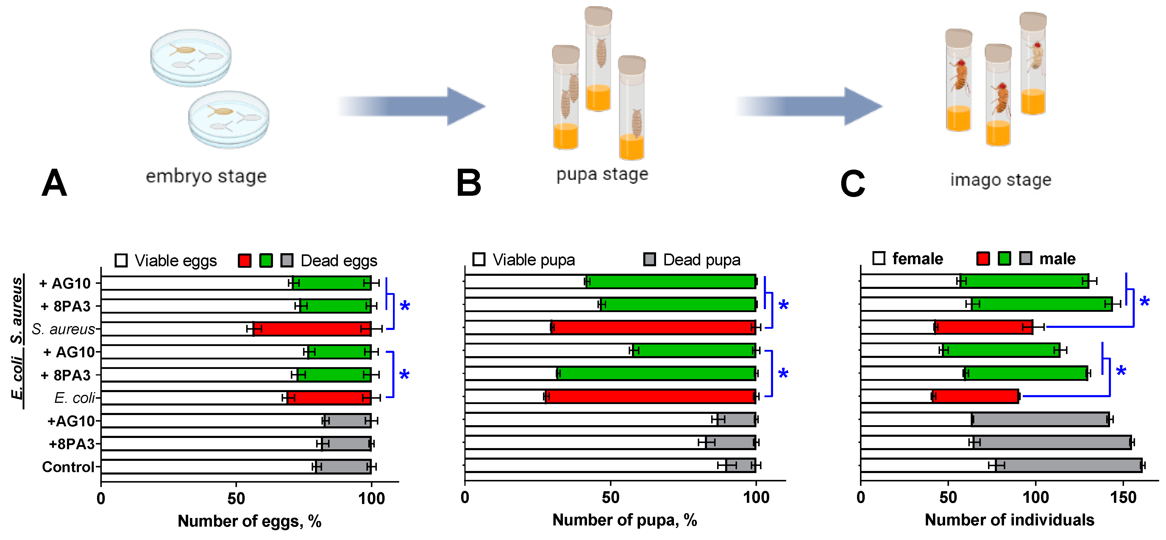
2.5. The Analysis of Flies Viability on Embryonic and Metamorphosis Stages

The frequency of lethal mutations during embryogenesis was used as an indicator of the changes occurring in gametes of imagoes. The frequency of embryo lethality was determined as the fraction of the eggs that stopped their development at a certain stage out of the total number of the eggs laid. To perform the experiment, virgin imagoes from all control and experimental groups were separated according to their sex within the first day after eclosion and kept separately in vials with temporary culture medium until they reached a sexually mature age (three days). Then, males and females were put together for 12 h for mating. Subsequently, inseminated females were placed in Petri dishes (d = 10 cm) with temporary medium (5 individuals per dish) for 12 h to obtain eggs. In the next day, the eggs were counted using a stereoscopic microscope and then placed in a thermostat (t = 24 °C) for 48 h. The sample size was 10 Petri dishes for each experimental group. The embryo lethality level was calculated according to [82].
Viability at the pupa stage was estimated as a percentage of those that did not hatch at the end of the period of emergence of adults from puparia in the offspring of five parental pairs. At the same time, the preimaginal death was assessed by changes in the morphology of the pupa.

2.6. Fertility Analysis

For the fertility analysis, five newly emerged females and five males were placed in vials with 10 mL of standard medium (control) and medium containing bacteria of all experimental groups for the oviposition period of 7 days. The vials were kept in an incubator until progeny appeared. Then, parental insects were removed from the tube. The number of adult males and females was fixed. For each experimental group, 10 vials were analyzed [83].

2.7. DNA Comet Assay

To assess DNA damage in the gut of flies, an alkaline variant of the DNA comet assay was used; this allows for the determination of single-strand DNA breaks in cells [84]. For all experimental groups, the gut of third-in star Drosophila larvae was isolated and mechanically suspended in the Poel’s salt solution (15 mMNaCl, 6.4 mM NaH2PO4, 42 mMKCl, 7.9 mM CaCl2, 1.8 mM KHCO3, 20.8 mM MgSO4; pH 6.95). Enterocytes were embedded in 0.75% agarose on slides. To prepare each single slide, a total of 5 guts were used. The slides were placed in a lysis solution (2.5 M NaCl, 100 mM EDTA, 10 mMTris, 1% Triton X-100; pH 10) for 1 h at 4 °C. Then the slides were incubated in an alkaline electrophoresis buffer (0.3 M NaOH, 1 mM EDTA, pH 13) for 10 min followed by electrophoresis at 15 V/300 mA for 10 min at 4 °C. Slides were washed 3 times in 20 mMTris (pH 7.5) and 3 times in distilled water and then fixed in ethanol for 10 min. Fluorescence microscopy (Carl Zeiss Axio Imager M2, Oberkochen, Germany) was used to visualize and rank the DNA comets.

2.8. SMART Test

The genotoxicity of L. plantarum strains and strains of E. coli and S. aureus was assessed using standard cross versions of SMART on D. melanogaster. Females from the flr3/In(3LR)TM3 line were mated with mwh/mwh males [85,86,87]. The lines were kindly provided by O. N. Antosyuk, Ural Federal University (UrFU, Ekaterinburg, Russia). The emergent adults were collected and preserved in 70% ethanol. The preserved fly wings were placed under a stereoscopic microscope using entomological pincers and the wing pairs were spread over codified slides. A Faure solution (30 g gum Arabic, 20 mL glycerol, 50 g chloral hydrate, and 50 mL distilled water) was used to fix the wings. After mounting, the slides remained on a warm plate (60 °C) for 1 h. The spots (single or twin) on the wings were counted by using a Carl Zeiss Observer 1.0 microscope (Carl Zeiss, Oberkochen, Germany) at 400× magnification.

2.9. DNA Extraction

A single colony of L. plantarum AG10 was grown in Man–Rogosa–Sharpe (MRS) Broth (SigmaAldrich, St. Louis, MO, USA) under microaerophilic conditions at 37 °C overnight. Then, bacterial cells were harvested by centrifugation at 10,000 rpm for 5 min and the genomic DNA was extracted using GeneJET Genomic DNA Purification Kit (ThermoFisher, Waltham, MA, USA) according to the manufacturer’s protocol. The quality and purity of DNA was checked by using 0.7% agarose gel electrophoresis and OD260/OD280 ratio on Nanodrop2000 systems.

2.10. Genome Sequencing and Assembling

The L. plantarum AG10 whole genome sequencing was performed using a high-throughput IlluminaMiSeq platform. For IlluminaMiSeq sequencing, DNA was sheared to fragments ranging between 300 and 500 bp using the Covaris S220 (Covaris, Woburn, MA, USA) The fragmented DNA sample was end-paired, dA-tailed and ligated to multiple adapters. The ligated products were purified and further enriched using PCR, and paired-end sequencing was performed by using IlluminaMiseq (Illumina, San Diego, CA, USA). The quality of sequence reads was assessed using FastQC (version 0.11.9) [88]; the genome was assembled using SPAdes 3.15.3 [89]. The raw data of complete genome sequence of L. plantarum AG10 are available in NCBI GenBank database as BioProject PRJNA953814.

2.11. Bioinformatic Analysis

The assembled genome was annotated using Prokka [90]. Distribution of genes into subsystem categories was performed using the RAST server [91].
Genomic and proteomic alignments were performed using a Blast Global Alignment tool [92]. In silico screening for antimicrobial peptides was performed using online algorithms for the analysis of antimicrobial activity: dbAMP (https://awi.cuhk.edu.cn/dbAMP/ampfinder.php, accessed on 10 April 2023) [93], CAMPR3 (http://www.camp3.bicnirrh.res.in/, accessed on 10 April 2023) [94]. The VFDB database (http://www.mgc.ac.cn/VFs/main.htm, accessed on 12 April 2023) and VRprofile 2.0 (https://tool-mml.sjtu.edu.cn/STEP/STEP_VR.html, accessed on 13 April 2023) were used to search for proteins associated with bacterial virulence.

2.12. Statistical Analysis

All experiments were performed in biological triplicates with three repeats in each unless otherwise specifically stated. Statistical data analysis was performed using one-factor analysis of variance (one-way ANOVA) statistical test with Holm–Sidak correction for multiple testing in GraphPad Prism version 6.0 for Windows (GraphPad Software). For each indicator, the arithmetic mean, its estimation error and standard deviation were calculated. For data with non-Gaussian distribution, a non-parametric one-way analysis of variance (Kruskal–Wallis) test has been performed and median values with interquartile ranges were shown.

3. Results

3.1. In Vitro Antagonistic Activity of L. plantarum AG10

The antagonistic activity of L. plantarum AG10 was tested by the agar block method against Bacillus cereus, Escherichia coli MG1655, Staphylococcus aureus ATCC 29213, Klebsiella pneumonia, Serratia marcescens and Pseudomonas aeruginosa ATCC 27853 (see Table 1). L. plantarum 8PA3 was used as control strain. P. aeruginosa revealed highest sensitivity to antimicrobial substances produced by L. plantarum AG10, while K. pneumoniae and B. cereus were almost insusceptible. E. coli, S. aureus and S. marcescens exhibited weak susceptibility. By contrast, L. plantarum 8PA3 demonstrated antagonistic activity against P. aeruginosa only.
Further, the ability of L. plantarum AG10 to repress E. coli and S. aureus was evaluated in the skim milk fermentation model followed by storage over 21 days. To this end, pathogens were inoculated together with a starter culture and milk contamination was monitored. A probiotic strain of L. plantarum 8PA3 was used as a relevant control. The milk was incubated for 20 h at 40 °C under static conditions and then stored at 4 °C for 21 days. The number of viable cells of both lactobacilli and pathogenic microorganisms was evaluated by CFU counting after end of fermentation and after 3, 7 and 21 days of storage.
Overall, on the 21st day of storage, the amount of L. plantarum AG10 was slightly higher when compared to L. plantarum 8PA3, suggesting increased survival potential of the strain. In the presence of L. plantarum 8PA3, the viable E. coli completely disappeared up to the 21st day of the storage (Figure 1B). By contrast, while no complete death of the pathogen could be observed in the presence of L. plantarum AG10, the growth of E. coli was suppressed even during fermentation time, since the amount of CFUs was 105 at the storage start (Figure 1A). Similarly, the number of viable S. aureus in the presence of L. plantarum AG10 was reduced 10-fold when compared to the L. plantarum 8PA3 co-culture, suggesting that L. plantarum AG10 is able to repress the growth of other bacteria during the fermentation period. During storage, CFUs of S. aureus continuously decreased in the co-culture of both Lactobacilli strains, with no significant differences in their growth repression rates.

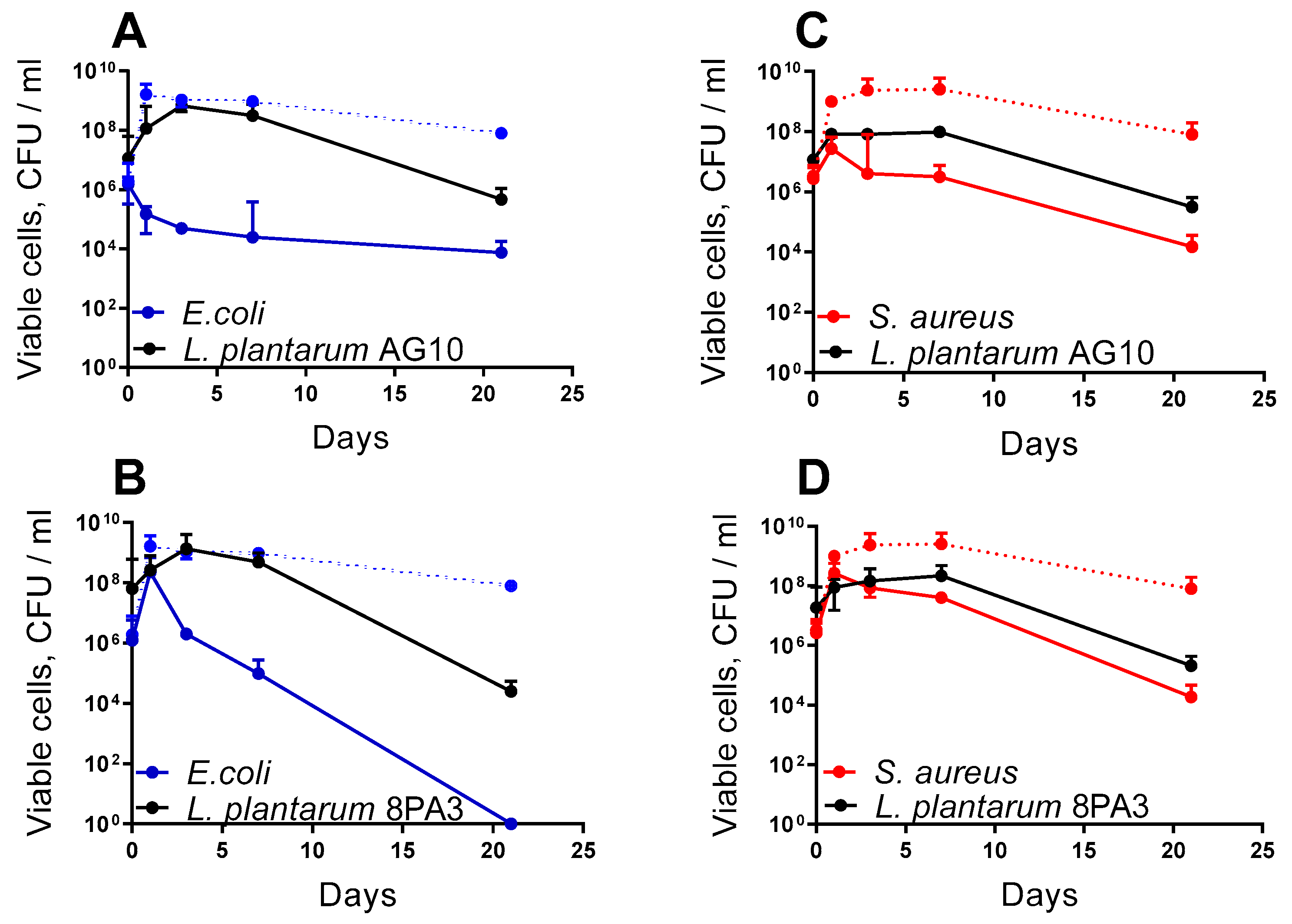
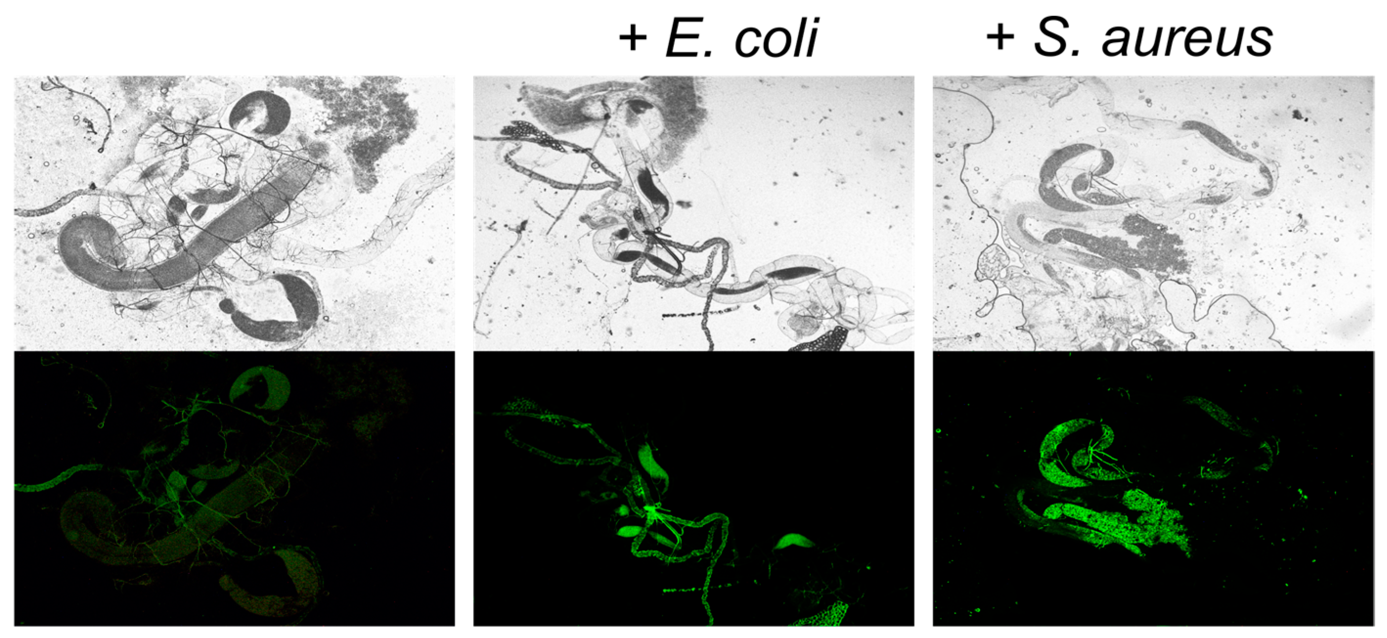
3.2. Antagonistic Activity of Lactobacillus in the Drosophila Model

Tthe ability of L. plantarum strains to repress the growth of S. aureus and E. coli in vivo was evaluated on flies infected per-oral with these pathogens. Synchronous embryo clutches were transferred onto Drosophila nutrient medium; 100 μL of either S. aureus or E. coli suspension (107–108 CFU/mL in sterile PBS) were added as this concentration of viable pathogens has been used in some similar investigations [95,96,97,98]. Then, viability and reproduction were evaluated by counting the number of eggs laid, embryonic death, pupa death and fertility (number of the imagoes).
To confirm the uptake of S. aureus and E. coli by insects, the E. coli and S. aureus strains carrying pCtuf Amp Gfpr plasmid providing constitutive GFP synthesis were added to the growth medium with larvae. During the third stage of development, the larvaes’ gut was extracted and analyzed using fluorescence microscopy. As can be seen in Figure 2, strong fluorescence can be observed in the guts of larvae grown in medium with the addition of E. coli and S. aureus strains carrying pCtuf Amp Gfpr plasmid, suggesting the uptake of bacteria by larvae.
Neither the embryonic stage nor further development of the fruit flies was affected by the presence of L. plantarum 8PA3 and L. plantarum AG10 in the growth medium (Figure 3A greys). By contrast, the addition of either E. coli or S. aureus to the medium increased the frequency of embryonic death 1.5-fold (p < 0.05) and 2.2-fold (p < 0.05), respectively (Figure 3A reds). The simultaneous introduction of Lactobacilli with pathogens to the medium partially restored the survival of eggs (Figure 3A greens). A significant effect of the L. plantarum AG10 strain was observed, with the reduction of the embryonic death rate by 7% (p < 0.05) when co-cultured with E. coli and by 15%, (p < 0.05) when co-cultured with S. aureus. In the case of L. plantarum 8PA3, a significant effect could be observed only when co-cultured with S. aureus cells (reduction of embryonic death by 17%, p < 0.05).
Similar effects of E. coli and S. aureus on Drosophila viability, as well as its recovery following the addition of L. plantarum strains, have been observed at the pupa stage (Figure 3B). The presence of either E. coli or S. aureus in the medium increased the mortality rate of insects in the pupal stage by 60–62% when compared to the control (p < 0.05). However, in the presence of L. plantarum AG10, the mortality at the pupal stage was reliably reduced by 30% when compared to the group with solely E. coli, and by 12% when compared to the group with solely Staphylococci (p < 0.05). L. plantarum 8PA3 was able to reduce the mortality rate at the pupa stage by 17% (p < 0.05) but only in the group infected with E. coli.
Finally, Figure 3C exemplifies the effect of bacteria on the viability of insects at the imago stage. The number of viable individuals was reduced by 45% and 40% in the groups infected with either E. coli or S. aureus when compared to the control group, respectively. Similarly, while no significant effect of either of the L. plantarum strains could be observed when added solely to the flies’ growth medium, their presence could level out the negative effect of both pathogens, providing roughly similar amount of adults as in non-treated group.

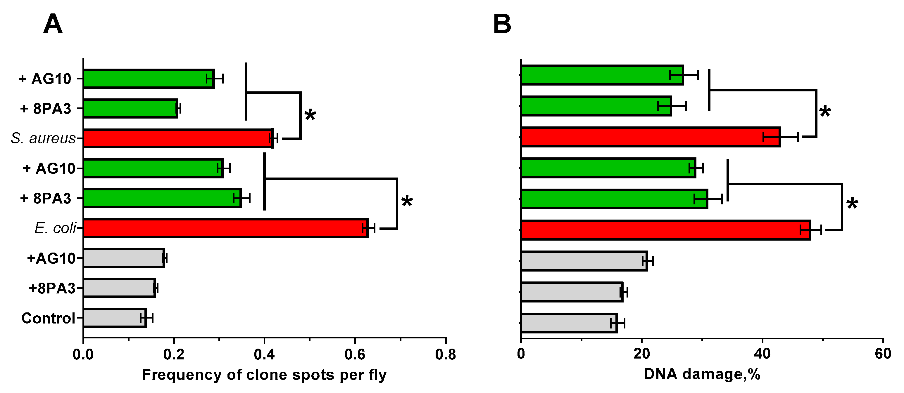
3.3. Genotoxicity of Lactobacillus Strains in the Drosophila Model

The potential genotoxicity of the strains [99] was evaluated by counting (i) the number of recombinant spots in the SMART test (definition of mutational/recombinant events) and (ii) the number of enterocytes of flies with DNA damage and the index of DNA comets (IDC, determination of damage in DNA). As can be seen in Figure 4, the L. plantarum strains alone did not demonstrate reliable genotoxic effect on the insects, while cultivation of flies in the presence of E. coli and S. aureus led to significant genotoxic effects (3–4 fold increase of spots frequency in the SMART test and DNA damage rate). The addition of either L. plantarum 8PA3 or L. plantarum AG10 together with the pathogens reduced the frequency of mutation and recombination events 1.5–2-fold.

3.4. Whole Genome Sequence of L. plantarum AG10

The whole genome of L. plantarum AG10 was sequenced using the high-throughput IlluminaMiSeq platform with overall coverage of 79× and sequence reads quality of 98.1% (probability of incorrect base call 0.1%). The L. plantarum AG10 genome consists of 109 contigs with the total length of 3,479,919 bp and 44.5% G + C content. The NCBI Bioproject has been deposited at NCBI under the accession number PRJNA953814 (https://www.ncbi.nlm.nih.gov/bioproject/PRJNA953814/, accessed on 8 April 2023) and consists of annotated genome and raw sequence data.
The genome annotation predicted 3467 coding sequences. Among them, 2129 open reading frames encode hypothetical proteins with unknown function; 1338 coding sequences encode proteins with known or predicted functions, 1222 of which can be distributed into 27 general metabolic groups (Table 2). The largest number of genes belongs to the carbohydrates (262 genes), the metabolism of amino acids and their derivatives (188 genes) and proteins (127 genes).

3.5. In Silico Identification of Antimicrobial Peptides and Virulence Factors in LAB Genomes

It was previously reported that LAB strains produce various antimicrobial peptides as antagonistic tools [31]. To evaluate whether the antagonistic properties of L. plantarum AG10 are associated with the production of bacteriocins or bacteriocin-like peptides, the whole genome of L. plantarum AG10 was analyzed in order to search for linear ribosomal peptides by using the AMP finder algorithm on the dbAMP server. Finally, three peptides (LLCBMPMO_01958, LLCBMPMO_01967 and LLCBMPMO_01968) with identity higher than 90% with peptides characterized by predicted antimicrobial potential were found (Table 3). In the L. plantarum 8PA3 genome, four peptides (WP_003641985.1, WP_003643800.1, WP_003643811.1 and WP_046947768.1) with high identity with antimicrobial peptides were found (Table 4). Further evaluation of antimicrobial properties of these peptides using CAMPR3 server indicated that the protein LLCBMPMO_01968 has the highest probability to exhibit antimicrobial properties (see Table 5). WP_003641985.1 has the highest probability of exhibiting antimicrobial properties according to the CAMPR3 server, while active only against S. aureus (see Table 5). WP_046947768.1 has lower scores in CAMPR3 but is predicted as active by DBAASP server. Thus, the activity of these peptides should be independently validated using an artificially synthesized peptide; this is planned as a part of our future work.
Further analysis of the L. plantarum AG10 genome using the VFDB database revealed, in silico, only three putative virulence factors (Table 6), while 30 were predicted for L. plantarum 8PA3 [100]; this apparently suggests the safety of L. plantarum AG10 as a probiotic strain.

4. Discussion

The widespread emergence of pathogenic bacteria resistant to conventional antimicrobials represents one of the key health challenges worldwide, and thus active screening of new approaches for the prevention and treatment of diseases is essential. Probiotic bacteria, such as lactic acid bacteria, Bacilli and their metabolites, have been intensively investigated as promising natural and eco-friendly substitutes to antibiotics [18,19,20,25,26].
Recent data indicate that the L. plantarum AG10 strain, isolated previously from clover silage, demonstrates promising antagonistic activity and probiotic properties in vitro [50,52]. In this work, we also show that it competes with pathogens in vivo, as well as diminishes their negative effects on the host. Besides the pronounced repression of most foodborne pathogens in the classic agar drop diffusion test (Table 1), this bacterium was able to suppress the growth of S. aureus and E. coli during milk fermentation. Thus, the amount of viable E. coli cells in the final product decreased from 106 to 105 after 8 h of milk fermentation by L. plantarum AG10 (Figure 1A); in the milk fermented by approved probiotic L. plantarum 8PA3, the viable E. coli cells increased until 108 (Figure 1B). Similar suppression has been observed for S. aureus under the same conditions (Compare Figure 1C,D), suggesting antimicrobial potential of the strain. In addition, low amounts of putative virulence factors have been detected in silico in the genome of L. plantarum AG10 by using the VFDB database (in contrast to 30 found in L. plantarum 8PA3 [100]), suggesting its prominent safety profile as a potential probiotic.
The ability of L. plantarum AG10 to compete with pathogenic flora has been further demonstrated in vivo using a Drosophila melanogaster model. AG10 could diminish the negative effects of both S. aureus and E. coli on the survival characteristics of Drosophila melanogaster at both embryonic (larvae) and pupa stages, while the reference strain was active mainly against S. aureus only. Thus, the addition of either L. plantarum AG10 or the reference L. plantarum 8PA3 strain significantly reduced the mortality of Drosophila embryo and pupa infected with S. aureus, while L. plantarum AG10 could also level out the effects of E. coli on the survival of insects (Figure 3A,B). Consequently, in groups infected with pathogens in the presence of Lactobacilli, the amount of imago was similar to the untreated group or groups treated solely with Lactobacilli (Figure 3). Since bacteria added to the flies’ growth media were preliminary washed by PBS, the observed effects are apparently driven by antimicrobial substances produced by Lactobacilli de novo. Moreover, the experiment with S. aureus and E. coli overexpressing GFP confirmed that pathogens are captured by larvae (Figure 2), whereas successful introduction of Lactobacilli to the larvaes’ gut was shown previously [100], suggesting that the repression of pathogens occurs in vivo, while ex vivo interactions remain also possible.
The exact mechanism of the observed antimicrobial activity remains open to discussion. Lactic acid bacteria (LAB) compete with other bacteria by using various mechanisms such as synthesis of organic acids (mainly lactic acid) and acidification of the broth [31], hydrogen peroxide production [32] and secretion of antimicrobial peptides, as well as bacteriocins [33,34]. While broth acidification seems to be the main factor of antagonism in both agar drop diffusion and milk fermentation tests, antimicrobial peptides could play the leading role in vivo [33,34]. Moreover, it has been reported previously that L. plantarum AG10 acidifies the milk in a less pronounced manner than the industrial strains [52,53], assuming that the production of antimicrobial peptides could be apparently responsible for the observed effects both in vitro and in vivo. Indeed, the screening of putative antimicrobial peptides in the genome of L. plantarum AG10 in comparison with the dbAMP database allowed identification of three peptides with over 90% homology with peptides characterized by presumable antimicrobial activity (Table 3). Among them, LLCBMPMO_01967 seems to be active only against S. aureus, while LLCBMPMO_01968 exhibit antimicrobial properties against both S. aureus and E. coli with a high probability (Table 5). These findings agree with observations of the apparent repression of pathogenic bacteria in Drosophila (Figure 3 and Figure 4). The reference strain L. plantarum 8PA3 also carries four putative AMPs in the genome (Table 4) with WP_046947768.1, with predicted activity against both pathogens for the latter (Table 5). Nevertheless, during in vivo experiments, L. plantarum 8PA3 did not reduce the mortality in E. coli infected group (Figure 3 and Figure 4), suggesting that this AMP is apparently either inactive or non-expressed under experimental conditions. Thus, despite the relatively high possibility of antimicrobial properties predicted for AMPs in silico (Table 4), an independent in vitro verification of their activity is required in further investigations.

5. Conclusions

Taken together, these data allow the suggestion that the L. plantarum AG10 strain is a promising candidate for use in dairy production, to suppress the growth of pathogens during milk fermentation, and in livestock food supplements, as a preservative from foodborne infections. While our data demonstrated that the L. plantarum AG10 strain is capable of reducing the negative effects of S. aureus and E. coli in vivo in the survival model of Drosophila melanogaster at the embryonic (larval) and pupal stages, multiple additional tests are required to validate the safety and efficiency of lactobacilli as a natural and eco-friendly alternative to antimicrobials. We plan to examine this in our future research. The whole genome sequencing allowed identification, with high probability, of the putative antimicrobial peptide responsible for the above activity. The genome contains extremely low amounts of putative virulence factors, indicating that L. plantarum AG10 represents a promising probiotic.

Author Contributions

This study was made possible through the collaboration of all authors. Conceptualization, E.N. and A.K.; methodology, E.N. and A.K.; software, I.Z., M.B. and A.K.; validation, A.K.; formal analysis, E.N., D.Y., A.K. and A.E.; investigation, E.G., V.K., I.Z. and D.K.; resources, A.K., D.Y. and E.N.; data curation, E.N., D.Y., A.K. and M.B.; writing—original draft preparation, E.N., E.G., V.K., D.Y. and A.K.; writing—review and editing, E.N., E.G., V.K., D.Y. and A.K.; visualization, E.G., V.K., I.Z. and A.K.; supervision, D.Y., A.K. and E.N.; project administration, D.Y., E.N. and A.K.; funding acquisition, D.Y. All authors have read and agreed to the published version of the manuscript.

Funding

This research was funded by Russian Science Foundation (grant N 22-16-00040).

Data Availability Statement

All data are available in manuscript.

Acknowledgments

F. Götz, University Tübingen, Germany is kindly acknowledged for providing the pCtuf Amp Gfpr plasmid. The work was performed in frames of the Kazan Federal University Strategic Academic Leadership Program (PRIORITY-2030) and using the equipment of the Interdisciplinary Center for Shared Use of the Kazan Federal University.

Conflicts of Interest

The authors declare no conflict of interest.

References

  1. Sneeringer, S.; MacDonald, J.M.; Key, N.; McBride, W.D.; Mathews, K. Economics of antibiotic use in US livestock production. USDA Econ. Res. Rep. 2015, 200. [Google Scholar]
  2. Manaia, C.M.; Vaz-Moreira, I.; Nunes, O.C. Antibiotic resistance in waste water and surface water and human health implications. Emerg. Org. Contam. Hum. Health 2012, 20, 173–212. [Google Scholar] [CrossRef]
  3. Schmieder, R.; Edwards, R. Insights into antibiotic resistance through metagenomic approaches. Future Microbiol. 2012, 7, 73–89. [Google Scholar] [CrossRef] [PubMed]
  4. Smits, C.H.; Li, D.; Patience, J.F.; den Hartog, L. Animal nutrition strategies and options to reduce the use of antimicrobials in animal production. Food Agric. Organ. U. N. 2021, 2021, 184. [Google Scholar]
  5. Hawkey, P.M. The growing burden of antimicrobial resistance. J. Antimicrob. Chemother. 2008, 62 (Suppl. S1), i1–i9. [Google Scholar] [CrossRef]
  6. Qiao, M.; Ying, G.G.; Singer, A.C.; Zhu, Y.G. Review of antibiotic resistance in China and its environment. Environ. Int. 2018, 110, 160–172. [Google Scholar] [CrossRef]
  7. Hinton, M.; Linton, A.H. Control of salmonella infections in broiler chickens by the acid treatment of their feed. Vet. Rec. 1988, 123, 416–421. [Google Scholar] [CrossRef]
  8. Van Immerseel, F.; De Buck, J.; Boyen, F.; Bohez, L.; Pasmans, F.; Volf, J.; Sevcik, M.; Rychlik, I.; Haesebrouck, F.; Ducatelle, R. Medium-chain fatty acids decrease colonization and invasion through hilA suppression shortly after infection of chickens with Salmonella enterica serovar Enteritidis. Appl. Environ. Microbiol. 2004, 70, 3582–3587. [Google Scholar] [CrossRef]
  9. Thompson, J.L.; Hinton, M. Antibacterial activity of formic and propionic acids in the diet of hens on Salmonellas in the crop. Br. Poult. Sci. 1997, 38, 59–65. [Google Scholar] [CrossRef] [PubMed]
  10. Mitsch, P.; Zitterl-Eglseer, K.; Köhler, B.; Gabler, C.; Losa, R.; Zimpernik, I. The effect of two different blends of essential oil components on the proliferation of Clostridium perfringens in the intestines of broiler chickens. Poult. Sci. 2004, 83, 669–675. [Google Scholar] [CrossRef] [PubMed]
  11. Hammer, K.A.; Carson, C.F.; Riley, T.V. Antimicrobial activity of essential oils and other plant extracts. J. Appl. Microbiol. 1999, 86, 985–990. [Google Scholar] [CrossRef]
  12. Hernández, F.; Madrid, J.; García, V.; Orengo, J.; Megías, M.D. Influence of two plant extracts on broilers performance, digestibility, and digestive organ size. Poult. Sci. 2004, 83, 169–174. [Google Scholar] [CrossRef] [PubMed]
  13. Friedman, M.; Buick, R.; Elliott, C.T. Antibacterial activities of naturally occurring compounds against antibiotic-resistant Bacillus cereus vegetative cells and spores, Escherichia coli, and Staphylococcus aureus. J. Food Prot. 2004, 67, 1774–1778. [Google Scholar] [CrossRef]
  14. Lewis, K.; Ausubel, F.M. Prospects for plant-derived antibacterials. Nat. Biotechnol. 2006, 24, 1504–1507. [Google Scholar] [CrossRef] [PubMed]
  15. Cowan, M.M. Plant products as antimicrobial agents. Clin. Microbiol. Rev. 1999, 12, 564–582. [Google Scholar] [CrossRef] [PubMed]
  16. Vaou, N.; Stavropoulou, E.; Voidarou, C.; Tsigalou, C.; Bezirtzoglou, E. Towards Advances in Medicinal Plant Antimicrobial Activity: A Review Study on Challenges and Future Perspectives. Microorganisms 2021, 9, 9102041. [Google Scholar] [CrossRef]
  17. Wang, S.; Zeng, X.; Yang, Q.; Qiao, S. Antimicrobial Peptides as Potential Alternatives to Antibiotics in Food Animal Industry. Int. J. Mol. Sci. 2016, 17, 17050603. [Google Scholar] [CrossRef]
  18. Anee, I.J.; Alam, S.; Begum, R.A.; Shahjahan, R.M.; Khandaker, A.M. The role of probiotics on animal health and nutrition. J. Basic Appl. Zool. 2021, 82, 52. [Google Scholar] [CrossRef]
  19. Alagawany, M.; Abd El-Hack, M.E.; Farag, M.R.; Sachan, S.; Karthik, K.; Dhama, K. The use of probiotics as eco-friendly alternatives for antibiotics in poultry nutrition. Environ. Sci. Pollut. Res. Int. 2018, 25, 10611–10618. [Google Scholar] [CrossRef]
  20. Vasile, N.; Ghindea, R.; Vassu, T. Probiotics—An alternative treatment for various diseases. Roum. Arch. Microbiol. Immunol. 2011, 70, 54–59. [Google Scholar]
  21. Hotel, A.C.P.; Cordoba, A. Health and nutritional properties of probiotics in food including powder milk with live lactic acid bacteria. Prevention 2001, 5, 1–10. [Google Scholar]
  22. Hill, C.; Guarner, F.; Reid, G.; Gibson, G.R.; Merenstein, D.J.; Pot, B.; Morelli, L.; Canani, R.B.; Flint, H.J.; Salminen, S.; et al. Expert consensus document. The International Scientific Association for Probiotics and Prebiotics consensus statement on the scope and appropriate use of the term probiotic. Nat. Rev. Gastroenterol. Hepatol. 2014, 11, 506–514. [Google Scholar] [CrossRef]
  23. Simon, O.; Jadamus, A.; Vahjen, W. Probiotic feed additives-effectiveness and expected modes of action. J. Anim. Feed Sci. 2001, 10, 51–68. [Google Scholar] [CrossRef]
  24. Tan, L.T.; Chan, K.G.; Lee, L.H.; Goh, B.H. Streptomyces Bacteria as Potential Probiotics in Aquaculture. Front. Microbiol. 2016, 7, 79. [Google Scholar] [CrossRef]
  25. Markowiak, P.; Śliżewska, K. Effects of Probiotics, Prebiotics, and Synbiotics on Human Health. Nutrients 2017, 9, 9091021. [Google Scholar] [CrossRef]
  26. Soccol, C.R.; de Souza Vandenberghe, L.P.; Spier, M.R.; Medeiros, A.P.; Yamaguishi, C.T.; De Dea Lindner, J.; Pandey, A. The potential of probiotics: A review. Food Technol. Biotechnol. 2010, 48, 413–434. [Google Scholar]
  27. Hammes, W.P.; Hertel, C. The genera lactobacillus and carnobacterium. Prokaryotes 2006, 4, 320–403. [Google Scholar]
  28. Wyszyńska, A.; Kobierecka, P.; Bardowski, J.; Jagusztyn-Krynicka, E.K. Lactic acid bacteria—20 years exploring their potential as live vectors for mucosal vaccination. Appl. Microbiol. Biotechnol. 2015, 99, 2967–2977. [Google Scholar] [CrossRef]
  29. Cui, C.; Shen, C.J.; Jia, G.; Wang, K.N. Effect of dietary Bacillus subtilis on proportion of Bacteroidetes and Firmicutes in swine intestine and lipid metabolism. Genet. Mol. Res. 2013, 12, 1766–1776. [Google Scholar] [CrossRef]
  30. Sanders, M.E.; Morelli, L.; Tompkins, T.A. Sporeformers as Human Probiotics: Bacillus, Sporolactobacillus, and Brevibacillus. Compr. Rev. Food Sci. Food Saf. 2003, 2, 101–110. [Google Scholar] [CrossRef]
  31. Moshood, Y.; Tengku, A. Lactic Acid Bacteria: Bacteriocin Producer: A Mini Review. IOSR J. Pharm. 2013, 3, 44–55. [Google Scholar]
  32. Piard, J.C.; Desmazeaud, M. Inhibiting Factors Produced by Lactic-acid Bacteria 1. Oxygen Metabolites and Catabolism end-Products. Lait 1991, 71, 525–541. [Google Scholar] [CrossRef]
  33. Giacometti, A.; Cirioni, O.; Ancarani, F.; Del Prete, M.S.; Fortuna, M.; Scalise, G. In Vitro activities of polycationic peptides alone and in combination with clinically used antimicrobial agents against Rhodococcus equi. Antimicrob. Agents Chemother. 1999, 43, 2093–2096. [Google Scholar] [CrossRef] [PubMed]
  34. Vaara, M.; Porro, M. Group of peptides that act synergistically with hydrophobic antibiotics against gram-negative enteric bacteria. Antimicrob. Agents Chemother. 1996, 40, 1801–1805. [Google Scholar] [CrossRef] [PubMed]
  35. Martín, V.; Cárdenas, N.; Ocaña, S.; Marín, M.; Arroyo, R.; Beltrán, D.; Badiola, C.; Fernández, L.; Rodríguez, J.M. Rectal and vaginal eradication of Streptococcus agalactiae (GBS) in pregnant women by using Lactobacillus salivarius CECT 9145, a target-specific probiotic strain. Nutrients 2019, 11, 810. [Google Scholar] [CrossRef]
  36. Abatemarco Júnior, M.; Sandes, S.H.C.; Ricci, M.F.; Arantes, R.M.E.; Nunes, Á.C.; Nicoli, J.R.; Neumann, E. Protective effect of Lactobacillus diolivorans 1Z, isolated from Brazilian kefir, against Salmonella enterica serovar typhimurium in experimental murine models. Front. Microbiol. 2018, 9, 2856. [Google Scholar] [CrossRef]
  37. Zheng, Y.-D.; Pan, Y.; He, K.; Li, N.; Yang, D.; Du, G.-F.; Ge, R.; He, Q.-Y.; Sun, X. SPD_1495 contributes to capsular polysaccharide synthesis and virulence in Streptococcus pneumoniae. Msystems 2020, 5, e00025-20. [Google Scholar] [CrossRef]
  38. Singh, A.K.; Tiwari, U.P.; Berrocoso, J.D.; Dersjant-Li, Y.; Awati, A.; Jha, R. Effects of a combination of xylanase, amylase and protease, and probiotics on major nutrients including amino acids and non-starch polysaccharides utilization in broilers fed different level of fibers. Poult. Sci. 2019, 98, 5571–5581. [Google Scholar] [CrossRef]
  39. Corcionivoschi, N.; Drinceanu, D.; Pop, I.M.; Stack, D.; Stef, L.; Julean, C.; Bourke, B. The effect of probiotics on animal health. Anim. Sci. Biotechnol. 2010, 43, 35–41. [Google Scholar]
  40. Yoo, J.Y.; Kim, S.S. Probiotics and Prebiotics: Present Status and Future Perspectives on Metabolic Disorders. Nutrients 2016, 8, 173. [Google Scholar] [CrossRef]
  41. Linares, D.M.; Ross, P.; Stanton, C. Beneficial Microbes: The pharmacy in the gut. Bioengineered 2016, 7, 11–20. [Google Scholar] [CrossRef] [PubMed]
  42. Oelschlaeger, T.A. Mechanisms of probiotic actions—A review. Int. J. Med. Microbiol. 2010, 300, 57–62. [Google Scholar] [CrossRef]
  43. Lee, I.K.; Kye, Y.C.; Kim, G.; Kim, H.W.; Gu, M.J.; Umboh, J.; Maaruf, K.; Kim, S.W.; Yun, C.H. Stress, Nutrition, and Intestinal Immune Responses in Pigs—A Review. Asian-Australas. J. Anim. Sci. 2016, 29, 1075–1082. [Google Scholar] [CrossRef]
  44. Bajagai, Y.S.; Klieve, A.V.; Dart, P.J.; Bryden, W.L. Probiotics in Animal Nutrition: Production, Impact and Regulation; FAO: Rome, Italy, 2016. [Google Scholar]
  45. Thelestam, M.; Frisan, T. Cytolethal distending toxins. Rev. Physiol. Biochem. Pharm. 2004, 152, 111–133. [Google Scholar] [CrossRef]
  46. Chumduri, C.; Gurumurthy, R.K.; Zietlow, R.; Meyer, T.F. Subversion of host genome integrity by bacterial pathogens. Nat. Rev. Mol. Cell Biol. 2016, 17, 659–673. [Google Scholar] [CrossRef] [PubMed]
  47. Deplanche, M.; Mouhali, N.; Nguyen, M.T.; Cauty, C.; Ezan, F.; Diot, A.; Raulin, L.; Dutertre, S.; Langouet, S.; Legembre, P.; et al. Staphylococcus aureus induces DNA damage in host cell. Sci. Rep. 2019, 9, 7694. [Google Scholar] [CrossRef] [PubMed]
  48. Prazdnova, E.V.; Mazanko, M.S.; Chistyakov, V.A.; Bogdanova, A.A.; Refeld, A.G.; Kharchenko, E.Y.; Chikindas, M.L. Antimutagenic Activity as a Criterion of Potential Probiotic Properties. Probiotics Antimicrob. Proteins 2022, 14, 1094–1109. [Google Scholar] [CrossRef]
  49. Wu, J.; Zhang, Y.; Ye, L.; Wang, C. The anti-cancer effects and mechanisms of lactic acid bacteria exopolysaccharides in vitro: A review. Carbohydr. Polym. 2021, 253, 117308. [Google Scholar] [CrossRef] [PubMed]
  50. Gavrilova, E.; Anisimova, E.; Gabdelkhadieva, A.; Nikitina, E.; Vafina, A.; Yarullina, D.; Bogachev, M.; Kayumov, A. Newly isolated lactic acid bacteria from silage targeting biofilms of foodborne pathogens during milk fermentation. BMC Microbiol. 2019, 19, 248. [Google Scholar] [CrossRef]
  51. Nikitina, E.; Petrova, T.; Vafina, A.; Ezhkova, A.; Nait Yahia, M.; Kayumov, A. Textural and Functional Properties of Skimmed and Whole Milk Fermented by Novel Lactiplantibacillus plantarum AG10 Strain Isolated from Silage. Fermentation 2022, 8, 290. [Google Scholar] [CrossRef]
  52. Nikitina, E.; Petrova, T.; Sungatullina, A.; Bondar, O.; Kharina, M.; Mikshina, P.; Gavrilova, E.; Kayumov, A. The Profile of Exopolysaccharides Produced by Various Lactobacillus Species from Silage during Not-Fat Milk Fermentation. Fermentation 2023, 9, 197. [Google Scholar] [CrossRef]
  53. Nikitina, E. Potential use of Lactiplantibacillus plantarum strains for fermentation of fat milk. IOP Conf. Ser. Earth Environ. Sci. 2022, 978, 012047. [Google Scholar] [CrossRef]
  54. Trinder, M.; Daisley, B.A.; Dube, J.S.; Reid, G. as a High-Throughput Model for Host-Microbiota Interactions. Front. Microbiol. 2017, 8, 751. [Google Scholar] [CrossRef] [PubMed]
  55. Lemaitre, B.; Miguel-Aliaga, I. The digestive tract of Drosophila melanogaster. Annu. Rev. Genet. 2013, 47, 377–404. [Google Scholar]
  56. Sahu, S.; Jaysingh, P.; Mishra, M. Drosophila melanogaster as an In Vivo Model for the Investigation of Host-Microbiota Interaction. In Prebiotics, Probiotics and Nutraceuticals; Springer: Berlin/Heidelberg, Germany, 2022; pp. 275–300. [Google Scholar]
  57. Broderick, N.A.; Buchon, N.; Lemaitre, B. Microbiota-induced changes in Drosophila melanogaster host gene expression and gut morphology. mBio 2014, 5, e01117-14. [Google Scholar] [CrossRef]
  58. Vallet-Gely, I.; Lemaitre, B.; Boccard, F. Bacterial strategies to overcome insect defences. Nat. Rev. Microbiol. 2008, 6, 302–313. [Google Scholar] [CrossRef]
  59. Hedges, L.M.; Johnson, K.N. Induction of host defence responses by Drosophila C virus. J. Gen. Virol. 2008, 89, 1497–1501. [Google Scholar] [CrossRef]
  60. Chamilos, G.; Lewis, R.E.; Hu, J.; Xiao, L.; Zal, T.; Gilliet, M.; Halder, G.; Kontoyiannis, D.P. Drosophila melanogaster as a model host to dissect the immunopathogenesis of zygomycosis. Proc. Natl. Acad. Sci. USA 2008, 105, 9367–9372. [Google Scholar] [CrossRef]
  61. Chen, K.; Luan, X.; Liu, Q.; Wang, J.; Chang, X.; Snijders, A.M.; Mao, J.-H.; Secombe, J.; Dan, Z.; Chen, J.-H. Drosophila histone demethylase KDM5 regulates social behavior through immune control and gut microbiota maintenance. Cell Host Microbe 2019, 25, 537–552. [Google Scholar] [CrossRef] [PubMed]
  62. Slankster, E.; Lee, C.; Hess, K.M.; Odell, S.; Mathew, D. Effect of gut microbes on olfactory behavior of Drosophila melanogaster larva. Bios 2019, 90, 227. [Google Scholar] [CrossRef] [PubMed]
  63. Casadevall, A.; Pirofski, L.-A. The damage-response framework of microbial pathogenesis. Nat. Rev. Microbiol. 2003, 1, 17–24. [Google Scholar] [CrossRef] [PubMed]
  64. Zhou, Y.; Cui, Y.; Qu, X. Exopolysaccharides of lactic acid bacteria: Structure, bioactivity and associations: A review. Carbohydr. Polym. 2019, 207, 317–332. [Google Scholar] [CrossRef] [PubMed]
  65. Storelli, G.; Defaye, A.; Erkosar, B.; Hols, P.; Royet, J.; Leulier, F. Lactobacillus plantarum promotes Drosophila systemic growth by modulating hormonal signals through TOR-dependent nutrient sensing. Cell Metab. 2011, 14, 403–414. [Google Scholar] [CrossRef] [PubMed]
  66. Poinsot, P.; Penhoat, A.; Mitchell, M.; Sauvinet, V.; Meiller, L.; Louche-Pélissier, C.; Meugnier, E.; Ruiz, M.; Schwarzer, M.; Michalski, M.-C. Probiotic from human breast milk, Lactobacillus fermentum, promotes growth in animal model of chronic malnutrition. Pediatr. Res. 2020, 88, 374–381. [Google Scholar] [CrossRef]
  67. Yamada, R.; Deshpande, S.A.; Bruce, K.D.; Mak, E.M.; William, W.J. Microbes promote amino acid harvest to rescue undernutrition in Drosophila. Cell Rep. 2015, 10, 865–872. [Google Scholar] [CrossRef]
  68. Consuegra, J.; Grenier, T.; Baa-Puyoulet, P.; Rahioui, I.; Akherraz, H.; Gervais, H.; Parisot, N.; Da Silva, P.; Charles, H.; Calevro, F. Drosophila-associated bacteria differentially shape the nutritional requirements of their host during juvenile growth. PLoS Biol. 2020, 18, e3000681. [Google Scholar] [CrossRef]
  69. Newell, P.D.; Douglas, A.E. Interspecies interactions determine the impact of the gut microbiota on nutrient allocation in Drosophila melanogaster. Appl. Environ. Microbiol. 2014, 80, 788–796. [Google Scholar] [CrossRef]
  70. Deshpande, S.A.; Carvalho, G.B.; Amador, A.; Phillips, A.M.; Hoxha, S.; Lizotte, K.J.; Ja, W.W. Quantifying Drosophila food intake: Comparative analysis of current methodology. Nat. Methods 2014, 11, 535–540. [Google Scholar] [CrossRef]
  71. Leitão-Gonçalves, R.; Carvalho-Santos, Z.; Francisco, A.P.; Fioreze, G.T.; Anjos, M.; Baltazar, C.; Elias, A.P.; Itskov, P.M.; Piper, M.D.; Ribeiro, C. Commensal bacteria and essential amino acids control food choice behavior and reproduction. PLoS Biol. 2017, 15, e2000862. [Google Scholar] [CrossRef]
  72. Jones, R.M.; Luo, L.; Ardita, C.S.; Richardson, A.N.; Kwon, Y.M.; Mercante, J.W.; Alam, A.; Gates, C.L.; Wu, H.; Swanson, P.A. Symbiotic lactobacilli stimulate gut epithelial proliferation via Nox-mediated generation of reactive oxygen species. EMBO J. 2013, 32, 3017–3028. [Google Scholar] [CrossRef]
  73. Jones, R.M.; Desai, C.; Darby, T.M.; Luo, L.; Wolfarth, A.A.; Scharer, C.D.; Ardita, C.S.; Reedy, A.R.; Keebaugh, E.S.; Neish, A.S. Lactobacilli modulate epithelial cytoprotection through the Nrf2 pathway. Cell Rep. 2015, 12, 1217–1225. [Google Scholar] [CrossRef]
  74. Kasozi, K.I.; Bukenya, A.; Eze, E.D.; Kasolo, J.; Tayebwa, D.S.; Ssempijja, F.; Suubo, J.; Tamale, A.; Echoru, I.; Ntulume, I. Low concentrations of Lactobacillus rhamnosus GG (Yoba®) are safe in male Drosophila melanogaster. BMC Res. Notes 2019, 12, 269. [Google Scholar] [CrossRef]
  75. Hegan, P.; Mooseker, M.; Handelsman, J.; Miles, J. Effect of probiotic and pathogenic bacteria on Drosophila intestinal pathology. J. Biomol. Tech. 2014, 25, S26. [Google Scholar]
  76. Ramírez-Camejo, L.; García-Alicea, M.; Maldonado-Morales, G.; Bayman, P. Probiotics may protect Drosophila from infection by Aspergillus flavus. Int. J. Pharm. Sci. Res. 2017, 8, 1624. [Google Scholar]
  77. Tsapieva, A.; Duplik, N.; Suvorov, A. Structure of plantaricin locus of Lactobacillus plantarum 8P-A3. Benef. Microbes 2011, 2, 255–261. [Google Scholar] [CrossRef] [PubMed]
  78. Stern, N.J.; Svetoch, E.A.; Eruslanov, B.V.; Perelygin, V.V.; Mitsevich, E.V.; Mitsevich, I.P.; Pokhilenko, V.D.; Levchuk, V.P.; Svetoch, O.E.; Seal, B.S. Isolation of a Lactobacillus salivarius strain and purification of its bacteriocin, which is inhibitory to Campylobacter jejuni in the chicken gastrointestinal system. Antimicrob. Agents Chemother. 2006, 50, 3111–3116. [Google Scholar] [CrossRef] [PubMed]
  79. Herigstad, B.; Hamilton, M.; Heersink, J. How to optimize the drop plate method for enumerating bacteria. J. Microbiol. Methods 2001, 44, 121–129. [Google Scholar] [CrossRef]
  80. Baidamshina, D.R.; Trizna, E.Y.; Holyavka, M.G.; Bogachev, M.I.; Artyukhov, V.G.; Akhatova, F.S.; Rozhina, E.V.; Fakhrullin, R.F.; Kayumov, A.R. Targeting microbial biofilms using Ficin, a nonspecific plant protease. Sci. Rep. 2017, 7, 46068. [Google Scholar] [CrossRef]
  81. Sharafutdinov, I.S.; Trizna, E.Y.; Baidamshina, D.R.; Ryzhikova, M.N.; Sibgatullina, R.R.; Khabibrakhmanova, A.M.; Latypova, L.Z.; Kurbangalieva, A.R.; Rozhina, E.V. Klinger-Strobel, M. Antimicrobial Effects of Sulfonyl Derivative of 2(5H)-Furanone against Planktonic and Biofilm Associated Methicillin-Resistant and -Susceptible Staphylococcus aureus. Front. Microbiol. 2017, 8, 2246. [Google Scholar] [CrossRef]
  82. Tikhomirova, M. Geneticheskii analiz; Izdadelstvo LGU: Leningrad, Russia, 1990. [Google Scholar]
  83. Adonyeva, N.V.; Menshanov, P.N.; Gruntenko, N. A link between atmospheric pressure and fertility of drosophila laboratory strains. Insects 2021, 12, 947. [Google Scholar] [CrossRef]
  84. Mukhopadhyay, I.; Chowdhuri, D.K.; Bajpayee, M.; Dhawan, A. Evaluation of in vivo genotoxicity of cypermethrin in Drosophila melanogaster using the alkaline Comet assay. Mutagenesis 2004, 19, 85–90. [Google Scholar] [CrossRef] [PubMed]
  85. Do Amaral, V.S.; da Silva, R.M.; Reguly, M.L.; de Andrade, H.H.R. Drosophila wing-spot test for genotoxic assessment of pollutants in water samples from urban and industrial origin. Mutat. Res./Genet. Toxicol. Environ. Mutagen. 2005, 583, 67–74. [Google Scholar] [CrossRef] [PubMed]
  86. Kaya, B.; Creus, A.; Yanikoğlu, A.; Cabré, O.; Marcos, R. Use of the Drosophila wing spot test in the genotoxicity testing of different herbicides. Environ. Mol. Mutagen. 2000, 36, 40–46. [Google Scholar] [CrossRef]
  87. Graf, U.; Frei, H.; Kägi, A.; Katz, A.J.; Würgler, F.E. Thirty compounds tested in the Drosophila wing spot test. Mutat. Res. 1989, 222, 359–373. [Google Scholar] [CrossRef] [PubMed]
  88. FastQC: A Quality Control Tool for High Throughput Sequence Data [Online]. 2010. Available online: http://www.bioinformatics.babraham.ac.uk/projects/fastqc/ (accessed on 15 March 2023).
  89. Bankevich, A.; Nurk, S.; Antipov, D.; Gurevich, A.A.; Dvorkin, M.; Kulikov, A.S.; Lesin, V.M.; Nikolenko, S.I.; Pham, S.; Prjibelski, A.D. SPAdes: A new genome assembly algorithm and its applications to single-cell sequencing. J. Comput. Biol. 2012, 19, 455–477. [Google Scholar] [CrossRef]
  90. Seemann, T. Prokka: Rapid prokaryotic genome annotation. Bioinformatics 2014, 30, 2068–2069. [Google Scholar] [CrossRef]
  91. Aziz, R.K.; Bartels, D.; Best, A.A.; DeJongh, M.; Disz, T.; Edwards, R.A.; Formsma, K.; Gerdes, S.; Glass, E.M.; Kubal, M.; et al. The RAST Server: Rapid annotations using subsystems technology. BMC Genom. 2008, 9, 75. [Google Scholar] [CrossRef]
  92. Camacho, C.; Coulouris, G.; Avagyan, V.; Ma, N.; Papadopoulos, J.; Bealer, K.; Madden, T.L. BLAST+: Architecture and applications. BMC Bioinform. 2009, 10, 421. [Google Scholar] [CrossRef]
  93. Jhong, J.-H.; Yao, L.; Pang, Y.; Li, Z.; Chung, C.-R.; Wang, R.; Li, S.; Li, W.; Luo, M.; Ma, R. dbAMP 2.0: Updated resource for antimicrobial peptides with an enhanced scanning method for genomic and proteomic data. Nucleic Acids Res. 2022, 50, D460–D470. [Google Scholar] [CrossRef]
  94. Waghu, F.H.; Barai, R.S.; Gurung, P.; Idicula-Thomas, S. CAMP(R3): A database on sequences, structures and signatures of antimicrobial peptides. Nucleic Acids Res. 2016, 44, D1094–D1097. [Google Scholar] [CrossRef]
  95. Ramond, E.; Jamet, A.; Ding, X.; Euphrasie, D.; Bouvier, C.; Lallemant, L.; He, X.; Arbibe, L.; Coureuil, M.; Charbit, A. Reactive Oxygen Species-Dependent Innate Immune Mechanisms Control Methicillin-Resistant Staphylococcus aureus Virulence in the. mBio 2021, 12, e0027621. [Google Scholar] [CrossRef] [PubMed]
  96. Needham, A.J.; Kibart, M.; Crossley, H.; Ingham, P.W.; Foster, S.J. Drosophila melanogaster as a model host for Staphylococcus aureus infection. Microbiology 2004, 150 Pt 7, 2347–2355. [Google Scholar] [CrossRef]
  97. Kim, E.K.; Lee, K.A.; Hyeon, D.Y.; Kyung, M.; Jun, K.Y.; Seo, S.H.; Hwang, D.; Kwon, Y.; Lee, W.J. Bacterial Nucleoside Catabolism Controls Quorum Sensing and Commensal-to-Pathogen Transition in the Drosophila Gut. Cell Host Microbe 2020, 27, 345–357. [Google Scholar] [CrossRef] [PubMed]
  98. Ott, L.C.; Engelken, M.; Scott, S.M.; McNeill, E.M.; Mellata, M. Model for Gut-Mediated Horizontal Transfer of Narrow- and Broad-Host-Range Plasmids. mSphere 2021, 6, e0069821. [Google Scholar] [CrossRef]
  99. Fast, D.; Duggal, A.; Foley, E. Monoassociation with Lactobacillus plantarum disrupts intestinal homeostasis in adult Drosophila melanogaster. mBio 2018, 9, e01114-18. [Google Scholar] [CrossRef] [PubMed]
  100. Kostenko, V.; Mouzykantov, A.; Baranova, N.; Boulygina, E.; Markelova, M.; Khusnutdinova, D.; Trushin, M.; Chernova, O.; Chernov, V. Development of Resistance to Clarithromycin and Amoxicillin-Clavulanic Acid in Lactiplantibacillus plantarum In Vitro Is Followed by Genomic Rearrangements and Evolution of Virulence. Microbiol. Spectr. 2022, 10, e02360-21. [Google Scholar] [CrossRef]
Figure 1. Antimicrobial activity against E. coli (A,B) and S. aureus (C,D)and repression of pathogenic bacteria by either L. plantarum AG10 (A,C) or L. plantarum 8PA3 (B,D) during co-cultivation in fermented milk and subsequent storage at a temperature of 4 degrees for 21 days with control points on 1, 3, 7 and 21 days by the CFU counting method. Median values with interquartile ranges are shown.
Microorganisms 11 01297 g001
Figure 2. Fluorescence microscopy of gut of third stage D. melanogaster larvae of L. plantarum strains developed on media with either E. coli or S. aureus. Representative images of gut samples taken from five larvae in each group in three independent experiments are shown.
Microorganisms 11 01297 g002
Figure 3. Antagonistic effects of L. plantarum strains against pathogens (E. coli and S. aureus) at embryo development (A), pupa morphogenesis (B) and fertility (C) stages of D. melanogaster development. Insects were grown on medium either without or with solely Lactobacilli (greys), solely pathogens (reds) or pathogens with Lactobacilli (greens) as indicated on the left. Asterisks indicate statistically significant differences (p < 0.05) determined using one-factor analysis of variance (one-way ANOVA) statistical test with Holm-Sidak correction for multiple testing between flies grown either in the presence of solely pathogen or with additional presence of L. plantarum strain as indicated (n = 10).
Microorganisms 11 01297 g003
Figure 4. Suppression of E. coli and S. aureus genotoxicity by L. plantarum strains against D. melanogaster. (A) frequency of spots (SMART-test), (B) DNA damage index (DNA comet test). Insects were grown on medium either without or with solely Lactobacilli (greys), solely pathogens (reds) or pathogens with Lactobacilli (greens), as indicated on the left. Asterisks show statistically significant differences (p < 0.05) determined using one-factor analysis of variance (one-way ANOVA) statistical test with Holm–Sidak correction for multiple testing between flies grown in presence of solely pathogen and in addition of L. plantarum strain as indicated ((A) n = 10, (B) n = 30–50).
Microorganisms 11 01297 g004
Table 1. Antimicrobial activity of LAB strains evaluated by agar block method (inhibition zones, mm). Data are present as averages ± SD from three biological triplicates with three repeats in each.
LAB StrainGrowth Inhibition, mm
B. cereusE. coliS. aureusK. pneumoniaeS. marcescensP. aeruginosa
L. plantarum AG101.0 ± 0.092.3 ± 0.202.8 ± 0.241.0 ± 0.703.0 ± 0.245.0 ± 0.36
L. plantarum 8PA31.0 ± 0.111.5 ± 0.131.5 ± 0.131.0 ± 0.801.5 ± 0.602.5 ± 0.14
Table 2. The genes distribution over 27 general COG functional categories.
Number of GenesDescription
262Carbohydrates
188Amino acids and derivatives
127Protein Metabolism
112Cofactors, vitamins, prosthetic groups, pigments
90Nucleosides and nucleotides
77Cell wall and capsule
65DNA metabolism
41RNA metabolism
39Virulence, disease and defense
38Fatty acids, lipids, and isoprenoids
37Membrane transport
30Phages, prophages, transposable elements, plasmids
21Stress response
16Regulation and cell signaling
15Respiration
14Miscellaneous
9Nitrogen metabolism
8Metabolism of aromatic compounds
6Dormancy and sporulation
6Phosphorus metabolism
5Potassium metabolism
5Iron acquisition and metabolism
4Cell division and cell cycle
4Secondary metabolism
3Sulfur metabolism
0Photosynthesis
0Motility and chemotaxis
Table 3. Putative antimicrobial peptides of L. plantarum AG10 genome detected in silico with the dbAMP server.
Protein IDL. plantarum AG10 Protein SequenceAMP IDIdentity, %Alignment Length
LLCBMPMO_01958MNKMIKNLDVVDASAPISNNKLNGVVGGDAWKNFWSSLRKGFYDGEAGRANPSLINGLKLRRAYSGNQINYdbAMP_095519250
dbAMP_0244010021
LLCBMPMO_01967MKKFLVLRDRELNAISGGVFHAYSARGVRNNYKSAVGPADWVISAVRGFIHGdbAMP_1171710034
LLCBMPMO_01968MLQFEKLQYSRLPQKKLAKISGGFNRGGYNFGKSVRHVVDAIGSVAGIRGILKSIRdbAMP_0843310056
dbAMP_0225810033
dbAMP_022579133
Table 4. Putative antimicrobial peptides of L. plantarum 8PA3 genome detected in silico with the dbAMP server.
Protein IDL. plantarum 8PA3 Protein SequenceAMP IDIdentityAlignment Length
WP_003641985.1MLQFEKLQYSRLPQKKLAKISGGFNRGGYNFGKSVRHVVDAIGSVAGIRGILKSIRdbAMP_0843310056
dbAMP_0225810033
dbAMP_022579133
WP_003643800.1MDKFEKISTSNLEKISGGDLTTKLWSSWGYYLGKKARWNLKHPYVQFdbAMP_0606610028
WP_003643811.1MKKFLVLRDRELNAISGGVFHAYSARGVRNNYKSAVGPADWVISAVRGFIHGdbAMP_1171710034
WP_046947768.1MKIQIKSMKQLSNKEMQKIVGGKSSAYSLQMGATAIKQVKKLFKKWGWdbAMP_079329848
dbAMP_0536310026
dbAMP_0077310023
dbAMP_1228710022
dbAMP_122869521
dbAMP_0243810017
Table 5. Evaluation of antimicrobial properties for L. plantarum AG10 and L. plantarum 8PA3 peptides with the CAMPR3 and DBAASP servers.
NThe Protein PrecursorCAMPR3DBAASP
SVMRandom ForestDACE. coli ATCC 25922S. aureus ATCC 25923
1LLCBMPMO_019580.3990.5560.399Not Active (0.70) Not Active (0.78)
2LLCBMPMO_019670.2300.347Koheцфoрмы0.230Active (0.51) Not Active (0.61)
3LLCBMPMO_019680.7570.8120.757Active (0.53) Active (0.50)
4WP_003641985.10.7160.7570.812Active (0.54)Not Active (0.51)
5WP_003643800.10.0960.1220.023Not Active (0.63)Not Active (0.69)
6WP_003643811.10.1890.2300.347Not Active (0.60)Not Active (0.62)
7WP_046947768.10.4770.6100.500Active (0.74)Active (0.60)
Table 6. Virulence factors detected in silico in L. plantarum AG10 genome (based on the VFDB database).
L. plantarum AG10 ProteinGene LocusBacterial Virulence Factor
ATP-dependent Clp endopeptidase proteolytic subunit ClpPQBL19_01375ATP-dependent Clp protease proteolytic subunit (Listeria)
UTP-glucose-1-phosphate uridylyltransferase GalUQBL19_01520UTP-glucose-1-phosphate uridylyltransferase HasC (product);
Hyaluronic acid capsule (relative VF)
(Streptococcus)
choloylglycine hydrolaseQBL19_03780Bile salt hydrolase (Listeria)
Disclaimer/Publisher’s Note: The statements, opinions and data contained in all publications are solely those of the individual author(s) and contributor(s) and not of MDPI and/or the editor(s). MDPI and/or the editor(s) disclaim responsibility for any injury to people or property resulting from any ideas, methods, instructions or products referred to in the content.

