
Corynebacterium striatum Prosthetic Joint Infection Successfully Treated with Long-Term Dalbavancin


Abstract
1. Introduction
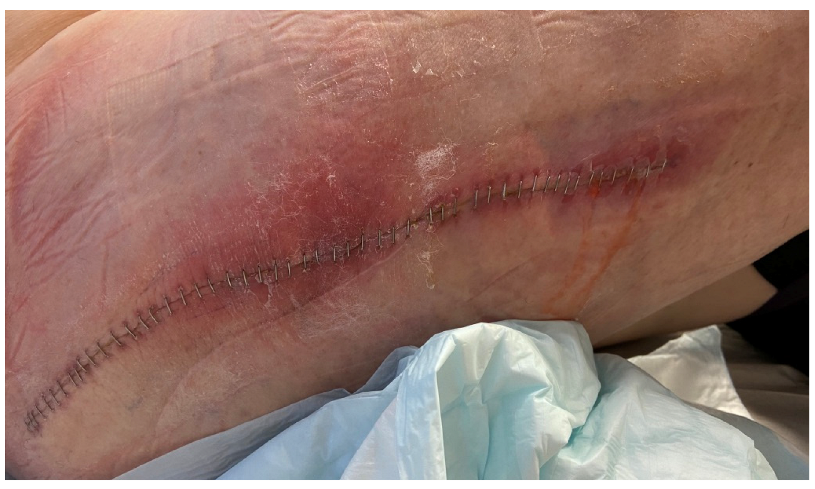
Case Presentation
2. Materials and Methods
2.1. Bacterial Cultures
2.2. Population Analysis Profile–Area under the Curve (PAP-AUC) Method
2.3. Genome Sequencing and Analyses
3. Results
3.1. Clinical Course
3.2. MIC
3.3. PAP-AUC
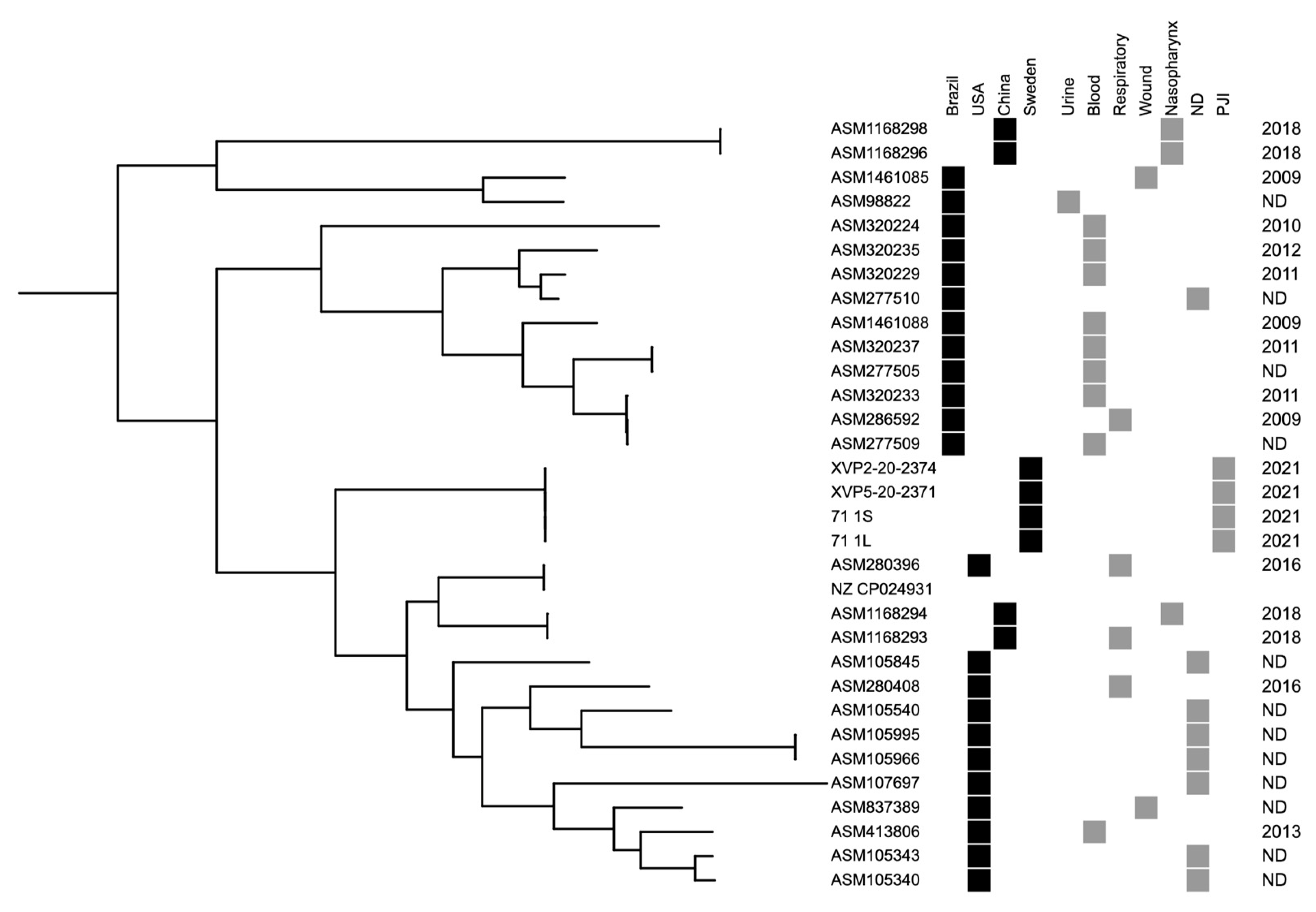
3.4. Genome Sequencing of C. striatum Isolates
4. Discussion
5. Conclusions
Author Contributions
Funding
Informed Consent Statement
Conflicts of Interest
References
- Lindgren, V.; Gordon, M.; Wretenberg, P.; Kärrholm, J.; Garellick, G. Deep Infection after Total Hip Replacement: A Method for National Incidence Surveillance. Infect. Control. Hosp. Epidemiol. 2014, 35, 1491–1496. [Google Scholar] [CrossRef] [PubMed]
- Thompson, O.; W-Dahl, A.; Lindgren, V.; Gordon, M.; Robertsson, O.; Stefánsdóttir, A. Similar Periprosthetic Joint Infection Rates after and before a National Infection Control Program: A Study of 45,438 Primary Total Knee Arthroplasties. Acta Orthop. 2022, 93, 3–10. [Google Scholar] [CrossRef] [PubMed]
- Engesæter, L.B.; Dale, H.; Schrama, J.C.; Hallan, G.; Lie, S.A. Surgical Procedures in the Treatment of 784 Infected THAs Reported to the Norwegian Arthroplasty Register: Best Survival with 2-Stage Exchange Revision, but Also Good Results with Debridement and Retention of the Fixed Implant. Acta Orthop. 2011, 82, 530–537. [Google Scholar] [CrossRef] [PubMed]
- Wildeman, P.; Rolfson, O.; Söderquist, B.; Wretenberg, P.; Lindgren, V. What Are the Long-Term Outcomes of Mortality, Quality of Life, and Hip Function after Prosthetic Joint Infection of the Hip? A 10-Year Follow-up from Sweden. Clin. Orthop. Relat. Res. 2021, 479, 2203–2213. [Google Scholar] [CrossRef]
- Kapadia, B.H.; Berg, R.A.; Daley, J.A.; Fritz, J.; Bhave, A.; Mont, M.A. Periprosthetic Joint Infection. Lancet 2016, 387, 386–394. [Google Scholar] [CrossRef]
- Tande, A.J.; Patel, R. Prosthetic Joint Infection. Clin. Microbiol. Rev. 2014, 27, 302–345. [Google Scholar] [CrossRef]
- Flurin, L.; Greenwood-Quaintance, K.E.; Patel, R. Microbiology of Polymicrobial Prosthetic Joint Infection. Diagn. Microbiol. Infect Dis. 2019, 94, 255–259. [Google Scholar] [CrossRef]
- Månsson, E.; Bech Johannesen, T.; Nilsdotter-Augustinsson, Å.; Söderquist, B.; Stegger, M. Comparative Genomics of Staphylococcus Epidermidis from Prosthetic-Joint Infections and Nares Highlights Genetic Traits Associated with Antimicrobial Resistance, Not Virulence. Microb. Genom. 2021, 7, 000504. [Google Scholar] [CrossRef]
- Cazanave, C.; Greenwood-Quaintance, K.E.; Hanssen, A.D.; Patel, R. Corynebacterium Prosthetic Joint Infection. J. Clin. Microbiol. 2012, 50, 1518–1523. [Google Scholar] [CrossRef]
- Kalt, F.; Schulthess, B.; Sidler, F.; Herren, S.; Fucentese, S.F.; Zingg, P.O.; Berli, M.; Zinkernagel, A.S.; Zbinden, R.; Achermann, Y. Corynebacterium Species Rarely Cause Orthopedic Infections. J. Clin. Microbiol. 2018, 56, e01200-18. [Google Scholar] [CrossRef]
- Noussair, L.; Salomon, E.; El Sayed, F.; Duran, C.; Bouchand, F.; Roux, A.-L.; Gaillard, J.-L.; Bauer, T.; Rottman, M.; Dinh, A. Monomicrobial Bone and Joint Infection Due to Corynebacterium Striatum: Literature Review and Amoxicillin-Rifampin Combination as Treatment Perspective. Eur. J. Clin. Microbiol. Infect Dis. 2019, 38, 1269–1278. [Google Scholar] [CrossRef] [PubMed]
- Tai, D.B.G.; Patel, R.; Abdel, M.P.; Berbari, E.F.; Tande, A.J. Microbiology of Hip and Knee Periprosthetic Joint Infections: A Database Study. Clin. Microbiol. Infect 2022, 28, 255–259. [Google Scholar] [CrossRef]
- Bernard, K.A. Coryneform Gram-Positive Rods. In Manual of Clinical Microbiology, 12th ed.; ASM Press: Washington, DC, USA, 2019; Volume 1, pp. 413–442. [Google Scholar]
- Silva-Santana, G.; Silva, C.M.F.; Olivella, J.G.B.; Silva, I.F.; Fernandes, L.M.O.; Sued-Karam, B.R.; Santos, C.S.; Souza, C.; Mattos-Guaraldi, A.L. Worldwide Survey of Corynebacterium Striatum Increasingly Associated with Human Invasive Infections, Nosocomial Outbreak, and Antimicrobial Multidrug-Resistance, 1976–2020. Arch. Microbiol. 2021, 203, 1863–1880. [Google Scholar] [CrossRef] [PubMed]
- Chauvelot, P.; Ferry, T.; Tafani, V.; Diot, A.; Tasse, J.; Conrad, A.; Chidiac, C.; Braun, E.; Lustig, S.; Laurent, F.; et al. Bone and Joint Infection Involving Corynebacterium Spp.: From Clinical Features to Pathophysiological Pathways. Front. Med. 2021, 7, 539501. [Google Scholar] [CrossRef]
- Hines, K.M.; Waalkes, A.; Penewit, K.; Holmes, E.A.; Salipante, S.J.; Werth, B.J.; Xu, L. Characterization of the Mechanisms of Daptomycin Resistance among Gram-Positive Bacterial Pathogens by Multidimensional Lipidomics. mSphere 2017, 2, e00492-17. [Google Scholar] [CrossRef]
- Wootton, M.; Howe, R.A.; Hillman, R.; Walsh, T.R.; Bennett, P.M.; MacGowan, A.P. A Modified Population Analysis Profile (PAP) Method to Detect Hetero-Resistance to Vancomycin in Staphylococcus Aureus in a UK Hospital. J. Antimicrob. Chemother. 2001, 47, 399–403. [Google Scholar] [CrossRef]
- Al Janabi, J.; Tevell, S.; Sieber, R.N.; Stegger, M.; Söderquist, B. Emerging Resistance in Staphylococcus Epidermidis during Dalbavancin Exposure: A Case Report and in Vitro Analysis of Isolates from Prosthetic Joint Infections. J. Antimicrob. Chemother. 2023, dkac434. [Google Scholar] [CrossRef]
- Bortolaia, V.; Kaas, R.S.; Ruppe, E.; Roberts, M.C.; Schwarz, S.; Cattoir, V.; Philippon, A.; Allesoe, R.L.; Rebelo, A.R.; Florensa, A.F.; et al. ResFinder 4.0 for Predictions of Phenotypes from Genotypes. J. Antimicrob. Chemother. 2020, 75, 3491–3500. [Google Scholar] [CrossRef]
- Scheper, H.; Gerritsen, L.M.; Pijls, B.G.; Van Asten, S.A.; Visser, L.G.; De Boer, M.G.J. Outcome of Debridement, Antibiotics, and Implant Retention for Staphylococcal Hip and Knee Prosthetic Joint Infections, Focused on Rifampicin Use: A Systematic Review and Meta-Analysis. Open Forum Infect. Dis. 2021, 8, ofab298. [Google Scholar] [CrossRef]
- Oliva, A.; Stefani, S.; Venditti, M.; Di Domenico, E.G. Biofilm-Related Infections in Gram-Positive Bacteria and the Potential Role of the Long-Acting Agent Dalbavancin. Front. Microbiol. 2021, 12, 749685. [Google Scholar] [CrossRef] [PubMed]
- Matt, M.; Duran, C.; Courjon, J.; Lotte, R.; Moing, V.L.; Monnin, B.; Pavese, P.; Chavanet, P.; Khatchatourian, L.; Tattevin, P.; et al. Dalbavancin Treatment for Prosthetic Joint Infections in Real-Life: A National Cohort Study and Literature Review. J. Glob. Antimicrob. Resist 2021, 25, 341–345. [Google Scholar] [CrossRef]
- Simon, S.; Frank, B.J.H.; Hartmann, S.; Hinterhuber, L.; Reitsamer, M.; Aichmair, A.; Dominkus, M.; Söderquist, B.; Hofstaetter, J.G. Dalbavancin in Gram-Positive Periprosthetic Joint Infections. J. Antimicrob. Chemother. 2022, 77, 2274–2277. [Google Scholar] [CrossRef]
- Buzón-Martín, L.; Zollner-Schwetz, I.; Tobudic, S.; Cercenado, E.; Lora-Tamayo, J. Dalbavancin for the Treatment of Prosthetic Joint Infections: A Narrative Review. Antibiotics 2021, 10, 656. [Google Scholar] [CrossRef] [PubMed]
- Wunsch, S.; Krause, R.; Valentin, T.; Prattes, J.; Janata, O.; Lenger, A.; Bellmann-Weiler, R.; Weiss, G.; Zollner-Schwetz, I. Multicenter Clinical Experience of Real Life Dalbavancin Use in Gram-Positive Infections. Int. J. Infect Dis. 2019, 81, 210–214. [Google Scholar] [CrossRef]
- Dunne, M.W.; Puttagunta, S.; Sprenger, C.R.; Rubino, C.; Van Wart, S.; Baldassarre, J. Extended-Duration Dosing and Distribution of Dalbavancin into Bone and Articular Tissue. Antimicrob. Agents Chemother. 2015, 59, 1849–1855. [Google Scholar] [CrossRef] [PubMed]
- Cojutti, P.G.; Tedeschi, S.; Gatti, M.; Zamparini, E.; Meschiari, M.; Siega, P.D.; Mazzitelli, M.; Soavi, L.; Binazzi, R.; Erne, E.M.; et al. Population Pharmacokinetic and Pharmacodynamic Analysis of Dalbavancin for Long-Term Treatment of Subacute and/or Chronic Infectious Diseases: The Major Role of Therapeutic Drug Monitoring. Antibiotics 2022, 11, 996. [Google Scholar] [CrossRef] [PubMed]
- Spaziante, M.; Franchi, C.; Taliani, G.; D’Avolio, A.; Pietropaolo, V.; Biliotti, E.; Esvan, R.; Venditti, M. Serum Bactericidal Activity Levels Monitor to Guide Intravenous Dalbavancin Chronic Suppressive Therapy of Inoperable Staphylococcal Prosthetic Valve Endocarditis: A Case Report. Open Forum Infect. Dis. 2019, 6, ofz427. [Google Scholar] [CrossRef] [PubMed]
- Campanile, F.; Carretto, E.; Barbarini, D.; Grigis, A.; Falcone, M.; Goglio, A.; Venditti, M.; Stefani, S. Clonal Multidrug-Resistant Corynebacterium Striatum Strains, Italy. Emerg. Infect. Dis. 2009, 15, 75–78. [Google Scholar] [CrossRef]
- de Souza, C.; Faria, Y.V.; Sant’Anna, L.D.O.; Viana, V.G.; Seabra, S.H.; Souza, M.C.D.; Vieira, V.V.; Hirata Júnior, R.; Moreira, L.D.O.; Mattos-Guaraldi, A.L.D. Biofilm Production by Multiresistant Corynebacterium Striatum Associated with Nosocomial Outbreak. Mem. Inst. Oswaldo Cruz. 2015, 110, 242–248. [Google Scholar] [CrossRef]
- Fernández, J.; Greenwood-Quaintance, K.E.; Patel, R. In Vitro Activity of Dalbavancin against Biofilms of Staphylococci Isolated from Prosthetic Joint Infections. Diagn. Microbiol. Infect. Dis. 2016, 85, 449–451. [Google Scholar] [CrossRef]
- El Haj, C.; Benavent, E.; Sierra, Y.; Soldevila, L.; Rigo-Bonnin, R.; Torrejón, B.; Gomez-Junyent, J.; Rosselló, I.; Murillo, O. Comparative Efficacy of Dalbavancin Alone and with Rifampicin against in Vitro Biofilms in a Pharmacodynamic Model with Methicillin-Resistant Staphylococcus Aureus. Int. J. Antimicrob. Agents 2022, 60, 106664. [Google Scholar] [CrossRef] [PubMed]


Disclaimer/Publisher’s Note: The statements, opinions and data contained in all publications are solely those of the individual author(s) and contributor(s) and not of MDPI and/or the editor(s). MDPI and/or the editor(s) disclaim responsibility for any injury to people or property resulting from any ideas, methods, instructions or products referred to in the content. |
© 2023 by the authors. Licensee MDPI, Basel, Switzerland. This article is an open access article distributed under the terms and conditions of the Creative Commons Attribution (CC BY) license (https://creativecommons.org/licenses/by/4.0/).
Share and Cite
Söderquist, B.; Henningsson, T.; Stegger, M. Corynebacterium striatum Prosthetic Joint Infection Successfully Treated with Long-Term Dalbavancin. Microorganisms 2023, 11, 550. https://doi.org/10.3390/microorganisms11030550
Söderquist B, Henningsson T, Stegger M. Corynebacterium striatum Prosthetic Joint Infection Successfully Treated with Long-Term Dalbavancin. Microorganisms. 2023; 11(3):550. https://doi.org/10.3390/microorganisms11030550
Chicago/Turabian StyleSöderquist, Bo, Thomas Henningsson, and Marc Stegger. 2023. "Corynebacterium striatum Prosthetic Joint Infection Successfully Treated with Long-Term Dalbavancin" Microorganisms 11, no. 3: 550. https://doi.org/10.3390/microorganisms11030550
APA StyleSöderquist, B., Henningsson, T., & Stegger, M. (2023). Corynebacterium striatum Prosthetic Joint Infection Successfully Treated with Long-Term Dalbavancin. Microorganisms, 11(3), 550. https://doi.org/10.3390/microorganisms11030550