Article

Risk Assessment for the Spread of Flavescence Dorée-Related Phytoplasmas from Alder to Grapevine by Alternative Insect Vectors in Germany

1
Julius Kühn Institute, Federal Research Centre for Cultivated Plants, Institute for Plant Protection in Fruit Crops and Viticulture, Geilweilerhof, 76833 Siebeldingen, Germany
2
RLP AgroScience, Breitenweg 71, 67435 Neustadt an der Weinstrasse, Germany
*
Author to whom correspondence should be addressed.
Microorganisms 2023, 11(11), 2766; https://doi.org/10.3390/microorganisms11112766
Submission received: 30 September 2023 / Revised: 7 November 2023 / Accepted: 10 November 2023 / Published: 14 November 2023

Abstract

:
“Flavescence dorée” (FD)-related phytoplasmas are widespread in alder in Germany and their transmission to grapevine represents a high risk for FD outbreaks when the primary vector, Scaphoideus titanus, becomes present in the future. Therefore, the potential role of the Deltocephalinae leafhopper species in transmitting FD-related phytoplasmas from alder to grapevine was studied in extensive transmission trials conducted between 2017 and 2020. The transmission capacity of autochthonous Allygus spp. and the invasive Orientus ishidae captured on infected alder trees was tested under controlled conditions using various test designs, including grouped insects and single-insect studies. The latter experiments were analyzed in terms of survival probability, transmission success and phytoplasma load in the insects, measured by quantitative PCR. A minimum inoculation titer (MIT) required for successful transmission to alder was defined for both Allygus spp. and O. ishidae. While Allygus spp. exhibited slightly better survival on Vitis vinifera compared to O. ishidae, the latter displayed higher phytoplasma loads and greater transmission success. Although all species were capable of infecting alder seedlings, O. ishidae was able to transmit 16SrV-phytoplasmas directly to single grapevines. Infective adults of O. ishidae were captured from the beginning of July until the end of August, while Allygus spp. were only considered infective towards the end of the season. Thus, O. ishidae likely poses a higher risk for FD transmission from alder to grapevine, albeit at a very low level, as only five out of 90 transmission trials to V. vinifera were successful.

1. Introduction

“Flavescence dorée” (FD) is a threatening disease of grapevine (Vitis vinifera L.) associated with phytoplasmas belonging to the 16SrV ribosomal group [1,2], comprising the subgroups 16SrV-C and 16SrV-D [3]. Based on the map genetic locus, the subgroups can be further divided into clusters FD1, FD2 and FD3, with FD1 and FD3 including strains from subgroup 16SrV-C and cluster FD2 including strains from both subgroups [4,5]. To date, FD is transmitted from vine to vine by the Neartic leafhopper Scaphoideus titanus Ball, introduced into Europe in the first half of the 20th century [6]. Scaphoideus titanus is monophagous on Vitis vinifera and is the main source for the epidemic spread of the disease within vineyards.
However, it has been shown that other Auchenorryncha species may harbour FD phytoplasmas, e.g., the invasive mosaic leafhopper Orientus ishidae (Matsumara) [7] or the Palaearctic Dictyophara europaea (Linnaeus), which have been confirmed to transmit FD phytoplasmas from Clematis vitalba L. to grapevine [8]. Some years later, the capability of O. ishidae to acquire it from an infected source and transmit it to grapevine under laboratory conditions was proven [9].
The “flavescence dorée” phytoplasma epidemiology is not limited to the ‘grapevine–S.titanus’ pathosystem within the vineyard but may represent a complex system at a broader landscape scale involving additional vectors and reservoir plants [9,10,11,12]. In Europe, in addition to this phytoplasma, other members of the 16SrV group infect grapevine, alder (Alnus glutinosa (L.) Gaertn.) and other plant species [5,13]. The 16SrV group phytoplasmas not transmitted by S. titanus and infecting alder were referred to as alder yellows phytoplasma (AldY), while those detected in grapevine were called Palatinate grapevine yellows phytoplasma (PGY) [14]. PGY and AldY were found to be genetically closely related to “flavescence dorée” phytoplasmas [15,16]. Further comparative and phylogenetic analyses revealed that these three phytoplasmas were members of the same phylogenetic subclade [3,4]. AldY and PGY were shown to be transmitted from alder to alder and occasionally from alder to grapevine by the Macropsinae leafhopper Oncopsis alni Schrank [13,17] without causing epidemic outbreaks. Malembic-Maher et al. [11] demonstrated that none of the PGY-isolates transmitted to Vicia faba L. by Oncopsis alni were acquired or transmitted by S. titanus. In contrast, the Deltocephalinae species Allygus modestus/mixtus and O. ishidae collected from alder were capable of transmitting phytoplasma strains to Vicia faba L., which could then be further transmitted by S.titanus. The authors elucidated that the “flavescence dorée” phytoplasmas are of European origin and are widespread in natural habitats in hosts like alders. In Germany, alder trees are frequently infected with various 16SrV phytoplasmas, and preliminary studies showed that FD-like phytoplasmas are widespread [18]. Although S. titanus has not yet been detected in Germany, a single grapevine infected by “flavescence dorée” phytoplasmas was found in the vicinity of an alder stand and Deltocephalinae species collected from these alders harboured the same phytoplasma strain as the grapevine [19]. During a previous survey of the leafhopper fauna on alders, it was discovered that the autochthonous Allygus mixtus (Fabricius) and Allygus modestus Scott, as well as the invasive O. ishidae, were the most abundant Deltocephalinae species on alder, and they were frequently infected with 16SrV phytoplasmas [20].
The objective of this study was to evaluate the role of Allygus spp. and O. ishidae in the transmission of “flavescence dorée” phytoplasmas from alder to grapevine and, consequently, to assess the role of these species in the possible spread of this pathogen into vineyards. This issue is of great concern in the context of the possible appearance of S. titanus in Germany. In this regard, the transmission capacity of Allygus spp. and O. ishidae collected from alder in Germany was investigated in more detail. Different types of transmission trials and survival experiments were carried out with these species. Furthermore, their infection status was analyzed, a species-specific minimum titer for successful transmission was defined, and the variation of the phytoplasma load during the season was also assessed.

2. Materials and Methods

2.1. Insect and Plant Material

Adult Cicadellidae leafhoppers were collected in different vine-growing areas in southwestern Germany by sweep-netting from infected alder trees adjacent to vineyards, from the beginning of June until September in the years 2017–2020. Specimens of the Deltocephalinae Allygus modestus, Allygus mixtus and Orientus ishidae were collected using a mouth aspirator and transported to the laboratory in cages, where they were either stored at −20 °C or used alive for transmission trials. Dead insects were identified at the species level and sexed based on morphological characteristics according to Kunz et al. [21] and Biedermann and Niedringhaus [22]. As Allygus spp. are difficult to distinguish by morphological means, a subset of samples was analyzed by COI barcoding according to the EPPO standard protocol [23], confirming morphological identification results that both Allygus modestus and Allygus mixtus were present in the samples.
Alder seedlings, hot water-treated grapevine cuttings of cv. Chardonnay and micropropagated phytoplasma-free plantlets of cv. Scheurebe were grown in 2 L pots at the Julius Kühn-Institute, Siebeldingen, Germany, under insect-proof conditions. Glasshouse conditions were 25  ±  5 °C temperature, 70  ±  10% relative humidity, a photoperiod of 16:8 h light/dark and a minimum of 2500 lx light intensity.

2.2. Transmission Trials

Insects collected in the field from infected A. glutinosa were caged on recipient plants either in a dual-choice approach or in single plant assays with or without transfer. The experimental set-up is illustrated in Figure S1a,b (Supplementary Materials).
For dual choice experiments, leafhoppers of the same species (variable group size: 5–20, depending on collections) were caged with seedlings of A. glutinosa (8–10 fully developed leaves) and hot-water treated V. vinifera cv. Chardonnay test plants (8–10 fully developed leaves) in mesh cages (32.5 cm × 32.5 cm × 32.5 cm, Watkins & Doncaster, Leominster, UK) (Figure S1a) until death.
In single-plant tests, the insects were caged on potted plants in cylindrical acrylic insect-proof cages covered with a nylon mesh (height 35 cm, diameter 13 cm). For assays with transfer, batches of 5–15 insects were put on alder seedlings (6–10 fully developed leaves) and after an inoculation access period (IAP) of 3–7 days, living specimens were transferred to micropropagated V. vinifera plantlets cv. Scheurebe (6–10 fully developed leaves) as second test plants until their death (Figure S1b). Single plant-single insect trials were carried out on seedlings of A. glutinosa (6–8 fully developed leaves) and micropropagated V. vinifera cv. Scheurebe (6–8 fully developed leaves).
All tests were carried out under the same conditions in a walk-in climatic chamber Fitotron type SGR233 (Weiss Technik Ltd., Loughborough, UK) at a photoperiod of 16:8 h (1 h each of dusk at dawn), 23:19 ± 2 °C and 75 ± 5% relative humidity. Cylindrical cages were checked daily and dead individuals were removed and stored at −20 °C until final morphological identification and molecular analyses. At the end of the trial, inoculated test plants were removed from the cage, treated once with an insecticide (Spruzit Schädlingsfrei, W. Neudorff GmbH KG, Emmerthal, Germany) and transferred into the glasshouse. Plants were tested for phytoplasma presence three times to confirm successful or failed transmission: (i) approximately 8 weeks after the end of the trial, (ii) after hibernation and dormancy in the cold house (about 6 months after IAP) and (iii) about 12 months after IAP.

2.3. Survival Experiments

In combination with single insect-single plant transmission trials in 2019 and 2020, the survival of Allygus spp. and O. ishidae specimens on A. glutinosa and V. vinifera cv. Scheurebe was recorded daily. Cages containing a water source but without any plant served as negative controls. Dead insects were removed from the cages, morphologically identified, sexed and stored at −20 °C until DNA extraction and examination of phytoplasma presence.

2.4. DNA Extraction

Total nucleic acid (TNA) of the test plants was extracted from 120 to 150 mg leaf midribs and petioles with the CTAB method by Doyle and Doyle [24], modified according to Boudon-Padieu et al. [25]. The final pellet was resuspended in 150 µL TE buffer. TNA was extracted from healthy plantlets that were raised in the glasshouse with extraction procedures as negative control.
TNA of insects was extracted according to the CTAB protocol by Maixner et al. [14]. Individuals were homogenized in 250 µL CTAB buffer using a cell mill (TissueLyser II, Qiagen GmbH, Hilden, Germany) with two carbide beads per tube. After precipitation and washing procedures, the final pellet was resuspended in 150 µL TE buffer.

2.5. Phytoplasma Detection

Generic phytoplasma detection was carried out by PCR with the universal primers U5/P7 [26,27], using a 20 µL reaction volume with a final concentration of 0.025 U/µL DreamTaq Green DNA-Polymerase (Thermo Fisher Scientific GmbH, Waltham, MA, USA), 0.2 mM dNTPs, 0.5 µM (each primer), 1X DreamTaq Green Buffer (Thermo Fisher Scientific GmbH, Waltham, MA, USA) and 2 µL template DNA. Cycle conditions were 2 min at 94 °C followed by 5 cycles of (30 s at 94 °C, 30 s at 59 °C and 90 s at 72 °C), 5 cycles of (30 s at 94 °C, 30 s at 58 °C and 90 s at 72 °C), 20 cycles of (30 s at 94 °C, 30 s at 57 °C and 90 s at 72 °C) and a final elongation step of 30 s at 72 °C.
To identify phytoplasmas of the 16SrV ribosomal group, PCR was carried out with the group-specific primers fAY/rEY as described by Maixner and Reinert [17].

2.6. Amplification and Sequencing of Genetic Markers

PCR-positive samples of insects or inoculated plants were further characterized by amplification of the secY-map locus (map gene) by nested PCR and sequencing as described by Arnaud et al. [4] and by the vmpA gene marker (vmp gene) according to the protocol of Malembic-Maher et al. [11]. Primers and PCR conditions are described in Table S3 in Malembic-Maher et al. [11] The products of both amplifications were purified with the QIAquick PCR Purification Kit (Qiagen GmbH, Hilden, Germany). Sequencing reactions were performed by SeqLab (Microsynth GmbH, Balgach, Switzerland) via Sanger sequencing in both directions on the map gene using the primers fFD9F6/rMapR2, and upstream on the vmpA gene with primers VMPA-F3/VMPA-R5 [11]. Assembling of raw sequences and multiple alignments were performed with CLC Main Workbench 22.0.2. (Qiagen GmbH, Hilden, Germany) and phylogenetic reconstructions using maximum parsimony were performed by MEGA7 [28] with randomized bootstrapping evaluation.

2.7. Phytoplasma Quantification

The phytoplasma concentration in infected insects was determined by quantitative real-time PCR using a newly developed SYBR Green™ assay. The housekeeping gene map was used for primer selection. Forward primer MAP U1 (5′-ATCGTTATAATGAAAGAGGC-3′) and reverse primer MAP U2 (5′-TGTTTAATACCTATATCTAAAG-3′) were selected in two conserved regions among the 121 map genotypes published by Malembic-Maher et al. [11]. A 272 bp gene fragment of 16SrV phytoplasmas was amplified with these primers (0.22 µM each) using 1× TEMPase Mastermix (VWR International GmbH, Darmstadt, Germany) with 3 mM Mg2+ and SYBR Green™ I (Lumiprobe GmbH, Hannover, Germany) diluted to 1:66,000 in 20 µL reactions. Cycle conditions were 15 min at 95 °C for hot start followed by 40 cycles of 15 s at 95 °C, 15 s at 54 °C and 30 s at 72 °C. After the final elongation step of 4 min at 72 °C, a melting curve analysis ranging from 50 °C to 90 °C was performed. Each sample was diluted 1:5 and 1 µL was amplified in duplicate in two independent runs in a Chromo4 Real-Time PCR detector (Bio-Rad Laboratories GmbH, Feldkirchen, Germany). Absolute quantification was performed by the standard curve method using a plasmid containing the map sequence of map-genotype M38 in serial 10-fold dilutions ranging from 1 × 10⁸ target copies per µL to 1 copy per µL. The cycle threshold (Ct) values of the standard dilutions were plotted and verified to give a linear relationship, which served as the standard curve. The quality of the standard curve was adjusted manually for each run according to the best linear regression coefficient. Then, the Ct-values of each sample were compared to this standard curve and the copy number in each sample was calculated. The phytoplasma concentrations were normalized to the ng DNA per µL as measured by NanoDrop™ 2000c (Thermo Fisher Scientific GmbH, Waltham, MA, USA).

2.8. Statistical Analysis

The effect of the host plant species and/or phytoplasma load of the insect on the survival of Allygus spp. and O. ishidae was assessed through survival curves, estimated by the Kaplan–Meier method for censored data [29]. A marginal Cox proportional-hazards model was applied where robust standard errors were obtained [30]. The Cox model was validated by checking the proportional hazards assumptions with a Schoenfeld residual analysis [29]. The dependent variable was the survival time of each insect; the categorical explanatory variables were the sex, the phytoplasma infection status, the insect taxon (Allygus spp. or O. ishidae), the transmission success (unsuccessful/successful) and the test plant species (A. glutinosa/V. vinifera). Pairwise comparisons were performed using Log-rank test with Bonferroni correction. Statistical differences between plants or insect taxa were indicated when p < 0.05.
To test the effect of the phytoplasma load on the transmission success to the test plants, a Welch’s t-test was performed for each insect taxon as the data violate the assumption of homogeneity of variances. The response variable was the transmission success, and the categorical explanatory variable was the phytoplasma load.
Seasonal variations of the phytoplasma load within the species Allygus spp. and O. ishidae were calculated by Welch’s ANOVA, followed by the Games–Howell Post Hoc Test. The response variable was the period within the years, and the categorical explanatory variable was the phytoplasma load.
All analyses were performed in R version 4.2.2 [31]. Cox models were developed and validated using the ‘survival’ package [32]. Kaplan–Meier curves were plotted using the ‘survminer’ package [33]. Pairwise comparisons were run using the ‘emmeans’ package [34]. Boxplots and barplots were generated with packages ‘ggplot2′ [35] and ‘ggstatsplot’ [36], respectively.

3. Results

3.1. Dual-Choice Test

The results of the dual choice trials are summarized in Table 1. In 2017, O. ishidae was able to transmit 16SrV-group phytoplasmas from field-grown alder to a high percentage of alder seedlings but was also able to inoculate one grapevine plantlet in the same cage where an alder seedling had been inoculated. In all infected individuals of O. ishidae and in all infected test plants (alder and grapevine), the FD-map genotype M38 was identified by sequence analysis. In contrast, in the following year 2018, Allygus spp. as well as O. ishidae inoculated alder seedlings, but no transmission occurred to Vitis vinifera during dual choice experiments.

3.2. Transmission Test with Plant-Transferred Insects

In a second experimental design, insects were initially placed on A. glutinosa and then transferred to V. vinifera. As depicted in Table 2, a high transmission rate to alder seedlings was observed with both O. ishidae and Allygus spp. in all years. However, there was no successful inoculation of grapevine plantlets by the same batches of insects that previously inoculated alder. Despite annual fluctuations, infection rates in alders inoculated by O. ishidae were consistently higher than those inoculated by Allygus spp. This is consistent with the higher infection rate in O. ishidae compared to Allygus spp., but also with the greater number of O. ishidae per test plant. As expected, the infection rates of insects collected from the first test plant (alder) and subsequently from the second test plant (grapevine) were closely matched. Infection rates in Allygus spp. showed more variation, with a notable difference in 2019.

3.3. Single Insect—Single Plant Transmission Tests

The third experimental setup was designed as single insect-single plant trials and aimed to explore transmission parameters in a one-by-one insect-plant assignment. The results of the transmission trials are summarized in Table 3. The transmission rates to alder in 2019 and 2020 were relatively consistent for Allygus spp. (12% vs. 14%), but varied considerably between the years for O. ishidae (6% vs. 46%). These variations between years and species are also evident in the transmission efficiency, which represents the proportion of infected leafhoppers that successfully transmitted (Allygus spp.: 18% and 19%; O. ishidae: 7% and 46%). Nevertheless, individual O. ishidae were capable of transmitting the phytoplasma to grapevine with an efficiency of 13%, while there was no successful inoculation by Allygus spp. Figure S2 depicts an infected micropropagated plantlet cv. ‘Scheurebe’ three months after inoculation with O. ishidae.

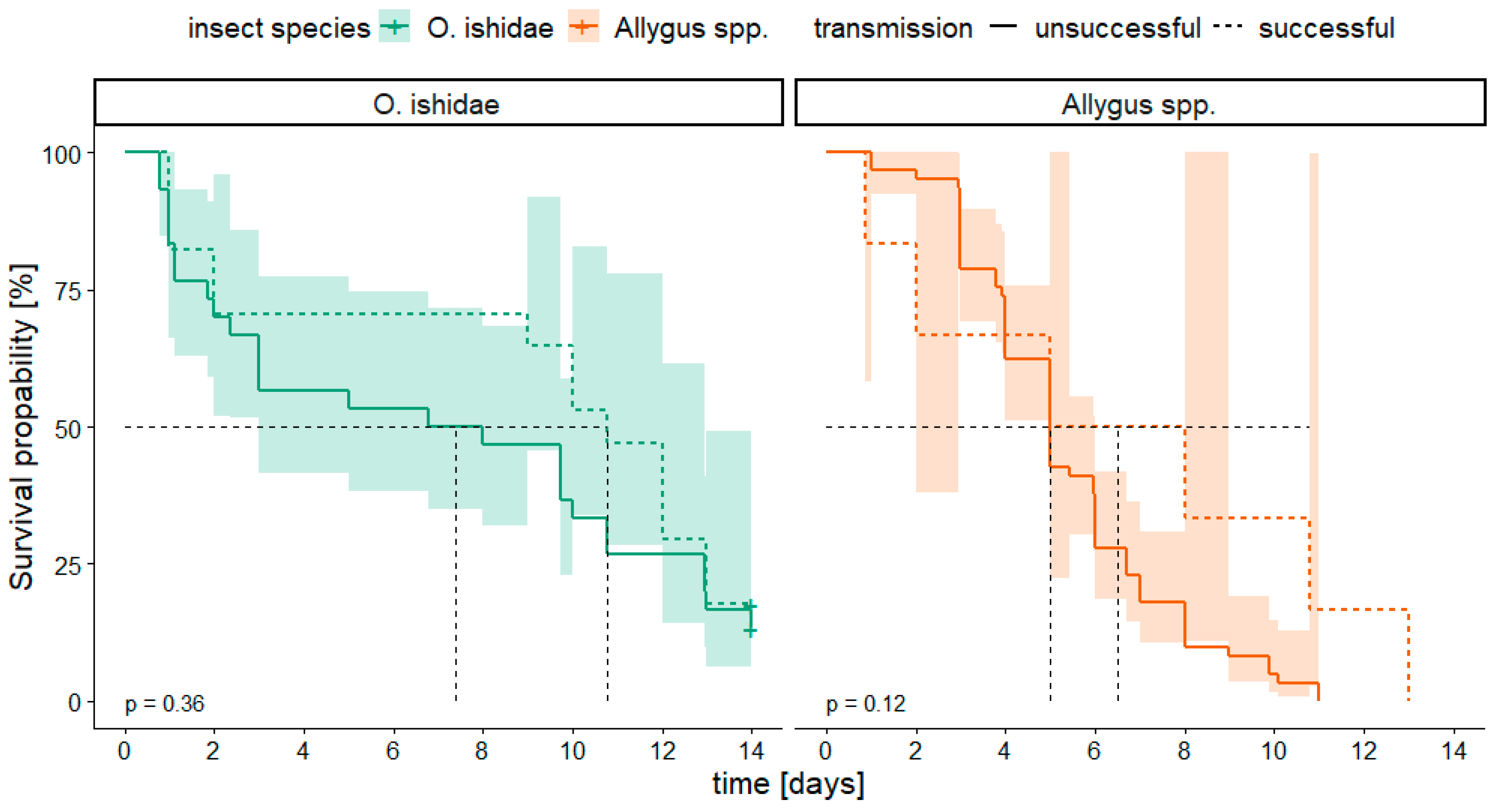
3.4. Survival Analysis

To assess the suitability of a plant species as feeding host and the influence on the transmission success, we combined the transmission trials with a survival analysis during single insect-single plant assays. In a first step, we verified that there was no significant difference between the two years of the investigation, 2019 and 2020 (p > 0.05). Consequently, we pooled the data for further analyses.
Figure 1 illustrates the survival probability of both taxa, Allygus spp. and O. ishidae, on the two test plants A. glutinosa and V. vinifera, compared to a negative control without food source. Regarding the test plants no relevant difference occurred between the survival of Allygus spp. on alder compared to grapevine (p < 0.05), while O. ishidae performed statistically significantly better on alder than on grapevine (p < 0.001). When comparing the insect species, O. ishidae showed a significantly longer survival probability on alder compared to Allygus spp. (Figure 1, left). Conversely, the survival of Allygus spp. was significantly better on grapevine than that of O. ishidae (Figure 1, middle). None of the specimens could survive without any food source for more than 3 days (Figure 1, right).
The influence of the sex of the individuals on the survival probability was also examined (Figure S3, Supplementary Materials). With the exception of the species O. ishidae on alder, females exhibited a higher survival probability than males.
The effect of the phytoplasma infection status of the insects (infected compared to non-infected) on their survival probability was analyzed for both species. Single insect-single plant trials conducted in 2019 and 2020 revealed that the infection status did not significantly impair the survival probability of any species tested in our study (p > 0.05, Figure 2).
It was also investigated whether the inoculation success was influenced by the longevity of the insects on the test plants. Therefore, the survival of infected individuals was analyzed in relation to their ability to inoculate alder seedlings. While the dataset for transmission to V. vinifera was too limited for statistical analysis (five transmissions only), in the case of alder, we observed no effect of the longevity on the inoculation success, since there was no significant difference in the survival probability between insects that transmitted the phytoplasma successfully and those that did not (Figure 3).

3.5. Phytoplasma Quantification

The phytoplasma load was quantified in all infected specimens, and it was also differentiated among individuals, which transmitted successfully and those that did not. In both species, insects that successfully transmitted had a significantly higher titer compared to infected specimens without transmission success (Figure 4). Single O. ishidae generally had a titer approximately 10 times higher than Allygus spp. (see scaling in Figure 4). Based on the quantitative data, it was possible to define a minimum inoculation titer (MIT) for transmission to alder for both species. The MIT was determined from the lowest titer of Allygus spp. or O. ishidae individuals that still successfully transmitted. The titer of successfully transmitting Allygus spp. was at least 62 phytoplasma genomic units/ng DNA. Single O. ishidae transmitted at a minimum titer of 218 phytoplasma genomic units/ng DNA. Individuals with a phytoplasma load above the species-specific MIT were therefore classified as potentially infective. All of the four O. ishidae specimens that successfully inoculated V. vinifera in the single insect-single plant trials had a high phytoplasma load.
Based on the MIT, the transmission success was examined in relation to the inoculation access period (IAP) of potentially infective insects. The results, summarized in Table 4, illustrate that O. ishidae is able to successfully transmit to alder within only one day after inoculation. In contrast, for Allygus spp., no individual reached the MIT after one day, but they exceeded the MIT after two days, followed by successful transmission to alder. A majority of the insects (Allygus spp. 78% and O. ishidae 75%) classified as infective transmitted the phytoplasma after an extended IAP of up to 10 days.
Furthermore, the variation of the phytoplasma load of field-collected leafhoppers of both taxa during the season and the proportion of specimens classified as infective (having a phytoplasma load exceeding MIT) was calculated and related to the collection time (Figure 5, note the different scaling between species). Interestingly, the trend for both species is different. In Allygus spp., the phytoplasma load steadily increased from June to July, with individuals reaching the MIT not before the end of July. In contrast, O. ishidae specimens were already classified as infective from the first sampling date at the end of June. However, it is worth noting that even though O. ishidae were already infective at the beginning of the season, their phytoplasma load showed high variability.

3.6. Genotyping of Phytoplasmas in Insects and Plants

The phytoplasma genotypes in infected leafhoppers and test plants were determined based on the gene markers map and vmp. As shown in Table 5, the map type M38, which belongs to the cluster FD2, is predominant in both insect species and in both test plant species. One alder seedling became infected by the PGY genotype M48 after inoculation with Allygus spp. While Allygus spp. carried different map PGY genotypes, O. ishidae was almost exclusively loaded with the M38 genotype, the same genotype that was also transmitted to V. vinifera by this species in the transmission trials. The differentiation on the vmpA gene confirmed the expected allocation of all PGY-associated map genotypes to the VmpA-I cluster and the map genotype M38 to the VmpA-II cluster.

4. Discussion

The European origin of FD phytoplasmas is well documented [11]. In particular, alder trees are considered a main source of FD phytoplasmas. Rigamonti et al. [12] reported infection rates of black alder in northwestern Italy of about 91%, which fits well with infection rates in Germany or France [11,18]. While the presence of S. titanus, the main vector of “flavescence dorée” phytoplasmas, is the prerequisite for FD outbreaks in vineyards, autochthonous Deltocephalinae leafhoppers may occasionally transmit “flavescence dorée” phytoplasmas from alder to grapevine, thereby establishing sources of inoculum in the vineyards that could increase the risk of outbreaks once S. titanus emerges. However, the probability and the parameters of “flavescence dorée” phytoplasma transmission from alder to grapevine by Deltocephalinae species remain poorly understood, although this information is required for risk analysis, particularly for regions where S. titanus is approaching. Since Allygus spp. and O. ishidae are confirmed vectors of “flavescence dorée” phytoplasmas to alder under experimental conditions [11,20], this study focused on the transmission parameters of field-captured adults of these two taxa in transmission trials to V. vinifera. Malembic-Maher et al. [11] reported high infection rates by 16SrV phytoplasmas in the Deltocephalinae Allygus spp., O. ishidae and the Macropsinae Oncopsis alni. Only the Deltocephalinae transmitted “flavescence dorée” phytoplasmas, while O. alni transmitted PGY strains that are not transmitted by S. titanus.
The objective of this study was to elaborate if Allygus spp. and O. ishidae, collected from infected alder in the field, were able to infect grapevine and to elucidate the transmission parameters to assess the risk for FD outbreaks. As both species are not closely associated with grapevine and were only occasionally captured in vineyards, we tested three different experimental designs of transmission assays. First, dual choice experiments with both, the preferred host plant alder and the occasional host grapevine in the same cage, allowed the leafhoppers to move freely between the natural host and grapevine. Second, transfer trials where field-captured insects are adapted on alder before being forced to feed on grapevine. Third, a one-by-one assignment between single leafhoppers on their host alder and the occasional grapevine with three objectives: (i) to ensure feeding of the test insects on the plant, (ii) to record the individual survival time and (iii) to correlate transmission success with the individual insect’s phytoplasma titers.
The experimental set-ups of the transmission trials were adapted from our previous studies with O. alni, Allygus spp. and O. ishidae using groups of insects [11,13,19]. The data from all three experimental set-ups confirmed previous results, demonstrating that both Allygus spp. and O. ishidae efficiently inoculated alder seedlings, although transmission rates were lower in the third experiment where test plants were inoculated by only one insect vector. The transmission rates of Allygus spp. to alder (29–80%) were much higher than the 20% previously reported by Malembic-Maher et al. [11], while O. ishidae was confirmed as a very efficient vector of 16SrV phytoplasmas with transmission rates ranging from 67 to 100% in all studies. Thus, both taxa play a role in maintaining 16SrV phytoplasmas in European alder populations.
While larger hot-water treated grapevine cuttings cv. Chardonnay were used for the dual choice and transfer approaches with groups of insects, the single insect-single plan3 experiments were carried out with smaller ex vitro plants cv. Scheurebe. It is conceivable that the smaller plantlets with more delicate tissue react more sensitively to inoculation and, thus, the transmission success was higher.
Transmission to grapevine was only achieved by O. ishidae in the first (one successful transmission) and third (four successful transmissions) experimental set-ups, with comparable transmission rates of 17% and 13%, respectively. The general capability of O. ishidae to inoculate grapevine plantlets under forced experimental conditions had been previously demonstrated by Lessio et al. [9]. However, they were only successful in two trials with specimens that performed an acquisition access period (AAP) on infected plants under experimental conditions. We obtained successful inoculation of grapevine with naturally infected O. ishidae and proved that O. ishidae is able to transmit “flavescence dorée” phytoplasma from alder to grapevine, since the FD map genotype M38 was detected in both the successful vectors and the recipient grapevine plants. According to Malembic-Maher et al. [11] map genotype M38 is identical to grapevine map-FD2 genotypes detected in FD outbreaks and can be transmitted by S. titanus.
As it was worked with field-captured adults at different capture periods, infection rates of insects and transmission rates varied among the years of experimentation. In the transmission trails with dual choice and by transfer, infection rates of Allygus spp. and O. ishidae ranged from 14 to 72% and 32 to 80%, respectively, confirming the data presented by Malembic-Maher et al. [11], who found 60% infected Allygus spp. and 52% infected O. ishidae. Comparable infection rates for O. ishidae were reported from Slovenia [37] (25–50%), Switzerland [38] (85%) and the Alsace region in France [39] (56%).
The single insect-single plant approach allowed us to analyze the correlation between survival probability, infection status of the individuals and transmission success. The statistical analysis revealed a significantly higher survival probability of O. ishidae on alder compared to grapevine, indicating that alder is a true host plant for O. ishidae. Both species are more or less polyphagous with adults preferring some trees and shrubs [9,40,41,42,43]. An important difference between both taxa is the ability of O. ishidae to complete its whole life cycle on alder, whereas adult Allygus spp. move to alder after their larval instars have fed on herbaceous vegetation [44]. This observation was supported by recent data from Rizzoli et al. [38], who demonstrated that O. ishidae is able to complete its biological cycle on Alnus glutinosa and could acquire 16SrV phytoplasmas efficiently from alder. Accordingly, the low survival probability of both taxa on Vitis vinifera confirms the hypothesis that grapevine is not very attractive as food source for both species. The generally high transmission rates to alder indicate that both vector species can feed on alder in the adult stage. The survival times of Allygus spp. and O. ishidae on V. vinifera exceeded that of nonplant controls, which indicates that both species fed on grapevine recipient plants but were restricted in their ability to transmit the pathogen to these test plants. Additional electrical penetration graph (EPG) studies would be useful to understand and characterize the feeding mechanism underlying the varying transmission success between these species. Regarding FD, the feeding characteristics were only described for the vector S. titanus [45].
The survival probability was similar for transmitters and nontransmitters of both species and the infection status did not influence their survival, as it has been also demonstrated for infected S. titanus [46]. Reports on a reduced fitness of infected phytoplasma vectors, e.g., Macrosteles quadripunctulatus in the Chrysanthemun yellows model [47], showed that this is not always the case.
If the infection status does not affect the survival or fitness of the test insects, other parameters must be considered to explain the transmission success. The quantification of the phytoplasma load in all infected individuals revealed a significant difference in the phytoplasma titers of successfully transmitting specimens of both taxa, which allowed us to define a minimum inoculation titer (MIT) for transmission. As phytoplasmas have to multiply in the salivary glands of the insect during the latency period (LP) to render an individual infective, the phytoplasma load of an insect distinguishes between noninfective individuals that just acquired the phytoplasmas and infective individuals that can successfully transmit the phytoplasma with the saliva to the plant [48,49].
Interestingly, not only did O. ishidae have a 3-fold higher MIT than Allygus spp. but infective O. ishidae also generally had much higher phytoplasma loads, ranging predominantly from 103 to 104 phytoplasma genomic units (GU) per ng DNA, compared to infective Allygus spp. individuals with titers of 102 to 103 phytoplasma GU per ng DNA. However, the data are in the range of phytoplasma titers reported for Macrosteles quadripunctulatus (104 phytoplasma GU per ng total DNA) and Euscelidius variegatus (103 phytoplasma GU per ng total DNA) in the Chrysanthemun yellows model [47,50].
Only O. ishidae was able to transmit successfully to grapevine, and all these individuals had a high phytoplasma titer (104 phytoplasmas GU per ng DNA), a level that was never achieved in individuals of Allygus spp. From this observation, it can be assumed that the threshold titer for successful inoculation of grapevine might be higher than the MIT for alder.
The majority of individuals that reached the MIT successfully transmitted the pathogen to alder seedlings. The transmission probability generally increased with longer IAP, but O. ishidae succeeded in inoculating alder seedlings with an IAP of at least one day. In the case of Allygus spp., an IAP of at least two days resulted in transmission to alder, while a minimum of one day could not be tested in our studies. O. ishidae did not survive longer than 4 days on grapevine, but this was sufficient to transfer the agent to the plantlets. It can be assumed that highly infective individuals do not require a long incubation period on small plantlets as used for the transmission trials.
The differences between both taxa in terms of infection rate, incubation time and transmission success may be related to their different biological cycle [9,40,41,42,43]. Tracking the evolution of the mean phytoplasma titer and the percentage of infective individuals during the season, it was observed that already the first captured adults of O. ishidae showed a high phytoplasma load and the percentage of infective individuals remained at a high level throughout the entire season. The first instars of O. ishidae have the opportunity to acquire the pathogen from infected alder and the latency period can be completed by the end of nymphal development. In contrast, Allygus spp. only reached a substantial phytoplasma titer in the middle of the season, with the percentage decreasing toward the end. This corresponds with the life cycle of Allygus spp., which has the ability to acquire 16SrV phytoplasmas only when the adults move from the soil vegetation to the infected alder trees. A suspected latency period of four weeks thereafter could explain the observed low phytoplasma titers in Allygus spp. until mid-July. Therefore, it can be concluded that O. ishidae has a higher potential to acquire “flavescence dorée” phytoplasmas early from alder and to reach phytoplasma titers more relevant for transmission to other plants. In consequence, infective individuals of O. ishidae are present in the vicinity of vineyards for a much longer period than infective individuals of Allygus spp. increasing the probability of accidental “flavescence dorée” phytoplasma transmission to grapevine.
In 2017, in Alsace, a single grapevine infected with map genotype M38 was detected at a site where S. titanus was still absent [39]. The presence of Allygus spp. and O. ishidae infected by the M38 strain at the site led to the conclusion that these alternative vectors might be responsible for the transfer of M38 from reservoir alders to grapevine. A similar scenario occurred in Germany in 2020, with the detection of a M38 infected grapevine in a commercial vineyard in Rhineland-Palatinate, adjacent to an alder stand where specimens of Allygus and O. ishidae infected by M38 were sampled [19]. This study supports the conclusions by Malembic-Maher et al. [11] and Rizzoli et al. [38] that the coincidence of infected alder with Allygus spp. or O. ishidae and nearby vineyards may provoke the emergence of isolated FD cases that could trigger FD spread once S. titanus becomes established.
Especially in Germany, which is still assessed as FD-free but where the nonepidemic PGY variants exhibit the same symptoms as “flavescence dorée” phytoplasmas, the precise identification of the specific phytoplasma strain is crucial. Analyses of the phytoplasma genotypes in insects and test plants based on the housekeeping genes map and vmp showed that in all test plants as well as in almost all O. ishidae only map genotype M38 belonging to the genetic VmpA-II cluster was found. M38 was also the dominant genotype in infected Allygus spp. However, in Switzerland, Rizzoli et al. [38], and in northwestern Italy, Rigamonti et al. [12], evidenced a high prevalence of the map genotype M50 in O. ishidae as well as in alder trees. Similarly, Malembic-Maher et al. [11] obtained a high percentage of transmissions by O. ishidae with map genotype M50 originating from southwestern France. As M38 and M50 belong to different FD clusters, it might be concluded that the map-FD1 type M50 is more prevalent in regions south and west of the Alps whereas map-FD2 type M38 is dominant in regions north of the Alps. It remains to be elucidated what this means for the epidemiology of FD.
These data prove the possibility of “flavescence dorée” phytoplasma transmission from alder to grapevine. It may be considered a rare event, as transmissions from alder to grapevine under field conditions are likely less efficient than in our laboratory studies. However, the further spread of the invasive species O. ishidae could lead to a higher prevalence of M38 in alder, since it is shown in this study that O. ishidae is an efficient vector of the M38 strain from alder to alder. Preliminary data from Germany support this hypothesis [18] and demonstrate that alder trees are widespread in the vicinity of vineyards. This aspect might be similar in other viticultural regions with comparable natural habitats, e.g., Switzerland [38] and France [39]. Since it is of great importance for FD-free regions like Germany to identify and eradicate single FD-infected grapevines before the arrival of the vector S. titanus, monitoring should be focused on vineyards in the vicinity of alder stands. For this reason, currently risk maps of vineyards in the vicinity of alder stands are under preparation [18].

5. Conclusions

This study aimed to evaluate the risk posed by two Deltocephalinae leafhopper taxa for the spread of “flavescence dorée” from naturally infected alder into vineyards. Populations of Allygus spp. and Orientus ishidae collected from infected alder trees in southwestern Germany were infected by “flavescence dorée” phytoplasmas and readily transmitted the phytoplasma to alder seedlings, which confirms the results of previous studies. One to two days of inoculation access were sufficient for successful transmission to alder. Although both species survived and presumably fed on grapevine for several days, only O. ishidae transmitted “flavescence dorée” phytoplasmas successfully to grapevine in two of three modes of transmission trials, although with less efficiency compared to alder. In addition, O. ishidae carries higher titers of “flavescence dorée” phytoplasmas compared to Allygus spp. during a longer period within the season. This leads to the conclusion that the invasive leafhopper O. ishidae poses a higher risk for disseminating “flavescence dorée” phytoplasmas to vineyards than the two autochthonous Allygus species present on alder. However, the probability of O. ishidae spreading “flavescence dorée” phytoplasmas from alder to vineyards appears to be low, due to the feeding biology of this species and rather low transmission rates to grapevine even in controlled transmission trials. Nevertheless, every single grapevine infected by “flavescence dorée” phytoplasmas poses a risk of FD outbreaks in the presence of the vector S. titanus. Therefore, specific monitoring strategies of vineyards adjacent to alder stands need to be established in FD-free regions to prevent FD outbreaks.

Supplementary Materials

The following supporting information can be downloaded at: https://www.mdpi.com/article/10.3390/microorganisms11112766/s1, Figure S1: (a) Schematic diagram for a dual choice approach with several test plants in one cage. (b) Schematic diagram for a transmission trial assay with transfer among test plants. Figure S2: Symptom expression of an infected grapevine plantlet. Figure S3: Kaplan–Meier survival curves for females and males of Orientus ishidae and Allygus spp.

Author Contributions

Conceptualization, B.J., W.J. and M.M.; methodology, B.J.,W.J., S.B., S.K., M.R. and M.M.; software, A.M. and M.M.; validation, B.J., A.M., W.J. and M.M.; formal analysis, B.J., A.M., W.J. and M.M.; investigation, B.J., W.J., S.B., S.K. and M.R.; resources, B.J., W.J., S.B., S.K. and M.M.; data curation, A.M., W.J. and M.M.; writing—original draft preparation, B.J., A.M., W.J. and M.M.; writing—review and editing, B.J., A.M., W.J. and M.M.; visualization, B.J. and M.M.; supervision, B.J. and M.M.; project administration, B.J. and M.M.; funding acquisition, M.M. All authors have read and agreed to the published version of the manuscript.

Funding

This research was partly supported by funding from the European Regional Development Fund as part of the INTERREG V InvaProtect project Sustainable protection of plants against invasive pests and by funds of the Federal Ministry of Food and Agriculture (BMEL) based on a decision of the Parliament of the Federal Republic of Germany via the Federal Office for Agriculture and Food (BLE) under the innovation support programme.

Data Availability Statement

Data are included in the article or Supplementary Materials.

Acknowledgments

We thank Sweba Ghani and Rosalie Shresta for laboratory work during their internship and Anita Kramm for plant production and maintenance. Special thanks to Renee Moore for revising the English language.

Conflicts of Interest

The authors declare no conflict of interest.

References

  1. Lee, I.-M.; Davis, R.E.; Gundersen-Rindal, D.E. Phytoplasma: Phytopathogenic mollicutes. Annu. Rev. Microbiol. 2000, 54, 221–255. [Google Scholar] [CrossRef] [PubMed]
  2. Lee, I.-M.; Gundersen-Rindal, D.E.; Bertaccini, A. Phytoplasma: Ecology and genomic diversity. Phytopathology 1998, 88, 1359–1366. [Google Scholar] [CrossRef] [PubMed]
  3. Martini, M.; Murari, E.; Mori, N.; Bertaccini, A. Identification and epidemic distribution of two Flavescence dorée—related phytoplasmas in Veneto (Italy). Plant Dis. 1999, 83, 925–930. [Google Scholar] [CrossRef]
  4. Arnaud, G.; Malembic-Maher, S.; Salar, P.; Bonnet, P.; Maixner, M.; Marcone, C.; Boudon-Padieu, E.; Foissac, X. Multilocus sequence typing confirms the close genetic interrelatedness of three distinct Flavescence dorée phytoplasma strain clusters and group 16SrV phytoplasmas infecting grapevine and alder in Europe. Appl. Environ. Microbiol. 2007, 73, 4001–4010. [Google Scholar] [CrossRef] [PubMed]
  5. Malembic-Maher, S.; Salar, P.; Filippin, L.; Carle, P.; Angelini, E.; Foissac, X. Genetic diversity of European phytoplasmas of the 16SrV taxonomic group and proposal of ‘Candidatus Phytoplasma rubi’. Int. J. Syst. Evol. Microbiol. 2011, 61, 2129–2134. [Google Scholar] [CrossRef]
  6. Schvester, D.; Carle, P.; Moutous, G. Sur la transmission de la Flavescence dorée des vignes par une cicadelle. C. R. Séances Acad. Agric. Fr. 1961, 47, 1021–1024. [Google Scholar]
  7. Mehle, N.; Seljak, G.; Rupar, M.; Ravnikar, M.; Dermastia, M. The first detection of a phytoplasma from the 16SrV (Elm yellows) group in the mosaic leafhopper Orientus ishidae. New Dis. Rep. 2010, 22, 11. [Google Scholar] [CrossRef]
  8. Filippin, L.; Jović, J.; Cvrković, T.; Forte, V.; Clair, D.; Toševski, I.; Boudon-Padieu, E.; Borgo, M.; Angelini, E. Molecular characteristics of phytoplasmas associated with Flavescence dorée in clematis and grapevine and preliminary results on the role of Dictyophara europaea as a vector. Plant Pathol. 2009, 58, 826–837. [Google Scholar] [CrossRef]
  9. Lessio, F.; Picciau, L.; Gonella, E.; Mandrioli, M.; Tota, F.; Alma, A. The mosaic leafhopper Orientus ishidae: Host plants, spatial distribution, infectivity, and transmission of 16SrV phytoplasmas to vines. Bull. Insectol. 2016, 69, 277–289. [Google Scholar]
  10. Casati, P.; Jermini, M.; Quaglino, F.; Corbani, G.; Schaerer, S.; Passera, A.; Bianco, P.A.; Rigamonti, I.E. New insights on Flavescence dorée phytoplasma ecology in the vineyard agro-ecosystem in southern Switzerland. Ann. Appl. Biol. 2017, 171, 37–51. [Google Scholar] [CrossRef]
  11. Malembic-Maher, S.; Desqué, D.; Khalil, D.; Salar, P.; Bergey, B.; Danet, J.-L.; Duret, S.; Dubrana-Ourabah, M.-P.; Beven, L.; Ember, I.; et al. When a Palearctic bacterium meets a Nearctic insect vector: Genetic and ecological insights into the emergence of the grapevine Flavescence dorée epidemics in Europe. PLoS Pathog. 2020, 16, e1007967. [Google Scholar] [CrossRef] [PubMed]
  12. Rigamonti, I.E.; Salvetti, M.; Girgenti, P.; Bianco, P.A.; Quaglino, F. Investigation on Flavescence dorée in Nort-Western Italy Identifies Map-M54 (16SrV-D/Map-FD2) as the only Phytoplasma Genotype in Vitis vinifera L. and Reveals the Presence of New Putative Reservoir Plants. Biology 2023, 12, 1216. [Google Scholar] [CrossRef] [PubMed]
  13. Maixner, M.; Reinert, W.; Darimont, H. Transmission of grapevine yellows by Oncopsis alni (Schrank) (Auchenorrhyncha: Macropsinae). Vitis 2000, 39, 83–84. [Google Scholar]
  14. Maixner, M.; Ahrens, U.; Seemüller, E. Detection of the German Grapevine Yellows (Vergilbungskrankheit) Mlo in Grapevine, Alternative Hosts and a Vector by a Specific Pcr Procedure. Eur. J. Plant Pathol. 1995, 101, 241–250. [Google Scholar] [CrossRef]
  15. Angelini, E.; Clair, D.; Borgo, M.; Bertaccini, A.; Boudon-Padieu, E. Flavescence dorée in France and Italy—Occurrence of closely related phytoplasma isolates and their near relationships to Palatinate Grapevine Yellows and an Alder Yellows Phytoplasma. Vitis 2001, 40, 79–86. [Google Scholar]
  16. Angelini, E.; Negrisolo, E.; Clair, D.; Borgo, M.; Boudon-Padieu, E. Phylogenetic relationships among Flavescence dorée strains and related phytoplasmas determined by heteroduplex mobility assay and sequence of ribosomal and Nonribosomal DNA. Plant Pathol. 2003, 52, 663–672. [Google Scholar] [CrossRef]
  17. Maixner, M.; Reinert, W. Oncopsis alni (Schrank) (Auchenorrhyncha: Cicadellidae) as a vector of the alder yellows phytoplasma of Alnus glutinosa (L.) Gaertn. Eur. J. Plant Pathol. 1999, 105, 87–94. [Google Scholar] [CrossRef]
  18. Jarausch, W.; Bischoff, F.; Runne, M.; Trapp, M. GIS-basierte Risikoanalyse zur Ausbreitung von Flavescence dorée- Phytoplasmen von Wildhabitaten in angrenzende Weinberge. Julius-Kuehn-Archiv 2018, 461, 343–344. [Google Scholar]
  19. Jarausch, B.; Biancu, S.; Kugler, S.; Wetzel, T.; Baumann, M.; Winterhagen, P.; Jarausch, W.; Kortekamp, A.; Maixner, M. First Report of Flavescence dorée -Related Phytoplasma in a Productive Vineyard in Germany. Plant Dis. 2021, 105, 3285. [Google Scholar] [CrossRef]
  20. Jarausch, B.; Biancu, S.; Lang, F.; Desque, D.; Salar, P.; Jarausch, W.; Xavier, F.; Sylvie, M.-M.; Maixner, M. Study of the epidemiology of “Flavescence dorée(FD)-related phytoplasmas and potential vectors in a FD-free area. Phytopathog. Mollicutes 2019, 9, 59–60. [Google Scholar] [CrossRef]
  21. Kunz, G.; Nickel, H.; Niedringhaus, R. Fotoatlas der Zikaden Deutschlands; Wissenschaftlich-Akademischer-Buchvertrieb-Fründt: Osnabrück, Germany, 2011; p. 293. [Google Scholar]
  22. Biedermann, R.; Niedringhaus, R. Die Zikaden Deutschlands—Bestimmungstafeln für alle Arten; Wissenschaftlich-Akademischer-Buchvertrieb-Fündt: Osnabrück, Germany, 2004; p. 410. [Google Scholar]
  23. EPPO. PM 7/079: Grapevine Flavescence dorée phytoplasma. Bull. OEPP/EPPO 2016, 46, 78–83. [Google Scholar] [CrossRef]
  24. Doyle, J.J.; Doyle, J.L. Isolation of plant DNA from fresh tissue. Focus 1990, 12, 13–15. [Google Scholar]
  25. Boudon-Padieu, E.; Bejat, A.; Clair, D.; Larrue, J.; Borgo, M.; Bertotto, L.; Angelini, E. Grapevine yellows: Comparison of different procedures for DNA extraction and amplification with PCR for routine diagnosis of phytoplasmas in grapevine. Vitis 2003, 42, 141–149. [Google Scholar]
  26. Lorenz, K.-H.; Schneider, B.; Ahrens, U.; Seemuller, E. Detection of the apple proliferation and pear decline phytoplasmas by PCR amplification of ribosomal and nonribosomal DNA. Phytopathology 1995, 85, 771–776. [Google Scholar] [CrossRef]
  27. Schneider, B.; Seemüller, E.; Smart, C.D.; Kirkpatrick, B.C. Phylogenetic classification of plant pathogenic mycoplasma-like organisms or phytoplasmas. In Molecular and Diagnostic Procedures in Mycoplasmology; Razin, S., Tully, J.G., Eds.; Academic Press: San Diego, CA, USA, 1995; Volume I, pp. 369–380. [Google Scholar]
  28. Kumar, S.; Stecher, G.; Tamura, K. MEGA7: Molecular Evolutionary Genetics Analysis Version 7.0 for Bigger Datasets. Mol. Biol. Evol. 2016, 33, 1870–1874. [Google Scholar] [CrossRef] [PubMed]
  29. Klein, J.P.; Moeschberger, M.L. Survival Analysis—Techniques for Censored and Truncated Data; Springer Science & Business Media: New York, NY, USA, 2003. [Google Scholar]
  30. Therneau, T.M.; Grambsch, P.M. Modeling Survival Data: Extending the Cox Model; Springer: New York, NY, USA, 2000. [Google Scholar]
  31. R Core Team. R: A Language and Environment for Statistical Computing. Available online: https://www.R-project.org/ (accessed on 19 September 2023).
  32. Therneau, T. A Package for Survival Analysis in R. R Package Version 3.4-0. Available online: https://CRAN.R-project.org/package=survival (accessed on 20 September 2023).
  33. Kassambra, A. Survminer: Drawing Survival Curves Using ‘ggplot2’. R Package Version 0.4.9. Available online: https://CRAN.R-project.org/package=survminer (accessed on 20 September 2023).
  34. Lenth, R. Emmeans: Estimated Marginal Means, Aka Least-Squares Means. R Package Version 1.8.6. Available online: https://CRAN.R-project.org/package=emmeans (accessed on 20 September 2023).
  35. Wickham, H. ggplot2: Elegant Graphics for Data Analysis; Springer: New York, NY, USA, 2016. [Google Scholar]
  36. Patil, I. Visualizations with statistical details: The ‘ggstatsplot’ approach. J. Open Source Softw. 2021, 6, 3167. [Google Scholar] [CrossRef]
  37. Mehle, N.; Rupar, M.; Seljak, G.; Ravnikar, M.; Dermastia, M. Molucular diversity of ‘Flavescence dorée’ phytoplasma strains in Slovenia. Bull. Insectology 2011, 64, 29–30. [Google Scholar]
  38. Rizzoli, A.; Belgeri, E.; Jermini, M.; Conedera, M.; Filippin, L.; Angelini, E. Alnus glutinosa and Orientus ishidae (Matsumura, 1902) share phytoplasma genotypes linked to the ‘Flavescence dorée’ epidemics. J. Appl. Entomol. 2021, 145, 1015–1028. [Google Scholar] [CrossRef]
  39. Auriol, A.; Salar, P.; Pedemay, S.; Lusseau, T.; Desque, D.; Lacaze, D.; Bocquart, M.; Levillain, M.; Bey, J.-S.; Pienne, P.; et al. Origin of isolated cases of “flavescence doree” in North-East of France: Search for reservoir plants and insect vectors in semi-natural habitats near vineyards. Phytopathog. Mollicutes 2023, 13, 45–46. [Google Scholar] [CrossRef]
  40. Guglielmino, A. Observations on the genus Orientus (Rhynchota Cicadomorpha Cicadellidae) and description of a new species: O. amurensis n. sp. from Russia (Amur Region and Maritime Territory) and China (Liaoning Province). Marbg. Entomol. Publ. 2005, 3, 99–110. [Google Scholar]
  41. Klejdysz, T.; Zwolińska, A.; Walczak, M.; Kobiałka, M. The first record of a potential pest Orientus ishidae (Matsumura, 1902) (Hemiptera: Cicadellidae) in Poland. J. Plant Prot. Res. 2017, 57, 107–112. [Google Scholar] [CrossRef]
  42. Nickel, H. First addendum to the Leafhoppers and Planthoppers of Germany (Hemiptera: Auchenorrhyncha). Cicadina 2010, 11, 107–122. [Google Scholar]
  43. Parise, G. Notes on the biology of Orientus ishidae (Matsumura, 1902) in Piedmont (Italy): (Hemiptera: Cicadellidae: Deltocephalinae). Cicadina 2017, 17, 19–28. [Google Scholar]
  44. Nickel, H.; Remane, R. Artenliste der Zikaden Deutschlands, mit Angaben zu Nährpflanzen, Nahrungsbreite, Lebenszyklen, Areal und Gefährdung (Hemiptera, Fulgoromorpha et Cicadomorpha). Beitr. Zikadenkunde 2002, 5, 27–64. [Google Scholar]
  45. Chuche, J.; Backus, E.A.; Thiery, D.; Sauvion, N. First finding of a dual-meaning X wave for phloem and xylem fluid ingestion: Characterization of Scaphoideus titanus (Hemiptera: Cicadellidae) EPG waveforms. J. Insect Physiol. 2017, 102, 50–61. [Google Scholar] [CrossRef]
  46. Bressan, A.; Girolami, V.; Boudon-Padieu, É. Reduced fitness of the leafhopper vector Scaphoideus titanus exposed to Flavescence dorée phytoplasma. Entomol. Exp. Appl. 2005, 115, 283–290. [Google Scholar] [CrossRef]
  47. D’Amelio, R.; Palermo, S.; Marzachi, C.; Bosco, D. Influence of Chrysanthemum yellows phytoplasma on the fitness of two of its leafhopper vectors, Macrosteles quadripunctulatus and Euscelidius variegatus. Bull. Insectology 2008, 61, 349–354. [Google Scholar]
  48. Bosco, D.; D’Amelio, R. Transmission Specificity and Competition of Multiple Phytoplasmas in the Insect Vector; CAB International: Wallingford, UK, 2010. [Google Scholar]
  49. Weintraub, P.G.; Beanland, L. Insect vectors of phytoplasmas. Annu. Rev. Entomol. 2006, 51, 91–111. [Google Scholar] [CrossRef]
  50. Marzachi, C.; Bosco, D. Relative quantification of chrysanthemum yellows (16Sr I) phytoplasma in its plant and insect host using real-time polymerase chain reaction. Mol. Biotechnol. 2005, 30, 117–128. [Google Scholar] [CrossRef]
Figure 1. Kaplan–Meier survival curves for Orientus ishidae (green; nAlnus = 47, nVitis = 49, nControl = 10) and Allygus spp. (orange; nAlnus = 67, nVitis = 66, nControl = 20) on two different test plants (Alnus glutinosa and Vitis vinifera) and nonplant control. Confidence intervals of 95% are depicted by transparency. Censored data are marked with a plus (+). p-values < 0.05 represent significant differences between insect species according to Log-rank test with Bonferroni correction. Dashed black line indicates median survival time (i.e., time when 50% of the insects are still alive).
Microorganisms 11 02766 g001
Figure 2. Kaplan–Meier survival curves for phytoplasma positive (+) and negative (−) tested Orientus ishidae (green; nAlnus+ = 40, nVitis+ = 40, nAlnus− = 7, nVitis− = 9) and Allygus spp. (orange; nAlnus+ = 48, nVitis+ = 38, nAlnus−= 19, nVitis− = 28) on two different test plants (Alnus glutinosa and Vitis vinifera). Infestation status was determined by PCR with 16SrV group specific primers. Confidence intervals of 95% are reported by transparency. Censored data are marked with a plus (+). p-values < 0.05 represent significant differences between PCR positive and negative insects according to Log-rank test with Bonferroni correction. Dashed black line indicates median survival time (i.e., time when 50% of the insects are still alive).
Microorganisms 11 02766 g002
Figure 3. Kaplan–Meier survival curves of Orientus ishidae (green; nsucc = 17, nunsucc = 30) and Allygus spp. (orange; nsucc = 6, nunsucc+ = 61) related to the transmission success of infected individuals to the test plant Alnus glutinosa. Confidence intervals of 95% are reported by transparency. Censored data are marked with a plus (+). p-values < 0.05 represent significant differences between PCR positive and negative insects according to Log-rank test with Bonferroni correction. Dashed black line indicates median survival time (i.e., time when 50% of the insects are still alive).
Microorganisms 11 02766 g003
Figure 4. Comparison of the mean phytoplasma load (μmean) of Allygus spp. and Orientus ishidae which resulted in a successful (red) or unsuccessful transmission (green) to Alnus glutinosa. Statistically significant differences between successful and unsuccessful transmission to the test plant was estimated by Welch’s t-Test (* p < 0.05).
Microorganisms 11 02766 g004
Figure 5. (Left) Comparison of the estimated mean phytoplasma load (μmean) of (top) Orientus ishidae and (bottom) Allygus spp. between the beginning (=b) of June and the end (=e) of August (data pooled from 2019 and 2020). Statistically significant differences between periods were estimated by Welch’s ANOVA (* p < 0.05, ** p < 0.01 and *** p < 0.001). (Right) Proportion of Orientus ishidae (top) and Allygus spp. (bottom) classified as infective (red) and noninfective (green) during the season from July to August (data pooled from 2019 and 2020).
Microorganisms 11 02766 g005
Table 1. Phytoplasma transmission rate and infectivity of Allygus spp. and Orientus ishidae on test plants Alnus glutinosa and Vitis vinifera in dual choice experiments in years 2017 and 2018.
2017number of plants PCR+/
number of plants tested
number of insects cagednumber of insects PCR+/
number of insects tested
Allygus spp.-3614/28 (50%)
A. glutinosa2/4 (50%)--
V. vinifera0/4 (0%)--
O. ishidae-4113/26 (50%)
A. glutinosa5/6 (83%)--
V. vinifera1/6 (17%)--
2018number of plants PCR+/
number of plants tested
number of insects cagednumber of insects PCR+/number of insects tested
Allygus spp.-236/19 (32%)
A. glutinosa1/2 (50%)--
V. vinifera0/2 (0%)--
O. ishidae-5214/44 (32%)
A. glutinosa1/4 (25%)--
V. vinifera0/3 (0%)--
Table 2. Phytoplasma transmission rates and infectivity of Allygus spp. and Orientus ishidae in transfer trials from Alnus glutinosa to Vitis vinifera in the years 2017 to 2019.
2017number of plants PCR+/
number of plants tested
number of insects cagednumber of insects PCR+/
number of insects tested
1st test plant A. glutinosa
Allygus spp.4/5 (80%)427/15 (47%)
O. ishidae5/5 (100%)8515/18 (83%)
Transfer to 2nd test plant V. vinifera
Allygus spp.0/5 (0%)2714/27 (52%)
O. ishidae0/5 (0%)6748/60 (80%)
2018number of plants PCR+/
number of plants tested
number of insects cagednumber of insects PCR+/
number of insects tested
1st test plant A. glutinosa
Allygus spp.3/7 (43%)303/8 (38%)
O. ishidae8/12 (67%)9414/25 (56%)
Transfer to 2nd test plant V. vinifera
Allygus spp.0/7 (0%)198/15 (53%)
O. ishidae0/12 (0%)6427/55 (49%)
2019number of plants PCR+/
number of plants tested
number of insects cagednumber of insects PCR+/
number of insects tested
1st test plant A. glutinosa
Allygus spp.2/7 (29%)5513/18 (72%)
O. ishidae11/14 (79%)15215/38 (39%)
Transfer to 2nd test plant V. vinifera
Allygus spp.0/7 (0%)364/29 (14%)
O. ishidae0/14 (0%)11451/113 (45%)
Table 3. Phytoplasma transmission rates and infectivity of Allygus spp. and Orientus ishidae in single insect-single plant trials with Alnus glutinosa or Vitis vinifera in the years 2019 and 2020.
2019number of plants PCR+/
number of plants tested
number of insects PCR+/
number of insects tested
A. glutinosa
Allygus spp.2/17 (12%)11/17 (65%)
O. ishidae1/18 (16%)14/18 (78%)
V. vinifera
Allygus spp.0/17 (0%)12/17 (71%)
O. ishidae0/19 (0%)12/19 (63%)
2020number of plants PCR+/
number of plants tested
number of insects PCR+/
number of insects tested
A. glutinosa
Allygus spp.7/50 (14%)36/50 (72%)
O.ishidae13/30 (43%)28/30 (93%)
V. vinifera
Allygus spp.0/49 (0%)24/49 (49%)
O. ishidae4/30 (13%)26/30 (87%)
Table 4. Phytoplasma transmission rates to Alnus glutinosa seedlings by (a) Allygus spp and (b) Orientus ishidae with varying inoculation access periods (IAP).
IAP in DaysNumber of Trials with
Infected Insects
Number of
Infective Insects
Number of
Transmissions
Transmission Rate of
Infective Insects
(a) Allygus spp.
1200-
2211100%
3600-
44100%
5922100%
6–8134375%
8–10000-
>10111100%
(b) O. ishidae
154250%
2322100%
3000-
4000-
51100%
6–81100%
8–10333100%
>10139889%
Table 5. Genotyping of phytoplasma infected insects Allygus spp. and Orientus ishidae and the respective test plants Alnus glutinosa and Vitis vinifera based on the map gene.
Map Genotypes
FD2PGY
SpeciesNumber AnalyzedM38M14M39M47M48M53M110
Allygus spp.6556212121
O. ishidae189188 1
A. glutinosa4039 1
V. vinifera55
Disclaimer/Publisher’s Note: The statements, opinions and data contained in all publications are solely those of the individual author(s) and contributor(s) and not of MDPI and/or the editor(s). MDPI and/or the editor(s) disclaim responsibility for any injury to people or property resulting from any ideas, methods, instructions or products referred to in the content.

