Abstract
Streptococcus suis (S. suis) is a zoonotic bacterial pathogen causing lethal infections in pigs and humans. Identification of virulence-related genes (VRGs) is of great importance in understanding the pathobiology of a bacterial pathogen. To identify novel VRGs, a transposon (Tn) mutant library of S. suis strain SC19 was constructed in this study. The insertion sites of approximately 1700 mutants were identified by Tn-seq, which involved 417 different genes. A total of 32 attenuated strains were identified from the library by using a Galleria mellonella larvae infection model, and 30 novel VRGs were discovered, including transcription regulators, transporters, hypothetical proteins, etc. An isogenic deletion mutant of hxtR gene (ΔhxtR) and its complementary strain (CΔhxtR) were constructed, and their virulence was compared with the wild-type strain in G. mellonella larvae and mice, which showed that disruption of hxtR significantly attenuated the virulence. Moreover, the ΔhxtR strain displayed a reduced survival ability in whole blood, increased sensitivity to phagocytosis, increased chain length, and growth defect. Taken together, this study performed a high throughput screening for VRGs of S. suis using a G. mellonella larvae model and further characterized a novel critical virulence factor.
1. Introduction
Streptococcus suis (S. suis) is an important zoonotic pathogen causing serious diseases in pigs, including meningitis, endocarditis, and sepsis, and can also infect humans, leading to streptococcal toxic shock-like syndrome (STSLS) and even acute death [1,2,3]. Serotype 2 (SS2) is the most virulent and prevalent serotype in pigs and human beings among the numerous (at least 29) serotypes of S. suis [4]. More than one thousand cases of human S. suis infections have been reported all over the world, and two large-scale outbreaks in China led to 55 deaths of people and huge economic losses to the pig industry [5].
For decades, continuous efforts have been concentrated on studying the virulence of SS2, and numerous virulence factors (VFs) have been discovered, including suilysin (Sly) [6,7], capsular polysaccharide (CPS) [8], and arginine deiminase system (ADS) [9], etc. Nevertheless, the pathogenesis of S. suis remains to be unraveled, which reveals the necessity of the continued search for novel VFs and virulence-related genes (VRGs) in S. suis.
Transposon (Tn) is a mobile DNA element that can change its position in a genome. Insertion at the coding sequence or the promoter region of a gene can cause disruption of the gene. By using this feature, a large-scale random mutagenesis library can be generated efficiently. Tn mutagenesis has been extensively applied to identify functional genes in many species, including S. suis [10], Mycoplasma bovis [11], Pseudomonas aeruginosa [12], Salmonella Gallinarum [13], and Aeromonas hydrophila [14], etc.
Infection models are critical for identifying bacterial virulence factors. Various assays have been developed for virulence traits evaluation, including whole blood bactericidal assay, phagocytosis assay, adhesion and invasion assay, and classical animal infection models. However, the in vitro and ex vivo systems can not sufficiently reflect the complex environment in the host. Animal infection models, including pig [15], mouse [16], and zebrafish [17,18], provide valuable platforms to study the mechanism of pathogenesis of S. suis. However, they are time-consuming, expensive, and not animal-friendly. Therefore, a convenient and reliable surrogate animal model to rapidly assess the virulence of numerous S. suis mutants is urgently needed. Wax moth (Galleria mellonella) larvae have successfully served as a model to estimate the virulence of Streptococcus spp. [19,20], including S. suis [21]. It complies with the 3R principle of animal experiments.
Here, we constructed a Tn mutant library of S. suis and sought to identify more novel VRGs by screening a Tn library using the G. mellonella larvae infection model. A total of 32 mutants were identified that showed significantly attenuated virulence, involving 32 genes. A previously unreported gene encoding an XRE family transcriptional regulator HxtR was further selected and characterized.
2. Materials and Methods
2.1. Strains and Plasmids
The bacterial strains, plasmids, and primers used in this study are listed in Table 1. S. suis SC19 strain is a virulent strain of serotype 2 isolated from the 2005 S. suis outbreak in Sichuan Province, China [22]. Plasmid pTV408 containing a Tn917 transposon was kindly donated by Dr. Tracy Wang (University of Cambridge, Cambridge, UK); it was used for Tn mutagenesis in S. suis. S. suis SC19 and its derivatives were grown in tryptic soy broth (TSB; BD, Franklin Lakes, NJ, USA) or on tryptic soy agar (TSA; BD, Franklin Lakes, NJ, USA) with 10% (v/v) fetal bovine serum (FBS; Sijiqing, Hangzhou, China) at 37 °C. Erythromycin (1 µg/mL) was added to screen the transposants, and spectinomycin (100 µg/mL) was used to select a complemented strain. E. coli DH5α serving as the host strain for cloning was cultured in lysogeny broth (LB) or plated onto LB agar at 37 °C.

Table 1.
Bacterial strains, plasmids, and primers used in this study.
2.2. Construction of Tn Library
The transformant containing plasmid pTV408 was obtained by electrotransformation of pTV408 into the prepared competent cells of S. suis SC19, as described previously [25]. Loss of plasmid and integration of transposon was achieved through subsequent incubation of the transformants on CBA plates containing 5% (v/v) defibrinated sheep blood (DfSB; Yiqi, Zhengzhou, China) and 1 μg/mL erythromycin at the non-permissive temperature (37 °C) for the plasmid [26]. After five times incubation onto fresh CBA plates containing erythromycin at 37 °C, the erythromycin-resistant and kanamycin-sensitive clones were obtained, and transposon mutants were determined by PCR with primer pair JPM48/JPM49 to detect the presence of erythromycin-resistance cassette within the transposon and primer pair JPM50/JPM51 to detect the presence of kanamycin-resistance cassette located at the backbone of pTV408 plasmid (Table 1). All the erythromycin resistance cassette-positive and kanamycin resistance cassette-negative colonies (Tn library) were grown in TSB for 6–8 h and stored at −80 °C in 25% (v/v) glycerol.
2.3. Identification of Insertion Sites in Tn Library
The genomic DNA was extracted from each Tn mutant using the E.Z.N.A.® Bacterial DNA kit (Omega, Norcross, GA, USA), and the Tn insertion site was determined by using a linker PCR. Briefly, 25 µg genomic DNA was digested with 3 units of AluI for 1 h at 37 °C and cleaned up with a MiniElute PCR Purification kit (Qiagen, Hilden, Germany). The linker primers 254/256 were annealed via incubation at 95 °C for 2 min and slowly cooled on bench, and then, they were ligated to the digested genomic DNA using a DNA Ligation Kit Ver.2.1 (Takara, Japan) at 16 °C for at least 2 h. The ligation product was cleaned up with QiaQuick PCR purification kit (Qiagen, Hilden, Germany). Eventually, linker PCR with another pair of primers Tn917-seq/258 was performed, and all products were subjected to DNA sequencing by Quintara Bio, Wuhan, China. The Tn insertion sites were determined by mapping the sequencing results of the PCR products to the genome sequence of S. suis SC19 (GenBank accession number NZ_CP020863).
CGView Server was used to visualize the distribution of the inserted genes involved in the mutant library on the S. suis SC19 genome [27]. For KEGG pathway enrichment, BlastKOALA tool was first used to assign the K number to each gene [28]. The K number was then used to map each gene to the corresponding KEGG pathway using the Reconstruct tool of the KEGG mapper [29].
2.4. Screening the Virulence Attenuated S. suis Mutants Using G. mellonella Larvae
Mutants were revived by growing two generations on a TSA plate with 10% FBS and 1 μg/mL erythromycin at 37 °C and diluted to 0.5 MacFarland (1.5 × 10⁸ CFU/mL) using physiologic saline. The bacterial concentration of mutants was measured by using BD PhoenixSpec Nephelometer (BD, Franklin Lakes, NJ, USA). G. mellonella larvae, purchased from Yi Jia Yi Insect Breeding Ltd., were stored in the dark at 15 °C. The larvae with a weight between 0.4 and 0.5 g were injected with 20 μL of bacterial suspensions (approximately 3 × 10⁶ CFU) into the left posterior proleg, and the same amount of S. suis SC19 cells and physiologic saline were used as the positive and negative control, respectively. Six larvae were used for each group for the first-round screening. The survival of the larva was monitored at 12 h, 18 h, and 24 h post-infection (hpi).
2.5. Construction of hxtR Deletion Mutant and Complemented Strain
To construct hxtR deletion mutant, homologous recombination was performed as previously reported [23]. Briefly, the upstream and downstream fragments flanking of gene hxtR coding sequence (B9H01_RS05155) were amplified from the S. suis SC19 genomic DNA with primer pairs ΔhxtR-A/ΔhxtR-B and ΔhxtR-C/ΔhxtR-D, respectively (Table 1), and were cloned into plasmid pSET4s shuttle vector [23] through seamless cloning. Recombinant plasmids were electroporated into the S. suis SC19 strain, and the isogenic deletion strains were selected by double homologous recombination as described previously [30].
For complemented strain construction, the coding sequence and promoter of gene hxtR were amplified using primer pair CΔhxtR-F/CΔhxtR-R (Table 1) and cloned into the pSET2 vector [24]. The recombinant plasmid pSET2:hxtR was subsequently transformed into ΔhxtR to generate the complemented strain CΔhxtR.
To further confirm the mutant strain ΔhxtR and the complemented strain CΔhxtR, RNA was extracted from each strain using SV Total RNA Isolation System (Promega, Madison, WI, USA) according to the manufacturer’s instructions, and cDNA was synthesized using PrimeScriptTM RT reagent Kit (Takara, Kusatsu, Japan). The cDNA was used as the template to determine the expression of hxtR and its upstream and downstream genes by reverse transcription (RT)-PCR analysis with primer pairs hxtR-F/hxtR-R, 5150-F/5150-R, and 5160-F/5160-R, respectively (Table 1).
2.6. Mouse Infection Experiment
To further confirm the virulence results of ΔhxtR in G. mellonella larvae, a mouse infection model was used. Six-week-old specific-pathogen-free (SPF) Kunming mice with similar body weights (18~22 g) were randomly grouped into three groups (n = 8) and intraperitoneally infected with 8.5 × 10⁸ CFU of S. suis SC19 and ΔhxtR. A negative control group was set, which was injected with the same volume of physiological saline. Mouse survival after infection was recorded every 24 h for 7 d.
2.7. Whole Blood Bactericidal Assay
The whole blood bactericidal assay was modified from the previous report [31]. Cultures of S. suis SC19, ΔhxtR, and CΔhxtR in the mid-log phase were washed two times using physiological saline, then diluted to about 1~5 × 10⁷ CFU/mL. A total of 50 μL of bacterial suspensions was mixed with 450 μL fresh mouse blood and incubated at 37 °C for 30 min. The mixtures were serially diluted and plated on TSA plates. Bacterial survival was calculated as (CFU recovered/CFU in original inoculum) × 100%.
2.8. Phagocytosis Assay
The phagocytosis assay was performed according to a previous study [32]. Briefly, RAW264.7 murine macrophage cells were cultured at 37 °C and 5% CO2 overnight in RPMI 1640 medium with 10% FBS (Gibco, Invitrogen, Carlsbad, CA, USA) to form monolayers in 12-well plates with each well containing about 1.5 × 10⁶ cells. Bacterial cells of S. suis SC19, ΔhxtR, and CΔhxtR at the mid-log phase growth were added to the wells at a multiplicity of infection (MOI) of 10:1. After incubation at 37 °C for 30 min, the infected cells were washed, and fresh medium containing ampicillin (50 μg/mL) was added, followed by incubation for 1 h at 37 °C. Then, the cells were washed three times with phosphate-buffered saline (PBS) after the ampicillin treatment, and phagocytized bacterial cells were determined by lysing the RAW264.7 cells with sterile water and counting the number of released bacteria on TSA plates.
2.9. Morphology Observation
The indicated S. suis strains of overnight-grown culture were subcultured into fresh TSB and grown to the mid-log growth at 37 °C. For Gram staining, the cells were collected and washed three times with PBS and mounted on glass slides by flaming. The bacterial cells were then imaged following regular Gram staining under a light microscope.
For further morphological characterization, the bacterial cells were washed with PBS and resuspended in PBS containing fluorescent dye Alexa Fluor 647 (AF-647; Thermo, Waltham, MA, USA) at a final concentration of 20 nM and incubated at 37 °C for 30 min. Then, the cells were washed and imaged using a two-color structure illumination microscope (SIM; Nikon Instruments, Tokyo, Japan) with excitation at 651 nm and emission at 672 nm.
For chain length analysis, more than one hundred chains per sample were randomly selected in different fields to measure the length of the chain by using the software Image-Pro, and then, the average chain length was calculated (in µm) [33].
2.10. Growth Measurements
To evaluate the impact of hxtR knockout on the growth of S. suis, a Bioscreen C optical growth analyzer (Lab Systems Helsinki, Vantaa, Finland) was utilized to monitor the growth rates of S. suis SC19, ΔhxtR, and CΔhxtR. The overnight cultures of the strains were diluted to an initial OD600 of 0.01 with TSB, and then, the diluted culture was pipetted into a microplate. The plate was incubated at 37 °C with continuous shaking for 14 h, and the optical density at 600 nm was monitored every 1 h.
2.11. Statistical Analysis
Statistical differences between two groups in the whole blood bactericidal assay, phagocytosis assay, and chain length were analyzed using the Student’s t-test (unpaired, two-tailed) with GraphPad prism 7. The statistical difference between two groups in the G. mellonella larvae and mice infection assays were determined using the log-rank (Mantel–Cox) test with GraphPad prism 7.
3. Results
3.1. Mutants Construction and Insertion Sites Mapping
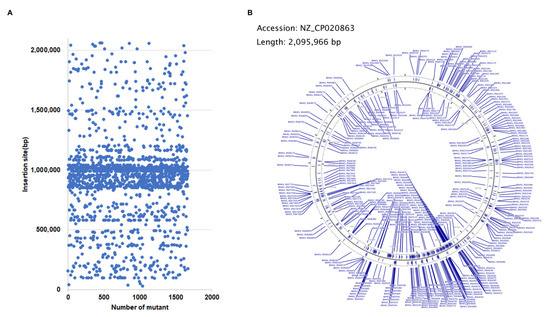
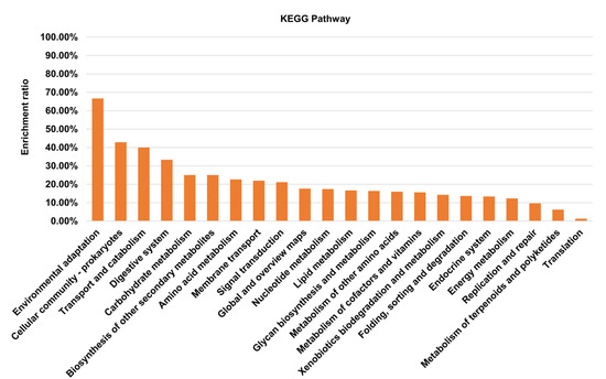
By using the Tn917 transposition system, a total of 1665 transposon mutants of S. suis were obtained and sequenced individually. The insertion sites were mapped to the genome of S. suis SC19 (GenBank accession number NZ_CP020863). As shown in Figure 1A, most insertion sites were located within the region ranging from 800 kbp to 1200 kbp of the genome. Among them, 1191 mutants contained a Tn insertion within the coding sequence of a gene, leading to the disruption of 417 different genes (Table S1), and 474 mutants contained an insertion in the intergenic region. The genes with a Tn insertion were shown on the genome (Figure 1B). These genes were distributed to 22 different KEGG pathways (Figure 2).

Figure 1.
Location of transposon insertion sites in the genome of S. suis SC19. (A) Scatter plot of the positions of the Tn insertion sites. Blue spots represent the position (bp) of the 1665 mutants in the SC19 genome where the transposition inserted sites are located. (B) Mapping of the Tn insertion sites to the genome of S. suis SC19 strain. CGView Server was used to visualize the distribution of the inserted genes involved in the mutant library on the S. suis SC19 genome.

Figure 2.
The KEGG pathway enrichment of the 417 Tn917-inserted genes. The 417 genes were assigned with a K number by using the BlastKOALA tool. The K number was then used to map each gene to its corresponding KEGG pathway using the Reconstruct tool of the KEGG mapper. The enrichment ratio indicates the number of genes identified from the Tn mutant library in each KEGG pathway to the number of total genes included in the S. suis SC19 genome in this pathway.
3.2. Virulence Assessment in G. mellonella Larvae
To do high throughput virulence evaluation with the Tn mutants, a G. mellonella larva infection model was used. Apart from the Tn mutants that were unable to be revived from the library, a total of 391 mutants were subject to the G. mellonella larva infection assay. The results showed that 32 mutants exhibited significantly attenuated virulence compared with the wild-type strain (Table 2). Among these genes, arcC and sspA were already reported virulence genes. These 32 strains were grouped into four categories according to their genetic loci, including 2 transcription regulators, 3 transporters, 6 hypothetical proteins, and 21 others (Table 2). Among these 32 genes, a novel transcription regulator gene hxtR (B9H01_RS05155) was selected for further study.

Table 2.
Transposants with reduced virulence in Galleria mellonella larvae.
3.3. Decreased Virulence of ΔhxtR in Larvae and Mice
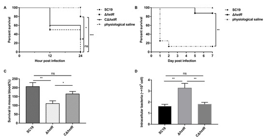
A deletion mutant of hxtR (ΔhxtR) and its complemented strain CΔhxtR were constructed and verified by PCR and RT-PCR (Figure S1). The RT-PCR results indicated that the knockout of hxtR did not affect the hxtR downstream gene transcription but led to the increased transcription of the upstream gene. Compared with S. suis SC19 and CΔhxtR, ΔhxtR showed a significant decrease in virulence in the G. mellonella larvae infection assay (Figure 3A).

Figure 3.
Virulence characterization of ΔhxtR, CΔhxtR, and SC19 strains. (A) G. mellonella larvae infection assay; 20 μL of bacterial cells (approximately 3 × 10⁶ CFU) of ΔhxtR, CΔhxtR, and SC19 strains at the mid-log phase were used to inject larva from the left posterior proleg. Each group contained 10 larvae. The survival was recorded at 12 and 24 hpi and statistically analyzed using the log-rank (Mantel–Cox) test. * indicates p < 0.05, and *** indicates p < 0.001. (B) Mouse infection assay; 500 μL of bacterial cells (8.5 × 10⁸ CFU) of ΔhxtR and SC19 strains at the mid-log phase were used to intraperitoneally inject mice. Each group contained 8 mice. The survival was recorded every 24 h post infection and statistically analyzed using the log-rank (Mantel–Cox) test. ** indicates p < 0.01. (C) Whole blood killing assay. Cells of ΔhxtR, CΔhxtR, and SC19 strains at the mid-log phase were mixed with freshly prepared anticoagulated mouse blood, followed by incubation at 37 °C for 30 min. The mixture was then plated on TSA plates for viable bacteria enumeration. The survival was calculated as (CFU recovered/CFU in original inoculum) × 100% and statistically analyzed using the Student’s t-test. ns indicates p > 0.05, * indicates p < 0.05, and ** indicates p < 0.01. (D) Macrophage phagocytosis assay. Cells of ΔhxtR, CΔhxtR, and SC19 strains were mixed with Raw264.7 macrophage cells with an MOI of 10:1, followed by incubation at 37 °C for 30 min. The cells were treated with ampicillin (50 μg/mL) to remove unphagocytized bacteria. The cells were then lysed, and the phagocytized bacteria were isolated and enumerated by plating on TSA plates. Statistical significances were determined by using the Student’s t-test. ns indicates p > 0.05, and ** indicates p < 0.01.
To further confirm the virulence attenuation of ΔhxtR, mouse infection assays were performed. It is shown in Figure 3B that all the S. suis SC19-infected mice exhibited obvious clinical symptoms, and most mice (six out of eight) died on the first day, and one died within 2 days post infection. However, only one mouse infected with ΔhxtR died within 5 days post infection, and the other mice (seven out of eight) survived until the end of the experiment (Figure 3B), indicating a dramatic decrease in virulence due to the deletion of hxtR.
3.4. Reduced Resistant Abilities of ΔhxtR to Whole Blood Killing and Phagocytosis
The whole blood killing and phagocytosis assays were performed with ΔhxtR, CΔhxtR, and SC19 strains. As shown in Figure 3C, the SC19 strain grew by 206% in mouse blood, while ΔhxtR only grew by 110%, indicating that deletion of hxtR resulted in a decreased ability to survive in blood. In the phagocytosis assay, compared to the CΔhxtR and SC19 strains, the ΔhxtR strain was much easier to be phagocytized by the macrophage cells, indicating an important role of hxtR in resisting phagocytosis by RAW264.7 macrophages (Figure 3D).
3.5. hxtR Is Involved in Cell Morphology and Growth of S. suis
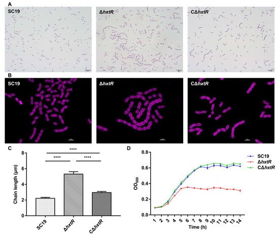
The impact of hxtR deletion on the morphology of S. suis was explored by Gram staining (Figure 4A) and Alexa Fluor 647 staining (Figure 4B). The results showed that hxtR inactivation induced extensive chain elongation and nearly spherical cells (Figure 4A–C). Subsequently, the growth of the strains was tested using a Bioscreen C optical growth analyzer. Growth curves showed that ΔhxtR grew more slowly during the exponential growth phase than SC19 and CΔhxtR in TSB. The ΔhxtR reached only half of the optical density value at the stationary phase of the SC19 and CΔhxtR (Figure 4D).

Figure 4.
Cell morphology and growth characterizations of ΔhxtR, CΔhxtR, and SC19 strains. (A) Morphological analysis by Gram staining. The cells of each indicated strain were grown to the mid-log phase, washed, subjected to regular Gram staining, and imaged under a light microscope. The scale bar is 10 μm. (B) Morphological analysis with SIM. The cells of each indicated strain were grown to the mid-log phase, washed, stained with fluorescent dye Alexa Fluor 647, and imaged using a two-color structure illumination microscope (SIM; Nikon Instruments, Tokyo, Japan) with excitation at 651 nm and emission at 672 nm. (C) Comparison of bacterial chain length. Over one hundred chains per sample were randomly selected in different fields from the Gram staining images to measure the length of the chain by using the software Image-Pro, and then the average chain length was calculated (in µm) and statistically analyzed using the Student’s t-test. **** indicates p < 0.0001. (D) Growth assay. The overnight cultures of the strains were diluted to an initial OD600 of 0.01 with TSB, and then, the diluted culture was pipetted into a microplate. The plate was incubated at 37 °C with continuous shaking for 14 h, and the optical density at 600 nm was monitored every 1 h.
4. Discussion
As an emerging zoonotic bacterial pathogen, S. suis can not only result in great economic losses in the pig industry [1] but also be transmitted to human beings through direct contact with high mortality [2]. VRGs assist bacterium against the defense of the mucosal immune, adhere to and invade the mucosal epithelial cells, survive in the bloodstream, and eventually attack multiple organs resulting in severe systemic diseases [38]. Therefore, elucidating the molecular mechanisms for S. suis pathogenesis is crucial for the development of novel and effective prophylactic and therapeutic approaches.
In order to identify novel VRGs of S. suis, a Tn mutant library of S. suis was generated. First, the Tn-inserted genes of 1665 mutants in the library were one-by-one sequenced, and the results revealed that most insertion sites were in the 800–1200 kbp region of the S. suis SC19 genome. The insertion of Tn917 was not completely random due to the choice of target determined by structural features of DNA, which has been observed in the S. equi Tn917 transposant pool [26,39]. Although there is a preference for insertion sites, the 417 different inserted genes identified from the 1665 mutants were scattered throughout the genome and involved in 22 KEGG pathways. Based on these analyses, we concluded that the mutant pool deserved further functional genomics research.
Efficient and appropriate experimental settings are important for gene function studies, especially when high throughput screening is needed. In recent decades, the use of invertebrates to explore microbial pathogenesis has made important contributions to biomedical research. In this study, we screened 391 mutants involved in different genes using the G. mellonella larvae infection model, and 32 mutants (32 gene loci) exhibited significantly attenuated virulence compared with S. suis SC19. Among these genes, two genes have been discovered to affect the virulence of S. suis. arcC, encoding a carbamate kinase ArcC, is considered a virulence-associated gene [37]. ArcA, ArcB, and ArcC compose the arginine deiminase system (ADS), which participates in the arginine metabolism [9]. In S. suis, the ADS has been reported previously to be involved in the pathogenesis and adaption to acidic conditions [9]. The sspA gene encodes a cell wall-associated subtilisin-like serine protease that is also critical for the virulence of S. suis. The deletion of sspA exhibited attenuated virulence compared with wild strain in pigs [35,40].
In addition to arcC and sspA, of which their roles in S. suis pathogenesis have been reported, the remaining 30 genes were identified for the first time to be related to the virulence of S. suis. Some of these genes, however, have been shown to be involved in the pathogenesis of other bacterial pathogens. For instance, hylA encodes a hyaluronidase, and its deletion mutant has been demonstrated to be significantly attenuated using a murine model in Staphylococcus aureus [41]. The formate C-acetyltransferase (PFL) is involved in the pyruvate metabolism, which has been shown to lead to a change in the lipid composition of the cell membrane and attenuate the virulence in mice when disrupted in Streptococcus pneumoniae [42]. These data suggest that our Tn library screening provides some novel candidate VRGs deserving further study in S. suis.
hxtR encodes an XRE family transcriptional regulator, and its function is unknown in S. suis. It was identified to influence the virulence of S. suis in our primary Tn library screening. To further confirm this, a deletion mutant of hxtR and its complemented strain were constructed. Independent G. mellonella larvae and mice infection assay validated that hxtR played an important role in the pathogenesis of S. suis. Our subsequent in vitro studies revealed that ΔhxtR displayed a reduced growth ability in whole blood and increased sensitivity to phagocytosis, further confirming the virulence phenotype. However, no significant change in the abilities to adhere to and invade porcine kidney-15 cells was observed when compared with the wild-type and complementary strains (data not shown). Moreover, the morphological study suggested that ΔhxtR presented abnormal cell division manifested by the formation of exceptional long chains of bacterial cells. Considering that bacterial cell division is one of the most important physiological processes, a possible explanation for the attenuated virulence of ΔhxtR could be attributed to its influence on the replication and growth of S. suis.
XRE family transcriptional regulators are widespread in bacteria, and they are involved in the regulation of virulence, antibiotic synthesis and resistance, and stress response [43,44]. In S. suis, there are multiple XRE family transcriptional regulators, among which SrtR has been previously confirmed to regulate the virulence in a murine model and oxidative stress tolerance [45], and XtrSs was shown to be involved in response to hydrogen peroxide stress [46]. Moreover, XRE family transcriptional regulators were shown to participate in the direct regulation of virulence factors. In Staphylococcus aureus, the XRE family transcriptional regulator XdrA was reported to control the expression of the virulence gene spa [43]. In group B streptococcus, XtgS was demonstrated to negatively regulate its virulence by directly repressing pseP transcription [47]. In our study, deletion of hxtR seemed to interfere with the expression of its upstream gene, which is a hypothetical gene. Whether this influence contributes to attenuated virulence of ΔhxtR needs further investigation. Furthermore, identifying the regulation targets will be crucial to revealing the underlying mechanism of how HxtR regulates the virulence of S. suis.
5. Conclusions
In this work, we constructed a transposon mutagenesis library of S. suis containing 1665 mutants and identified the Tn insertion sites. By using G. mellonella larvae infection assay, a total of 32 gene loci were identified from the Tn library that were involved in the virulence of S. suis. We further characterized a previously unreported VRG, hxtR, which showed that it had a significant influence on virulence in larvae and mice and affected bacterial growth and division. Overall, our study uncovers some new VRGs and provides valuable information for exploring S. suis pathogenesis.
Supplementary Materials
The following supporting information can be downloaded at: https://www.mdpi.com/article/10.3390/microorganisms10050868/s1, Figure S1: Verification of ΔhxtR and CΔhxtR by PCR and RT-PCR. (A) Identification of ΔhxtR and CΔhxtR by PCR. Lanes 1–4 show the amplification of the upstream gene of hxtR using the primer pair 5150-F/R. Lanes 5–8 show the amplification of hxtR using the primer pair hxtR-F/R. Lanes 9–12 show the amplification of the downstream gene of hxtR using the primer pair 5160-F/R. In lanes 1, 5, and 9, the genomic DNA of S. suis SC19 was used as the template for PCR. In lanes 2, 6, and 10, the genomic DNA of ΔhxtR was used as the template for PCR. In lanes 3, 7, and 11, the genomic DNA of CΔhxtR was used as the template for PCR. Lanes 4, 8, and 12 represent the negative control. (B) Identification of ΔhxtR and CΔhxtR by RT-PCR. Same primers were used as above, and the cDNA of SC19, ΔhxtR, and CΔhxtR were used as templates in the PCR.; Table S1: Tn mutants information.
Author Contributions
Funding acquisition, Conceptualization, Project administration, Writing—review and editing, R.Z. and Q.H. (Qi Huang); Investigation, Project administration, Visualization, Writing—original draft, J.F.; Performed the statistical analysis and contributed with reagents/materials/analysis tools, L.Z. (Lelin Zhao), Q.H. (Qiao Hu), S.L., H.L., Q.Z., G.Z. and L.Z. (Liangsheng Zhang); Writing—review and editing, L.L. All authors have read and agreed to the published version of the manuscript.
Funding
This research was funded by the National Key Research and Development Plans of China to Dr. Rui Zhou (No. 2021YFD1800401).
Institutional Review Board Statement
All animal experiments were performed according to animal welfare standards and were reviewed and approved by the Laboratory Animal Ethics Committee of Huazhong Agricultural University, China (HZAUMO-2022-0041).
Informed Consent Statement
Not applicable.
Data Availability Statement
Not applicable.
Acknowledgments
We thank Tracy Wang (University of Cambridge) for her kindly providing pTV408 and Takamatsu (Japan) for his pSET4s and pSET2 plasmids.
Conflicts of Interest
The authors declare no conflict of interest.
References
- Lun, Z.-R.; Wang, Q.-P.; Chen, X.-G.; Li, A.-X.; Zhu, X.-Q. Streptococcus suis: An emerging zoonotic pathogen. Lancet Infect. Dis. 2007, 7, 201–209. [Google Scholar] [CrossRef]
- Arends, J.P.; Zanen, H.C. Meningitis Caused by Streptococcus suis in Humans. Clin. Infect. Dis. 1988, 10, 131–137. [Google Scholar] [CrossRef] [PubMed]
- Wangkaew, S.; Chaiwarith, R.; Tharavichitkul, P.; Supparatpinyo, K. Streptococcus suis infection: A series of 41 cases from Chiang Mai University Hospital. J. Infect. 2006, 52, 455–460. [Google Scholar] [CrossRef] [PubMed]
- Goyette-Desjardins, G.; Auger, J.P.; Xu, J.; Segura, M.; Gottschalk, M. Streptococcus suis, an important pig pathogen and emerging zoonotic agent-an update on the worldwide distribution based on serotyping and sequence typing. Emerg. Microbes Infect. 2014, 3, e45. [Google Scholar] [CrossRef] [PubMed]
- Feng, Y.; Zhang, H.; Wu, Z.; Wang, S.; Cao, M.; Hu, D.; Wang, C. Streptococcus suis infection: An emerging/reemerging challenge of bacterial infectious diseases? Virulence 2014, 5, 477–497. [Google Scholar] [CrossRef]
- Allen, A.G.; Bolitho, S.; Lindsay, H.; Khan, S.; Bryant, C.; Norton, P.; Ward, P.; Leigh, J.; Morgan, J.; Riches, H.; et al. Generation and Characterization of a Defined Mutant of Streptococcus suis Lacking Suilysin. Infect. Immun. 2001, 69, 2732–2735. [Google Scholar] [CrossRef] [PubMed]
- Lun, S.; Perez-Casal, J.; Connor, W.; Willson, P. Role of suilysin in pathogenesis of Streptococcus suis capsular serotype 2. Microb. Pathog. 2003, 34, 27–37. [Google Scholar] [CrossRef]
- Smith, H.E.; Damman, M.; van der Velde, J.; Wagenaar, F.; Wisselink, H.J.; Stockhofe-Zurwieden, N.; Smits, M.A. Identification and characterization of the cps locus of Streptococcus suis serotype 2: The capsule protects against phagocytosis and is an important virulence factor. Infect. Immun. 1999, 67, 1750–1756. [Google Scholar] [CrossRef]
- Gruening, P.; Fulde, M.; Valentin-Weigand, P.; Goethe, R. Structure, Regulation, and Putative Function of the Arginine Deiminase System of Streptococcus suis. J. Bacteriol. 2006, 188, 361–369. [Google Scholar] [CrossRef]
- Pei, X.; Liu, M.; Zhou, H.; Fan, H. Screening for phagocytosis resistance-related genes via a transposon mutant library of Streptococcus suis serotype 2. Virulence 2020, 11, 825–838. [Google Scholar] [CrossRef]
- Josi, C.; Burki, S.; Vidal, S.; Dordet-Frisoni, E.; Citti, C.; Falquet, L.; Pilo, P. Large-Scale Analysis of the Mycoplasma bovis Genome Identified Non-essential, Adhesion- and Virulence-Related Genes. Front. Microbiol. 2019, 10, 2085. [Google Scholar] [CrossRef] [PubMed]
- Feinbaum, R.L.; Urbach, J.M.; Liberati, N.T.; Djonovic, S.; Adonizio, A.; Carvunis, A.-R.; Ausubel, F.M. Genome-Wide Identification of Pseudomonas aeruginosa Virulence-Related Genes Using a Caenorhabditis elegans Infection Model. PLoS Pathog. 2012, 8, e1002813. [Google Scholar] [CrossRef] [PubMed]
- Geng, S.; Jiao, X.; Barrow, P.; Pan, Z.; Chen, X. Virulence determinants of Salmonella Gallinarum biovar Pullorum identified by PCR signature-tagged mutagenesis and the spiC mutant as a candidate live attenuated vaccine. Vet. Microbiol. 2014, 168, 388–394. [Google Scholar] [CrossRef] [PubMed]
- Pang, M.; Xie, X.; Dong, Y.; Du, H.; Wang, N.; Lu, C.; Liu, Y. Identification of novel virulence-related genes in Aeromonas hydrophila by screening transposon mutants in a Tetrahymena infection model. Vet. Microbiol. 2017, 199, 36–46. [Google Scholar] [CrossRef]
- Wang, C.; Li, M.; Feng, Y.; Zheng, F.; Dong, Y.; Pan, X.; Cheng, G.; Dong, R.; Hu, D.; Feng, X.; et al. The involvement of sortase A in high virulence of STSS-causing Streptococcus suis serotype 2. Arch. Microbiol. 2009, 191, 23–33. [Google Scholar] [CrossRef]
- Domínguez-Punaro, M.C.; Segura, M.; Plante, M.-M.; Lacouture, S.; Rivest, S.; Gottschalk, M. Streptococcus suisSerotype 2, an Important Swine and Human Pathogen, Induces Strong Systemic and Cerebral Inflammatory Responses in a Mouse Model of Infection. J. Immunol. 2007, 179, 1842–1854. [Google Scholar] [CrossRef]
- Wu, Z.; Zhang, W.; Lu, Y.; Lu, C. Transcriptome profiling of zebrafish infected with Streptococcus suis. Microb. Pathog. 2010, 48, 178–187. [Google Scholar] [CrossRef]
- Zaccaria, E.; Cao, R.; Wells, J.M.; Van Baarlen, P. A Zebrafish Larval Model to Assess Virulence of Porcine Streptococcus suis Strains. PLoS ONE 2016, 11, e0151623. [Google Scholar] [CrossRef][Green Version]
- Evans, B.A.; Rozen, D. A Streptococcus pneumoniae infection model in larvae of the wax moth Galleria mellonella. Eur. J. Clin. Microbiol. 2012, 31, 2653–2660. [Google Scholar] [CrossRef]
- Loh, J.M.; Adenwalla, N.; Wiles, S.; Proft, T. Galleria mellonella larvae as an infection model for group A streptococcus. Virulence 2013, 4, 419–428. [Google Scholar] [CrossRef]
- Velikova, N.; Kavanagh, K.; Wells, J.M. Evaluation of Galleria mellonella larvae for studying the virulence of Streptococcus suis. BMC Microbiol. 2016, 16, 291. [Google Scholar] [CrossRef] [PubMed]
- Li, W.; Liu, L.; Chen, H.; Zhou, R. Identification of Streptococcus suis genes preferentially expressed under iron starvation by selective capture of transcribed sequences. FEMS Microbiol. Lett. 2009, 292, 123–133. [Google Scholar] [CrossRef] [PubMed]
- Takamatsu, D.; Osaki, M.; Sekizaki, T. Thermosensitive Suicide Vectors for Gene Replacement in Streptococcus suis. Plasmid 2001, 46, 140–148. [Google Scholar] [CrossRef] [PubMed]
- Takamatsu, D.; Osaki, M.; Sekizaki, T. Construction and Characterization of Streptococcus suis–Escherichia coli Shuttle Cloning Vectors. Plasmid 2001, 45, 101–113. [Google Scholar] [CrossRef] [PubMed]
- Zhang, T.; Ding, Y.; Li, T.; Wan, Y.; Li, W.; Chen, H.; Zhou, R. A Fur-like protein PerR regulates two oxidative stress response related operons dpr and metQIN in Streptococcus suis. BMC Microbiol. 2012, 12, 85. [Google Scholar] [CrossRef]
- Slater, J.; Allen, A.; May, J.; Bolitho, S.; Lindsay, H.; Maskell, D. Mutagenesis of Streptococcus equi and Streptococcus suis by transposon Tn917. Vet. Microbiol. 2003, 93, 197–206. [Google Scholar] [CrossRef]
- Stothard, P.; Wishart, D.S. Circular genome visualization and exploration using CGView. Bioinformatics 2005, 21, 537–539. [Google Scholar] [CrossRef]
- Kanehisa, M.; Sato, Y.; Kawashima, M.; Furumichi, M.; Tanabe, M. KEGG as a reference resource for gene and protein annotation. Nucleic Acids Res. 2016, 44, D457–D462. [Google Scholar] [CrossRef]
- Kanehisa, M.; Sato, Y. KEGG Mapper for inferring cellular functions from protein sequences. Protein Sci. 2020, 29, 28–35. [Google Scholar] [CrossRef]
- Monk, I.R.; Shah, I.M.; Xu, M.; Tan, M.-W.; Foster, T.J. Transforming the Untransformable: Application of Direct Transformation To Manipulate Genetically Staphylococcus aureus and Staphylococcus epidermidis. MBio 2012, 3, e00277-11. [Google Scholar] [CrossRef]
- Deng, S.; Xu, T.; Fang, Q.; Yu, L.; Zhu, J.; Chen, L.; Liu, J.; Zhou, R. The Surface-Exposed Protein SntA Contributes to Complement Evasion in Zoonotic Streptococcus suis. Front. Immunol. 2018, 9, 1063. [Google Scholar] [CrossRef] [PubMed]
- Li, W.; Wan, Y.; Tao, Z.; Chen, H.; Zhou, R. A novel fibronectin-binding protein of Streptococcus suis serotype 2 contributes to epithelial cell invasion and in vivo dissemination. Vet. Microbiol. 2013, 162, 186–194. [Google Scholar] [CrossRef] [PubMed]
- Dalia, A.B.; Weiser, J.N. Minimization of Bacterial Size Allows for Complement Evasion and Is Overcome by the Agglutinating Effect of Antibody. Cell Host Microbe 2011, 10, 486–496. [Google Scholar] [CrossRef] [PubMed]
- Haas, B.; Vaillancourt, K.; Bonifait, L.; Gottschalk, M.; Grenier, D. Hyaluronate lyase activity of Streptococcus suis serotype 2 and modulatory effects of hyaluronic acid on the bacterium’s virulence properties. BMC Res. Notes 2015, 8, 722. [Google Scholar] [CrossRef]
- Hu, Q.; Liu, P.; Yu, Z.; Zhao, G.; Li, J.; Teng, L.; Zhou, M.; Bei, W.; Chen, H.; Jin, M. Identification of a cell wall-associated subtilisin-like serine protease involved in the pathogenesis of Streptococcus suis serotype 2. Microb. Pathog. 2010, 48, 103–109. [Google Scholar] [CrossRef]
- Bonifait, L.; Dominguez-Punaro, M.D.L.C.; Vaillancourt, K.; Bart, C.; Slater, J.; Frenette, M.; Gottschalk, M.; Grenier, D. The cell envelope subtilisin-like proteinase is a virulence determinant for Streptococcus suis. BMC Microbiol. 2010, 10, 42. [Google Scholar] [CrossRef]
- Jing, H.-B.; Yuan, J.; Wang, J.; Yuan, Y.; Zhu, L.; Liu, X.-K.; Zheng, Y.-L.; Wei, K.-H.; Zhang, X.-M.; Geng, H.-R.; et al. Proteome analysis of Streptococcus suis serotype 2. Proteomics 2007, 8, 333–349. [Google Scholar] [CrossRef]
- Segura, M.; Fittipaldi, N.; Calzas, C.; Gottschalk, M. Critical Streptococcus suis Virulence Factors: Are They All Really Critical? Trends Microbiol. 2017, 25, 585–599. [Google Scholar] [CrossRef]
- Scott, J.R.; Churchward, G.G. Conjugative transposition. Annu. Rev. Microbiol. 2001, 49, 455–456. [Google Scholar] [CrossRef]
- Bonifait, L.; Vaillancourt, K.; Gottschalk, M.; Frenette, M.; Grenier, D. Purification and characterization of the subtilisin-like protease of Streptococcus suis that contributes to its virulence. Vet. Microbiol. 2011, 148, 333–340. [Google Scholar] [CrossRef]
- Ibberson, C.B.; Jones, C.L.; Singh, S.; Wise, M.C.; Hart, M.E.; Zurawski, D.V.; Horswill, A.R. Staphylococcus aureus hyaluronidase is a CodY-regulated virulence factor. Infect. Immun. 2014, 82, 4253–4264. [Google Scholar] [CrossRef] [PubMed]
- Yesilkaya, H.; Spissu, F.; Carvalho, S.M.; Terra, V.S.; Homer, K.A.; Benisty, R.; Porat, N.; Neves, A.R.; Andrew, P.W. Pyruvate Formate Lyase Is Required for Pneumococcal Fermentative Metabolism and Virulence. Infect. Immun. 2009, 77, 5418–5427. [Google Scholar] [CrossRef] [PubMed][Green Version]
- McCallum, N.; Hinds, J.; Ender, M.; Berger-Bachi, B.; Stutzmann Meier, P. Transcriptional Profiling of XdrA, a New Regulator of spa Transcription in Staphylococcus aureus. J. Bacteriol. 2010, 192, 5151–5164. [Google Scholar] [CrossRef] [PubMed]
- Si, M.; Chen, C.; Zhong, J.; Li, X.; Liu, Y.; Su, T.; Yang, G. MsrR is a thiol-based oxidation-sensing regulator of the XRE family that modulates C. glutamicum oxidative stress resistance. Microb. Cell Factories 2020, 19, 189. [Google Scholar] [CrossRef] [PubMed]
- Hu, Y.; Hu, Q.; Wei, R.; Li, R.; Zhao, D.; Ge, M.; Yao, Q.; Yu, X. The XRE Family Transcriptional Regulator SrtR in Streptococcus suis Is Involved in Oxidant Tolerance and Virulence. Front. Cell. Infect. Microbiol. 2019, 8, 452. [Google Scholar] [CrossRef]
- Zhang, Y.; Liang, S.; Pan, Z.; Yu, Y.; Yao, H.; Liu, Y.; Liu, G. XRE family transcriptional regulator XtrSs modulates Streptococcus suis fitness under hydrogen peroxide stress. Arch. Microbiol. 2022, 204, 244. [Google Scholar] [CrossRef]
- Liu, G.; Gao, T.; Zhong, X.; Ma, J.; Zhang, Y.; Zhang, S.; Wu, Z.; Pan, Z.; Zhu, Y.; Yao, H.; et al. The Novel Streptococcal Transcriptional Regulator XtgS Negatively Regulates Bacterial Virulence and Directly Represses PseP Transcription. Infect. Immun. 2020, 88, 88. [Google Scholar] [CrossRef]
Publisher’s Note: MDPI stays neutral with regard to jurisdictional claims in published maps and institutional affiliations. |
© 2022 by the authors. Licensee MDPI, Basel, Switzerland. This article is an open access article distributed under the terms and conditions of the Creative Commons Attribution (CC BY) license (https://creativecommons.org/licenses/by/4.0/).