In the original publication [1], there was a mistake in Figure 5 and Supplementary Figure S2A as published. There was an error in the sub-image of the first row, the fourth column the sub-image (B) in hippocampus day 5 in Figure 5. In Figure S2A, only change the value of granular layer thickness in the hippocampus on day 5. The corrected Figure 5 and Supplementary Figure S2A appear below.

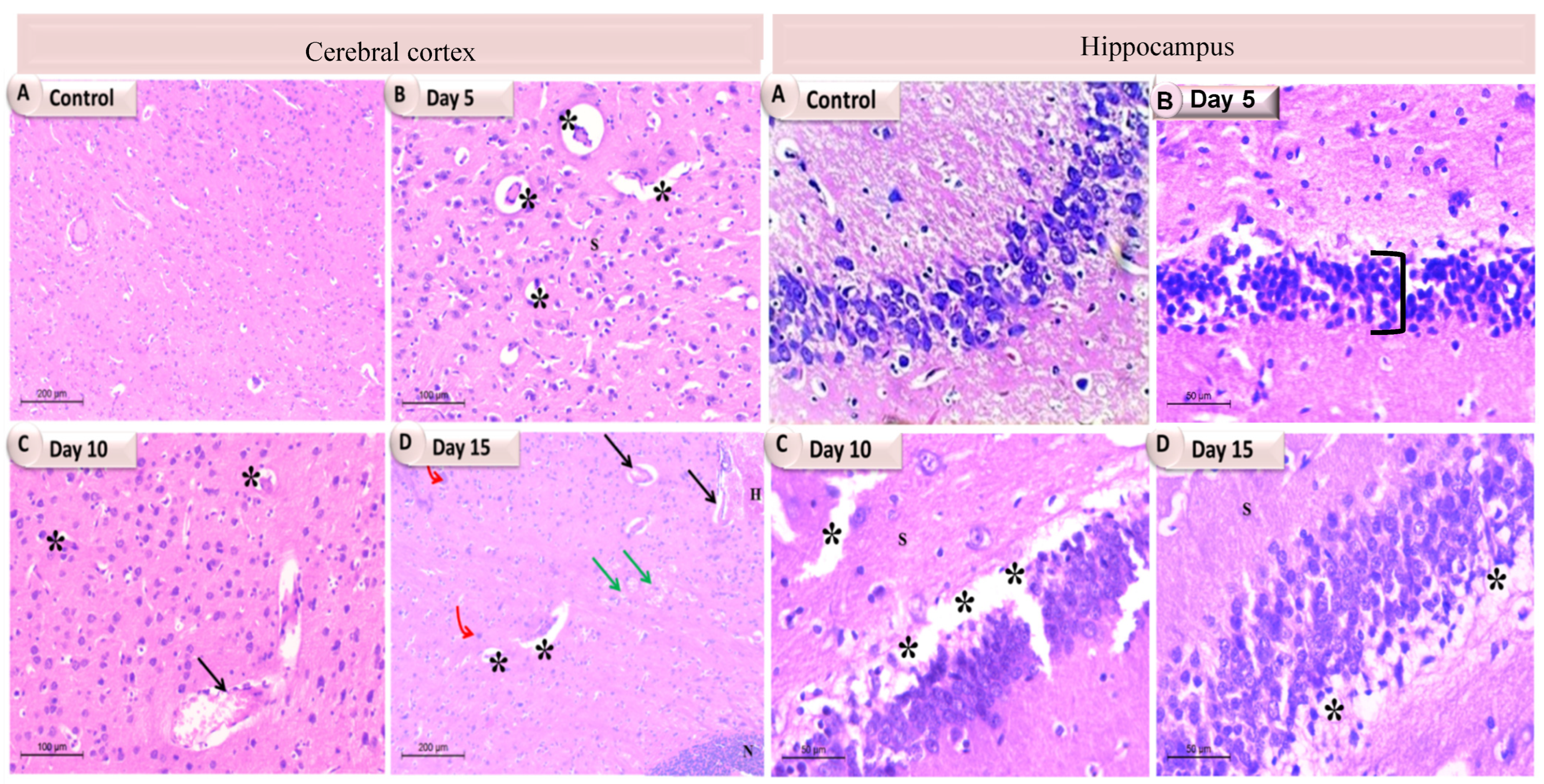
Figure 5.
A photomicrograph showing the individual brain tissues at different time intervals of K. pneumoniae infected rats. In the cerebral cortex, (A) The control group showing normal cytoarchitecture with normal neurons; (B) Day 5 showing vacuolations (stars); (C) Day 10 showing dilated congested blood vessel (arrow) with perivascular edema and vacuolations (stars); (D) Day 15 showing dilated blood vessels (black arrows), severe congestion (green arrows), vacuolations (stars) and gliosis (red bent arrows), a large area of necrotic foci in the brain parenchyma along with lymphocyte infiltration and the presence of degenerating and/or apoptotic neurons (N). While in the hippocampus: (A) The control group showing normal cytoarchitecture; (B) Day 5 showing decreased thickness of the pyramidal layer (bracket); (C) Day 10 showing degeneration and vacuolation (stars); (D) Day 15 exhibited a number of vacuolations (stars).
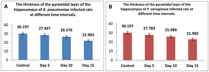
Supplementary Figure S2: The thickness of the pyramidal layer of the hippocampus at different time intervals of infected rats with either (A) K. pneumoniae, or (B) P. aeruginosa was assessed. Herein, five different fields in each photomicrograph from Figures 5 and 6 at each different time interval of infected rats were analyzed on Intel® Core I7® based computer using VideoTest Morphology® software (Russia) with a specific built-in routine for measuring the thickness of the pyramidal layer of the hippocampus. The difference between K. pneumoniae and P. aeruginosa in decreasing the thickness of the layer became more evident at day 15 after infection, and it is slightly higher in case of P. aeruginosa than K. pneumoniae.
The authors state that the scientific conclusions are unaffected. This correction was approved by the Academic Editor. The original publication has also been updated.
Reference
- Elwakil, B.H.; Bakr, B.A.; Aljeldah, M.M.; Shehata, N.S.; Shahin, Y.H.; Olama, Z.A.; Augustyniak, M.; Aboul-Soud, M.A.M.; El Wakil, A. Memory Impairment, Pro-Inflammatory Host Response and Brain Histopathologic Severity in Rats Infected with K. pneumoniae or P. aeruginosa Meningitis. Pathogens 2022, 11, 933. [Google Scholar] [CrossRef] [PubMed]
Disclaimer/Publisher’s Note: The statements, opinions and data contained in all publications are solely those of the individual author(s) and contributor(s) and not of MDPI and/or the editor(s). MDPI and/or the editor(s) disclaim responsibility for any injury to people or property resulting from any ideas, methods, instructions or products referred to in the content. |
© 2024 by the authors. Licensee MDPI, Basel, Switzerland. This article is an open access article distributed under the terms and conditions of the Creative Commons Attribution (CC BY) license (https://creativecommons.org/licenses/by/4.0/).