Abstract
The incessant need for the elimination of pathogenic viruses and multi-drug resistant bacteria has been a critical issue during recent decades, and requires the creation of new antimicrobial materials. Our study describes the production of silver-modified anodic alumina substrates by two methods, and estimation of their bactericidal activity. Aluminum oxide coatings were obtained via an anodization process of low-purity aluminum in an acidic bath for different time periods. The realization of silver infiltration into the pores of the alumina layers was carried out employing two different routes—electrochemical deposition, and in situ thermal reduction. The obtained films were characterized using scanning electron microscopy (SEM). Changes in the surface morphology and thickness of the initial oxide structures after hot water sealing procedure were observed. The presence of silver inside the pores of the alumina layers was also assessed. It was found that silver electrodeposition resulted in greater surface saturation. Large silver accumulations were observed on the thinner anodic films which experienced electroplating for longer time periods. Finally, the antibacterial activity of the modified alumina structures against Gram-negative (Escherichia coli) and Gram-positive (Bacillus cereus) bacteria was evaluated. The results demonstrate that silver deposits acquired by the electrochemical technique improve the bactericidal efficiency of the anodic aluminum oxide (AAO) layers. On the contrary, alumina structures with chemically embedded Ag particles did not show significant antibacterial properties. Overall, the present studies demonstrate that biological activity of silver-doped AAO films depends on the techniques used for their modification.
1. Introduction
During the past couple of decades, porous anodic alumina, fabricated via an anodization process, has received great attention [1]. Usually, the electrochemical oxidation of aluminum is performed in an acidic electrolyte (generally sulfuric, oxalic, or phosphoric acid). When voltage is applied in the acidic bath an oxide layer over the aluminum substrate is formed. The anodization conditions (type of electrolyte, pH, temperature) directly influence the potential properties of the resulting alumina templates. The duration of the electrochemical oxidation determines the oxide film thickness, and it can reach 120 μm. The diameter of the well-ordered pores, depending on the applied voltage, can change between 5 nm and 10 μm. The pH of the acid is reported to influence the inter-pore distances. The colder the bath is, the more ordered oxide structures are formed [2].
There are many studies focused on the optical properties, chemical stability, bio-inertness, and biocompatibility of anodic alumina. In fact, all materials introduced in the human body should be non-toxic, non-antigenic, non-carcinogenic, and non-magnetic. Aluminum oxide has thus far been considered as bio-inert [3]. It has already been used in orthopedics [4], dentistry [5], medical scaffolds for tissue engineering, and drug delivery systems [6]. Additionally, anodized aluminum details have been designed for applications in public environments—aerospace industry, decorative purposes, manufacture of nanomaterials, etc. [6]. It is essential that these anodic surfaces are under exposure in aggressive conditions and are subjected to many cleaning procedures. In fact, the type of coatings employed by technological and medical fields must be resistible to multiple disinfection and washing processes, repeated over time [7]. The sterilization of aluminum and other medicinal instruments, as well as bactericidal properties of components and auxiliary tools, has always been a matter of deep concern [8].
The porous structure of the anodic aluminum oxide (AAO) gives the opportunity for the incorporation of a variety of metal particles in different forms—nanowires, nanorods, and nanotubes [6]. Combination with other metals can provide new operating functions of the modified alumina matrixes, for example, better corrosion resistance, and an antibacterial efficiency that prevents the distribution of infections.
The most simple and inexpensive approaches for the deposition of metallic particles into the pores of alumina substrates are electrochemical and chemical methods [9]. The incorporation procedures have been reported in many investigations as effective and powerful techniques, resulting in composite materials with numerous hi-tech applications [10]. In the early 1970s, Hermann et al. proposed 93 examples of electrochemical deposition using different solutions of Pb, Cd, Cr, Fe, Au, Co, Cu, Mn, Ni, Se, Ag, Te, Zn, and Sn [11]. Now, this type of surface modification is widely used to promote new colors and the corrosion resistance of anodic alumina substrates [10].
At the same time, Ag nanoparticles are intensively investigated due to their specific optical, thermal, electrical, and biological features (anticancer, antifungal, antiviral, and antimicrobial activities). The excellent inhibitory effect of nanosilver against different types of bacteria—E. coli, M. luteus [12], P. aeruginosa, S. faecalis, S. aureus [13], K. pneumoniae, and B. pumilus [14]—is well established in the literature. Nanosized species have better bactericidal properties compared to microsized particles, and they have the ability to directly alter the cell permeability if they are smaller than 10 nm [7]. The highly developed surface area of particles with a small size enhances the antibacterial activity and bioavailability of biomaterials [15]. At the same time, there is evidence that nanosized silver can cause toxicity to liver and stem cells, human and mouse fibroblasts, and human carcinoma cells [16]. The physicochemical properties of nanosized particles could have a significant impact on their biological behavior, so a precise characterization of their size, shape, surface area, aggregation, biocompatibility, cytotoxicity, etc., is necessary. In order to prevent harmful effects of nanomaterials, they need to be evaluated in detail before application [17].
Alternating current (AC) polarization is the most common technique for Ag incorporation into the pores of anodic aluminum oxide films. Silver nanoparticles can also be produced by physical, chemical, and biological approaches [18]. The chemical method is based on the reduction of silver salts (silver nitrate, silver chlorate, and silver acetate). The morphology and size of the formed nanoparticles are strongly influenced by the nature of the reducing agent [19].
When a matrix is loaded with nanostructures, the resulting material is unique. For example, nanosized silver in a combination of porous alumina templates can be useful in different medical fields—in orthopedics, in dentistry, as an antibacterial drug release carrier, etc. [20]. However, differences in physicochemical properties, depending on the fabricating conditions, can cause different interactions with cells. Therefore, all products containing Ag need to be considered separately as compounds with a specific mode of action, and different mechanisms of resistance [21].
The aim of the current investigation was to produce silver-doped anodic alumina substrates by two different routes—electrochemical and chemical methods—and to evaluate their antibactericidal activity against Gram-negative (Escherichia coli) and Gram-positive (Bacillus cereus) bacteria. In order to complete the biological activity of these new composite materials, a comparison to our previously reported biocompatibility evaluations is needed.
2. Materials and Methods
2.1. Silver-Doped Anodic Alumina Preparation
All experimental procedures are schematically represented in detail in Figure 1. Aluminum sheets corresponding to standard EN 573-3/485-2 (Si ≤ 0.25 wt. %, Zn ≤ 0.07 wt. %, Mg ≤ 0.05 wt. %, Cu ≤ 0.05 wt. %, Ti ≤ 0.05 wt. %, Mn ≤ 0.25 wt. %, Fe ≤ 0.4 wt. %, other ≤0.03 wt. %, Al bal.) were cut into samples with active working area of 8 cm2, distributed on both sides of each specimen. Prior to anodization, three different pretreatment steps were performed. The anodic oxidation was completed in a two-electrode cell (with a lead plate as a counter electrode, placed symmetrically around Al anode) for 30 and 60 min. After that, two non-identical silver deposition techniques were used—AC-electrochemical incorporation and in situ thermal reduction. All specimens were repeatedly washed with double-distilled water and dried in air after each operation. For the preparation of all solutions, analytical-grade reagents were employed.
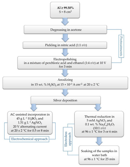
Figure 1.
Preparation procedures of silver-modified anodic alumina samples.
2.2. Microstructure Characterization of the Modified Structures
Morphological observations of the obtained anodic films with or without deposited silver were performed using scanning electron microscopy (PrismaTM E SEM by Thermo Fisher Scientific, with an attachment for element analysis—Energy Dispersive X-ray (EDX) analyzer, Waltham, MA, USA).
2.3. Evaluation of Antibacterial Activity
The antibacterial properties of silver-doped alumina were investigated using a Gram-positive Bacillus cereus strain American Type Culture Collection (ATCC) 11778, and a Gram-negative Escherichia coli strain ATCC 25922. The assays were based on the methodology applied by Calovi et al. with subtle modifications [7]. In brief, 20 µL of thawed bacterial stocks were inoculated on a nutrient agar and grown overnight at 37 °C. Then, 1 or 2 colonies were transferred to 10 mL of Mueller Hinton broth (Merck KGaA, Darmstadt, Germany) to achieve a starting bacterial concentration of approximately 1.5 × 10⁸ cells/mL (McFarland standard 0.5). The resulting suspension was diluted in sterile phosphate-buffered saline (PBS) to obtain an inoculum with ≈1.0 × 10⁶ cells/mL concentration. After that, 100 µL inoculum was applied on the surface of alumina test substrates, which had been pre-sterilized with 70% ethanol and horizontally placed into a sterile Petri dish. The contaminated specimens were covered with a sterile polypropylene membrane and incubated at 37 °C for 24 h, at a relative humidity of ≈90%. At the end of the incubation period, the samples were transferred to a sterile tube. Bacterial cells were recovered from the test specimens by adding 10 mL of sterile PBS and vortexing for 1–2 min to detach them. Then, 10-fold serial dilutions in PBS were performed, and 100 μL of each sample was inoculated on nutrient agar. The culture dishes were incubated at 37 °C for 24 or 48 h to determine the number of viable bacteria. The resulting colonies were counted, and the antibacterial rate was calculated by the following formula:
where Nt is the number of viable bacteria on the silver-modified test sample and N0 is the number of viable bacteria on the control non-modified alumina sample with the corresponding oxide film thickness.
Antibacterial rate % = [(N0 − Nt)/N0] × 100,
Additional experiments were performed to evaluate the relative duration of silver-doped alumina substrates antibacterial effects. Test specimens contaminated with 100 µL inoculum (≈1.0 × 10⁶ cells/mL) were incubated at 37 °C for 24 h and high humidity. Then, 10 mL of nutrient broth was added to the culture vessel, and the samples were incubated for 72 h at 37 °C. During this culture period, 500 µL samples were collected every 24 h in order to assess the number of viable bacteria over time—at 24 h, 48 h, and 72 h after addition of growth medium to the vessel containing contaminated alumina sample. The samples were pipetted onto a 96-well culture plate (Costar, Corning Inc., New York, NY, USA) in triplicates (100 μL/well). 3-(4,5-dimethylthiazol-2-yl)-2,5-diphenyltetrazolium bromide (MTT) solution (Merck KgaA, Darmstadt, Germany) to a final concentration of 0.5 mg/mL was added to each sample well. In the presence of viable bacteria MTT (yellow color) is reduced to a formazan product (purple color) [22,23]. The plates were incubated for 1 h at 37 °C and then absorbance at 570 nm was measured using a Synergy-2 reader (BioTek, Winooski, VT, USA).
For SEM analyses, the method described by Zhang et al., 2013 was used [24]. In summary, 100 µL E. coli suspension (≈1.0 × 10⁶ cells/mL) was inoculated onto the silver-modified alumina substrates and control specimens (anodic alumina layers), and cultured in a humidified incubator at 37 °C for 24 h. Then, the samples were gently washed with PBS and fixed for 2 h at room temperature in 2.5% glutaraldehyde PBS solution. After fixation, the specimens were rinsed three times with PBS and then dehydrated in a gradient ethanol/distilled water mixture (50%, 60%, 70%, 80%, 90%, and 100%). The specimens were incubated for 10 min at room temperature with each alcohol solution, and with absolute ethanol at the end of the dehydration procedure. The surfaces of the alumina samples were analyzed on a scanning electron microscope (Prisma™ E SEM, Thermo Fisher Scientific, Waltham, MA, USA).
3. Results and Discussion
All sets of alumina plates used in the current study and the corresponding treatments are presented in Table 1.

Table 1.
Abbreviations of specimen groups used in the manuscript.
3.1. Scanning Electron Microscope Observations
As the insulating properties of alumina are strongly dependent on its uniform distribution and thickness, it was of great interest to investigate these features of the resulting oxide layers after 30 and 60 min anodization processes at the side edge of the metal pads. Previously, the Al/Al2O3 interfaces in cross-sectional SEM analysis of other areas of anodized specimens revealed evenly distributed oxide film, well bonded to the Al substrate [10,25]. Additionally, the morphology and structure of oxide films subjected to the post-anodizing hot water sealing were evaluated. To ensure the reliability of these results, the SEM evaluations were performed using two identical specimens of each group. Three different areas of each sample were inspected.
The comparison between the images establishes a slightly lower thickness of the unsealed samples (Figure 2a,c). As can be seen in Figure 2a, the thinnest film (approximately 10 µm) is formed in the acidic bath for 30 min, and on the interface Al/oxide layer the appearance of cracks is observed. A detailed investigation of the surfaces, obtained by the means of this anodic regime, revealed that the oxide layer is irregularly distributed in the edge area, and at the same time, in some places, no cracks or crevices can be seen. Their occurrence is a result of the anodization process parameters and the presence of impurities in the aluminum alloy [10]. At the edges of the metal plates, greater local inhibition of the oxide growth was experienced. More interesting is the fact that when samples of group A were submitted to the sealing procedure, the oxide layer grew (Figure 2b). The same dependence occurred for the metal pads of group B—electroformed layers for 60 min (Figure 2c,d). It is already reported that sealing in aqueous media induces the incorporation of water in the structures [26]. Historically, this is the most common and effective way to improve the corrosion resistance of anodized aluminum. As a consequence of this process, the outer surface and the pore walls react to form a crystalline hydrate known as boehmite. Finally, this inert hydrated oxide fills all of the pores, and provides a protective layer all over the surface [6].
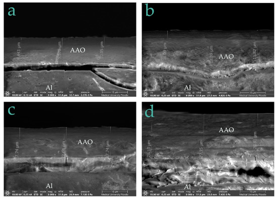
Figure 2.
Microphotographs at the edges of (a) unsealed specimen of group A, (b) sealed specimen of group A, (c) unsealed specimen of group B, and (d) sealed specimen of group B at ×8000 magnification.
In Table 2, the data results of the thickness measurements are summarized.
Table 2.
Anodic film thickness, acquired from SEM analyses.
In addition, quantitative EDX map examinations were performed to investigate the elemental distribution of the anodic layers. The data showed the presence of sulfur and carbon. In Figure 3, a typical composition of the AAO films is presented.
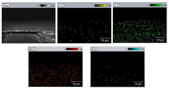
Figure 3.
EDX map of anodic layer.
It is important to highlight the fact that sealing also reduces the porosity of the anodic films, and induces morphology changes [26], so the initial alumina substrates which were thereafter submitted to silver deposition were not sealed. This procedure was performed as a final step only for the in situ electroless Ag incorporation method. The deposition approach was reported by Pornnumpa et al., providing the pore sealing of AAO, distributed with silver nanoparticles [27].
In this study, in situ thermal reduction of Ag+ was performed in order to obtain silver particles with bactericidal activity which were deposited into the porous layers. After that, a hot water sealing procedure was performed. In Figure 4, microphotographs of the as-obtained Ag-doped films are presented. For better detection of the silver, an inspection with the EDX analyzer of the top-view of the surfaces at different magnifications was performed.
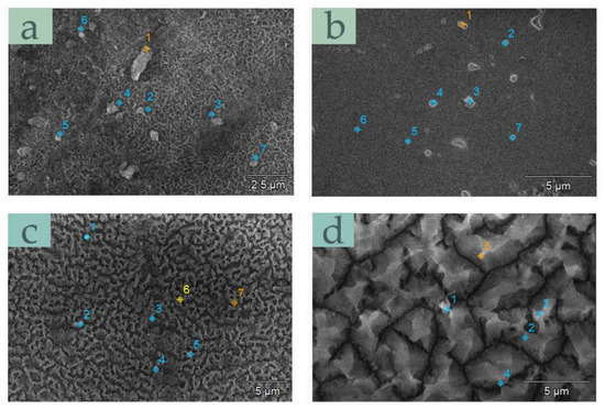
Figure 4.
SEM images of specimens of groups (a) AT-3, (b) AT-6, (c) BT-3, and (d) BT-6.
In all the depicted samples, the oxide layers are irregular. With the prolonged anodization and hydrothermal sealing at temperatures above 95 °C, the pores of the oxide skeleton are filled by a crystalline hydrate phase (Figure 4c,d) where silver particles are incorporated. However, uncovered areas were also observed. In Figure 4a,b, Ag is mainly located in the pits and crevice sites of the alumina films. It can be seen both as individual particles and agglomerates. In addition, the dimensions of some species were above nanoscale, and they were not evenly distributed in the anodic coatings. These findings are in agreement with our previous investigation [28]. However, an extended EDX topological analysis revealed small Ag quantities for all samples. In Figure 5, a top-view of the specimens of groups AT-3 and AT-6 at higher magnification are presented. Many silver particles with different sizes can be seen. However, at the same time, in different areas of the same samples, difficulties in locating Ag species occurred. This fact is proof for the low efficiency and repeatability of the electroless deposition method.
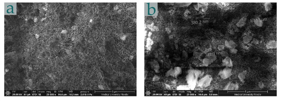
Figure 5.
Microphotographs of sample of groups (a) AT-3 and (b) AT-6 (anodic oxidation for 30 min and chemical deposition for 3 and 6 min) at ×25,000 magnification.
In comparison with the thermal reduction method, silver electroplating resulted in greater Ag content on the alumina substrates, regardless of the alumina thickness (Figure 6). Most saturated with silver surfaces were the oxide layers formed in the acidic bath for 30 min—sample groups AE-0.5 and AE-8. As expected, larger accumulations of Ag deposits were observed on the anodic films which experienced silvering for the longer time periods. Metallic silver clusters are well established in Figure 6b. The Ag content was confirmed by the EDX attachment. Typical EDX spectra are presented in Figure 7. The data observed agree with our preliminary investigations [25] and evince the reproducibility of the electroplating deposition method.
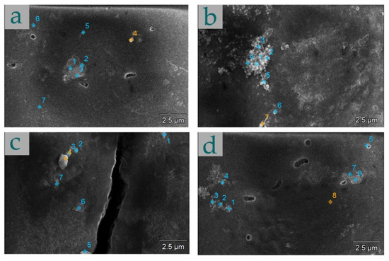
Figure 6.
Microphotographs of samples of groups (a) AE-0.5, (b) AE-8, (c) BE-0.5, and (d) BE-8.
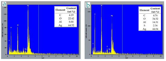
Figure 7.
EDX spectra of (a) AE-8 and (b) BE-8.
A detailed examination of the morphology of metal pads of group AE-8 showed non-homogenously distributed Ag species forming agglomerates with different shapes and sizes (Figure 8). Lack of crystallinity of the composite layers was observed, and they appeared amorphous.
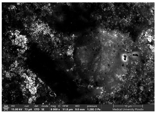
Figure 8.
Microphotograph of specimen of group AE-8 at ×8000 magnification.
Biocompatibility, cell growth, cytotoxicity, and bactericidal properties are in correlation with the morphological characteristics such as size, shape, concentration of silver particles, exposure time, and cell type [29,30,31]. Therefore, the first step in most studies in this field is to determine the quantity, distribution degree, and other physicochemical features of deposited Ag species into the matrixes. Simultaneously, the efficient synthesis of nanosized silver has always been a challenge. When applying two different deposition approaches, the as-obtained Ag-loaded alumina substrates exhibited differences in particle distribution and appearance. Therefore, antibacterial tests were conducted by means of the synthesis method and alumina film thickness.
3.2. Antibacterial Activity of Silver-Modified Alumina Substrates
There are different theories explaining the mechanism of the antibacterial action of silver. According to one of them, metallic silver is relatively inert, but can react with aqueous solutions, resulting in the release of highly reactive silver ions. Another mechanism is related to the formation of reactive oxygen species (ROS). The antimicrobial activity of silver ions (Ag+) is directly proportional to the environmental quantity of Ag, and it is known that they are powerful bactericidal agents even in low concentrations due to the oligodynamic effects [32]. The silver mode of action is related to binding strength, type of target, oxidation level, etc. [21]. Physical and chemical properties of composite materials indicate that they should be considered as different forms with specific mechanisms of toxicity. Our results on the antibacterial activity of silver-modified alumina specimens are presented in Table 3. As can be seen, the AAO layers loaded with silver by the electrochemical reaction achieved 100% bactericidal efficiency against both bacterial strains, irrespective of the oxide film thickness and the deposition time periods (specimen groups AE and BE). On the other hand, all the samples which underwent chemical deposition showed low levels or no inhibitory effect against E. coli and B. cereus. This circumstance is probably due to the low silver amount and poor adhesion on the surfaces. Moreover, the fact that the final sealing procedure leads to the hydration of pore mouths and the incorporation of silver particles in the hydrated porous structures, apparently accelerates bactericidal efficiency suppression. Furthermore, some studies state that silver ions produced by the electrolytic route exhibit better antibacterial activity when compared to those synthesized through dissolving silver compounds [21]. Additional evaluations in this direction, when silver is loaded on a matrix, are needed.
Table 3.
Antibacterial activity of silver-doped alumina substrates after contamination with E. coli or B. cereus and subsequent 24 h incubation.
Figure 9 demonstrates the relative levels of viable E. coli and B. cereus bacteria during culture for an extended period in the presence of silver-doped alumina pads. The aim of these experiments was to evaluate the duration and potential long-term bactericidal effect of the specimens. In accordance with the data for the antibacterial rate, the co-culture assays for longer periods demonstrated complete inhibition of the growth and viability of both Gram-positive (Figure 9b,d) and Gram-negative (Figure 9a,c) bacteria, in the presence of alumina substrates with silver incorporated by the electrochemical approach. The samples obtained by the method of thermal reduction showed an increase in the levels of viable bacteria over culture-time, indicating weak antibacterial properties.
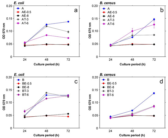
Figure 9.
Relative levels of viable E. coli (a,c) and B. cereus (b,d) bacteria during different culture periods in the presence of test specimens.
To support the bactericidal efficiency data, SEM analysis was performed. Figure 10 displays metal pads analyzed after antimicrobial assays. The total inhibition of bacterial growth is proven for the electrodeposited silver on the anodic films (Figure 10b). The attachment of E. coli cells after a 24 h cultivation on silver-modified alumina substrates by the electroless approach is presented in Figure 10a.
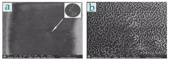
Figure 10.
Microphotographs of sample groups (a) BT-6 and (b) BE-8 at ×5000 magnification.
3.3. Silver-Modified Alumina Substrates—Comparison of Biocompatibility
In order to give a broader view of the biological features of these new composite materials, we linked this investigation to our previously reported biocompatibility evaluations of the same modified substrates [25,28]. It was already mentioned that silver can cause toxicity for different cell types, but there are findings which suggest that it does not exhibit any negative effects related to contact with the human body [7]. In Figure 11, the collected data from our previous findings are summarized, with the aim of showing the contrary effect of the silver deposition method on the bioassays and the antibacterial activity tests. It should be noted that silver particles synthesized by the in situ thermal reduction method augment the biocompatibility of the alumina substrates; at the same time, they are not able to make them bactericidal. On the contrary, the anodic films which sustained silver electrodeposition reveal a 100% bacteria inhibition rate, but slightly reduce the mouse National Institutes of Health (NIH) Swiss 3T3 fibroblast line (denoted as NIH/3T3) and human fibroblasts cell survival levels. These data are not surprising and correspond to the SEM and chemical analyses [25,28] which indicate efficient silver incorporation by the electrochemical method. Higher Ag particle levels result in better antibacterial activity and a slight reduction in biocompatibility. However, survival of human fibroblasts in co-culture with AE or BE samples was not markedly decreased, indicating a good tolerance rate and low toxic potential.
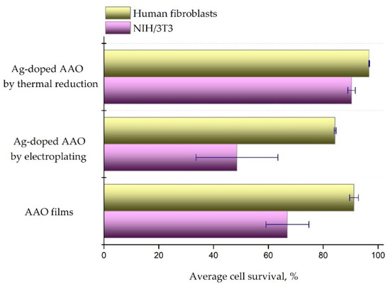
Figure 11.
Average cell survival levels determined after 48 h in vitro culture of NIH/3T3 and human fibroblast cells with modified alumina substrates. All samples were tested in duplicates (n = 4). Figure bars represent mean ± standard error of the mean, and ± SD is indicated for each group.
4. Conclusions
The current investigation aims to present two different routes for silver deposition on anodic alumina substrates—the electrochemical method and electroless thermal reduction. The assessments provided are specifically focused on evaluating the morphological and bactericidal features of the silver-containing alumina coatings, depending on the modification techniques. The topological analysis showed that electroplated silver, in comparison to the electroless Ag deposition, leads to greater surface saturation, and better reproducibility of the experiments. These combined layers demonstrated 100% inhibition of the growth of Gram-negative and Gram-positive bacteria. A significant bactericidal action of silver-doped structures obtained by the electroless method was not proven. Taking into account the performance of these composite layers in biocompatibility evaluations, depending on the modification technique, the final structures should be considered as materials with different characteristics, modes of action, and eventual fields of application.
Author Contributions
Conceptualization, D.K., N.M. and T.B.; Formal analysis, D.K. and T.B.; Funding acquisition, D.K. and N.M.; Investigation, D.K., N.M., T.B. and N.Z.; Methodology, D.K., N.M., T.B. and B.D.; Project administration, D.K. and T.B.; Software, D.K.; Supervision, D.K., N.M., T.B. and B.D.; Validation, D.K., N.M. and T.B.; Visualization, D.K., N.M. and T.B.; Writing—original draft, D.K., T.B., B.D. and R.M.; Writing—review and editing, D.K., N.M., T.B., B.D. and R.M. All authors have read and agreed to the published version of the manuscript.
Funding
The authors would like to thank the Medical University of Plovdiv for financial support.
Institutional Review Board Statement
Not applicable.
Informed Consent Statement
Not applicable.
Data Availability Statement
The datasets used and analyzed during the current study are available from the corresponding author on reasonable request. All data are in the form of tables and figures.
Conflicts of Interest
The authors declare that there is no conflict of interest regarding the publication of this paper. The funders had no role in the design of the study; in the collection, analyses, or interpretation of data; in the writing of the manuscript, or in the decision to publish the results.
References
- Yang, R.; Sui, C.; Gong, J.; Qu, L. Silver nanowires prepared by modified AAO template method. Mater. Lett. 2007, 61, 900–903. [Google Scholar] [CrossRef]
- Gultepe, E.; Nagesha, D.; Sridhar, S.; Amiji, M. Nanoporous inorganic membranes or coatings for sustained drug delivery in implantable devices. Adv. Drug Deliv. Rev. 2010, 62, 305–315. [Google Scholar] [CrossRef] [PubMed]
- Sedel, L. Evolution of alumina-on-alumina implants: A review. Clin. Orthop. Relat. Res. 2000, 379, 48–54. [Google Scholar] [CrossRef] [PubMed]
- Toccafondi, C.; Dante, S.; Reverberi, A.P.; Salerno, M. Biomedical applications of anodic porous alumina. Curr. Nanosci. 2015, 11, 572–580. [Google Scholar] [CrossRef]
- Kusy, R.P. Orthodontic Biomaterials: From the Past to the Present. Angle Orthod. 2002, 72, 501–512. [Google Scholar] [CrossRef]
- Poinern, G.E.J.; Ali, N.; Fawcett, D. Progress in nano-engineered anodic aluminum oxide membrane development. Materials 2011, 4, 487–526. [Google Scholar] [CrossRef] [PubMed] [Green Version]
- Calovi, M.; Furlan, B.; Coroneo, V.; Massidda, O.; Rossi, S. Facile Route to Effective Antimicrobial Aluminum Oxide Layer Realized by Co-Deposition with Silver Nitrate. Coatings 2022, 12, 28. [Google Scholar] [CrossRef]
- Dehghan, F.; Mardanpour, H.; Kamali, S.; Alirezaei, S. Synthesis and antibacterial properties of novel Al2O3-Ag anodised composite coating. Mater. Technol. 2021, 36, 721–730. [Google Scholar] [CrossRef]
- Tzaneva, B.R. Electrochemical and Electroless Deposition of Metal in Anodic Aluminium Oxide Nanoporous Template. Annu. J. Electron. 2013, 204–206. [Google Scholar]
- Girginov, C.; Kozhukharov, S.; Kiradzhiyska, D.; Mancheva, R. Characterization of porous anodic alumina with AC-incorporated silver. Electrochim. Acta 2018, 292, 614–627. [Google Scholar] [CrossRef]
- Hermann, E. Elektrolytisches Färben von Anodisiertem Aluminium. Galvanotechnik 1972, 63, 110–121. [Google Scholar]
- Jagminas, A.; Žalnėravičius, R.; Rėza, A.; Paškevičius, A.; Selskienė, A. Design, optical and antimicrobial properties of extremely thin alumina films colored with silver nanospecies. Dalton Trans. 2015, 44, 4512–4519. [Google Scholar] [CrossRef]
- Chi, G.J.; Yao, S.W.; Fan, J.; Zhang, W.G.; Wang, H.Z. Antibacterial activity of anodized aluminum with deposited silver. Surf. Coat. Technol. 2002, 157, 162–165. [Google Scholar] [CrossRef]
- Mokhena, T.C.; Luyt, A.S. Electrospun alginate nanofibres impregnated with silver nanoparticles: Preparation, morphology and antibacterial properties. Carbohydr. Polym. 2017, 165, 304–312. [Google Scholar] [CrossRef] [PubMed]
- Ying, J.Y. The era of nanotechnology. Nano Today 2008, 3, 1. [Google Scholar] [CrossRef]
- Marambio-Jones, C.; Hoek, E.M.V. A review of the antibacterial effects of silver nanomaterials and potential implications for human health and the environment. J. Nanopart. Res. 2010, 12, 1531–1551. [Google Scholar] [CrossRef]
- Zhang, X.-F.; Liu, Z.-G.; Shen, W.; Gurunathan, S. Silver nanoparticles: Synthesis, characterization, properties, applications, and therapeutic approaches. Int. J. Mol. Sci. 2016, 17, 1534. [Google Scholar] [CrossRef]
- Dhand, V.; Soumya, L.; Bharadwaj, S.; Chakra, S.; Bhatt, D.; Sreedhar, B. Green synthesis of silver nanoparticles using Coffea arabica seed extract and its antibacterial activity. Mater. Sci. Eng. C 2016, 58, 36–43. [Google Scholar] [CrossRef]
- Singh, A.; Kaur, K. Biological and physical applications of silver nanoparticles with emerging trends of green synthesis. In Engineered Nanomaterials-Health and Safety; InTech Open: London, UK, 2019. [Google Scholar]
- Thorat, S.; Diaspro, A.; Scarpellini, A.; Povia, M.; Salerno, M. Comparative study of loading of anodic porous alumina with silver nanoparticles using different methods. Materials 2013, 6, 206–216. [Google Scholar] [CrossRef] [Green Version]
- Kędziora, A.; Speruda, M.; Krzyżewska, E.; Rybka, J.; Łukowiak, A.; Bugla-Płoskońska, G. Similarities and differences between silver ions and silver in nanoforms as antibacterial agents. Int. J. Mol. Sci. 2018, 19, 444. [Google Scholar] [CrossRef] [Green Version]
- Mosmann, T. Rapid colorimetric assay for cellular growth and survival: Application to proliferation and cytotoxicity assays. J. Immunol. Methods 1983, 65, 55–63. [Google Scholar] [CrossRef]
- Moyo, B.; Mukanganyama, S. Antibacterial effects of Cissus welwitschii and Triumfetta welwitschii extracts against Escherichia coli and Bacillus cereus. Int. J. Bacteriol. 2015, 2015, 162028. [Google Scholar] [CrossRef]
- Zhang, D.; Ren, L.; Zhang, Y.; Xue, N.; Yang, K.; Zhong, M. Antibacterial activity against Porphyromonas gingivalis and biological characteristics of antibacterial stainless steel. Colloids Surf. B Biointerfaces 2013, 105, 51–57. [Google Scholar] [CrossRef] [PubMed]
- Kiradzhiyska, D.; Batsalova, T.; Dzhambazov, B.; Mancheva, R. In vitro Biocompatibility Evaluation of Anodic Alumina Substrates with Electrochemically Embedded Silver. Rev. Chim. 2020, 71, 81–88. [Google Scholar] [CrossRef]
- Ofoegbu, S.U.; Fernandes, F.A.O.; Pereira, A.B. The sealing step in aluminum anodizing: A focus on sustainable strategies for enhancing both energy efficiency and corrosion resistance. Coatings 2020, 10, 226. [Google Scholar] [CrossRef] [Green Version]
- Pornnumpa, N.; Jariyaboon, M. Antibacterial and Corrosion Resistance Properties of Anodized AA6061 Aluminum Alloy. Eng. J. 2019, 23, 171–181. [Google Scholar] [CrossRef]
- Kiradzhiyska, D.; Milcheva, N.; Mancheva, R.; Batsalova, T.; Dzhambazov, B.; Zahariev, N. Preparation and Preliminary Evaluation of Silver-Modified Anodic Alumina for Biomedical Applications. Metals 2022, 12, 51. [Google Scholar] [CrossRef]
- Lin, J.-J.; Lin, W.-C.; Li, S.-D.; Lin, C.-Y.; Hsu, S. Evaluation of the antibacterial activity and biocompatibility for silver nanoparticles immobilized on nano silicate platelets. ACS Appl. Mater. Interfaces 2013, 5, 433–443. [Google Scholar] [CrossRef]
- Martínez-Castañón, G.A.; Niño-Martínez, N.; Martínez-Gutierrez, F.; Martínez-Mendoza, J.R.; Ruiz, F. Synthesis and antibacterial activity of silver nanoparticles with different sizes. J. Nanopart. Res. 2008, 10, 1343–1348. [Google Scholar] [CrossRef]
- Maddinedi, S.B.; Mandal, B.K.; Anna, K.K. Environment friendly approach for size controllable synthesis of biocompatible Silver nanoparticles using diastase. Environ. Toxicol. Pharmacol. 2017, 49, 131–136. [Google Scholar] [CrossRef]
- Mohamed, D.S.; Abd El-Baky, R.M.; Sandle, T.; Mandour, S.A.; Ahmed, E.F. Antimicrobial Activity of Silver-Treated Bacteria against other Multi-Drug Resistant Pathogens in Their Environment. Antibiotics 2020, 9, 181. [Google Scholar] [CrossRef] [PubMed] [Green Version]
Publisher’s Note: MDPI stays neutral with regard to jurisdictional claims in published maps and institutional affiliations. |
© 2022 by the authors. Licensee MDPI, Basel, Switzerland. This article is an open access article distributed under the terms and conditions of the Creative Commons Attribution (CC BY) license (https://creativecommons.org/licenses/by/4.0/).