
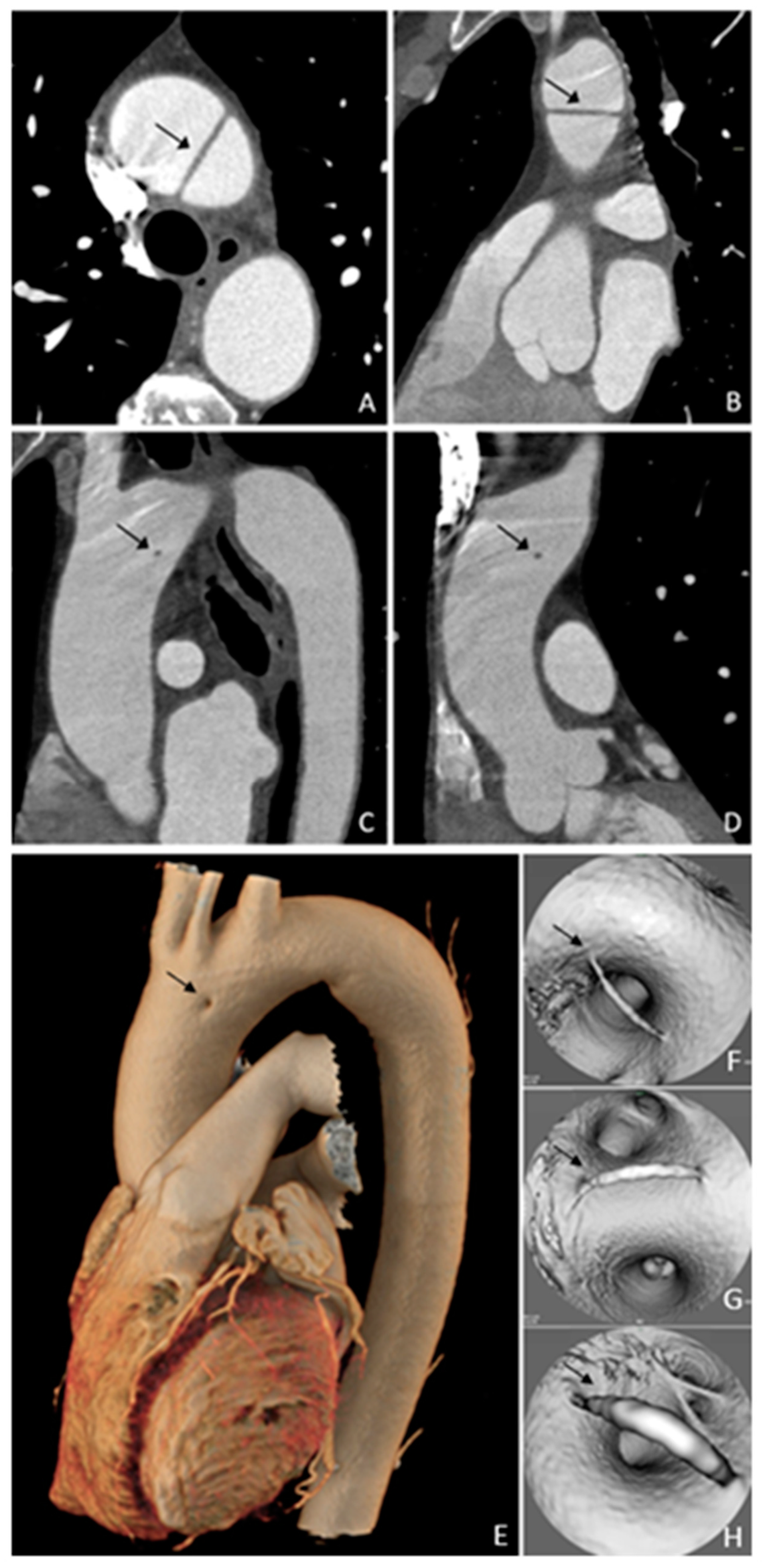
V Aortic Arch Remnant

 ,
,  , , , , ,
Abstract
Author Contributions
Funding
Institutional Review Board Statement
Informed Consent Statement
Data Availability Statement
Conflicts of Interest
References
- Schicchi, N.; Agliata, G.; Giovagnoni, A. CT imaging of a rare case of persistent fifth aortic arch in newborn. BJR Case Rep. 2016, 2, 20150048. [Google Scholar] [CrossRef] [PubMed]
- Liu, Y.; Zhang, H.; Ren, J.; Cao, A.; Guo, J.; Liu, B.; Bao, M.; Zheng, C. Persistent fifth aortic arch: A single-center experience, case series. Transl. Pediatr. 2021, 10, 1566–1572. [Google Scholar] [CrossRef] [PubMed]
- Weinberg, P.M. Aortic arch anomalies. J. Cardiovasc. Magn. Reson. 2006, 8, 633–643. [Google Scholar] [CrossRef] [PubMed]
- Oshitani, T.; Kawasaki, Y.; Murakami, Y.; Fujino, M.; Sasaki, T.; Nakamura, K.; Yoshida, Y.; Suzuki, T.; Ehara, E. A double-barrelled aorta with high aortic Arch. J. Cardiol. Cases 2021, 24, 284–286. [Google Scholar] [CrossRef] [PubMed]
- Shan, H.; Du, X.; Zheng, G.; Ke, T.; Liao, C.; Yang, H. Persistent fifth aortic arch: A comprehensive literature review. Front. Pediatr. 2023, 11, 1183345. [Google Scholar] [CrossRef] [PubMed]
- Naimo, P.S.; del Carmen Vazquez-Alvarez, M.; d’Udekem, Y.; Jones, B.; Konstantinov, I.E. Double-Lumen Aortic Arch: Persistence of the Fifth Aortic Arch. Ann. Thorac. Surg. 2016, 101, e155–e156. [Google Scholar] [CrossRef] [PubMed]
Disclaimer/Publisher’s Note: The statements, opinions and data contained in all publications are solely those of the individual author(s) and contributor(s) and not of MDPI and/or the editor(s). MDPI and/or the editor(s) disclaim responsibility for any injury to people or property resulting from any ideas, methods, instructions or products referred to in the content. |
© 2025 by the authors. Licensee MDPI, Basel, Switzerland. This article is an open access article distributed under the terms and conditions of the Creative Commons Attribution (CC BY) license (https://creativecommons.org/licenses/by/4.0/).
Share and Cite
Tagliati, C.; Fogante, M.; Lamja, S.; Cerimele, C.; Quaranta, A.; Matarrese, A.A.; Battista, D.; Bernardini, A.; Argalia, G.; Carbone, I.; et al. V Aortic Arch Remnant. Diagnostics 2025, 15, 1036. https://doi.org/10.3390/diagnostics15081036
Tagliati C, Fogante M, Lamja S, Cerimele C, Quaranta A, Matarrese AA, Battista D, Bernardini A, Argalia G, Carbone I, et al. V Aortic Arch Remnant. Diagnostics. 2025; 15(8):1036. https://doi.org/10.3390/diagnostics15081036
Chicago/Turabian StyleTagliati, Corrado, Marco Fogante, Stefania Lamja, Cecilia Cerimele, Alessia Quaranta, Alfonso Alberto Matarrese, Davide Battista, Antonio Bernardini, Giulio Argalia, Iacopo Carbone, and et al. 2025. "V Aortic Arch Remnant" Diagnostics 15, no. 8: 1036. https://doi.org/10.3390/diagnostics15081036
APA StyleTagliati, C., Fogante, M., Lamja, S., Cerimele, C., Quaranta, A., Matarrese, A. A., Battista, D., Bernardini, A., Argalia, G., Carbone, I., Di Cesare, E., Schicchi, N., & Lanni, G. (2025). V Aortic Arch Remnant. Diagnostics, 15(8), 1036. https://doi.org/10.3390/diagnostics15081036