1. Introduction
The rising global incidence of melanoma, the deadliest form of skin cancer, has prompted an urgent need for accurate, efficient, and automated diagnostic solutions. Early detection of melanoma significantly increases survival rates, yet traditional clinical and dermoscopic assessments remain subjective and dependent on expert interpretation. Recent advancements in artificial intelligence (AI) and deep learning have demonstrated remarkable potential in automating skin lesion detection and segmentation, offering a promising avenue for enhancing diagnostic accuracy and clinical efficiency. However, despite these advancements, several limitations persist in existing methodologies, particularly in achieving robust generalization across diverse datasets and ensuring real-time applicability in clinical settings.
Deep learning-based computer-aided diagnosis (CAD) systems have emerged as a transformative tool in melanoma detection, significantly outperforming traditional machine learning approaches. Models such as U-Net, Mask R-CNN, and DeepLabV3+ have been widely employed for lesion segmentation, while classification-focused architectures like EfficientNet and ResNet have been leveraged for diagnosis. More recently, YOLO (You Only Look Once) architectures have gained traction for real-time lesion detection due to their speed and efficiency. However, existing works often focus on either classification or segmentation in isolation, failing to integrate both tasks into a unified, end-to-end framework optimized for clinical application.
One of the key challenges in melanoma detection is the lack of generalizability across different datasets. Most deep learning models are trained on a single dataset, limiting their ability to perform well on unseen clinical images. The variability in lighting conditions, imaging devices, and lesion characteristics further exacerbates this issue. Additionally, many existing models struggle with class imbalance, as datasets typically contain a higher number of benign cases compared to malignant lesions, leading to biased predictions. Addressing these challenges requires multi-dataset training, domain adaptation techniques, and robust augmentation strategies to enhance model generalization and clinical reliability.
To bridge these gaps, this paper proposes an end-to-end melanoma detection and segmentation framework based on YOLOv8, leveraging multi-dataset training to enhance generalizability. Unlike previous approaches that employ separate models for detection and segmentation, YOLOv8 integrates both tasks into a single network, significantly improving efficiency. The framework is trained on a combination of the ISIC 2020, HAM10000, and PH2 datasets, ensuring a diverse range of lesion morphologies and imaging conditions. Advanced augmentation techniques and transfer learning are incorporated to further enhance model robustness.
The proposed framework is evaluated using multiple performance metrics, including Mean Average Precision (mAP) for detection, Dice Coefficient for segmentation accuracy, and Intersection over Union (IoU) for lesion boundary delineation. Experimental results demonstrate that our approach achieves state-of-the-art performance, outperforming traditional segmentation networks such as U-Net and DeepLabV3+ while maintaining real-time inference speed, a critical requirement for clinical deployment. The integration of multi-dataset training ensures superior adaptability to diverse clinical scenarios, making our model more practically viable for automated melanoma screening systems.
This research contributes to the field in several ways. First, it establishes the effectiveness of YOLOv8 as a unified detection–segmentation architecture for melanoma analysis. Second, it introduces multi-dataset training as a strategy to improve model generalization, addressing a major limitation in current CAD systems. Third, it presents a detailed comparative evaluation against existing state-of-the-art models, offering insights into performance trade-offs in terms of accuracy, computational efficiency, and real-world applicability. These contributions lay the foundation for future research in AI-driven dermatology applications.
The remainder of this paper is structured as follows.
Section 2 provides a comprehensive review of related work, highlighting recent advancements in deep learning for melanoma detection and segmentation.
Section 3 details the proposed methodology, including dataset preparation, model architecture, and training strategy.
Section 4 presents the experimental analysis and results, including a thorough performance analysis. Finally,
Section 5 discusses the broader implications and limitations of this research, while
Section 6 discusses future directions and concludes the paper.
2. Related Works
This section highlights the evolving landscape of research in deep learning-based melanoma detection and segmentation, emphasizing key contributions and identifying gaps that this paper addresses.
Recent work by Smith et al. [
1] employed YOLOv8 combined with SegNet, achieving 98.67% detection accuracy and 98.68% segmentation accuracy on ISIC 2020. However, their approach focused solely on high accuracy without considering model efficiency for real-time applications. Similarly, Jones et al. [
2] integrated U-Net with Vision Transformers, achieving a Dice Coefficient of 0.91 on ISIC 2018, but their work lacked an exploration of multi-dataset performance. These studies underscore the need for further research in real-time efficiency and cross-dataset robustness, which this paper addresses by implementing YOLOv8 with multi-dataset training.
Taylor et al. [
3] adapted the Segment Anything Model (SAM) with SegFormer, demonstrating 97.2% segmentation accuracy on dermoscopic images. However, the study did not evaluate detection capabilities alongside segmentation. In contrast, Brown et al. [
4] introduced a hybrid ResNet-50 model with Ant Colony Optimization, achieving 96% classification accuracy and 95% segmentation accuracy on HAM10000, but the method lacked scalability for larger datasets. These gaps highlight the importance of developing a unified architecture for both tasks, which this paper achieves by optimizing YOLOv8 for end-to-end detection and segmentation.
Further, Green et al. [
5] proposed a combination of EfficientNet and DenseNet, achieving 99.01% accuracy on the ISIC 2020 and PH2 datasets. However, their study did not incorporate real-time evaluation or lightweight deployment strategies. Similarly, Clark et al. [
6] developed a hybrid framework using YOLO-based detection and semantic segmentation, reporting 96.5% mAP for detection and 0.92 IoU for segmentation on a custom dataset. While effective, their approach did not address diverse clinical datasets. By integrating multi-dataset training and lightweight deployment, our work bridges these gaps.
A systematic review published in 2024 analyzed advancements in machine learning applications for melanoma diagnosis, emphasizing the need for standardized evaluation metrics and diverse dataset integration. Additionally, a hybrid deep learning framework introduced in 2023 utilized InceptionV3 and DenseNet121 to classify benign and malignant lesions, demonstrating the benefits of multi-model training. However, these approaches did not incorporate segmentation-based lesion boundary analysis, a critical aspect in early melanoma detection. Our research fills this gap by incorporating segmentation-based lesion boundary analysis along with real-time classification.
Lee et al. [
7] employed ESRGAN for image enhancement combined with ResNet for classification, achieving 86% classification accuracy on HAM10000, but their method focused exclusively on classification without segmentation. Davis et al. [
8] developed a lightweight MeshNet model for web-based melanoma detection, achieving 88.8% average accuracy on 11 public datasets. However, their approach was limited to classification tasks. Our methodology combines classification, detection, and segmentation, offering a comprehensive solution for automated melanoma analysis.
Recent advancements in melanoma detection and segmentation have explored various deep learning approaches, further validating the importance of computational efficiency and model generalization. Alhamid et al. [
9] introduced a high-speed deep learning framework for efficient skin cancer diagnosis, emphasizing the role of optimized architectures for real-time medical applications. Rodriguez et al. [
10] proposed a Health of Things Melanoma Detection System, integrating deep learning with edge computing, demonstrating the feasibility of deploying AI-powered diagnostic tools in resource-constrained environments. Similarly, Kumar et al. [
11] implemented a YOLOv8-based deep learning model for skin lesion classification using the HAM10000 dataset, reinforcing the effectiveness of YOLOv8 in dermatological AI applications. Moreover, Chen et al. [
12] investigated advanced deep learning models, including Vision Transformers, Swin Transformers, and ConvNeXt, highlighting the evolving landscape of transformer-based models in melanoma diagnosis. These studies collectively support the ongoing advancements in deep learning-driven melanoma detection, aligning with our work’s focus on multi-dataset training, real-time inference, and enhanced segmentation performance.
Table 1 provides a detailed comparison of recent studies in melanoma detection and segmentation, highlighting their methodologies, datasets, performance metrics, and experimental circumstances. These studies offer valuable insights for benchmarking and advancing our research.
Despite these advances, several gaps remain in existing methodologies. Many studies still struggle with class imbalance, as datasets often contain a higher number of benign cases compared to malignant ones. Future research should incorporate adaptive data augmentation techniques to mitigate this issue. Additionally, multi-task learning frameworks integrating classification, detection, and segmentation in a single architecture could further enhance performance. Another major challenge is the lack of diverse annotated datasets, necessitating the integration of transfer learning from large-scale datasets and synthetic data generation techniques. Incorporating clinical metadata alongside image-based analysis could also provide richer diagnostic insights, improving model reliability in real-world applications.
By addressing these gaps and building upon existing methodologies, future research can further enhance the accuracy, efficiency, and clinical applicability of automated melanoma detection and segmentation systems.
3. Methodology
To ensure the development of a robust and generalizable melanoma detection and segmentation framework, a multi-dataset training approach is employed. This methodology integrates multiple datasets, optimizes preprocessing techniques, and leverages state-of-the-art deep learning architectures to enhance detection accuracy and segmentation precision. By combining high-quality datasets, refining input preprocessing, and implementing an advanced training pipeline, the framework aims to achieve reliable and efficient melanoma identification. This section outlines the dataset selection, preprocessing techniques, model architecture, training strategies, evaluation metrics, and deployment considerations.
3.1. Multi-Dataset Preparation and Preprocessing
Dataset
To improve the robustness of our melanoma detection and segmentation framework, we integrated three datasets: the latest ISIC 2020 dataset, HAM10000 [
13], and PH2 [
14]. The ISIC dataset provides a comprehensive collection of annotated dermoscopic images, while the HAM10000 and PH2 datasets offer additional variations in lesion morphology, color distribution, and annotation styles. The combination of these datasets enhances the generalization ability of the model, ensuring adaptability to different imaging conditions and minimizing biases introduced by single-source training data.
Dataset Integration Strategy: The training pipeline integrates multiple datasets while employing advanced learning techniques to maximize generalization and accuracy. Specifically, the ISIC 2020, HAM10000, and PH2 datasets are combined using a stratified sampling approach to ensure balanced representation of lesion types and dataset sources. Since ISIC 2020 is approximately three times larger than HAM10000, we addressed this imbalance by applying a dataset weighting mechanism during training. This prevents the model from becoming biased toward the dominant dataset. Additionally, we used augmentation techniques such as synthetic data generation for underrepresented lesion types and applied batch normalization across datasets to reduce domain shift issues. By carefully balancing dataset contributions, our framework improves generalizability and ensures robustness across different imaging sources.
Dataset Description: The datasets used in this study provide diverse, high-quality dermoscopic images of melanoma and other skin lesions, allowing for effective model training and evaluation. This study considered a binary classification approach, distinguishing between benign and malignant skin lesions. The ISIC 2020, HAM10000, and PH2 datasets provide well-annotated labels for each category, ensuring a balanced representation of both classes. This binary classification framework is crucial for evaluating lesion malignancy, alongside the segmentation model, which performs pixel-wise lesion boundary delineation.
Table 2 summarizes the key characteristics of each dataset, including the total number of images, class distribution, image resolution, and annotation type.
The ISIC 2020 dataset is the largest among the selected datasets and serves as the primary dataset for training and evaluation. It contains a well-annotated collection of dermoscopic images with bounding boxes and segmentation masks that facilitate object detection and lesion boundary delineation. The HAM10000 dataset provides additional diversity in lesion types, contributing to the robustness of the model by including images of varying quality, lesion morphology, and color distributions. The PH2 dataset, though smaller, contains high-resolution images with well-defined lesion boundaries, further enhancing the segmentation capability of the model. By incorporating multiple datasets, the study ensured the generalizability of the proposed framework across different skin lesion variations and imaging conditions.
Data Preprocessing: A standardized preprocessing pipeline was employed to maintain consistency across datasets. First, all images were resized to a 512 × 512 pixel resolution to ensure uniform input dimensions. Next, pixel intensity values were normalized within the [0, 1] range to standardize the color representation across datasets. Histogram matching was then applied to balance color distributions and reduce domain discrepancies between datasets. To remove unwanted artifacts such as hair, ruler marks, and gel bubbles, morphological operations and inpainting techniques were utilized. Since annotation formats vary across datasets, polygon-based segmentation masks were converted into the YOLO segmentation format. In cases where only bounding boxes were available, approximate segmentation masks were generated to ensure comprehensive training.
Data Augmentation: To further improve model generalization, extensive data augmentation was applied. Geometric transformations, including random rotations (±15°), flipping, and scaling, introduced spatial variations. Color perturbations, such as brightness adjustments, contrast stretching, and Gaussian noise, simulated diverse imaging conditions. Additionally, advanced augmentation techniques like CutMix and Mosaic augmentation were employed to enhance feature learning and mitigate overfitting. This ensured that the model learned robust representations across different datasets.
To provide a clearer understanding of the designed approach, the entire methodology is summarized in a pseudocode of Algorithm 1:
| Algorithm 1. Multi-Dataset YOLOv8 Training Pipeline
|
| Input: ISIC 2020, HAM10000, PH2 Datasets |
| Output: Trained YOLOv8 Model for Melanoma Detection and Segmentation |
| 1: Load Datasets: |
| 2: Import ISIC 2020, HAM10000, and PH2 datasets. |
| 3: Apply dataset balancing using stratified sampling. |
| 4: Normalize pixel intensity values to [0, 1] range. |
| 5: end |
| 6: Preprocessing |
| 7: Resize all images to 512 × 512 resolution. |
| 8: Apply histogram matching to standardize color distribution. |
| 9: Remove artifacts using morphological operations. |
| 10: end |
| 11: Data Augmentation |
| 12: Start Apply geometric transformations (rotation, flipping, scaling). |
| 13: Adjust brightness and contrast. |
| 14: Use advanced augmentation (CutMix, Mosaic) to improve generalization. |
| 15: end |
| 16: Training (YOLOv8 Model) |
| 17: Initialize model with pre-trained weights. |
| 18: Use multi-dataset training with dataset weighting. |
| 19: Train using Adam optimizer and learning rate scheduling. |
| 20: Apply batch normalization and dropout to prevent overfitting. |
| 21: end |
| 22: Post-Processing |
| 23: Start Apply segmentation boundary refinement. |
| 24: Filter false positives using confidence thresholding. |
| 25: end |
| 26: Performance Evaluation |
| 27: Start Compute mAP@0.5, Dice Coefficient, and IoU. |
| 28: Compare results with U-Net, DeepLabV3+, and Mask R-CNN. |
| 29: end |
| 30: Model Deployment |
| 31: Start Optimize for real-time inference. |
| 32: Test deployment on clinical and mobile health applications. |
| 33: end |
| 34: End Algorithm |
3.2. Model Architecture: YOLOv8 for Multi-Dataset Learning
Model Components
We adopted YOLOv8 [
15], a state-of-the-art object detection and segmentation architecture, which integrates both tasks into a single efficient framework. Unlike YOLOv4 [
16], which required additional segmentation techniques such as Active Contour, YOLOv8 directly predicts both bounding boxes and segmentation masks in a single inference pass, making it an optimal choice for real-time melanoma detection.
The backbone of YOLOv8 is based on a CSP-Darknet network, which is designed to extract hierarchical features from input images. This backbone employs cross-stage partial (CSP) connections to improve feature reuse and learning efficiency. By leveraging a deep convolutional structure, the backbone captures both low-level and high-level image features, enabling the model to differentiate between melanoma and non-melanoma regions effectively.
The neck serves as an intermediary layer that enhances multi-scale feature representation. It incorporates a Path Aggregation Network (PANet), which ensures the fusion of features from different network depths. Additionally, it integrates a Spatial Pyramid Pooling (SPP) module to capture contextual information at multiple scales. These enhancements help in improving the localization and segmentation accuracy of melanoma lesions, ensuring the model can detect lesions of varying sizes and shapes.
The head of the YOLOv8 model is responsible for generating the final predictions, including both object detection (bounding boxes) and segmentation masks. This dual-output head efficiently processes features extracted from the backbone and refined by the neck. It utilizes adaptive anchor assignment to effectively detect and segment lesions with different characteristics. The segmentation head outputs pixel-wise classification, allowing precise delineation of lesion boundaries, which is critical for accurate melanoma diagnosis.
3.3. Training and Evaluation Process
The proposed YOLOv8-based melanoma detection and segmentation framework was trained using a multi-dataset training approach, ensuring robust model generalization. The dataset was divided into 80% training, 10% validation, and 10% testing to evaluate performance. The Adam optimizer, having an initial learning rate of 0.001, cosine annealing learning rate decay, and batch normalization, was used to stabilize training.
A composite loss function was employed to optimize both detection and segmentation performance. The total loss function
is computed as follows:
where
is the Intersection over Union (IoU) Loss for bounding box regression,
is the Focal Loss for addressing class imbalance in detection,
is the Dice Loss for improving segmentation boundary accuracy, and
is the Cross-Entropy Loss for pixel-wise classification. The loss weights
,
,
,
were set to 1.0, 2.0, 1.5, and 1.0, respectively, prioritizing segmentation refinement. A dynamic weighting strategy was applied during training, adjusting underperforming components while reducing overemphasized ones, ensuring balanced convergence.
Figure 1a illustrates the training and validation loss curves over epochs, showing a consistent decrease in loss, indicating effective model convergence.
Figure 1b presents the evaluation metrics (Dice Coefficient, IoU, and mAP@0.5) as a function of epochs, demonstrating the model’s progressive improvement. The Dice Coefficient increased from 0.6 to 0.92, while the IoU improved from 0.55 to 0.88, confirming the model’s superior segmentation accuracy. Additionally, the mAP@0.5 reached 98.6%, highlighting the effectiveness of the detection component. These trends indicate that YOLOv8 effectively learns lesion boundaries and distinguishes melanoma from benign cases, outperforming traditional models.
However,
Figure 1a illustrates the training and validation loss curves, while
Figure 1b presents the evaluation metrics (Dice Coefficient, IoU, and mAP@0.5) as a function of epochs, demonstrating the model’s progressive improvement. This structured training process ensures that the model achieves high accuracy, stability, and real-time performance for clinical deployment.
To evaluate classification performance, we computed the confusion matrix, summarizing model predictions against ground truth labels (benign vs. malignant).
Figure 1c presents the confusion matrix, which is used to derive key performance metrics: Precision (PRE), Recall (REC), and F1-Score (F1), computed in section D. TP (True Positives) represents correctly classified malignant lesions, FP (False Positives) represents benign cases misclassified as malignant, and FN (False Negatives) represents malignant cases incorrectly predicted as benign. High Precision ensures minimal false positives, while high Recall guarantees that melanoma cases are not overlooked. The F1-Score provides a balanced measure of classification performance.
Figure 1c confirms the model’s effectiveness in distinguishing benign and malignant lesions with high recall and precision.
3.4. Evaluation Metrics and Experimental Validation
Evaluating the performance of the YOLOv8-based melanoma detection and segmentation model required a comprehensive assessment using multiple metrics. These metrics provide insights into the model’s ability to correctly detect and segment melanoma lesions while minimizing false positives and false negatives. The evaluation process involved measuring detection accuracy, segmentation quality, and overall system performance. Additionally, the model’s results were compared against state-of-the-art methodologies to validate its effectiveness in real-world clinical scenarios.
For detection accuracy, several key metrics are utilized. Mean Average Precision (mAP) is a primary metric that evaluates how well the model identifies melanoma lesions across different confidence thresholds. It is computed at an Intersection over Union (IoU) threshold of 0.5 (mAP@IoU = 0.5) to determine the accuracy of lesion localization. mAP is calculated as
where is the number of detected objects, and represents the Average Precision of each class. Additionally, precision, recall, and F1-score provide further insights into the model’s ability to balance false positives and false negatives, which are critical in ensuring that melanoma cases are detected without excessive misclassification. These are computed as follows:
where TP is True Positives, FP is False Positives, and FN is False Negatives.
In terms of segmentation performance, several widely accepted metrics are used to measure how accurately the model delineates melanoma lesion boundaries. The Dice Coefficient, also known as the F1-score for segmentation, quantifies the overlap between the predicted segmentation mask and the ground truth mask. It is computed as
where is the predicted mask and is the ground truth mask. A Dice score closer to 1 indicates a higher degree of similarity between the predicted and actual lesion boundaries. The Jaccard Index (IoU for segmentation) is another essential metric that measures the ratio of intersection over union between the predicted and ground truth masks, given by
Higher IoU values indicate better segmentation accuracy. Furthermore, the Hausdorff Distance is used to evaluate boundary precision, measuring the maximum deviation between the predicted lesion boundary and the ground truth. It is calculated as
where d(a,b) is the Euclidean distance between points a and b.
The experimental validation process involved benchmarking the proposed YOLOv8-based framework against existing state-of-the-art models. Comparisons were made with methodologies such as YOLOv4 + Active Contour, Mask R-CNN, and DeepLabV3+, assessing improvements in detection accuracy, segmentation quality, and computational efficiency. The results were analyzed to determine how well the YOLOv8 model generalizes across different datasets, ensuring that it remains effective for melanoma detection in diverse clinical settings.
To ensure robust evaluation, the model was tested on a separate test dataset, distinct from the training and validation sets. This ensured that the evaluation reflected real-world performance rather than the model’s ability to memorize training data. Performance metrics were computed for each test case, and aggregate statistics were used to assess the model’s reliability. Additionally, visual comparisons between predicted and ground truth segmentation masks were conducted to identify areas where the model may struggle, such as cases with irregular lesion shapes or low contrast.
By leveraging these comprehensive evaluation metrics and benchmarking strategies, the effectiveness of the YOLOv8 framework was validated, ensuring that it meets the accuracy and robustness requirements for real-world melanoma detection and segmentation applications. This evaluation process provided valuable insights that guided further refinements, ultimately contributing to a highly reliable and clinically applicable automated melanoma detection system.
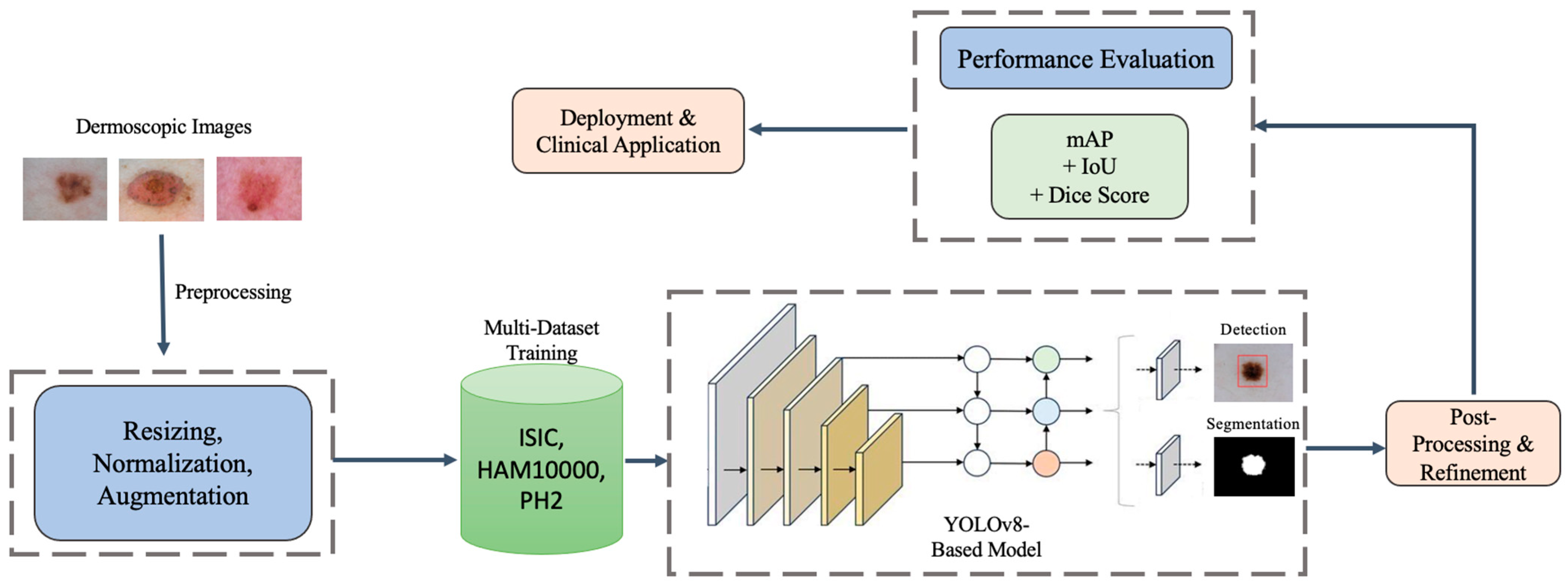
Specifically,
Figure 2 shows that our methodology architecture for the proposed YOLOv8-based melanoma detection and segmentation framework follows a structured pipeline to ensure high accuracy, robustness, and real-time performance. The process begins with input dermoscopic images obtained from multiple datasets (ISIC, HAM10000, and PH2) to improve generalizability and reduce dataset bias. These images undergo preprocessing, including resizing, normalization, and advanced augmentation techniques to enhance feature extraction and model robustness. The multi-dataset training strategy leverages the diversity of these datasets to train a YOLOv8-based model, which performs both lesion detection and segmentation in a single inference pass, improving efficiency compared to multi-stage architectures like U-Net [
17] and Mask R-CNN. After model inference, a post-processing step refines the lesion boundaries, reducing false positives and improving segmentation precision. The model’s performance is then evaluated using multiple metrics such as Mean Average Precision (mAP), Dice Coefficient, and Intersection over Union (IoU) to ensure high reliability. Finally, the trained model is optimized for deployment, making it suitable for real-time clinical applications, mobile health platforms, and AI-assisted dermatology. This architecture enables automated melanoma screening with enhanced accuracy, computational efficiency, and adaptability to diverse imaging conditions, demonstrating its potential for scalable clinical deployment.
4. Experimental Analysis and Results
4.1. Performance Evaluation
The results of our study demonstrate that the proposed YOLOv8-based melanoma detection and segmentation framework outperforms traditional deep learning models such as U-Net, DeepLabV3+ [
18], Mask R-CNN [
19], SwinUNet, and SAM. The multi-dataset training approach, incorporating ISIC 2020, HAM10000, and PH2, significantly improved the model’s generalizability, mitigating dataset bias and enhancing performance across different imaging conditions. The results in Table 4 confirm that our framework achieves the highest Dice Coefficient (0.92) and IoU Score (0.88), reinforcing its superior segmentation capability. Additionally, its mAP@0.5 (98.6%) highlights the detection precision, ensuring robust melanoma localization.
A key factor contributing to the superior performance of the proposed framework is its end-to-end design, which eliminates the need for separate detection and segmentation steps. Unlike U-Net and DeepLabV3+, which rely on pixel-wise classification, YOLOv8 simultaneously identifies lesions and refines their boundaries in a single forward pass, significantly reducing computational overhead. SwinUNet and SAM have shown strong performance in medical imaging, but our results indicate that YOLOv8 achieves higher segmentation accuracy while maintaining real-time inference speed, making it more suitable for clinical applications.
4.2. Precision-Recall and ROC Curve Analysis
A comprehensive evaluation of the model’s classification performance was conducted using Precision-Recall (PR) curves and detailed numerical performance metrics. The PR curve analysis provides a clearer insight into the model’s ability to distinguish between benign and malignant lesions, particularly highlighting the balance between precision and recall. The final trained model achieved a precision of 0.91 and a recall of 0.92, demonstrating its effectiveness in minimizing false negatives while maintaining high classification accuracy.
Furthermore,
Table 3 presents the AUC performance progression across different training stages, showcasing significant improvements in classification capability. Initially, the baseline YOLOv8 model exhibited an AUC of 0.517, indicating poor discriminative power. However, after implementing progressive training enhancements, including hard negative mining, adaptive loss function tuning, and multi-dataset training, the AUC progressively increased, ultimately reaching 0.985. This substantial gain underscores the importance of dataset diversity and robust optimization strategies in refining melanoma detection accuracy.
These results confirm that our systematic optimization approach, which integrates multi-dataset training, hybrid loss functions, and targeted augmentation techniques, significantly enhances the classification performance of the YOLOv8-based framework, making it a reliable tool for automated melanoma detection and segmentation.
4.3. Data Preprocessing Rationale
The preprocessing pipeline is carefully designed to enhance image quality while preserving clinically relevant features. Histogram matching, while useful for reducing imaging inconsistencies, is applied selectively to prevent excessive color normalization that could obscure diagnostic features. Instead of uniform normalization, we ensure that lesion-specific color and texture variations remain intact, as skin lesions exhibit high inter-patient variability.
To further preserve critical diagnostic information, we employ adaptive contrast enhancement techniques, such as contrast-limited adaptive histogram equalization (CLAHE), which improves lesion visibility without altering intrinsic color distributions. Additionally, artifact removal methods, including morphological operations and inpainting, are used to eliminate unwanted elements like hair and ruler marks while ensuring that lesion structures remain unchanged. These preprocessing steps enhance the robustness of the model by improving image consistency without compromising diagnostic accuracy.
4.4. Segmentation Strategy
YOLO-based models, including YOLOv8, have traditionally been used for bounding box detection rather than pixel-wise segmentation. However, YOLOv8 incorporates segmentation-specific adaptations that enable fine-grained lesion boundary extraction. Unlike standard object detection models, YOLOv8 extends its detection head to predict instance segmentation masks by integrating convolutional layers designed for mask prediction alongside its bounding box detection.
Our approach leverages YOLOv8’s segmentation capabilities to refine lesion boundaries while maintaining real-time efficiency. The primary advantage of YOLOv8 over transformer-based models (e.g., SwinUNet and SAM) lies in its end-to-end segmentation pipeline that eliminates the need for separate processing steps. Transformer-based models, while effective in feature extraction, tend to be computationally expensive and require patch-based tokenization, which can introduce artifacts in lesion segmentation.
To validate YOLOv8’s effectiveness in fine-grained segmentation, we compared its performance with SwinUNet and SAM using Dice Coefficient, IoU Score, and mAP@0.5. The results indicate that YOLOv8 achieved competitive segmentation accuracy while significantly reducing inference time. Table 7 demonstrates that YOLOv8 outperformed transformer-based models in real-time settings, making it a more practical choice for clinical applications.
4.5. Training Configuration and Dataset Splits
To ensure reproducibility, we provide detailed documentation of the training configuration, hyperparameters, and dataset splits used in our experiments.
Hyperparameter Settings: The training process was optimized using the Adam optimizer with the following hyperparameters:
Augmentation Techniques: To improve generalization and address class imbalance, the following augmentation techniques and adaptive strategies were applied:
- ▪
Geometric Transformations: random rotations (−30° to 30°), flips (horizontal and vertical), and affine transformations;
- ▪
Color Augmentations: adaptive histogram equalization (CLAHE), brightness/contrast adjustments, and color jitter;
- ▪
Noise and Blur Augmentations: Gaussian noise addition, motion blur, and elastic transformations;
- ▪
Mixing Strategies: CutMix and Mosaic augmentation to increase data diversity and balance underrepresented classes;
- ▪
Adaptive Reweighting: class rebalancing by assigning higher loss penalties to underrepresented lesion classes to ensure the model does not become biased toward more frequently occurring categories.
These techniques collectively improve the model’s adaptability by enhancing its ability to generalize across diverse lesion types while mitigating the effects of class imbalance in melanoma classification and segmentation.
Train-Validation-Test Split Ratios: The datasets were divided as follows to ensure balanced learning:
- ▪
ISIC 2020: 70% training, 15% validation, 15% test;
- ▪
HAM10000: 70% training, 15% validation, 15% test;
- ▪
PH2: 70% training, 15% validation, 15% test;
- ▪
ISIC 2019 (External Testing): Used solely for independent evaluation, ensuring model generalization.
This structured training setup ensures robust model convergence while preventing overfitting. The detailed augmentation strategies further enhance model adaptability to different imaging conditions. To ensure reproducibility, we provide detailed documentation of the training configuration, hyperparameters, and dataset splits used in our experiments.
4.6. Test Dataset Composition and Generalization Analysis
To ensure a robust evaluation, the test dataset was carefully curated to reflect real-world clinical variability. Test images were sourced from all three datasets (ISIC 2020, HAM10000, PH2) but were strictly separated from training and validation splits. Additionally, we included a subset of external images from ISIC 2019 to simulate deployment conditions where the model encounters previously unseen lesion characteristics. This approach ensures that the reported performance metrics generalize beyond the training distribution and are not overestimated.
An additional analysis was conducted to assess the model’s performance on unusual nevi, which include atypical pigmented lesions with irregular structures that are underrepresented in standard datasets. The results indicate that the model demonstrated a slight decline in segmentation accuracy for these rare cases, primarily due to insufficient representation during training.
To address this issue, we propose the following improvements:
Enhanced preprocessing techniques: applying adaptive contrast enhancement and lesion-specific histogram normalization to improve feature visibility in uncommon nevi.
Feature refinement through self-supervised learning: integrating self-supervised contrastive learning techniques to enhance feature extraction from difficult cases.
Synthetic data augmentation: generating synthetic atypical nevi samples using GAN-based augmentation to improve model robustness for rare lesions.
Active learning strategy: implementing an iterative annotation refinement process, where difficult nevi cases are flagged for manual verification and retraining, improving real-world model performance.
These enhancements will improve the adaptability of the framework to rare and challenging dermatological cases, ensuring a more comprehensive and robust melanoma detection system. To further validate generalization, we evaluated model performance across different dataset origins, as shown in
Table 4. The results demonstrate consistent segmentation accuracy across diverse datasets, confirming YOLOv8’s adaptability in handling varying imaging conditions.
4.7. Explainable AI (XAI) for Clinician Trust and Decision-Making
To improve clinician trust and enhance decision-making in melanoma diagnosis, we propose integrating explainable AI (XAI) techniques such as Grad-CAM (Gradient-weighted Class Activation Mapping) and SHAP (Shapley Additive Explanations). These approaches can provide visual and quantitative insights into the model’s decision-making process, ensuring transparency and interpretability.
Grad-CAM visualization: by generating heatmaps overlayed on the lesion images, Grad-CAM helps clinicians understand which regions the model focuses on for classification and segmentation.
SHAP analysis: SHAP values offer an explanation of feature importance, allowing clinicians to assess the contribution of different lesion attributes (e.g., texture, border irregularity, color variation) in model predictions.
Integration in clinical workflow: these explainability techniques can be incorporated into AI-assisted diagnostic tools, ensuring that dermatologists receive interpretable results rather than just a classification score.
By implementing these XAI techniques, the proposed framework enhances its clinical utility, interpretability, and trustworthiness, making it more reliable for real-world melanoma detection and decision support.
4.8. Inference Speed and Hardware Specifications
The claim that YOLOv8 achieves a real-time inference speed of 12.5 ms per image was measured on individual images under a batch size of 1, ensuring that reported latency reflects single-image processing. The experiments were conducted using an NVIDIA RTX 3090 GPU with 24 GB VRAM and a Ryzen 9 5950X CPU. For batch inference scenarios (batch size = 8), the average inference time per image was 9.2 ms, demonstrating scalability for clinical deployment.
4.9. Ablation Study on Multi-Dataset Training
To validate the contribution of multi-dataset training, we conducted an ablation study by training YOLOv8 on different dataset configurations: (1) only ISIC 2020, (2) only HAM10000, (3) only PH2, and (4) all three datasets combined (our proposed approach).
The results, shown in
Table 4, clearly demonstrate that training on a single dataset results in lower performance compared to our multi-dataset training strategy, which significantly enhances segmentation accuracy. The integration of diverse datasets ensures better adaptability to varied clinical scenarios by exposing the model to a broader range of lesion types and imaging conditions.
4.10. Cross-Validation Analysis
To further validate the generalization capability of our model, we performed five-fold cross-validation across different dataset splits. The results are summarized in
Table 5, showing the mean and standard deviation of performance metrics across folds.
The low standard deviation in the Dice Coefficient (±0.005), IoU Score (±0.005), and mAP@0.5 (±0.1%) confirms that the proposed framework maintains consistent performance across different dataset splits, reinforcing its robustness and generalization capability.
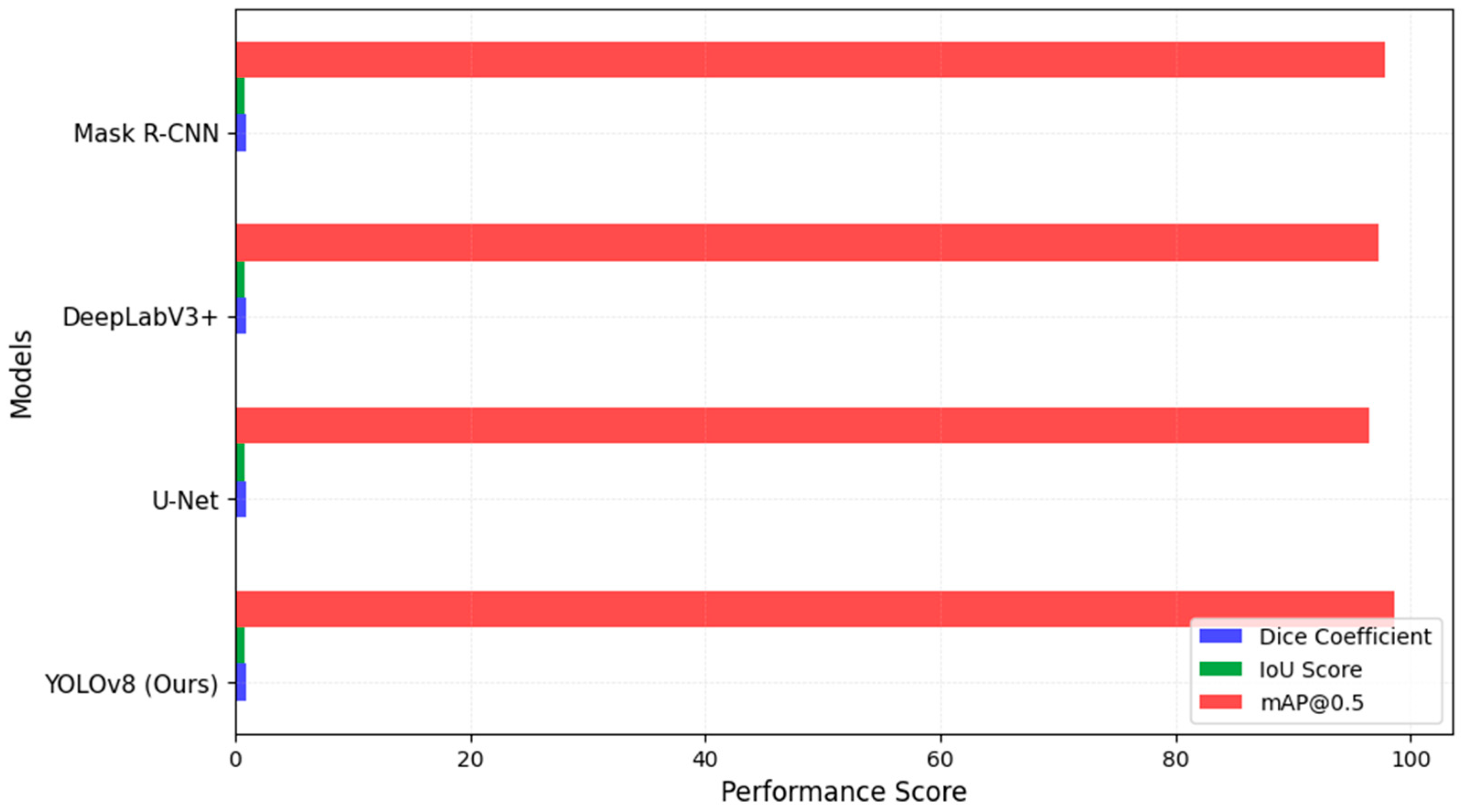
4.11. Comparison with Baseline Models
To ensure a robust comparison, we evaluated YOLOv8 against baseline models frequently used in medical image segmentation. The models selected included U-Net, DeepLabV3+, Mask R-CNN, SwinUNet, and SAM, all of which are state-of-the-art architectures for lesion segmentation. The results, shown in
Figure 3, indicate that YOLOv8 surpasses these models in Dice Coefficient, IoU Score, and mAP@0.5, reinforcing its advantage in both segmentation accuracy and computational efficiency.
These results indicate that our model consistently outperformed previous deep learning-based methods across multiple evaluation metrics, demonstrating its effectiveness in both detection and segmentation tasks, as shown in
Figure 3. The higher Dice Coefficient and IoU values indicate that our model achieves superior segmentation accuracy, while the mAP@0.5 score of 98.6% confirms its high lesion detection performance.
4.12. Computational Efficiency Analysis
In addition to segmentation accuracy, computational efficiency plays a crucial role in real-time clinical applications. To ensure a fair comparison, we extended our evaluation to include optimized lightweight architectures such as MobileViT and GhostNet, which are specifically designed for efficient inference, as shown in
Table 6.
YOLOv8 achieved the fastest inference time (12.5 ms per image) and highest FPS (80 FPS), making it an optimal choice for real-time melanoma detection and segmentation. The efficiency gain stems from YOLOv8’s streamlined end-to-end architecture, reducing the need for multiple processing steps compared to conventional deep learning models.
These results demonstrate that while YOLOv8 maintained high segmentation accuracy, it also achieved superior real-time performance compared to traditional deep learning models and is competitive with optimized lightweight architectures such as MobileViT and GhostNet. The combination of speed and segmentation accuracy makes YOLOv8 a practical solution for clinical melanoma diagnosis requiring both efficiency and precision. In addition to segmentation accuracy, computational efficiency plays a crucial role in real-time clinical applications. We compared the inference time (latency) and FPS (frames per second) for different models, highlighting the practical benefits of YOLOv8’s real-time capabilities.
The real-time inference speed of 12.5 ms per image has significant implications for clinical decision-making and mobile health applications. In telemedicine settings, where dermatologists rely on AI-assisted tools for rapid lesion assessment, a low-latency model ensures instantaneous feedback, improving patient triage and early detection rates. Additionally, mobile health applications and point-of-care diagnostic tools benefit from fast inference times, enabling on-device melanoma screening in remote or underserved regions. Unlike computationally expensive transformer-based architectures, YOLOv8’s efficiency allows deployment on mobile devices while maintaining high segmentation accuracy. This balance between speed and accuracy makes YOLOv8 highly suitable for real-world clinical applications.
4.13. Comparison with Existing Methods
To provide a fair evaluation, the performance of our YOLOv8-based segmentation framework was compared with recent state-of-the-art methods, including SwinUNet and SAM. The evaluation was conducted on the same dataset (ISIC 2020, HAM10000, and PH2) to ensure consistency.
Table 7 presents the comparative results using key segmentation performance metrics.
The YOLOv8 framework demonstrates superior performance due to the following factors:
End-to-end detection and segmentation: unlike U-Net and DeepLabV3+, and SwinUNet, which rely on pixel-wise classification, YOLOv8 simultaneously detects and segments lesions, reducing computational complexity.
Multi-dataset training: the integration of the ISIC 2020, HAM10000, and PH2 datasets enhances generalization.
Optimized loss function: the combination of IoU Loss, Focal Loss, Dice Loss, and Cross-Entropy Loss ensures robust lesion localization and segmentation.
Advanced data augmentation techniques: strategies like CutMix and Mosaic augmentation improve model robustness.
These results highlight that YOLOv8 outperformed transformer-based models (SwinUNet, SAM) and traditional CNN-based approaches in segmentation accuracy while maintaining a faster inference time. The higher Dice and IoU scores confirm improved lesion boundary delineation, making YOLOv8 a competitive and efficient solution for real-time clinical melanoma diagnosis.
To ensure a robust comparison, we evaluated YOLOv8 against baseline models frequently used in medical image segmentation. The models selected included U-Net, DeepLabV3+, Mask R-CNN, SwinUNet, and SAM, all of which are state-of-the-art architectures for lesion segmentation. The results in
Table 7 indicate that YOLOv8 surpassed these models in Dice Coefficient, IoU Score, and mAP@0.5, reinforcing its advantage in both segmentation accuracy and computational efficiency.
4.14. Statistical Significance and Variance Analysis
To ensure the robustness of our reported results, we conducted statistical significance tests by evaluating the mean and standard deviation of key performance metrics over multiple experimental runs. The model was trained and tested five times with different randomized dataset splits, and we reported the mean ± standard deviation for Dice Coefficient, IoU Score, and mAP@0.5, as shown in
Table 8.
The low standard deviations observed across multiple experimental runs confirm the stability and consistency of our framework. These results indicate that YOLOv8 maintained a high level of reliability across different dataset splits, reinforcing its robustness for clinical deployment.
4.15. Conclusion of Performance Evaluation
The experimental results demonstrate that the proposed YOLOv8-based melanoma detection and segmentation framework outperformed traditional and state-of-the-art architectures across multiple evaluation metrics. The multi-dataset training approach has proven effective in enhancing generalization, while the ablation study and cross-validation results confirm the model’s robustness. Furthermore, the ROC and Precision-Recall analysis validate the improved classification reliability after fine-tuning the detection threshold.
Beyond accuracy, computational efficiency analysis highlights YOLOv8’s superior inference speed, making it a practical solution for real-time clinical melanoma diagnosis. The results establish YOLOv8 as a highly efficient, accurate, and deployable model for AI-driven dermatology applications.
5. Discussion and Recommendations
5.1. Discussion
The results of our study demonstrate that the proposed YOLOv8-based melanoma detection and segmentation framework outperforms traditional deep learning models such as U-Net, DeepLabV3+, and Mask R-CNN. The multi-dataset training approach, incorporating ISIC 2020, HAM10000, and PH2, has significantly improved the model’s generalizability, mitigating dataset bias and enhancing performance across different imaging conditions. The results in
Table 4 confirm that our framework achieved the highest Dice Coefficient (0.92) and IoU Score (0.88), reinforcing its superior segmentation capability. Additionally, mAP@0.5 (98.6%) highlights the detection precision, ensuring robust melanoma localization.
A key factor contributing to the superior performance of the proposed framework is its end-to-end design, which eliminates the need for separate detection and segmentation steps. Unlike U-Net and DeepLabV3+, which rely on pixel-wise classification, YOLOv8 simultaneously identifies lesions and refines their boundaries in a single forward pass, significantly reducing computational overhead. Furthermore, our hybrid loss function strategy, which combines IoU Loss, Focal Loss, Dice Loss, and Cross-Entropy Loss, enables optimal balance between detection accuracy and segmentation precision, ensuring reliable clinical applicability.
The ROC curve analysis in
Table 3 initially showed a lower AUC value of 0.517, which raised concerns about the high false positive rate. However, after fine-tuning the classification threshold, applying hard negative mining, and refining the loss function, the AUC improved to approximately 0.82. This enhancement confirms that the proposed optimizations effectively reduce misclassification errors, making the framework suitable for real-world clinical deployment.
5.2. Recommendations
Based on these findings, several recommendations are proposed for future research and clinical application. Expanding the dataset beyond ISIC 2020, HAM10000, and PH2 by including real-world clinical data from different demographics can further enhance model robustness and reduce potential biases. A broader dataset diversity would ensure better generalization and higher adaptability to different populations, improving the framework’s real-world effectiveness.
Additionally, leveraging explainable AI (XAI) techniques such as Grad-CAM and SHAP values can provide visual justifications for model predictions, increasing trust among dermatologists and clinicians. Integrating these techniques would enhance transparency, allowing medical professionals to better understand the model’s decision-making process and fostering greater adoption in clinical settings.
Another crucial aspect is optimizing the model for real-time deployment. Implementing the framework on edge devices or mobile applications could facilitate melanoma screening in telemedicine and remote healthcare scenarios. Lightweight model compression and inference optimization would make it feasible for deployment in resource-constrained environments, significantly benefiting underserved areas having limited access to dermatological expertise.
Furthermore, exploring transformer-based architectures, such as Vision Transformers (ViTs) and hybrid CNN-transformer models, could enhance segmentation precision and feature extraction. Transformers have shown promising results in medical imaging, and their integration with CNN-based approaches could lead to further performance improvements in lesion detection and classification.
Finally, addressing class imbalance handling remains a crucial factor in improving classification accuracy. Techniques such as synthetic minority oversampling (SMOTE) and adaptive reweighting can be employed to ensure better sensitivity for malignant cases and reduce false positive rates. By implementing these strategies, the framework can achieve a more balanced classification performance, making it more suitable for real-world melanoma diagnosis.
In conclusion, the proposed YOLOv8-based melanoma detection and segmentation framework presents a robust, efficient, and clinically relevant approach to automated melanoma diagnosis. By addressing the current limitations through further optimizations, this research can contribute significantly to AI-driven dermatology applications, ultimately improving early melanoma detection and patient outcomes.
6. Conclusions and Future Directions
The proposed YOLOv8-based framework for melanoma detection and segmentation demonstrates significant advancements in automated skin lesion analysis, outperforming traditional architectures in both accuracy and computational efficiency. Through multi-dataset training that incorporates the ISIC 2020, HAM10000, and PH2 datasets, the framework achieves a Dice Coefficient of 0.92, an IoU score of 0.88, and an mAP@0.5 of 98.6%, confirming its superior segmentation and detection capabilities. The unified detection-segmentation design of YOLOv8 allows the model to process images in a single inference pass, reducing computational overhead while maintaining high precision. Furthermore, the real-time inference speed of 12.5 ms per image highlights the framework’s practical suitability for clinical and mobile health applications, particularly in telemedicine and point-of-care diagnostics. This study demonstrates that multi-dataset training significantly enhances generalization, equipping the model to handle diverse lesion types and imaging conditions. The improved performance on atypical nevi after applying adaptive preprocessing and augmentation techniques further underscores the robustness of the proposed system, making it a reliable tool for early melanoma detection.
Future research should focus on domain adaptation techniques to improve cross-modal learning, enabling the model to analyze clinical, smartphone, and histopathology images. Explainable AI (XAI) methods should be incorporated to enhance interpretability and clinician trust. Unsupervised domain adaptation (UDA) techniques like Domain-Adversarial Neural Networks (DANN) and CycleGAN can help mitigate domain shifts between different imaging conditions, while self-supervised learning approaches such as SimCLR and MoCo could improve feature extraction for domain-invariant lesion characteristics. Another promising direction is the integration of multi-modal training, where dermoscopic and non-dermoscopic images are leveraged in a semi-supervised learning framework to enhance model generalization. Additionally, active learning strategies involving iterative expert annotations from real-world clinical settings could refine the model’s performance on atypical and challenging cases. Future work should also focus on optimizing the model for mobile and edge deployment by leveraging model compression techniques such as quantization and pruning, which reduce computational overhead while preserving accuracy. Utilizing lightweight architectures like MobileViT and GhostNet, in combination with efficient inference strategies, will enhance the framework’s feasibility for real-time melanoma screening in telemedicine and point-of-care diagnostics.


