Abstract
The sternalis muscle, a well-documented anatomical variation in the chest muscles, has garnered attention in anatomical research but remains relatively unfamiliar to clinicians and radiologists. This variation exhibits a wide array of descriptions and classifications in the literature, emphasizing its highly variable characteristics. This study presents a new variant of the sternalis muscle with seven muscle bellies in a 79-year-old male donor. Bilateral accessory heads of the sternocleidomastoid muscles gave rise to two superior heads. Furthermore, five additional heads originated from the pectoralis major fascia, with three on the left and two on the right, together having widths of 6.6 cm on the left and 5.3 cm on the right. Innervation of the inferior heads was provided by the intercostal nerves. The configuration of the sternalis muscle with seven heads found in this study is exceptionally distinctive and has never been reported. This unique anatomical variation, coupled with three-dimensional imaging using photogrammetry, offers valuable insights for clinicians, especially in the context of breast surgery.

Figure 1.
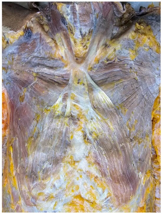
During the dissection of 79-year-old at death male donor, an unusual form of the sternalis [1] muscle was found. The donor was obtained through the body donation program of the Department of Anatomy, Faculty of Medicine, Khon Kaen University, and was embalmed using Thiel solutions [2]. The donor died of natural causes.

Figure 2.
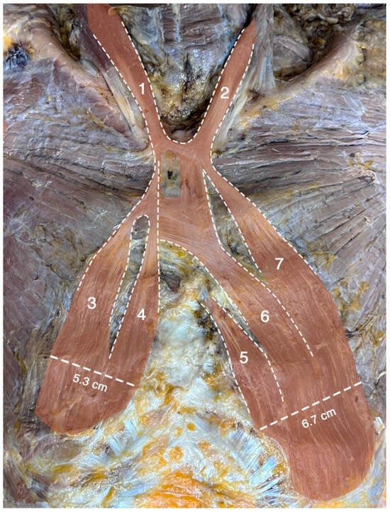
Dissection of the chest wall revealed a large sternalis muscle with seven heads. Two superior heads (#1 and #2) originated bilaterally as accessory heads of the sternocleidomastoid muscles. The widths of these two heads were 3.6 cm and 3.5 cm on the left and on the right sides, respectively. Five additional (#3–#7) heads originated inferiorly from the pectoralis fascia, three on the left side and two on the right side. The three heads on the left side (#5–#7) were attached along the costal margin near the rectus abdominis attachment. The widths of these pectoral heads were 6.7 cm on the left and 5.3 cm on the right. Intertendinous bands were present over the manubrium and the upper part of the sternal body, connecting both sides of the muscle. The muscle occupied nearly one-third of the chest wall area. The five inferior heads (#3–#7) received innervation from the intercostal nerves, while the innervation of the two superior heads (#1–#2) could not be confidently identified.

Figure 3.
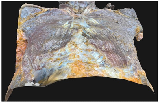
Three-dimensional (3D) image of the anterior chest wall of the present donor was captured using photogrammetry [3,4]. The 3D object was created using Adobe Substance 3D sampler (Adobe Inc., San Jose, CA, USA). The resulting 3D model of this donor in OBJ (Object file) format is available on Figshare via the following link: https://doi.org/10.6084/m9.figshare.25310815.
Author Contributions
Conceptualization, T.B., N.S. and L.Y.; Data curation, T.B., N.S., N.T. (Napawan Taradolpisut), R.S. and L.Y.; Formal analysis, T.S. and T.B.; Writing—original draft preparation, T.B., A.S. and L.Y.; Writing—review and editing, N.T. (Nareelak Tangsrisakda), N.S. and T.B. All authors have read and agreed to the published version of the manuscript.
Funding
This research received no external funding.
Institutional Review Board Statement
This study was approved by the Khon Kaen University Ethics Committees for Human Research (approval number: HE671124; date: 5 March 2024).
Informed Consent Statement
The study was exempted by the Ethics Committee, which oversighted all relevant documentation, including consent processes.
Data Availability Statement
The original data presented in the study are openly available at https://doi.org/10.6084/m9.figshare.25310815.
Acknowledgments
The authors state that every effort was made to follow all local and international ethical guidelines and laws that pertain to the use of human cadaveric donors in anatomical research [5]. The authors sincerely thank those who donated their bodies to science so that anatomical research could be performed. Results from such research can potentially increase humankind’s overall knowledge that can then improve patient care. Therefore, these donors and their families deserve our highest gratitude [6].
Conflicts of Interest
The authors declare no conflicts of interest.
References
- Bailey, P.M.; Tzarnas, C.D. The sternalis muscle: A normal finding encountered during breast surgery. Plast. Reconstr. Surg. 1999, 103, 1189–1190. [Google Scholar] [CrossRef] [PubMed]
- Taradolpisut, N.; Weerachatyanukul, W.; Suphamungmee, W.; Asuvapongpatana, S.; Luanphaisarnnont, T.; Chaiyamoon, A.; Berkban, T.; Halliwell, T.; Wilkinson, T.; Suwannakhan, A. The use of technical grade chemicals and on-site production of ammonium nitrate: A cost-effective and safer approach to Thiel embalming. Surg. Radiol. Anat. 2025, 47, 157. [Google Scholar] [CrossRef] [PubMed]
- Prasatkaew, W.; Kruepunga, N.; Yurasakpong, L.; Korkong, R.; Ardsawang, S.; Ronglakorn, S.; Sananpanich, K.; Suksri, S.; Suwannakhan, A. A reverse form of Linburg–Comstock variation with comments on its etiology and demonstration of interactive 3D portable document format. Surg. Radiol. Anat. 2021, 44, 227–232. [Google Scholar] [CrossRef] [PubMed]
- Suwannakhan, A.; Yurasakpong, L.; Taradolpisut, N.; Somrit, M.; Chaiyamoon, A.; Georgiev, G.P.; Iwanaga, J.; Tubbs, R.S. An accessory head of the extensor indicis: A rare case report. Surg. Radiol. Anat. 2024, 46, 1465–1468. [Google Scholar] [CrossRef] [PubMed]
- Iwanaga, J.; Singh, V.; Takeda, S.; Ogeng’o, J.; Kim, H.-J.; Moryś, J.; Ravi, K.S.; Ribatti, D.; Trainor, P.A.; Sañudo, J.R.; et al. Standardized statement for the ethical use of human cadaveric tissues in anatomy research papers: Recommendations from Anatomical Journal Editors-in-Chief. Clin. Anat. 2022, 35, 526–528. [Google Scholar] [CrossRef] [PubMed]
- Iwanaga, J.; Singh, V.; Ohtsuka, A.; Hwang, Y.; Kim, H.J.; Moryś, J.; Ravi, K.S.; Ribatti, D.; Trainor, P.A.; Sañudo, J.R.; et al. Acknowledging the use of human cadaveric tissues in research papers: Recommendations from anatomical journal editors. Clin. Anat. 2021, 34, 2–4. [Google Scholar] [CrossRef] [PubMed]
Disclaimer/Publisher’s Note: The statements, opinions and data contained in all publications are solely those of the individual author(s) and contributor(s) and not of MDPI and/or the editor(s). MDPI and/or the editor(s) disclaim responsibility for any injury to people or property resulting from any ideas, methods, instructions or products referred to in the content. |
© 2025 by the authors. Licensee MDPI, Basel, Switzerland. This article is an open access article distributed under the terms and conditions of the Creative Commons Attribution (CC BY) license (https://creativecommons.org/licenses/by/4.0/).