


Automated Quantitative Image-Derived Input Function for the Estimation of Cerebral Blood Flow Using Oxygen-15-Labelled Water on a Long-Axial Field-of-View PET/CT Scanner

 , , and
Abstract
1. Introduction
2. Materials and Methods
2.1. Phantom Data
2.2. Human Data
3. Results
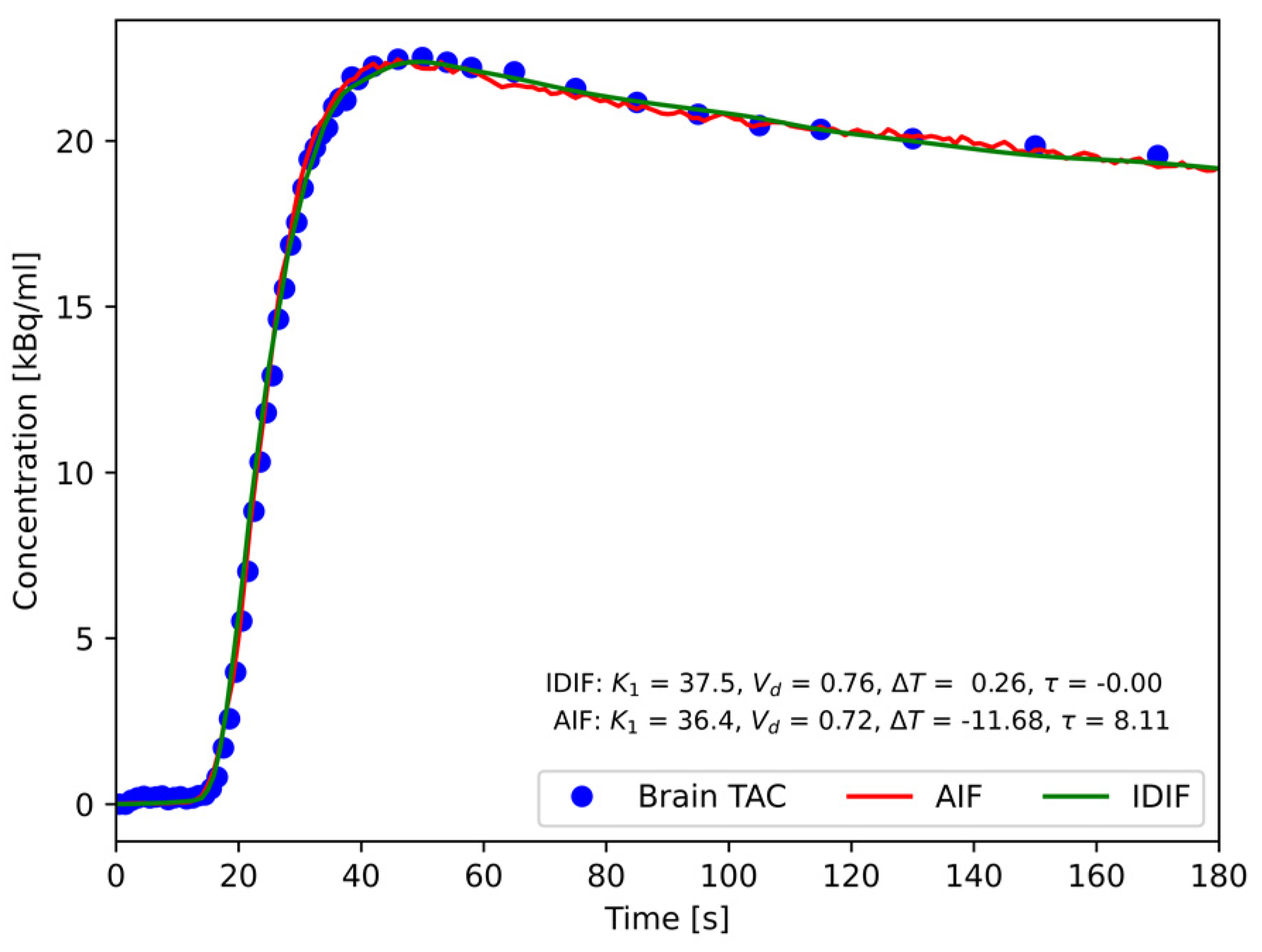
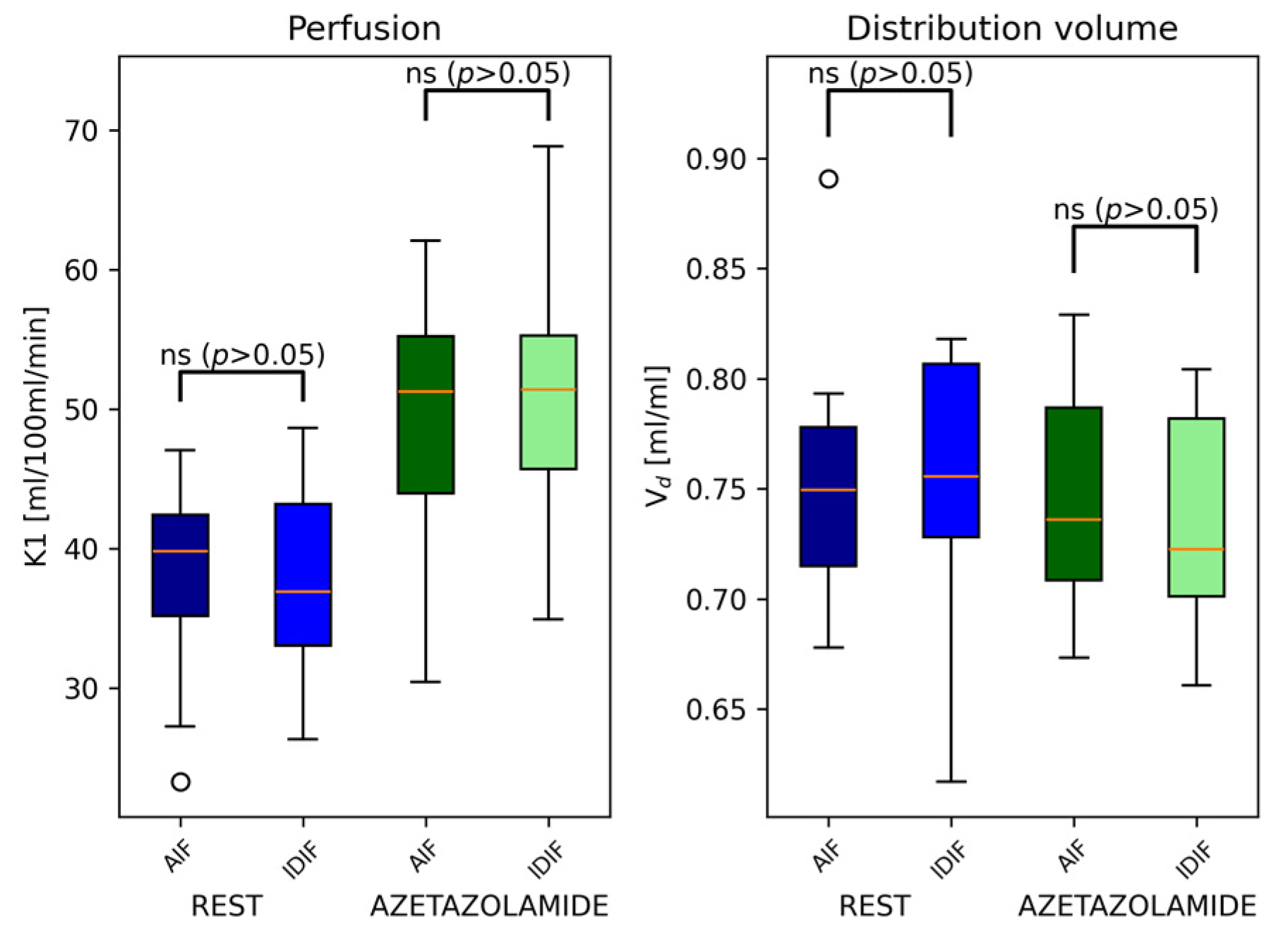
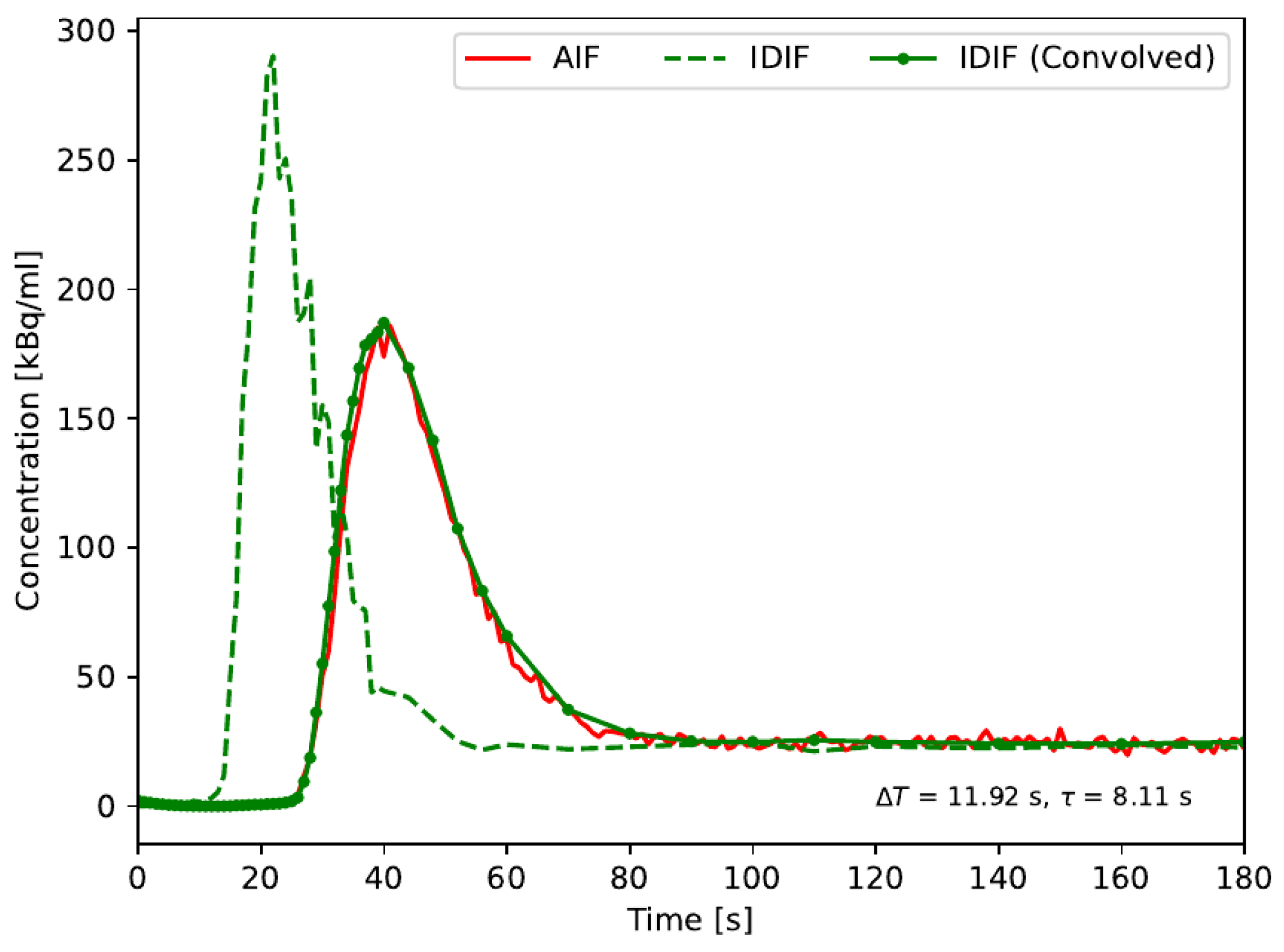
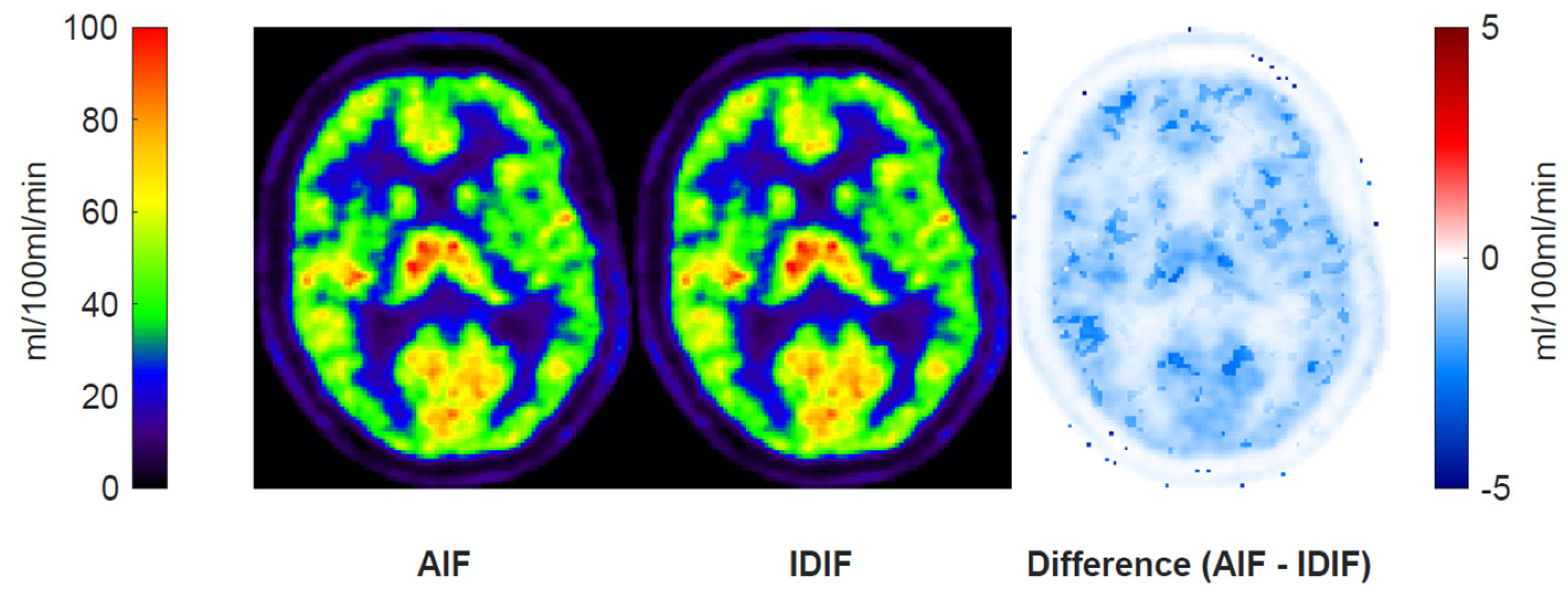
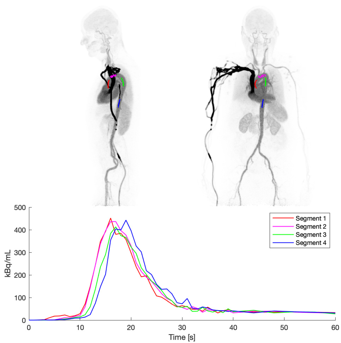
3.1. Blood Input Function Comparisons
3.2. Aorta Segmentation
3.3. Phantom Data
4. Discussion
5. Conclusions
Supplementary Materials
Author Contributions
Funding
Institutional Review Board Statement
Informed Consent Statement
Data Availability Statement
Conflicts of Interest
References
- Phelps, M.E. Positron emission tomography provides molecular imaging of biological processes. Proc. Natl. Acad. Sci. USA 2000, 97, 9226–9233. [Google Scholar] [CrossRef] [PubMed]
- Klunk, W.E.; Engler, H.; Nordberg, A.; Wang, Y.; Blomqvist, G.; Holt, D.P.; Bergström, M.; Savitcheva, I.; Huang, G.; Estrada, S.; et al. Imaging brain amyloid in Alzheimer’s disease with Pittsburgh Compound-B. Ann. Neurol. 2004, 55, 306–319. [Google Scholar] [CrossRef] [PubMed]
- Andersen, J.B.; Lindberg, U.; Olesen, O.V.; Benoit, D.; Ladefoged, C.N.; Larsson, H.B.; Højgaard, L.; Greisen, G.; Law, I. Hybrid PET/MRI imaging in healthy unsedated newborn infants with quantitative rCBF measurements using15O-water PET. J. Cereb. Blood Flow Metab. 2019, 39, 782–793. [Google Scholar] [CrossRef] [PubMed]
- Logan, J.; Fowler, J.S.; Volkow, N.D.; Wang, G.-J.; Ding, Y.-S.; Alexoff, D.L. Distribution Volume Ratios without Blood Sampling from Graphical Analysis of PET Data. J. Cereb. Blood Flow Metab. 1996, 16, 834–840. [Google Scholar] [CrossRef]
- Ichise, M.; Liow, J.-S.; Lu, J.-Q.; Takano, A.; Model, K.; Toyama, H.; Suhara, T.; Suzuki, K.; Innis, R.B.; Carson, R.E. Linearized reference tissue parametric imaging methods: Application to [11C]DASB positron emission tomography studies of the serotonin transporter in human brain. J. Cereb. Blood Flow Metab. 2003, 23, 1096–1112. [Google Scholar] [CrossRef] [PubMed]
- Carson, R.E.; Breier, A.; de Bartolomeis, A.; Saunders, R.C.; Su, T.P.; Schmall, B.; Der, M.G.; Pickar, D.; Eckelman, W.C. Quantification of Amphetamine-Induced Changes in [11C]Raclopride Binding with Continuous Infusion. J. Cereb. Blood Flow Metab. 1996, 17, 437–447. [Google Scholar] [CrossRef] [PubMed]
- Wang, G.; Rahmim, A.; Gunn, R.N. PET parametric imaging: Past, present, and future. IEEE Trans. Radiat. Plasma Med. Sci. 2020, 4, 663–675. [Google Scholar] [CrossRef] [PubMed]
- Iida, H.; Kanno, I.; Miura, S.; Murakami, M.; Takahashi, K.; Uemura, K. Error analysis of a quantitative cerebral blood flow measurement using H215O Autoradiography and positron emission tomography, with respect to the dispersion of the input function. J. Cereb. Blood Flow Metab. 1986, 6, 536–545. [Google Scholar] [CrossRef] [PubMed]
- Vestergaard, M.B.; Calvo, O.P.; Hansen, A.E.; Rosenbaum, S.; Larsson, H.B.; Henriksen, O.M.; Law, I. Validation of kinetic modeling of [15O]H2O PET using an image derived input function on hybrid PET/MRI. NeuroImage 2021, 233, 117950. [Google Scholar] [CrossRef]
- Moradi, H.; Vegh, V.; Reutens, D. Non-invasive input function extraction from dynamic PET using machine learning along with an iterative approach. J. Nucl. Med. 2021, 62 (Suppl. S1), 1461. [Google Scholar]
- Choi, Y.; Hawkins, R.A.; Huang, S.C.; Gambhir, S.S.; Brunken, R.C.; Phelps, M.E.; Schelbert, H.R. Parametric images of myocardial metabolic rate of glucose generated from dynamic cardiac PET and 2-[18F]fluoro-2-deoxy-d-glucose studies. J. Nucl. Med. 1991, 32, 733–738. [Google Scholar] [PubMed]
- Van Der Weerdt, A.P.; Klein, L.J.; Boellaard, R.; Visser, C.A.; Visser, F.C.; Lammertsma, A.A. Image-derived input functions for determination of MRGlu in cardiac (18)F-FDG PET scans. J. Nucl. Med. 2001, 42, 1622–1629. [Google Scholar] [PubMed]
- Huang, S.C.; Barrio, J.R.; Yu, D.; Chen, B.; Grafton, S.; Melega, W.P.; Hoffman, J.M.; Satyamurthy, N.; Mazziotta, J.C.; Phelps, M.E. Modelling approach for separating blood time activity curves in positron emission tomographic studies. Phys. Med. Biol. 1991, 36, 749–761. [Google Scholar] [CrossRef] [PubMed]
- Iida, H.; Jones, T.; Miura, S. Modeling Approach to Eliminate the Need to Separate Arterial Plasma in Oxygen-15 Inhalation Positron Emission Tomography. J. Nucl. Med. 1993, 34, 1333–1340. [Google Scholar]
- Kuttner, S.; Wickstrøm, K.K.; Lubberink, M.; Tolf, A.; Burman, J.; Sundset, R.; Jenssen, R.; Appel, L.; Axelsson, J. Cerebral blood flow measurements with 15O-water PET using a non-invasive machine-learning-derived arterial input function. J. Cereb. Blood Flow Metab. 2021, 41, 2229–2241. [Google Scholar] [CrossRef] [PubMed]
- Volpi, T.; Maccioni, L.; Colpo, M.; Debiasi, G.; Capotosti, A.; Ciceri, T.; Carson, R.E.; DeLorenzo, C.; Hahn, A.; Knudsen, G.M.; et al. An update on the use of image-derived input functions for human PET studies: New hopes or old illusions? EJNMMI Res. 2023, 13, 97. [Google Scholar] [CrossRef]
- Prenosil, G.A.; Sari, H.; Fürstner, M.; Afshar-Oromieh, A.; Shi, K.; Rominger, A.; Hentschel, M. Performance Characteristics of the Biograph Vision Quadra PET/CT System with a Long Axial Field of View Using the NEMA NU 2-2018 Standard. J. Nucl. Med. 2022, 63, 476–484. [Google Scholar] [CrossRef] [PubMed]
- Zhang, X.; Cherry, S.R.; Xie, Z.; Shi, H.; Badawi, R.D.; Qi, J. Subsecond total-body imaging using ultrasensitive positron emission tomography. Proc. Natl. Acad. Sci. USA 2020, 117, 2265–2267. [Google Scholar] [CrossRef]
- Yonas, H.; Darby, J.M.; Marks, E.C.; Durham, S.R.; Maxwell, C. CBF Measured by Xe-CT: Approach to Analysis and Normal Values. J. Cereb. Blood Flow Metab. 1991, 11, 716–725. [Google Scholar] [CrossRef]
- Vorstrup, S.; Henriksen, L.; Paulson, O.B. Effect of acetazolamide on cerebral blood flow and cerebral metabolic rate for oxygen. J. Clin. Investig. 1984, 74, 1634–1639. [Google Scholar] [CrossRef]
- Zirak, P.; Delgado-Mederos, R.; Martí-Fàbregas, J.; Durduran, T. Effects of acetazolamide on the micro- and macro-vascular cerebral hemodynamics: A diffuse optical and transcranial doppler ultrasound study. Biomed. Opt. Express 2010, 1, 1443–1459. [Google Scholar] [CrossRef] [PubMed]
- Wasserthal, J.; Breit, H.-C.; Meyer, M.T.; Pradella, M.; Hinck, D.; Sauter, A.W.; Heye, T.; Boll, D.T.; Cyriac, J.; Yang, S.; et al. TotalSegmentator: Robust Segmentation of 104 Anatomic Structures in CT Images. Radiol. Artif. Intell. 2023, 5, e230024. [Google Scholar] [CrossRef]
- Isensee, F.; Jaeger, P.F.; Kohl, S.A.A.; Petersen, J.; Maier-Hein, K.H. nnU-Net: A self-configuring method for deep learning-based biomedical image segmentation. Nat. Methods 2021, 18, 203–211. [Google Scholar] [CrossRef]
- Meyer, E. Simultaneous correction for tracer arrival delay and dispersion in CBF measurements by the H215O autoradio-graphic method and dynamic PET. J. Nucl. Med. 1989, 30, 1069–1078. [Google Scholar] [PubMed]
- Bland, J.M.; Altman, D.G. Statistics Notes: Measurement error proportional to the mean. BMJ 1996, 313, 106. [Google Scholar] [CrossRef] [PubMed]
- Gamechi, Z.S.; Bons, L.R.; Giordano, M.; Bos, D.; Budde, R.P.J.; Kofoed, K.F.; Pedersen, J.H.; Roos-Hesselink, J.W.; de Bruijne, M. Automated 3D segmentation and diameter measurement of the thoracic aorta on non-contrast enhanced CT. Eur. Radiol. 2019, 29, 4613–4623. [Google Scholar] [CrossRef] [PubMed]
- Zanotti-Fregonara, P.; Chen, K.; Liow, J.-S.; Fujita, M.; Innis, R.B. Image-Derived input function for brain pet studies: Many challenges and few opportunities. J. Cereb. Blood Flow Metab. 2011, 31, 1986–1998. [Google Scholar] [CrossRef]
- Toussaint, P.-J.; Meyer, E. A sensivity analysis of model parameters in dynamic blood flow studies using H215O and PET. In Quantification of Brain Function Using PET; Myers, R., Cunningham, V., Bailey, D., Jones, T., Eds.; Academic Press: San Diego, CA, USA, 1996; pp. 196–200. [Google Scholar]
- de Nijs, R. A novel model-based equation for size dependent mean recovery coefficients for spheres and other shapes. Phys. Medica 2023, 116, 103174. [Google Scholar] [CrossRef] [PubMed]
- Hawkins, R.A.; Choi, Y.; Huang, S.C.; Hoh, C.K.; Dahlbom, M.; Schiepers, C.; Satyamurthy, N.; Barrio, J.R.; Phelps, M.E. Evaluation of the skeletal kinetics of fluorine-18-fluoride ion with PET. J. Nucl. Med. 1992, 33, 633–642. [Google Scholar]
- Phelps, M.E.; Huang, S.C.; Hoffman, E.J.; Selin, C.; Sokoloff, L.; Kuhl, D.E. Tomographic measurement of local cerebral glucose metabolic rate in humans with (F-18)2-fluoro-2-deoxy-D-glucose: Validation of method. Ann. Neurol. 1979, 6, 371–388. [Google Scholar] [CrossRef]
- Larsson, H.B.W.; Law, I.; Andersen, T.L.; Andersen, F.L.; Fischer, B.M.; Vestergaard, M.B.; Larsson, T.S.W.; Lindberg, U. Brain perfusion estimation by Tikhonov model-free deconvolution in a long axial field of view PET/CT scanner exploring five different PET tracers. Eur. J. Nucl. Med. 2024, 51, 707–720. [Google Scholar] [CrossRef] [PubMed]
- Fung, E.K.; Carson, R.E. Cerebral blood flow with [15O]water PET studies using an image-derived input function and MR-defined carotid centerlines. Phys. Med. Biol. 2013, 58, 1903–1923. [Google Scholar] [CrossRef] [PubMed]
- Puig, O.; Henriksen, O.M.; Vestergaard, M.B.; Hansen, A.E.; Andersen, F.L.; Ladefoged, C.N.; Rostrup, E.; Larsson, H.B.; Lindberg, U.; Law, I. Comparison of simultaneous arterial spin labeling MRI and 15O-H2O PET measurements of regional cerebral blood flow in rest and altered perfusion states. J. Cereb. Blood Flow Metab. 2020, 40, 1621–1633. [Google Scholar] [CrossRef] [PubMed]
- Vestergaard, M.B.; Lindberg, U.; Aachmann-Andersen, N.J.; Lisbjerg, K.; Christensen, S.J.; Rasmussen, P.; Olsen, N.V.; Law, I.; Larsson, H.B.W.; Henriksen, O.M. Comparison of global cerebral blood flow measured by phase-contrast mapping MRI with 15O-H2O positron emission tomography. J. Magn. Reson. Imaging 2017, 45, 692–699. [Google Scholar] [CrossRef]
- Bremmer, J.P.; van Berckel, B.N.M.; Persoon, S.; Kappelle, L.J.; Lammertsma, A.A.; Kloet, R.; Luurtsema, G.; Rijbroek, A.; Klijn, C.J.M.; Boellaard, R. Day-to-day test–retest variability of CBF, CMRO2, and OEF measurements using dynamic 15O PET studies. Mol. Imaging Biol. 2011, 13, 759–768. [Google Scholar] [CrossRef] [PubMed]

| Diameter of Syringe/FWHM | 0 mm | 2 mm | 4 mm | 6 mm |
|---|---|---|---|---|
| 26.5 mm | 1.00 | 1.01 | 1.01 | 1.02 |
| 19.1 mm | 1.01 | 1.01 | 1.00 | 0.97 |
| 14.6 mm | 0.91 | 0.90 | 0.86 | 0.78 |
| 12.0 mm | 0.83 | 0.80 | 0.73 | 0.63 |
| 8.7 mm | 0.63 | 0.60 | 0.50 | 0.41 |
Disclaimer/Publisher’s Note: The statements, opinions and data contained in all publications are solely those of the individual author(s) and contributor(s) and not of MDPI and/or the editor(s). MDPI and/or the editor(s) disclaim responsibility for any injury to people or property resulting from any ideas, methods, instructions or products referred to in the content. |
© 2024 by the authors. Licensee MDPI, Basel, Switzerland. This article is an open access article distributed under the terms and conditions of the Creative Commons Attribution (CC BY) license (https://creativecommons.org/licenses/by/4.0/).
Share and Cite
Andersen, T.L.; Andersen, F.L.; Haddock, B.; Rosenbaum, S.; Larsson, H.B.W.; Law, I.; Lindberg, U. Automated Quantitative Image-Derived Input Function for the Estimation of Cerebral Blood Flow Using Oxygen-15-Labelled Water on a Long-Axial Field-of-View PET/CT Scanner. Diagnostics 2024, 14, 1590. https://doi.org/10.3390/diagnostics14151590
Andersen TL, Andersen FL, Haddock B, Rosenbaum S, Larsson HBW, Law I, Lindberg U. Automated Quantitative Image-Derived Input Function for the Estimation of Cerebral Blood Flow Using Oxygen-15-Labelled Water on a Long-Axial Field-of-View PET/CT Scanner. Diagnostics. 2024; 14(15):1590. https://doi.org/10.3390/diagnostics14151590
Chicago/Turabian StyleAndersen, Thomas Lund, Flemming Littrup Andersen, Bryan Haddock, Sverre Rosenbaum, Henrik Bo Wiberg Larsson, Ian Law, and Ulrich Lindberg. 2024. "Automated Quantitative Image-Derived Input Function for the Estimation of Cerebral Blood Flow Using Oxygen-15-Labelled Water on a Long-Axial Field-of-View PET/CT Scanner" Diagnostics 14, no. 15: 1590. https://doi.org/10.3390/diagnostics14151590
APA StyleAndersen, T. L., Andersen, F. L., Haddock, B., Rosenbaum, S., Larsson, H. B. W., Law, I., & Lindberg, U. (2024). Automated Quantitative Image-Derived Input Function for the Estimation of Cerebral Blood Flow Using Oxygen-15-Labelled Water on a Long-Axial Field-of-View PET/CT Scanner. Diagnostics, 14(15), 1590. https://doi.org/10.3390/diagnostics14151590