


Could Spinal Epidural Lipomatosis Be the Hallmark of Metabolic Syndrome on the Spine? A Literature Review with Emphasis on Etiology

 ,
, 

Abstract
1. Introduction
2. SEL’s Etiology
2.1. Excessive Amount of Corticosteroids
2.2. SEL and Obesity
2.3. Non-Alcoholic Fatty Liver Disease (NAFLD)
2.4. Miscellaneous
2.5. Idiopathic
2.6. Epidemiological Analysis of SEL’s Etiologies
3. SEL and Metabolic Syndrome
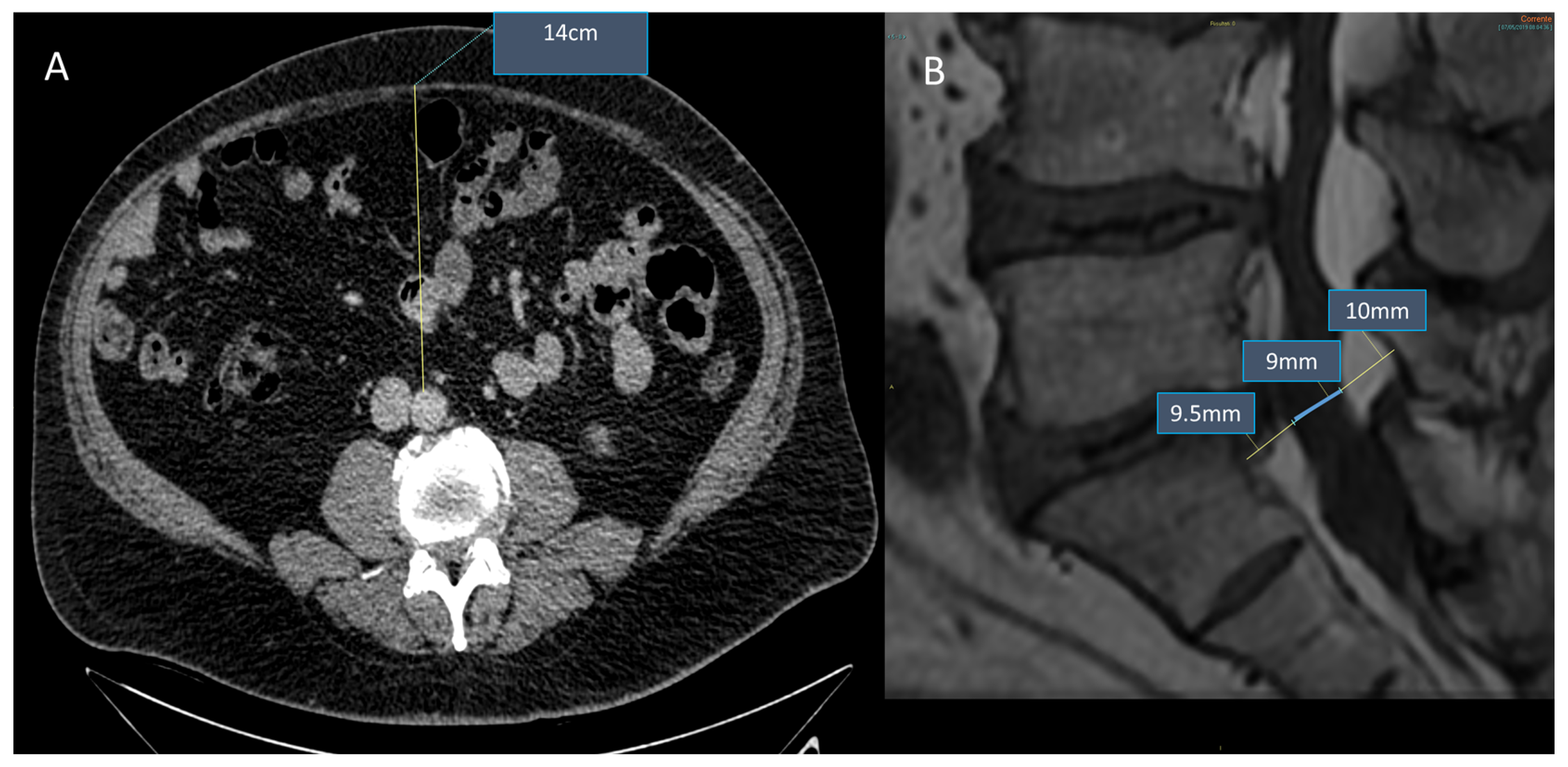
4. Diagnosis of SEL: The Role of Imaging
5. SEL Treatment
6. Conclusions
Author Contributions
Funding
Institutional Review Board Statement
Informed Consent Statement
Data Availability Statement
Conflicts of Interest
References
- Ferlic, P.W.; Mannion, A.F.; Jeszenszky, D.; Porchet, F.; Fekete, T.F.; Kleinstück, F.; Haschtmann, D. Patient-reported outcome of surgical treatment for lumbar spinal epidural lipomatosis. Spine J. 2016, 16, 1333–1341. [Google Scholar] [CrossRef]
- Fogel, G.R.; Cunningham, P.Y., III; Esses, S.I. Spinal epidural lipomatosis: Case reports, literature review and meta-analysis. Spine J. 2005, 5, 202–211. [Google Scholar] [CrossRef]
- Greenish, D.; Watura, K.; Harding, I. Spinal epidural lipomatosis following bilateral spinal decompression surgery. BMJ Case Rep. 2019, 12, e226985. [Google Scholar] [CrossRef]
- Choi, K.-C.; Kang, B.-U.; Lee, C.D.; Lee, S.-H. Rapid progression of spinal epidural lipomatosis. Eur. Spine J. 2011, 21, 408–412. [Google Scholar] [CrossRef]
- Youn, M.S.; Woo, Y.H.; Shin, J.K. Rapid progression of spinal epidural lipomatosis after percutaneous endoscopic spine surgery mimicking disc herniation. Int. J. Surg. Case Rep. 2020, 73, 1–4. [Google Scholar] [CrossRef]
- Praver, M.; Kennedy, B.C.; Ellis, J.A.; D’Amico, R.; Mandigo, C.E. Severity of presentation is associated with time to recovery in spinal epidural lipomatosis. J. Clin. Neurosci. 2015, 22, 1244–1249. [Google Scholar] [CrossRef]
- Al-Khawaja, D.; Seex, K.; Eslick, G.D. Spinal epidural lipomatosis—A brief review. J. Clin. Neurosci. 2008, 15, 1323–1326. [Google Scholar] [CrossRef]
- Robertson, S.C.; Traynelis, V.C.; Follett, K.A.; Menezes, A.H. Idiopathic spinal epidural lipomatosis. Neurosurgery 1997, 41, 68–74. [Google Scholar] [CrossRef]
- Papastefan, S.T.; Bhimani, A.D.; Denyer, S.; Khan, S.R.; Esfahani, D.R.; Nikas, D.C.; Mehta, A.I. Management of idiopathic spinal epidural lipomatosis: A case report and review of the literature. Childs Nerv. Syst. 2018, 34, 757–763. [Google Scholar] [CrossRef]
- Caruba, T.; Brunie, V.; Bousseau, V.; Guillemain, R.; Prognon, P.; Bégué, D.; Sabatier, B. Substitution of corticosteroid with everolimus after lung transplantation: A pediatric case report. Pharm. World Sci. 2010, 32, 347–349. [Google Scholar] [CrossRef]
- Möller, J.; Girschick, H.; Hahn, G.; Pessler, F. Steroid-induced spinal epidural lipomatosis in pediatric patients. Z. Rheumatol. 2010, 69, 447–449. [Google Scholar] [CrossRef]
- Vazquez, L.; Ellis, A.; Saint-Genez, D.; Patino, J.; Nogues, M. Epidural lipomatosis after renal transplantation—Complete recovery without surgery. Transplantation 1988, 46, 773–774. [Google Scholar] [CrossRef]
- Theyskens, N.C.; Paulino Pereira, N.R.; Janssen, S.J.; Bono, C.M.; Schwab, J.H.; Cha, T.D. The prevalence of spinal epidural lipomatosis on magnetic resonance imaging. Spine J. 2017, 17, 969–976. [Google Scholar] [CrossRef]
- Spinnato, P.; D’Agostino, V.; Fiorenzo, D.; Barakat, M.; Vara, G.; Ponti, F.; Filonzi, G.; Crombé, A.; Tetta, C.; Miceli, M. Underreporting of spinal epidural lipomatosis: A retrospective analysis of lumbosacral MRI examinations from different radiological settings. Diagn. Interv. Imaging 2022, 103, 251–257. [Google Scholar] [CrossRef]
- Borré, D.G.; Borré, G.E.; Aude, F.; Palmieri, G.N. Lumbosacral epidural lipomatosis: MRI grading. Eur. Radiol. 2003, 13, 1709–1721. [Google Scholar] [CrossRef]
- Fassett, D.R.; Schmidt, M.H. Spinal epidural lipomatosis: A review of its causes and recommendations for treatment. Neurosurg. Focus 2004, 16, 1–3. [Google Scholar] [CrossRef]
- Bodelier, A.; Groeneveld, W.; Van Der Linden, A.; Haak, H. Symptomatic epidural lipomatosis in ectopic Cushing’s syndrome. Eur. J. Endocrinol. 2004, 151, 765–769. [Google Scholar] [CrossRef]
- Schurmann, D.; Rademaker, J.; Trottenberg, T.; Bergmann, F.; Wesselmann, H.; Suttorp, N. Spinal epidural lipomatosis: A manifestation of HAART-associated lipodystrophy. AIDS 2005, 19, 2052–2054. [Google Scholar] [CrossRef]
- Lee, M.; Lekias, J.; Gubbay, S.S.; Hurst, P.E. Spinal cord compression by extradural fat after renal transplantation. Med. J. Aust. 1975, 1, 201–203. [Google Scholar] [CrossRef]
- Noh, E. An unusual complication of morbid obesity: Epidural lipomatosis. Am. J. Emerg. Med. 2015, 33, 742.e3–742.e4. [Google Scholar] [CrossRef]
- López-González, A.; Giner, M.R. Idiopathic spinal epidural lipomatosis: Urgent decompression in an atypical case. Eur. Spine J. 2008, 17, 225–227. [Google Scholar] [CrossRef] [PubMed]
- Mallard, F.; Buni, M.; Nolet, P.S.; Emary, P.; Taylor, J.A.; Moammer, G. Lumbar spinal epidural lipomatosis: A case report and review of the literature. Int. J. Surg. Case Rep. 2021, 78, 71–75. [Google Scholar] [CrossRef] [PubMed]
- Fujita, N.; Ishihara, S.; Michikawa, T.; Suzuki, S.; Tsuji, O.; Nagoshi, N.; Okada, E.; Yagi, M.; Tsuji, T.; Kono, H.; et al. Negative impact of spinal epidural lipomatosis on the surgical outcome of posterior lumbar spinous-splitting decompression surgery: A multicenter retrospective study. Spine J. 2019, 19, 1977–1985. [Google Scholar] [CrossRef] [PubMed]
- Jaimes, R., III; Rocco, A.G. Multiple epidural steroid injections and body mass index linked with the occurrence of epidural lipomatosis: A case series. BMC Anesthesiol. 2014, 14, 70. [Google Scholar] [CrossRef]
- Zentner, J.; Buchbender, K.; Vahlensieck, M. Spinal epidural lipomatosis is a complication of prolonged corticosteroid therapy. J. Neurosurg. Sci. 1995, 39, 81–85. [Google Scholar]
- Pinsker, M.O.; Kinzel, D.; Lumenta, C.B. Epidural thoracic lipomatosis induced by long-term steroid treatment case illustration. Acta Neurochir. 1998, 140, 991–992. [Google Scholar] [CrossRef]
- Kotilainen, E.; Hohenthal, U.; Karhu, J.; Kotilainen, P. Spinal epidural lipomatosis caused by corticosteroid treatment in ulcerative colitis. Eur. J. Intern. Med. 2006, 17, 138–140. [Google Scholar] [CrossRef]
- Koch, C.A.; Doppman, J.L.; Patronas, N.J.; Nieman, L.K.; Chrousos, G.P. Do glucocorticoids cause spinal epidural lipomatosis? When endocrinology and spinal surgery meet. Trends Endocrinol. Metab. 2000, 11, 86–90. [Google Scholar] [CrossRef]
- Mukhtar, N.; Alzahrani, A.S. Spinal epidural lipomatosis: A rare and frequently unrecognized complication of Cushing syndrome. Endocrine 2022, 76, 218–223. [Google Scholar] [CrossRef]
- Benamou, P.H.; Hilliquin, P.; Chemla, N.; Chevrot, A.; Cormier, C.; Menkès, C.J. Epidural lipomatosis not induced by corticosteroid therapy. Three cases including one in a patient with primary Cushing’s disease (review of the literature). Rev. Rhum. Engl. Ed. 1996, 63, 207–212. [Google Scholar]
- Stern, J.D.; Quint, D.J.; Sweasey, T.A.; Hoff, J.T. Spinal epidural lipomatosis: Two new idiopathic cases and a review of the literature. J. Spinal Disord. 1994, 7, 343–349. [Google Scholar] [CrossRef]
- Fessler, R.G.; Johnson, D.L.; Brown, F.D.; Erickson, R.K.; Reid, S.A.; Kranzler, L. Epidural lipomatosis in steroid-treated patients. Spine 1992, 17, 183–188. [Google Scholar] [CrossRef]
- Tobler, W.D.; Weil, S. Epidural lipomatosis and renal transplantation. Surg. Neurol. 1988, 29, 141–144. [Google Scholar] [CrossRef]
- Muñoz, A.; Barkovich, J.A.; Mateos, F.; Simón, R. Symptomatic epidural lipomatosis of the spinal cord in a child: MR demonstration of spinal cord injury. Pediatr. Radiol. 2002, 32, 865–868. [Google Scholar] [CrossRef]
- McCullen, G.M.; Spurling, G.R.; Webster, J.S. Epidural lipomatosis complicating lumbar steroid injections. J. Spinal Disord. 1999, 12, 526–529. [Google Scholar] [CrossRef]
- Roy-Camille, R.; Mazel, C.; Husson, J.L.; Saillant, G. Symptomatic spinal epidural lipomatosis induced by long-term steroid treatment. Review of the literature and report of two additional cases. Spine 1991, 16, 1365–1371. [Google Scholar] [CrossRef]
- Sandberg, D.I.; Lavyne, M.H. Symptomatic spinal epidural lipomatosis after local epidural corticosteroid injections: Case report. Neurosurgery 1999, 45, 162–165. [Google Scholar]
- Koch, C.A.; Doppman, J.L.; Watson, J.C.; Patronas, N.J.; Nieman, L.K. Spinal epidural lipomatosis in a patient with the ectopic corticotropin syndrome. N. Engl. J. Med. 1999, 341, 1399–1400. [Google Scholar] [CrossRef]
- Bhatia, K.; Frydenberg, E.; Steel, T.; Ow-Yang, M.; Ho, K.; Grainger, E. Spinal epidural lipomatosis due to a bronchial ACTH-secreting carcinoid tumour. J. Clin. Neurosci. 2010, 17, 1461–1462. [Google Scholar] [CrossRef]
- Ahmad, S.; Best, T.; Lansdown, A.; Hayhurst, C.; Smeeton, F.; Davies, S.; Rees, A. Spinal epidural lipomatosis: A rare association of Cushing’s disease. Endocrinol. Diabetes Metab. Case Rep. 2020, 2020, 20-0111. [Google Scholar] [CrossRef]
- Zhang, B.; Yuan, H.; Hu, L.; Saad, M. Obesity is a risk factor for epidural lipomatosis. A meta-analysis. J. Orthop. Surg. 2021, 29, 1–8. [Google Scholar] [CrossRef] [PubMed]
- Shimada, Y.; Ishikawa, Y.; Miyakoshi, N.; Suzuki, T.; Hongo, M.; Kasukawa, Y.; Okada, K.; Itoi, E. Decompression of idiopathic lumbar epidural lipomatosis: Diagnostic magnetic resonance imaging evaluation and review of the literature. J. Neurosurg. Spine 2006, 4, 24–30. [Google Scholar]
- Kumar, K.; Nath, R.K.; Nair, C.P.V.; Tchang, S.P. Symptomatic epidural lipomatosis secondary to obesity. Case report. J. Neurosurg. 1996, 85, 348–350. [Google Scholar] [CrossRef]
- Sugaya, H.; Tanaka, T.; Ogawa, T.; Mishima, H. Spinal epidural lipomatosis in lumbar magnetic resonance imaging scans. Orthopedics 2014, 37, e362–e366. [Google Scholar] [CrossRef]
- Spinnato, P.; Ponti, F.; De Pasqua, S. MRI Diagnosis of Obesity-Related Spinal Epidural Lipomatosis. Can. J. Neurol. Sci. 2020, 47, 124–125. [Google Scholar] [CrossRef] [PubMed]
- Yildirim, B.; Puvanesarajah, V.; An, H.S.; Novicoff, W.M.; Jain, A.; Shen, F.H.; Hassanzadeh, H. Lumbosacral Epidural Lipomatosis: A Retrospective Matched Case-Control Database Study. World Neurosurg. 2016, 96, 209–214. [Google Scholar] [CrossRef] [PubMed]
- Yki-Järvinen, H. Non-alcoholic fatty liver disease as a cause and a consequence of metabolic syndrome. Lancet Diabetes Endocrinol. 2014, 2, 901–910. [Google Scholar] [CrossRef]
- Abe, T.; Miyazaki, M.; Ishihara, T.; Kanezaki, S.; Notani, N.; Kataoka, M.; Tsumura, H. Spinal epidural lipomatosis is associated with liver fat deposition and dysfunction. Clin. Neurol. Neurosurg. 2019, 185, 105480. [Google Scholar] [CrossRef]
- Billings, F.; Hoyt, M. Epidural lipomatosis causing new debilitating back pain in a patient with human immunodeficiency virus on highly active antiretroviral therapy. Int. J. Obstet. Anesth. 2012, 21, 367–370. [Google Scholar] [CrossRef]
- Vince, G.H.; Brucker, C.; Langmann, P.; Herbold, C.; Solymosi, L.; Roosen, K. Epidural spinal lipomatosis with acute onset of paraplegia in an HIV-positive patient treated with corticosteroids and protease inhibitor: Case report. Spine 2005, 30, E524–E527. [Google Scholar] [CrossRef]
- Cersósimo, M.G.; Lasala, B.; Folgar, S.; Micheli, F. Epidural lipomatosis secondary to indinavir in an HIV-positive patient. Clin. Neuropharmacol. 2002, 25, 51–54. [Google Scholar] [CrossRef] [PubMed]
- Ebright, J.R.; Stellini, M.A.; Tselis, A.C. Spinal epidural lipomatosis in a human immunodeficiency virus-positive patient receiving steroids and protease inhibitor therapy. Clin. Infect. Dis. 2001, 32, e90–e91. [Google Scholar] [CrossRef] [PubMed]
- Mattei, T.A.; Goulart, C.R.; Rai, S.S.; Rehman, A.A.; Williams, M.; Mendel, E. Rapid development of spinal epidural lipomatosis after treatment of metastatic castration-resistant prostate cancer with second-generation androgen receptor antagonists. World Neurosurg. 2019, 125, 222–227. [Google Scholar] [CrossRef]
- Awwad, A.; Adam, R.E.I.; Patel, C.; Thomas, J.D. Symptomatic spinal epidural lipomatosis after combined hormonal and steroidal palliative therapy of prostate cancer. Spinal Cord Ser. Cases 2018, 4, 75. [Google Scholar] [CrossRef]
- Tulloch, I.; Laban, J.T.; Martin, A.J. A proposed link between spinal epidural lipomatosis, prostate cancer, and androgen deprivation therapy. J. Clin. Urol. 2018, 11, 299–301. [Google Scholar] [CrossRef]
- Grayling, M.; Jardine, D.L.; Mcclintock, A.D.; Spar, J.; Wilton, G.N. Symptomatic epidural lipomatosis following cyproterone acetate therapy. Aust. N. Z. J. Surg. 2000, 70, 233–235. [Google Scholar] [CrossRef] [PubMed]
- Han, S.R. Scoliosis associated with idiopathic lumbosacral epidural lipomatosis. J. Spine Surg. 2016, 2, 72–75. [Google Scholar] [CrossRef]
- Sabharwal, S.; Mahmood, F. Thoracic spinal epidural lipomatosis associated with adolescent scoliosis. J. Spinal Disord. Tech. 2006, 19, 217–221. [Google Scholar] [CrossRef]
- Kurt, E.; Bakker-Niezen, S.H. Neurogenic claudication by epidural lipomatosis: A case report and review of literature. Clin. Neurol. Neurosurg. 1995, 97, 354–357. [Google Scholar] [CrossRef]
- Neal, M.T.; Patra, D.P.; Lyons, M.K. Surgical management of thoracic myelopathy from long-segment epidural lipomatosis with skip hemilaminotomies: Illustrative case. J. Neurosurg. Case Lessons 2021, 2, CASE21595. [Google Scholar] [CrossRef]
- Okada, E.; Ishihara, S.; Azuma, K.; Michikawa, T.; Suzuki, S.; Tsuji, O.; Nori, S.; Nagoshi, N.; Yagi, M.; Takayama, M.; et al. Metabolic Syndrome is a Predisposing Factor for Diffuse Idiopathic Skeletal Hyperostosis. Neurospine 2021, 18, 109–116. [Google Scholar] [CrossRef]
- Zhang, Z.; Liu, Z.; Zhu, Z.; Qiu, Y. Spinal epidural lipomatosis—An easily ignored secondary intraspinal disorder in spinal kyphotic deformities. BMC Musculoskelet. Disord. 2017, 18, 112. [Google Scholar] [CrossRef] [PubMed]
- Abul-Kasim, K.; Schlenzka, D.; Selariu, E.; Ohlin, A. Spinal epidural lipomatosis: A common imaging feature in Scheuermann disease. J. Spinal Disord. Tech. 2012, 25, 356–361. [Google Scholar] [CrossRef]
- Oikonomou, A.; Birbilis, T.; Gymnopoulou, E.; Prassopoulos, P. Paget disease of the spine manifested by thoracic and lumbar epidural lipomatosis: Magnetic resonance imaging findings. Spine 2007, 32, E789–E792. [Google Scholar] [CrossRef] [PubMed]
- Koziarz, P.; Avruch, L. Spinal epidural lipomatosis associated with Paget’s disease of bone. Neuroradiology 2002, 44, 858–860. [Google Scholar] [CrossRef]
- Toshniwal, P.K.; Glick, R.P. Spinal epidural lipomatosis: Report of a case secondary to hypothyroidism and review of literature. J. Neurol. 1987, 234, 172–176. [Google Scholar] [CrossRef] [PubMed]
- Kim, S.-S.; Lim, D.-J. Epidural lipomatosis with cauda equina syndrome in chronic alcoholic patient: A case report. Int. J. Surg. Case Rep. 2017, 33, 12–15. [Google Scholar] [CrossRef]
- Lotan, I.; Charlson, R.W.; Fatterpekar, G.M.; Shapiro, M.; Smith, M.L.; William, C.; Kister, I. Progressive myelopathy associated with spinal epidural lipomatosis in three nonobese patients with type 1 diabetes mellitus. J. Neurol. Sci. 2020, 411, 116688. [Google Scholar] [CrossRef] [PubMed]
- Badami, J.P.; Hinck, V.C. Symptomatic deposition of epidural fat in a morbidly obese woman. Am. J. Neuroradiol. 1991, 3, 664–665. [Google Scholar]
- Haddad, S.F.; Hitchon, P.W.; Godersky, J.C. Idiopathic and glucocorticoid-induced spinal epidural lipomatosis. J. Neurosurg. 1991, 74, 38–42. [Google Scholar] [CrossRef]
- Miyakoshi, N.; Shimada, Y.; Suzuki, T.; Hongo, M.; Kasukawa, Y.; Okada, K.; Itoi, E. Factors related to long-term outcome after decompressive surgery for ossification of the ligamentum flavum of the thoracic spine. J. Neurosurg. 2003, 99, 251–256. [Google Scholar] [CrossRef] [PubMed]
- Eap, C.; Litre, C.F.; Duntze, J.; Theret, E.; Noudel, R.; Graftieaux, J.-P.; Rousseaux, P. Compression médullaire lente par lipomatose épidurale thoracique idiopathique: Cas clinique et revue de la littérature [Spinal cord compression caused by idiopathic dorsal epidural lipomatosis: Case report and critical review of the literature]. Neurochirurgie 2010, 56, 55–58. [Google Scholar] [CrossRef] [PubMed]
- Sato, M.; Yamashita, K.; Aoki, Y.; Hiroshima, K. Idiopathic spinal epidural lipomatosis. Case report and review of literature. Clin. Orthop. Relat. Res. 1995, 320, 129–134. [Google Scholar]
- Al-Yafeai, R.; Maghrabi, Y.; Malibary, H.; Baeesa, S. Spinal cord compression secondary to idiopathic thoracic epidural lipomatosis in an adolescent: A case report and review of literature. Int. J. Surg. Case Rep. 2017, 37, 225–229. [Google Scholar] [CrossRef] [PubMed]
- Lee, S.-B.; Park, H.-K.; Chang, J.-C.; Jin, S.-Y. Idiopathic thoracic epidural lipomatosis with chest pain. J. Korean Neurosurg. Soc. 2011, 50, 130–133. [Google Scholar] [CrossRef]
- Yoo, J.C.; Choi, J.J.; Lee, D.W.; Lee, S.P. Spinal epidural lipomatosis in Korean. J. Korean Neurosurg. Soc. 2014, 55, 365–369. [Google Scholar] [CrossRef] [PubMed]
- Alomari, S.; Lubelski, D.; Khalifeh, J.M.; Sacino, A.N.; Theodore, N.; Witham, T.; Bydon, A. Etiologies and Outcomes of Spinal Epidural Lipomatosis: Systematic Review of the Literature and Meta-Analysis of Reported Cases. Clin. Spine Surg. 2022, 35, 383–387. [Google Scholar] [CrossRef] [PubMed]
- Alberti, K.G.; Zimmet, P.; Shaw, J. IDF Epidemiology Task Force Consensus Group. The metabolic syndrome—A new worldwide definition. Lancet 2005, 366, 1059–1062. [Google Scholar] [CrossRef]
- Matsuzawa, Y.; Funahashi, T.; Nakamura, T. The concept of metabolic syndrome: Contribution of visceral fat accumulation and its molecular mechanism. J. Atheroscler. Thromb. 2011, 18, 629–639. [Google Scholar] [CrossRef]
- Zając-Gawlak, I.; Kłapcińska, B.; Kroemeke, A.; Pośpiech, D.; Pelclová, J.; Přidalová, M. Associations of visceral fat area and physical activity levels with the risk of metabolic syndrome in postmenopausal women. Biogerontology 2017, 18, 357–366. [Google Scholar] [CrossRef]
- Kvist, H.; Chowdhury, B.; Sjöström, L.; Tylén, U.; Cederblad, A. Adipose tissue volume determination in males by computed tomography and 40K. Int. J. Obes. 1988, 12, 249–266. [Google Scholar] [PubMed]
- Ishihara, S.; Fujita, N.; Azuma, K.; Michikawa, T.; Yagi, M.; Tsuji, T.; Takayama, M.; Matsumoto, H.; Nakamura, M.; Matsumoto, M.; et al. Spinal epidural lipomatosis is a previously unrecognized manifestation of metabolic syndrome. Spine J. 2019, 19, 493–500. [Google Scholar] [CrossRef] [PubMed]
- Shimabukuro, M.; Kozuka, C.; Taira, S.; Yabiku, K.; Dagvasumberel, M.; Ishida, M.; Matsumoto, S.; Yagi, S.; Fukuda, D.; Yamakawa, K.; et al. Ectopic fat deposition and global cardiometabolic risk: New paradigm in cardiovascular medicine. J. Med. Investig. 2013, 60, 1–14. [Google Scholar] [CrossRef] [PubMed]
- Oka, R.; Yagi, K.; Sakurai, M.; Nakamura, K.; Nagasawa, S.Y.; Miyamoto, S.; Nohara, A.; Kawashiri, M.; Hayashi, K.; Takeda, Y.; et al. Impact of visceral adipose tissue and subcutaneous adipose tissue on insulin resistance in middle-aged Japanese. J. Atheroscler. Thromb. 2012, 19, 814–822. [Google Scholar] [CrossRef]
- Fujita, N. Lumbar Spinal Canal Stenosis from the Perspective of Locomotive Syndrome and Metabolic Syndrome: A Narrative Review. Spine Surg. Relat. Res. 2020, 5, 61–67. [Google Scholar] [CrossRef]
- Ishihara, S.; Fujita, N.; Yagi, M.; Tsuji, T.; Michikawa, T.; Nishiwaki, Y.; Fukui, Y.; Horiuchi, K.; Ishii, K.; Nakamura, M.; et al. Idiopathic Spinal Epidural Fat Accumulation Is Associated with Hyperlipidemia. Spine 2018, 43, E468–E473. [Google Scholar] [CrossRef]
- Cushnie, D.; Urquhart, J.C.; Gurr, K.R.; Siddiqi, F.; Bailey, C.S. Obesity and spinal epidural lipomatosis in cauda equina syndrome. Spine J. 2018, 18, 407–413. [Google Scholar] [CrossRef]
- Walker, P.B.; Sark, C.; Brennan, G.; Smith, T.; Sherman, W.F.; Kaye, A.D. Spinal Epidural Lipomatosis: A Comprehensive Review. Orthop. Rev. 2021, 13, 25571. [Google Scholar] [CrossRef]
- Sasagasako, T.; Hanakita, J.; Minami, M.; Kanematsu, R.; Tomita, Y. Clinical Implications of the Epidural Fat Thickness in the Management of Lumbar Spinal Stenosis. World Neurosurg. 2021, 146, e205–e213. [Google Scholar] [CrossRef]
- Park, S.K.; Han, J.M.; Lee, K.; Cho, W.J.; Oh, J.H.; Choi, Y.S. The Clinical Characteristics of Spinal Epidural Lipomatosis in the Lumbar Spine. Anesthesiol. Pain Med. 2018, 8, e83069. [Google Scholar] [CrossRef]
- Morishita, S.; Arai, Y.; Yoshii, T.; Sakai, K.; Hirai, T.; Okawa, A. Lumbar epidural lipomatosis is associated with visceral fat and metabolic disorders. Eur. Spine J. 2018, 27, 1653–1661. [Google Scholar] [CrossRef] [PubMed]
- Tok, C.H.; Kaur, S.; Gangi, A. Symptomatic spinal epidural lipomatosis after a single local epidural steroid injection. Cardiovasc. Interv. Radiol. 2011, 34 (Suppl. 2), S250–S255. [Google Scholar] [CrossRef] [PubMed]
- Spinnato, P.; Barakat, M.; Lotrecchiano, L.; Giusti, D.; Filonzi, G.; Spinelli, D.; Pipola, V.; Moio, A.; Tetta, C.; Ponti, F. MRI Features and Clinical Significance of Spinal Epidural Lipomatosis: All You Should Know. Curr. Med. Imaging 2022, 18, 208–215. [Google Scholar] [CrossRef] [PubMed]
- Spinnato, P.; Lotrecchiano, L.; Ponti, F. “Y” sign in spinal epidural lipomatosis. Jt. Bone Spine 2020, 88, 105056. [Google Scholar] [CrossRef]
- Kuhn, M.J.; Youssef, H.T.; Swan, T.L.; Swenson, L.C. Lumbar epidural lipomatosis: The “Y” sign of thecal sac compression. Comput. Med. Imaging Graph. 1994, 18, 367–372. [Google Scholar] [CrossRef]
- Geers, C.; Lecouvet, F.E.; Behets, C.; Malghem, J.; Cosnard, G.; Lengelé, B.G. Polygonal deformation of the dural sac in lumbar epidural lipomatosis: Anatomic explanation by the presence of meningovertebral ligaments. Am. J. Neuroradiol. 2003, 24, 1276–1282. [Google Scholar]
- Reina, M.A.; Franco, C.D.; López, A.; Andrés, J.A.D.; Van Zundert, A. Clinical implications of epidural fat in the spinal canal. A scanning electron microscopic study. Acta Anaesthesiol. Belg. 2009, 60, 7–17. [Google Scholar]
- Trungu, S.; Forcato, S.; Raco, A. Spinal Epidural Lipomatosis: Weight Loss Cure. World Neurosurg. 2019, 125, 368–370. [Google Scholar] [CrossRef]
- Yu, Y.; Jiang, Y.; Xu, F.; Yuan, L.; Mao, Y.; Li, C. Percutaneous full-endoscopic uniportal decompression for the treatment of symptomatic idiopathic lumbar spinal epidural lipomatosis: Technical note. Front. Surg. 2022, 9, 894662. [Google Scholar] [CrossRef]
- Fuentes, A.M.; Housley, S.B.; Starling, R.V.; Mullin, J.P. Extensive Symptomatic Thoracolumbar Epidural Lipomatosis Treated With Minimally Invasive Hemilaminectomies: Technical Case Report. Oper. Neurosurg. 2022, 23, e184–e188. [Google Scholar] [CrossRef]
- Manjila, S.; Fana, M.; Medani, K.; Kay, M.D.; Manjila, R.; Bazil, T.G.; Udayasankar, U. Spinal Epidural Lipomatosis Causing Lumbar Canal Stenosis: A Pictorial Essay on Radiological Grading and the Role of Bariatric Surgery Versus Laminectomy. Cureus 2022, 14, e26492. [Google Scholar] [CrossRef] [PubMed]
- Mosch, M.H.; De Jong, L.D.; Hazebroek, E.J.; Van Susante, J.L. Lumbar Epidural Lipomatosis is Increased in Patients with Morbid Obesity and Subsequently Decreases after Bariatric Surgery. World Neurosurg. 2022, 158, e495–e500. [Google Scholar] [CrossRef] [PubMed]


| Literature Review (Time Period 1990–2020)—Causes of SEL | % |
|---|---|
| Obesity | 52 |
| Exogenous steroid use | 22 |
| Idiopathic | 26 |
| Literature Review (Time Period 1975–2003)—Causes of SEL | % |
| Exogenous steroid use | 55.3 |
| Obesity | 24.5 |
| Idiopathic | 17 |
| Endogenous steroid hormonal disease | 3.2 |
| Authors | Risk Factor | Number of Patients |
|---|---|---|
| Ishihara et al. [86] | Metabolic Syndrome (Obesity and Hyperlipidemia) | 166 |
| Yildirim et al. [46] | Metabolic Syndrome | 199 |
| Theyskens et al. [13] | Cushing’s Syndrome * | 731 |
| Bhatia et al. [39] | Cushing’s Syndrome * | 1 |
| Ahmad et al. [40] | Cushing’s Disease * | 1 |
| Ishihara et al. [82] | Hyperlipidemia * | 324 |
| Zhang et al. [41] | Obesity * | 807 |
| Cushnie et al. [87] | Obesity * | 99 |
| Walker et al. [88] | Obesity * | 25 |
| Fogel et al. [2] | Obesity * | 104 |
| Spinnato et al. [45] | Obesity * | 1 |
| Sasagasako et al. [89] | Subcutaneous Fat Thickness * | 500 |
| Lotan et al. [67] | Diabetes Type 1 * | 3 |
| Park et al. [90] | Diabetes Type 2 * | 42 |
| Morishita et al. [91] | Visceral Fat * | 218 |
| Abe et al. [48] | NAFLD * | 102 |
| Okada et al. [61] | DISH * | 327 |
| Khawaja et al. [7] | Hypertension * | 2 |
| Fasset et al. [16] | Exogenous Steroids | 2 |
| Tok et al. [92] | Epidural Steroid Injection | 1 |
| Billings et al. [49] | HAART-associated Lipodystrophy | 1 |
| Mattei et al. [53] | Androgen Deprivation Therapy | 1 |
| Papastefan et al. [9] | Idiopathic | 1 |
| Stern et al. [31] | Idiopathic | 2 |
| SEL MRI Grading System | |||
|---|---|---|---|
| MRI GRADE | DuS/EF Index | EF/SC Index (%) | Meaning |
| NORMAL (0) | ≥1.5 | ≤40 | Normal amount of EF |
| GRADE I | 1.49–1 | 41–50 | Mild overgrowth of EF |
| GRADE II | 0.99–0.34 | 51–74 | Moderate overgrowth of EF |
| GRADE III | ≤0.33 | ≥75 | Severe overgrowth of EF |
Disclaimer/Publisher’s Note: The statements, opinions and data contained in all publications are solely those of the individual author(s) and contributor(s) and not of MDPI and/or the editor(s). MDPI and/or the editor(s) disclaim responsibility for any injury to people or property resulting from any ideas, methods, instructions or products referred to in the content. |
© 2023 by the authors. Licensee MDPI, Basel, Switzerland. This article is an open access article distributed under the terms and conditions of the Creative Commons Attribution (CC BY) license (https://creativecommons.org/licenses/by/4.0/).
Share and Cite
D’Agostino, V.; Petrera, M.R.; Tedesco, G.; Pipola, V.; Ponti, F.; Spinnato, P. Could Spinal Epidural Lipomatosis Be the Hallmark of Metabolic Syndrome on the Spine? A Literature Review with Emphasis on Etiology. Diagnostics 2023, 13, 322. https://doi.org/10.3390/diagnostics13020322
D’Agostino V, Petrera MR, Tedesco G, Pipola V, Ponti F, Spinnato P. Could Spinal Epidural Lipomatosis Be the Hallmark of Metabolic Syndrome on the Spine? A Literature Review with Emphasis on Etiology. Diagnostics. 2023; 13(2):322. https://doi.org/10.3390/diagnostics13020322
Chicago/Turabian StyleD’Agostino, Valerio, Miriana Rosaria Petrera, Giuseppe Tedesco, Valerio Pipola, Federico Ponti, and Paolo Spinnato. 2023. "Could Spinal Epidural Lipomatosis Be the Hallmark of Metabolic Syndrome on the Spine? A Literature Review with Emphasis on Etiology" Diagnostics 13, no. 2: 322. https://doi.org/10.3390/diagnostics13020322
APA StyleD’Agostino, V., Petrera, M. R., Tedesco, G., Pipola, V., Ponti, F., & Spinnato, P. (2023). Could Spinal Epidural Lipomatosis Be the Hallmark of Metabolic Syndrome on the Spine? A Literature Review with Emphasis on Etiology. Diagnostics, 13(2), 322. https://doi.org/10.3390/diagnostics13020322