
Comparison of Metabolites and Gut Microbes between Patients with Ulcerative Colitis and Healthy Individuals for an Integrative Medicine Approach to Ulcerative Colitis—A Pilot Observational Clinical Study (STROBE Compliant)

 ,
,  , and
, and
Abstract
:1. Introduction
2. Materials and Methods
2.1. Study Design
2.2. Subjects
2.2.1. Sample Size Calculation
2.2.2. Inclusion and Exclusion Criteria for the UC Group
2.2.3. Inclusion and Exclusion Criteria for the HC Group
2.3. Variables
2.3.1. Metabolite Analysis
Blood Collection Method
Metabolite Analysis Method
Metabolite Pattern Analysis
2.3.2. Gut Microbe Analysis
Meal Adjustment Guide
Stool Collection and Specimen Delivery
Gut Microbe Analysis
2.3.3. Statistical Analysis
3. Results
3.1. Subject Characteristics
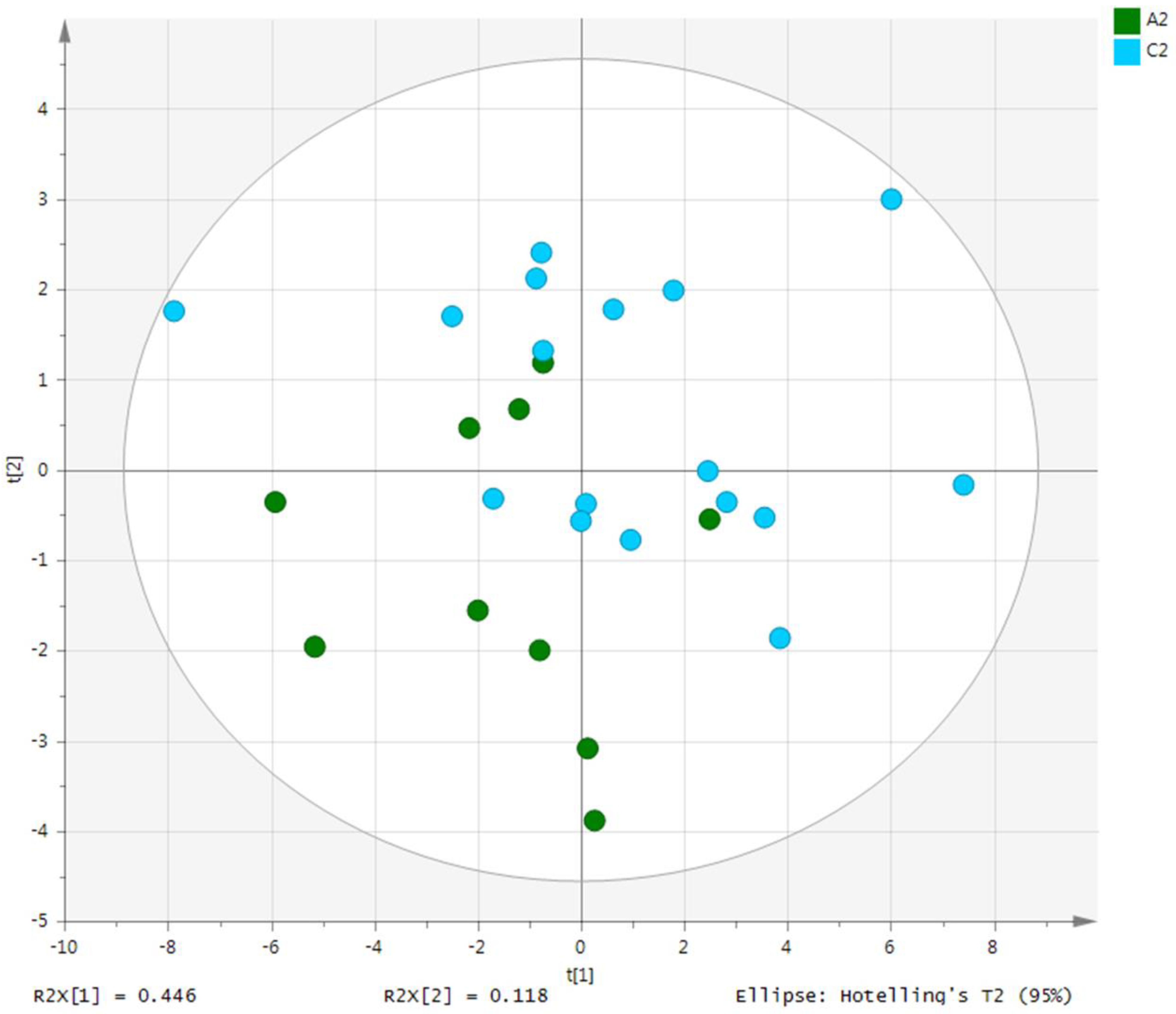
3.2. Metabolite Analysis
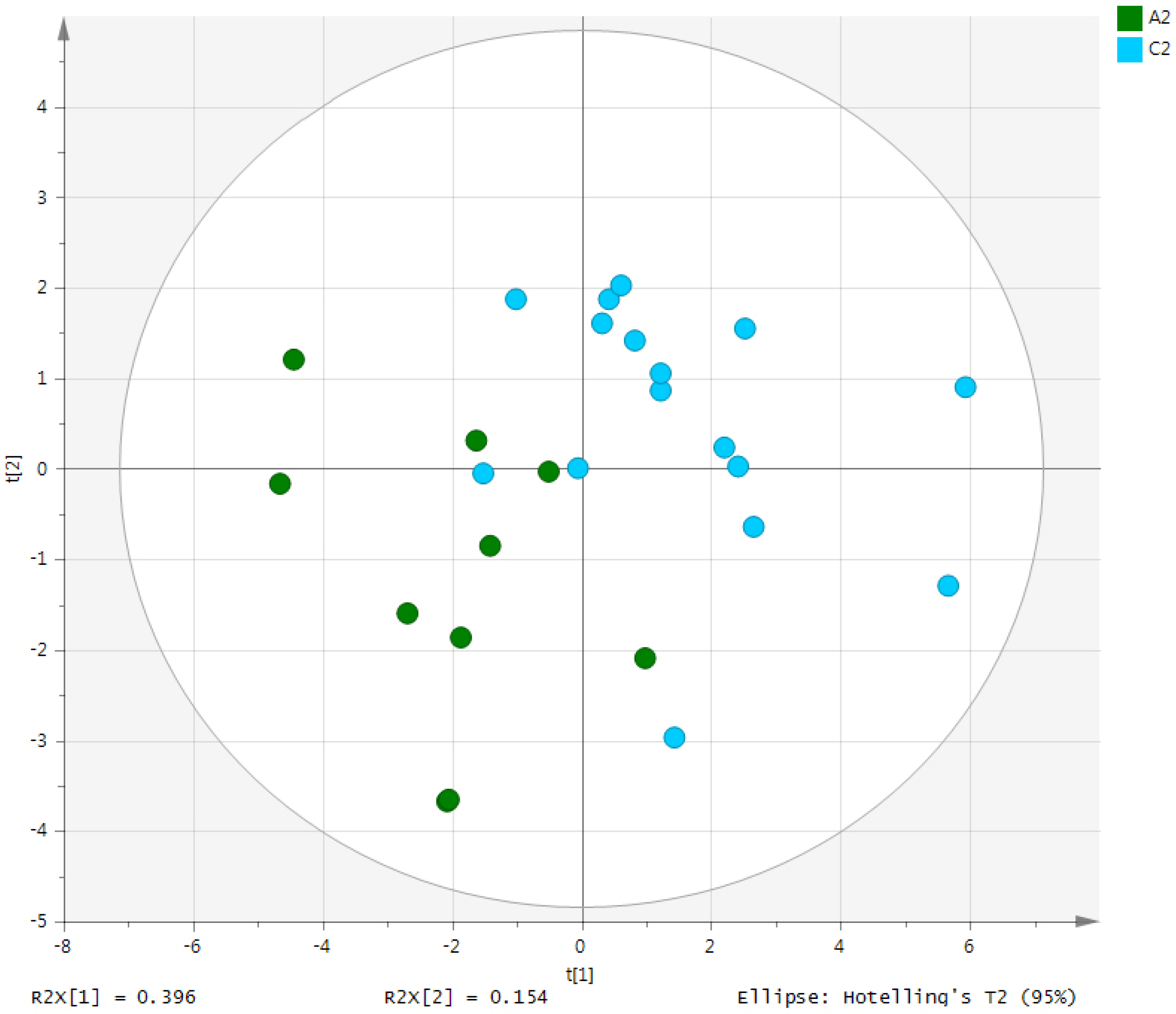
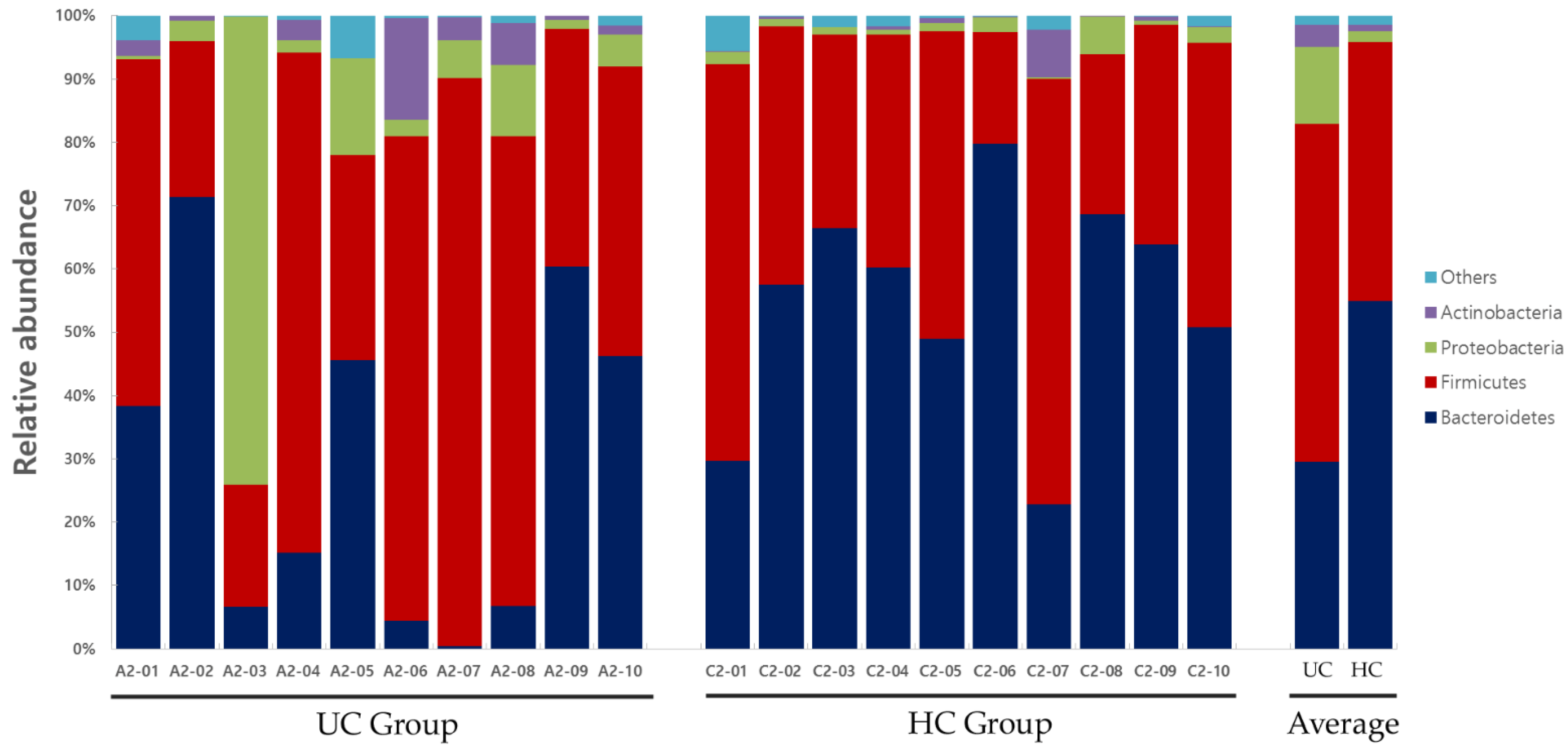
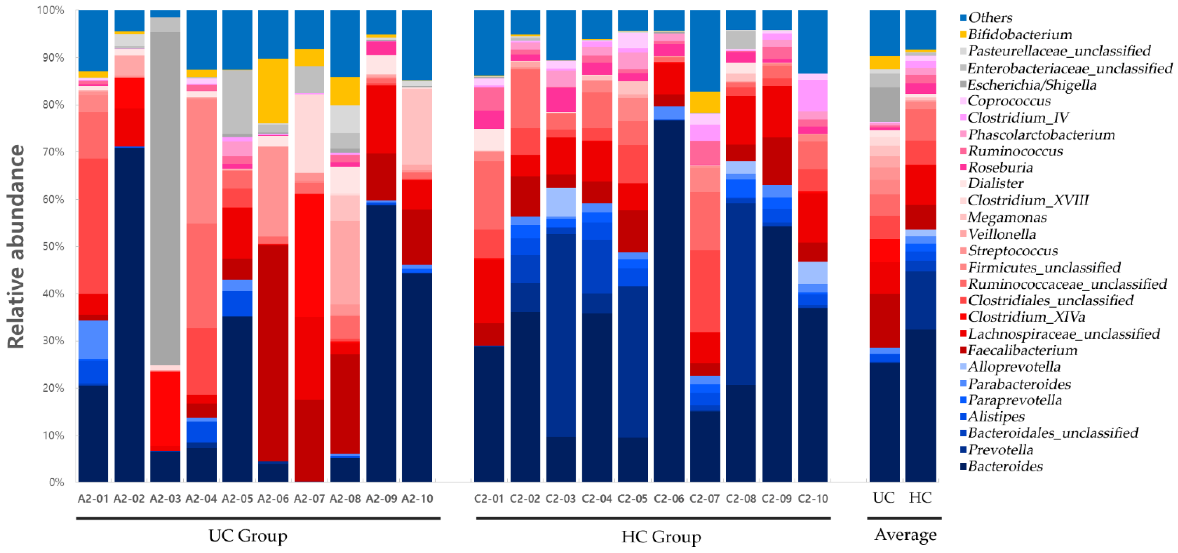
3.3. Gut Microbe Analysis
4. Discussion
Author Contributions
Funding
Institutional Review Board Statement
Informed Consent Statement
Data Availability Statement
Conflicts of Interest
References
- Reiff, C.; Kelly, D. Inflammatory Bowel Disease, Gut Bacteria and Probiotic Therapy. Int. J. Med. Microbiol. 2010, 300, 25–33. [Google Scholar] [CrossRef] [PubMed]
- Schee Genannt Halfmann, S.; Mählmann, L.; Leyens, L.; Reumann, M.; Brand, A. Personalized Medicine: What’s in It for Rare Diseases? Adv. Exp. Med. Biol. 2017, 1031, 387–404. [Google Scholar] [CrossRef]
- Czerska, I.; Skweres-Kuchta, M. Integrative Medicine as a New Treatment Model and the Future of Health Care Systems in the World in the Context of Rare Diseases. Eur. Res. Stud. 2021, 24, 800–809. [Google Scholar] [CrossRef]
- Schork, N.J.; Nazor, K. Integrated Genomic Medicine: A Paradigm for Rare Diseases and Beyond. Adv. Genet. 2017, 97, 81–113. [Google Scholar] [CrossRef] [PubMed]
- Gyngell, C.; Bowman-Smart, H.; Savulescu, J. Moral Reasons to Edit the Human Genome: Picking up from the Nuffield Report. J. Med. Ethics. 2019, 45, 514–523. [Google Scholar] [CrossRef] [PubMed]
- Ridaura, V.K.; Faith, J.J.; Rey, F.E.; Cheng, J.; Duncan, A.E.; Kau, A.L.; Griffin, N.W.; Lombard, V.; Henrissat, B.; Bain, J.R.; et al. Gut Microbiota from Twins Discordant for Obesity Modulate Metabolism in Mice. Science 2013, 341, 1241214. [Google Scholar] [CrossRef] [PubMed]
- Wink, M. Modes of Action of Herbal Medicines and Plant Secondary Metabolites. Medicines 2015, 2, 251–286. [Google Scholar] [CrossRef] [PubMed]
- An, X.; Bao, Q.; Di, S.; Zhao, Y.; Zhao, S.; Zhang, H.; Lian, F.; Tong, X. The Interaction between the Gut Microbiota and Herbal Medicines. Biomed. Pharmacother. 2019, 118, 109252. [Google Scholar] [CrossRef]
- Fan, Y.; Pedersen, O. Gut Microbiota in Human Metabolic Health and Disease. Nat. Rev. Microbiol. 2021, 19, 55–71. [Google Scholar] [CrossRef] [PubMed]
- Lavelle, A.; Sokol, H. Gut Microbiota-Derived Metabolites as Key Actors in Inflammatory Bowel Disease. Nat. Rev. Gastroenterol. Hepatol. 2020, 17, 223–237. [Google Scholar] [CrossRef] [PubMed]
- Zitomersky, N.L.; Atkinson, B.J.; Franklin, S.W.; Mitchell, P.D.; Snapper, S.B.; Comstock, L.E.; Bousvaros, A. Characterization of Adherent Bacteroidales from Intestinal Biopsies of Children and Young Adults with Inflammatory Bowel Disease. PLoS ONE 2013, 8, e63686. [Google Scholar] [CrossRef] [PubMed]
- Marchesi, J.R.; Holmes, E.; Khan, F.; Kochhar, S.; Scanlan, P.; Shanahan, F.; Wilson, I.D.; Wang, Y. Rapid and Noninvasive Metabonomic Characterization of Inflammatory Bowel Disease. J. Proteome Res. 2007, 6, 546–551. [Google Scholar] [CrossRef] [PubMed]
- Aldars-García, L.; Chaparro, M.; Gisbert, J.P. Systematic Review: The Gut Microbiome and Its Potential Clinical Application in Inflammatory Bowel Disease. Microorganisms 2021, 9, 977. [Google Scholar] [CrossRef] [PubMed]
- Kieser, M.; Wassmer, G. On the Use of the Upper Confidence Limit for the Variance from a Pilot Sample for Sample Size Determination. Biom. J. 1996, 38, 941–949. [Google Scholar] [CrossRef]
- Pietzke, M.; Meiser, J.; Vazquez, A. Formate Metabolism in Health and Disease. Mol. Metab. 2020, 33, 23–37. [Google Scholar] [CrossRef] [PubMed]
- Faber, F.; Bäumler, A.J. The Impact of Intestinal Inflammation on the Nutritional Environment of the Gut Microbiota. Immunol. Lett. 2014, 162, 48–53. [Google Scholar] [CrossRef] [PubMed]
- Baker, P.R.; Cramer, S.D.; Kennedy, M.; Assimos, D.G.; Holmes, R.P. Glycolate and Glyoxylate Metabolism in HepG2 Cells. Am. J. Physiol. Cell Physiol. 2004, 287, C1359–C1365. [Google Scholar] [CrossRef]
- Marengo, S.R.; Romani, A.M. Oxalate in Renal Stone Disease: The Terminal Metabolite That Just Won’t Go Away. Nat. Clin. Pract. Nephrol. 2008, 4, 368–377. [Google Scholar] [CrossRef]
- Caudarella, R.; Rizzoli, E.; Pironi, L.; Malavolta, N.; Martelli, G.; Poggioli, G.; Gozzetti, G.; Miglioli, M. Renal Stone Formation in Patients with Inflammatory Bowel Disease. Scanning Microsc. 1993, 7, 371–379, discussion 379. [Google Scholar]
- Chhibber-Goel, J.; Gaur, A.; Singhal, V.; Parakh, N.; Bhargava, B.; Sharma, A. The Complex Metabolism of Trimethylamine in Humans: Endogenous and Exogenous Sources. Expert Rev. Mol. Med. 2016, 18, e8. [Google Scholar] [CrossRef] [PubMed]
- Papa, A.; Danese, S.; Urgesi, R.; Grillo, A.; Guglielmo, S.; Roberto, I.; Bonizzi, M.; Guidi, L.; De Vitis, I.; Santoliquido, A.; et al. Early Atherosclerosis in Patients with Inflammatory Bowel Disease. Eur. Rev. Med. Pharmacol. Sci. 2006, 10, 7–11. [Google Scholar] [PubMed]
- Murín, R.; Mohammadi, G.; Leibfritz, D.; Hamprecht, B. Glial Metabolism of Valine. Neurochem. Res. 2009, 34, 1195–1203. [Google Scholar] [CrossRef]
- Lupton, J.R.; Brooks, J.; Butte, N.; Caballero, B.; Flatt, J.; Fried, S. Dietary Reference Intakes for Energy, Carbohydrate, Fiber, Fat, Fatty Acids, Cholesterol, Protein, and Amino Acids; National Academy Press: Washington, DC, USA, 2002; Volume 5, pp. 589–768. [Google Scholar]
- Gray, L.R.; Tompkins, S.C.; Taylor, E.B. Regulation of Pyruvate Metabolism and Human Disease. Cell. Mol. Life Sci. 2014, 71, 2577–2604. [Google Scholar] [CrossRef] [PubMed]
- Trompette, A.; Gollwitzer, E.S.; Yadava, K.; Sichelstiel, A.K.; Sprenger, N.; Ngom-Bru, C.; Blanchard, C.; Junt, T.; Nicod, L.P.; Harris, N.L.; et al. Gut Microbiota Metabolism of Dietary Fiber Influences Allergic Airway Disease and Hematopoiesis. Nat. Med. 2014, 20, 159–166. [Google Scholar] [CrossRef] [PubMed]
- Xu, M.; Jiang, Z.; Wang, C.; Li, N.; Bo, L.; Zha, Y.; Bian, J.; Zhang, Y.; Deng, X. Acetate attenuates inflammasome activation through GPR43-mediated Ca2+-dependent NLRP3 ubiquitination. Exp Mol. Med. 2019, 51, 1–13. [Google Scholar] [CrossRef] [PubMed]
- Sara, D.; Kathleen, M.; Jeroen, R.; Kristin, V.; Séverine, V. Short chain fatty acids and its producing organisms: An overlooked therapy for IBD? EBioMedicine 2021, 66, 103293. [Google Scholar] [CrossRef]
- Milan, H. Histidine in Health and Disease: Metabolism, Physiological Importance, and Use as a Supplement. Nutrients 2020, 12, 848. [Google Scholar] [CrossRef]
- Wang, D.; Ma, X.; Guo, S.; Wang, Y.; Li, T.; Zou, D.; Song, H.; Yang, W.; Ge, Y. Effect of Huangqin Tang on Urine Metabolic Profile in Rats with Ulcerative Colitis Based on UPLC-Q-Exactive Orbitrap MS. Evid. Based Complement. Altern. Med. 2020, 2020, 1874065. [Google Scholar] [CrossRef]
- Kim, C.H.; Jung, J.; Lee, Y.U.; Kim, K.H.; Kang, S.; Kang, G.H.; Chu, H.; Kim, S.Y.; Lee, S. Comparison of Metabolites and Gut Microbes between Patients with Parkinson’s Disease and Healthy Individuals-A Pilot Clinical Observational Study (STROBE Compliant). Healthcare 2022, 10, 302. [Google Scholar] [CrossRef] [PubMed]
- Coyne, M.J.; Comstock, L.E. Niche-Specific Features of the Intestinal Bacteroidales. J. Bacteriol. 2008, 190, 736–742. [Google Scholar] [CrossRef]
- Waksman, S.A.; Schatz, A.; Reynolds, D.M. Production of Antibiotic Substances by Actinomycetes. Ann. N. Y. Acad. Sci. 2010, 1213, 112–124. [Google Scholar] [CrossRef]
- Ganji, L.; Alebouyeh, M.; Shirazi, M.H.; Eshraghi, S.S.; Mirshafiey, A.; Ebrahimi Daryani, N.; Zali, M.R. Dysbiosis of Fecal Microbiota and High Frequency of Citrobacter, Klebsiella spp., and Actinomycetes in Patients with Irritable Bowel Syndrome and Gastroenteritis. Gastroenterol. Hepatol. Bed Bench. 2016, 9, 325–330. [Google Scholar]
- Yap, G.; Hong, P.; Lee, B. Microflora of the Intestine. Encycl. Food Microbiol. 2014, 2, 634–665. [Google Scholar]
- Chatterjee, K.; Banerjee, S. Microbiome and Motor Neuron Diseases. Prog. Mol. Biol. Transl. Sci. 2020, 176, 111–122. [Google Scholar] [CrossRef]
- Liu, B.; Piao, X.; Niu, W.; Zhang, Q.; Ma, C.; Wu, T.; Gu, Q.; Cui, T.; Li, S. Kuijieyuan Decoction Improved Intestinal Barrier Injury of Ulcerative Colitis by Affecting TLR4-Dependent PI3K/AKT/NF-κB Oxidative and Inflammatory Signaling and Gut Microbiota. Front. Pharmacol. 2020, 11, 1036. [Google Scholar] [CrossRef]
- Acharya, C.; Bajaj, J.S. Altered Microbiome in Patients with Cirrhosis and Complications. Clin. Gastroenterol. Hepatol. 2019, 17, 307–321. [Google Scholar] [CrossRef]
- Forbes, J.D.; Van Domselaar, G.; Bernstein, C.N. The Gut Microbiota in Immune-Mediated Inflammatory Diseases. Front. Microbiol. 2016, 7, 1081. [Google Scholar] [CrossRef]
- Morgan, X.C.; Tickle, T.L.; Sokol, H.; Gevers, D.; Devaney, K.L.; Ward, D.V.; Reyes, J.A.; Shah, S.A.; LeLeiko, N.; Snapper, S.B.; et al. Dysfunction of the Intestinal Microbiome in Inflammatory Bowel Disease and Treatment. Genome Biol. 2012, 13, R79. [Google Scholar] [CrossRef]
- Eisenberg, T.; Fawzy, A.; Nicklas, W.; Semmler, T.; Ewers, C. Phylogenetic and Comparative Genomics of the Family Leptotrichiaceae and Introduction of a Novel Fingerprinting MLVA for Streptobacillus moniliformis. BMC Genom. 2016, 17, 864. [Google Scholar] [CrossRef]
- Larsen, J.M. The Immune Response to Prevotella Bacteria in Chronic Inflammatory Disease. Immunology 2017, 151, 363–374. [Google Scholar] [CrossRef]
- Lewis, J.D.; Chen, E.Z.; Baldassano, R.N.; Otley, A.; Griffiths, A.; Lee, D.; Bittinger, K.; Bailey, A.; Friedman, E.; Hoffmann, C.; et al. Inflammation, Antibiotics, and Diet as Environmental Stressors of the Gut Microbiome in Pediatric Crohn’s Disease. Cell Host Microbe 2015, 18, 489–500. [Google Scholar] [CrossRef]
- Zhu, C.; Song, K.; Shen, Z.; Quan, Y.; Tan, B.; Luo, W.; Wu, S.; Tang, K.; Yang, Z.; Wang, X. Roseburia intestinalis Inhibits Interleukin-17 Excretion and Promotes Regulatory T Cells Differentiation in Colitis. Mol. Med. Rep. 2018, 17, 7567–7574. [Google Scholar] [CrossRef]
- Mills, E.; O’Neill, L.A. Succinate: A Metabolic Signal in Inflammation. Trends Cell Biol. 2014, 24, 313–320. [Google Scholar] [CrossRef] [PubMed]
- Yang, H.; Meng, L.; Ai, D.; Hou, N.; Li, H.; Shuai, X.; Peng, X. Acetic Acid Alleviates the Inflammatory Response and Liver Injury in Septic Mice by Increasing the Expression of TRIM40. Exp. Ther. Med. 2019, 17, 2789–2798. [Google Scholar] [CrossRef]
- Knights, D.; Lassen, K.G.; Xavier, R.J. Advances in Inflammatory Bowel Disease Pathogenesis: Linking Host Genetics and the Microbiome. Gut 2013, 62, 1505–1510. [Google Scholar] [CrossRef]
- Tedelind, S.; Westberg, F.; Kjerrulf, M.; Vidal, A. Anti-Inflammatory Properties of the Short-Chain Fatty Acids Acetate and Propionate: A Study with Relevance to Inflammatory Bowel Disease. World J. Gastroenterol. 2007, 13, 2826–2832. [Google Scholar] [CrossRef]
- Nagao-Kitamoto, H.; Kamada, N. Host-Microbial Cross-Talk in Inflammatory Bowel Disease. Immune Netw. 2017, 17, 1–12. [Google Scholar] [CrossRef]
- La Reau, A.J.; Suen, G. The Ruminococci: Key Symbionts of the Gut Ecosystem. J. Microbiol. 2018, 56, 199–208. [Google Scholar] [CrossRef]
- Shaw, K.A.; Bertha, M.; Hofmekler, T.; Chopra, P.; Vatanen, T.; Srivatsa, A.; Prince, J.; Kumar, A.; Sauer, C.; Zwick, M.E.; et al. Dysbiosis, Inflammation, and Response to Treatment: A Longitudinal Study of Pediatric Subjects with Newly Diagnosed Inflammatory Bowel Disease. Genome Med. 2016, 8, 75. [Google Scholar] [CrossRef]
- Bernstein, C.N.; Forbes, J.D. Gut Microbiome in Inflammatory Bowel Disease and Other Chronic Immune-Mediated Inflammatory Diseases. Inflamm. Intest. Dis. 2017, 2, 116–123. [Google Scholar] [CrossRef]
- Carretta, M.D.; Quiroga, J.; López, R.A.; Hidalgo, M.A.; Burgos, R.A. Participation of Short-Chain Fatty Acids and Their Receptors in Gut Inflammation and Colon Cancer. Front. Physiol. 2021, 12, 662739. [Google Scholar] [CrossRef] [PubMed]
- Labus, J.S.; Osadchiy, V.; Hsiao, E.Y.; Tap, J.; Derrien, M.; Gupta, A.; Tillisch, K.; Le Nevé, B.; Grinsvall, C.; Ljungberg, M.; et al. Evidence for an Association of Gut Microbial Clostridia with Brain Functional Connectivity and Gastrointestinal Sensorimotor Function in Patients with Irritable Bowel Syndrome, Based on Tripartite Network Analysis. Microbiome 2019, 7, 45. [Google Scholar] [CrossRef]
- Jørandli, J.W.; Thorsvik, S.; Skovdahl, H.K.; Kornfeld, B.; Sæterstad, S.; Gustafsson, B.I.; Sandvik, A.K.; van Beelen Granlund, A. The Serotonin Reuptake Transporter Is Reduced in the Epithelium of Active Crohn’s Disease and Ulcerative Colitis. Am. J. Physiol. Gastrointest. Liver Physiol. 2020, 319, G761–G768. [Google Scholar] [CrossRef] [PubMed]
- Qi, Y.; Zang, S.Q.; Wei, J.; Yu, H.C.; Yang, Z.; Wu, H.M.; Kang, Y.; Tao, H.; Yang, M.F.; Jin, L.; et al. High-Throughput Sequencing Provides Insights into Oral Microbiota Dysbiosis in Association with Inflammatory Bowel Disease. Genomics 2021, 113, 664–676. [Google Scholar] [CrossRef] [PubMed]
- van Kessel, S.P.; El Aidy, S. Bacterial Metabolites Mirror Altered Gut Microbiota Composition in Patients with Parkinson’s Disease. J. Parkinsons Dis. 2019, 9, S359–S370. [Google Scholar] [CrossRef]
- Wang, H.; Ye, J. Regulation of Energy Balance by Inflammation: Common Theme in Physiology and Pathology. Rev. Endocr. Metab. Disord. 2015, 16, 47–54. [Google Scholar] [CrossRef]
- Sugito, R.; Son, D. Obstacles to the Use of Complementary and Alternative Medicine by Primary Care Physicians: Preliminary Study. Trad. Kampo Med. 2019, 6, 173–177. [Google Scholar] [CrossRef]
- Suzuki, Y.; Yoshimura, N.; Saniabadi, A.R.; Saito, Y. Selective Granulocyte and Monocyte Adsorptive Apheresis as a First-Line Treatment for Steroid Naïve Patients with Active Ulcerative Colitis: A Prospective Uncontrolled Study. Dig. Dis. Sci. 2004, 49, 565–571. [Google Scholar] [CrossRef]
- Seo, S.H. Effect of Jakyakgamcho-Tang on Inflammatory Bowel Disease Using GC/MS-Based Metabolic Profiling Analysis. Ph.D. Dissertation, Dongshin University, Naju-Si, Korea, 2019. Available online: http://www.riss.kr/link?id=T15092756&outLink=K (accessed on 12 December 2018).


| Classification | UC Group | HC Group | p Value | |
|---|---|---|---|---|
| Total | 10 | 10 | ||
| Sex | Male | 5 | 5 | p > 0.05 |
| Female | 5 | 5 | ||
| Age (years) | Minimum | 33 | 33 | p > 0.05 |
| Maximum | 77 | 72 | ||
| Average | 59.4 | 53.9 | ||
| Disease duration (years) | Minimum | 2 | - | |
| Maximum | 18 | - | ||
| Average | 9.4 | |||
| Comorbidities | Hypertension | 5 | - | |
| Dyslipidemia | 1 | - | ||
| Prostatic hypertrophy | 1 | - | ||
| None | 5 | 10 | ||
| Active ingredients in the medications taken | Mesalazine | 8 | - | |
| Sulfasalazine | 2 | - | ||
| Rebamipide | 6 | - | ||
| Pinaverium bromide | 4 | - | ||
| Itopride hydrochloride | 3 | - | ||
| Mosapride citrate hydrate | 1 | - | ||
| Telmisartan | 1 | - | ||
| Amlodipine besylate | 2 | - | ||
| Losartan potassium | 2 | - | ||
| Carvedilol | 1 | - | ||
| Olmesartan medoxomil | 1 | - | ||
| Atorvastatin calcium trihydrate | 1 | - | ||
| Finasteride | 1 | |||
| None | 0 | 10 | ||
| Classification | Gut Microbes | UC Group vs. HC Group | ||
|---|---|---|---|---|
| ↑/↓ § | Significance (p < 0.05) | |||
| Stool | Phylum level | Bacteroidetes | ↓ | 0.022 |
| Class level | Bacteroidia | ↓ | 0.023 | |
| Order level | Bacteroidales | ↓ | 0.023 | |
| Actinomycetales | ↑ | 0.044 | ||
| Family level | Prevotellaceae | ↓ | 0.020 | |
| Acidaminococcaceae | ↓ | 0.015 | ||
| Leptotrichiaceae | ↑ | 0.025 | ||
| Genus level | Prevotella | ↓ | 0.049 | |
| Roseburia | ↓ | 0.016 | ||
| Paraprevotella | ↓ | 0.011 | ||
| Phascolarctobacterium | ↓ | 0.016 | ||
| Ruminococcus | ↓ | 0.015 | ||
| Coprococcus | ↓ | 0.028 | ||
| Clostridium_XIVB | ↓ | 0.049 | ||
| Atopobium | ↓ | 0.015 | ||
| Leptotrichia | ↑ | 0.038 | ||
| Classification | Description | ||
|---|---|---|---|
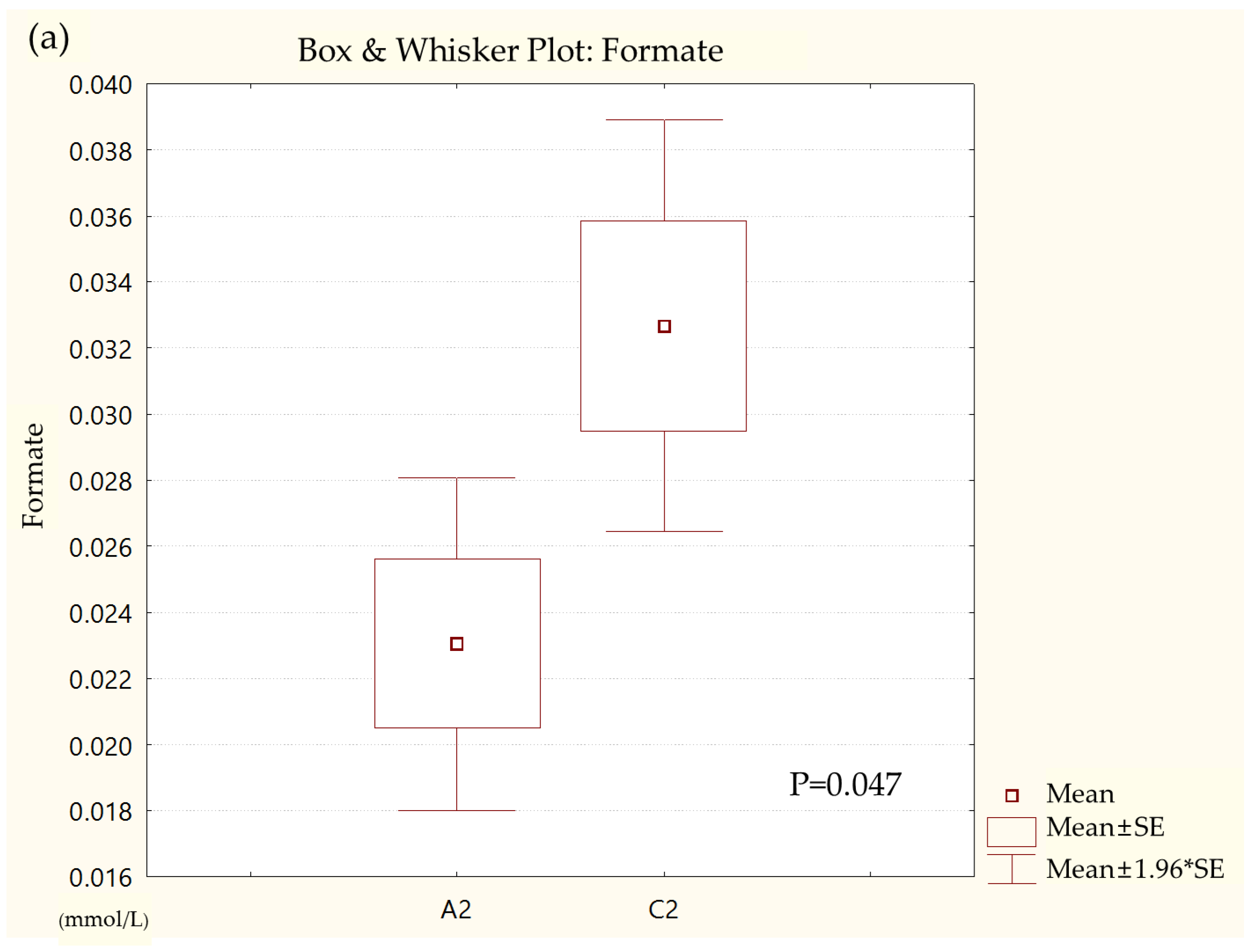
| Metabolites | Formate | Formate is associated with glucose-lactate metabolism. Immunologically, it is related to the decline of naïve T cells [15]. Formate also plays a role in producing energy through anaerobic respiration as an electron donor [16]. | |
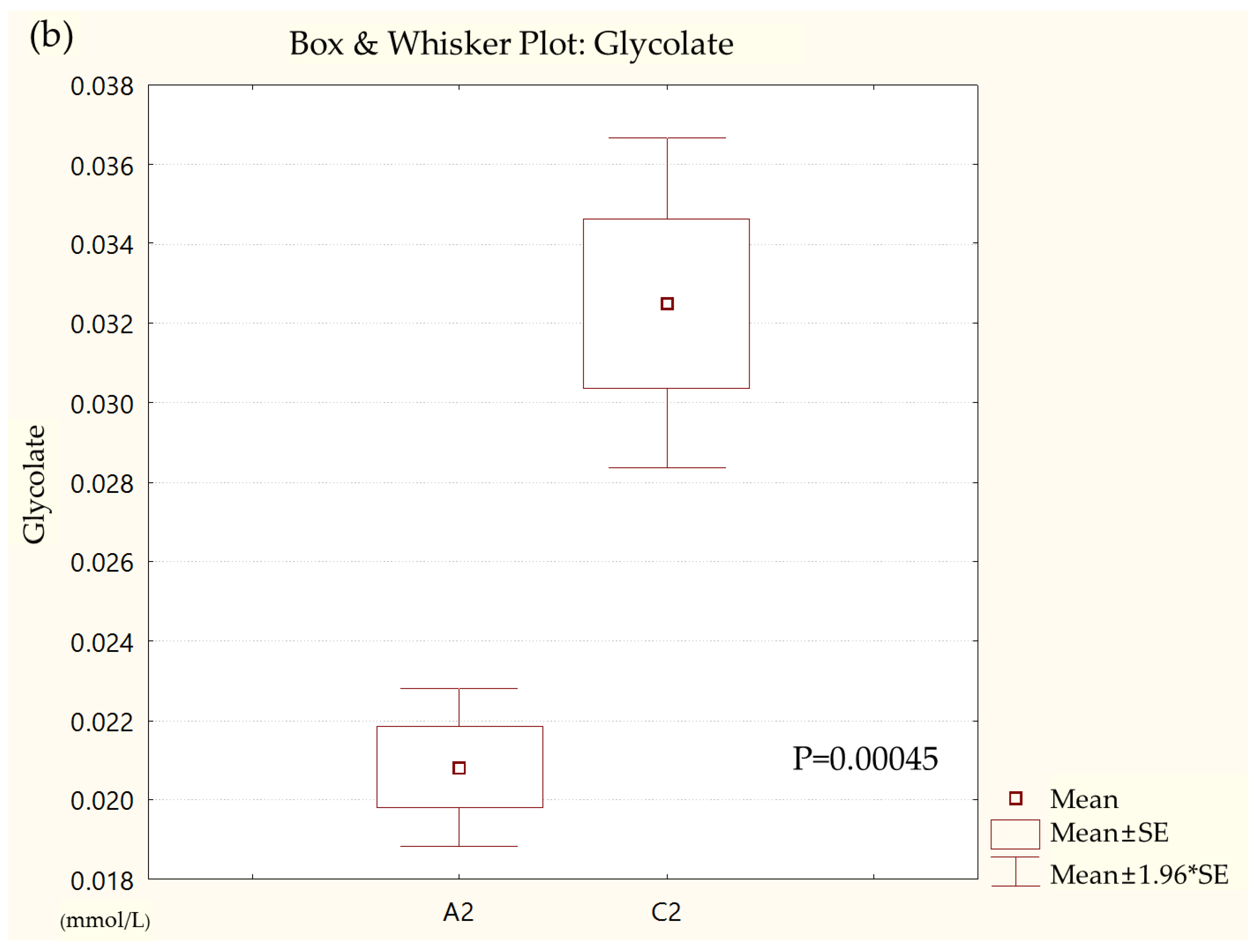
| Glycolate | Glycolate is a major precursor to oxalate [17], which is closely related to stone disease [18], and according to a report by Caudarella et al., stone disease occurs more commonly in patients with IBD [19]. | ||
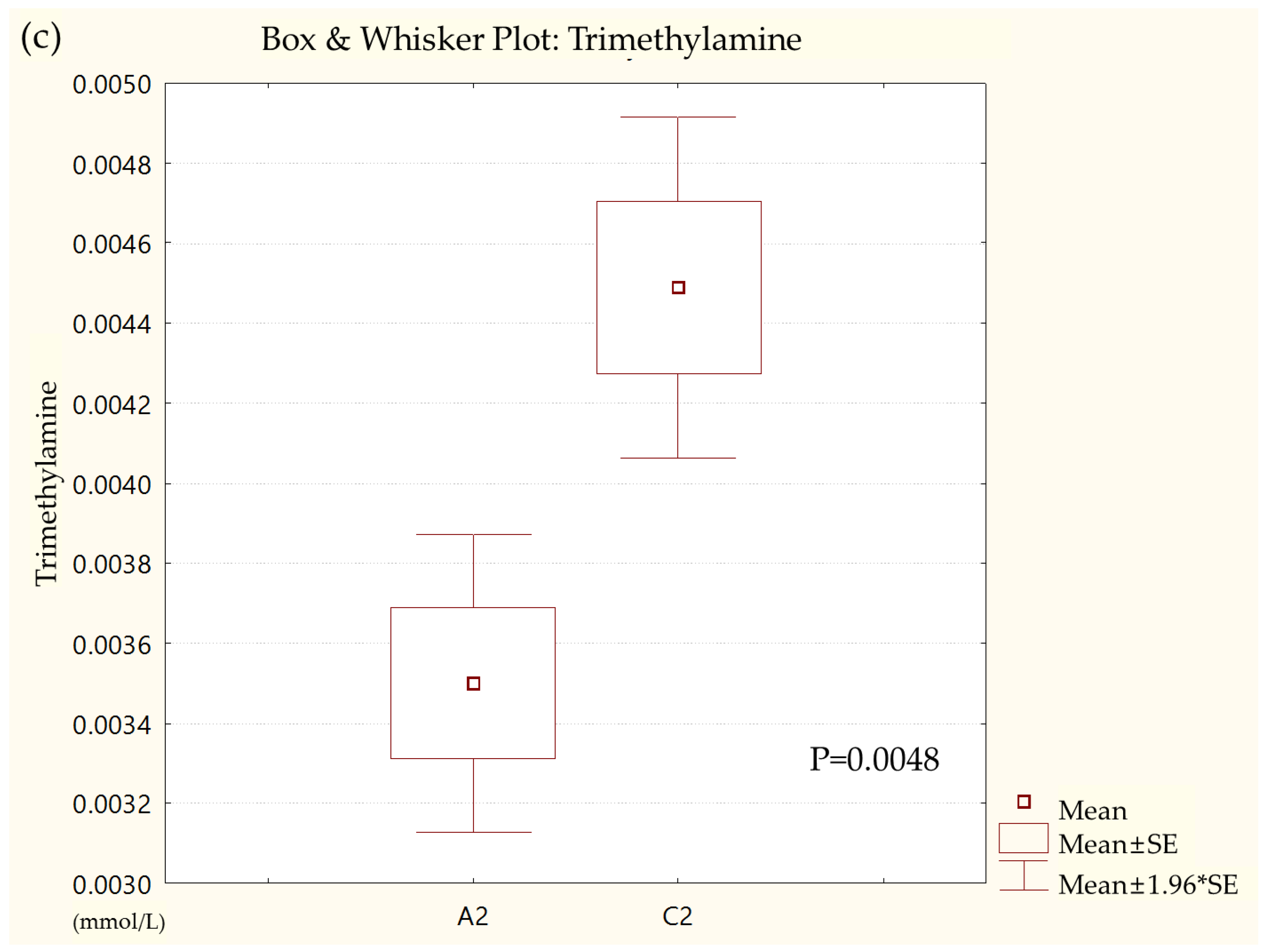
| Trimethylamine | Trimethylamine is caused by the intestinal degradation of dietary constituents such as choline and carnitine by microbial enzymes [20]. Trimethylamine is also a precursor to trimethylamine-N-oxide, which is associated with the risk of athero-thrombogenesis [20]. According to a study by Alfredo et al., IBD is closely associated with the risk of thrombotic complications [21]. Marchesi et al. also analyzed the metabolites of patients with IBD through fecal samples and found a decrease in trimethylamine, which is consistent with our study [12]. | ||
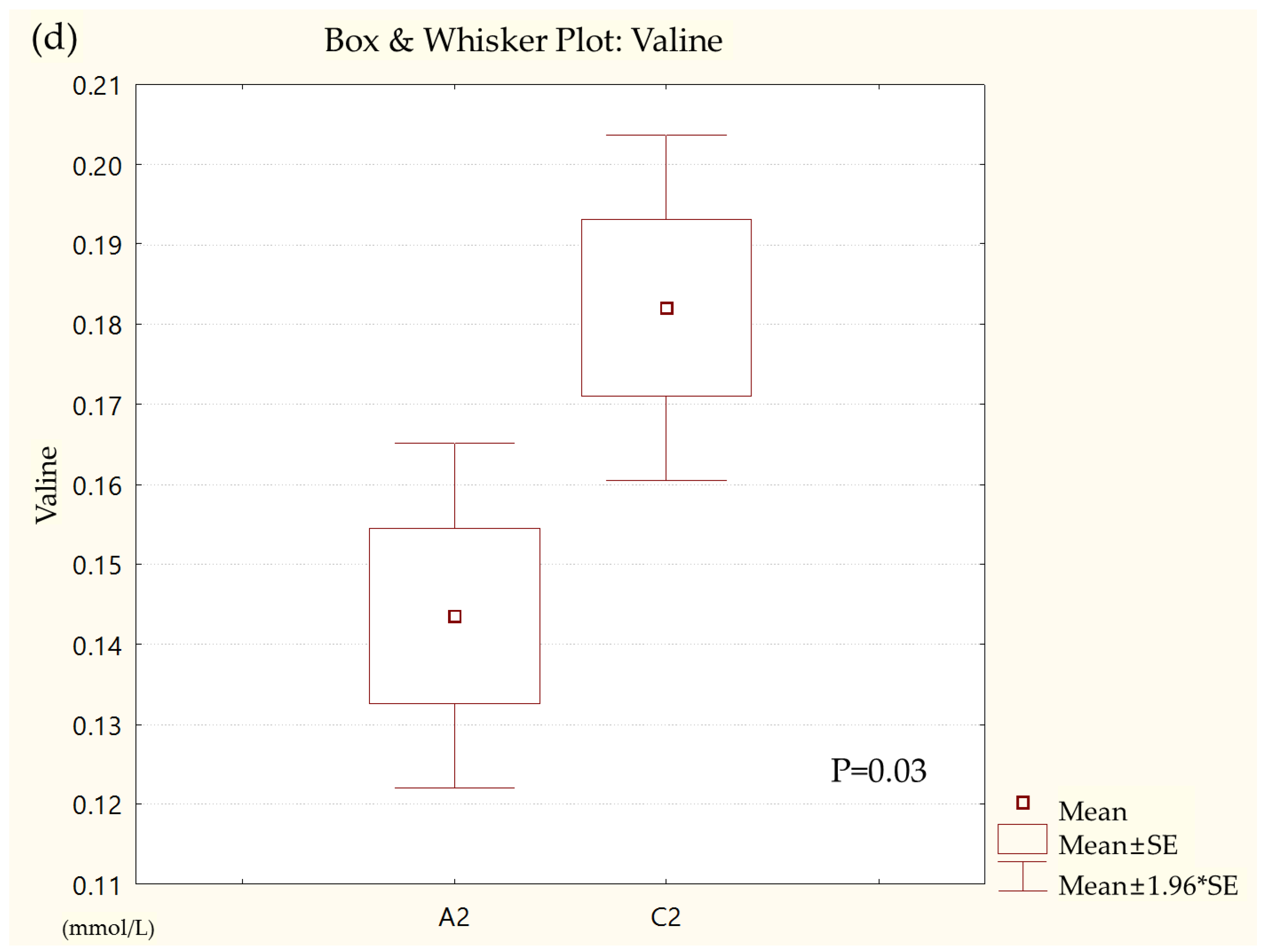
| Valine | Valine is a minor substrate of brain energy metabolism. During glutamatergic signaling, valine metabolism appears to be particularly crucial in the process of glutamate translocation between astrocytes and neurons [22]. Valine is an essential amino acid in animals, including humans, and must be ingested into the diet [23]. | ||
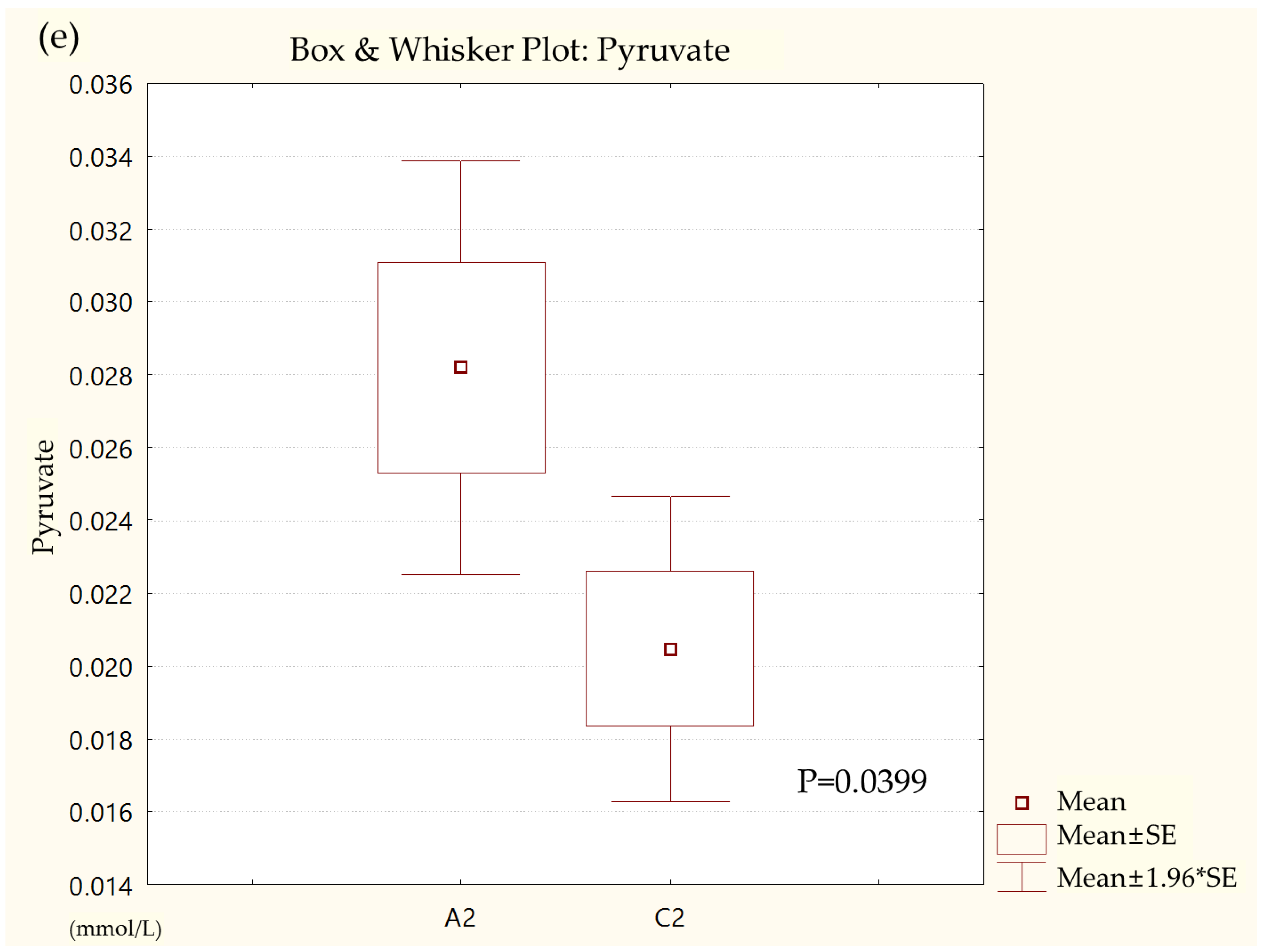
| Pyruvate | Pyruvate is the end-product of glycolysis. Abnormal pyruvate metabolism plays an especially prominent role in cancer, heart failure, and neurodegeneration. It is also associated with chronic obstructive pulmonary disease, obesity, diabetes, and aging [24]. | ||
| Acetate | Acetate is a short-chain fatty acid (SCFA) produced by gut microbes, which regulates inflammation in inflammatory and metabolic diseases [25]. Deleu et al. reported that SCFAs, including acetate, are closely related to IBD [26]. | ||
| τ-Methylhistidine | τ-Methylhistidine is associated with the degradation of intestinal proteins [27]. Wang et al. suggested that τ-methylhistidine is one of the potential biomarkers for ulcerative colitis [28]. | ||
| Gut microbes | Phylum level | Bacteroidetes | Bacteroidetes are known to produce anti-inflammatory metabolites such as SCFAs [29]. Our research team has previously confirmed that Bacteroidetes levels are lower in patients with Parkinson’s disease than in healthy individuals, which is related to neuroinflammation [30]. |
| Class level | Bacteroidia | Bacteroidia dominate microbial communities inhabiting the anaerobic environment of the lower gastrointestinal tract. Metabolic end products generated by Bacteroidia change the nutritional environment for both the host and other intestinal microbes. Formate, which was significant in the results of this study, is also a metabolic end product of Bacteroidia [16]. | |
| Order level | Bacteroidales | Bacteroidales have been found to modulate host immunological and intestinal activities such as mucosal barrier fortification, intestinal immune maturation, and angiogenesis by occupying a vital niche at the mucosal surface of the intestine. Bacteroidales species can have positive or harmful effects on their hosts, depending on their genetic content. In patients with IBD, more severe inflammation has been correlated with lower Bacteroidales diversity [31]. | |
| Actinomycetales | Many Actinomycetales found in natural substrates can prevent bacteria and other microbes from growing [32]. In one study, Actinomycetales were higher in patients with irritable bowel syndrome than in normal subjects [33]. Based on these studies, the decreased intestinal microbial diversity in patients with IBD may be related to the abundance of Actinomycetales. | ||
| Family level | Prevotellaceae | The Prevotellaceae family is associated with antibiotic biosynthesis and the transport of secondary metabolites [34]. Generally, Prevotellaceae produce SCFAs through the fermentation of dairy products. Reduced SCFAs cause increased gut permeability, which exposes the intestine to bacterial endotoxins [35]. In another study, the number of Prevotellaceae and Prevotella was significantly lower in patients with UC than in controls [36]. | |
| Acidaminococcaceae | The family Acidaminococcaceae is now called Veillonellaceae. The Veillonellaceae family is implicated in regulating systemic inflammation [37] and is therefore presumed to be closely related to immune-mediated inflammatory disease, including IBD [38]. In one study, it was suggested that Veillonellaceae might be a gut microbe closely related to IBD [39]. | ||
| Leptotrichiaceae | Leptotrichiaceae generally inhabit mucous membranes, but when introduced into different tissue or host sites, they can shift their pathogenic potential and produce severe and even life-threatening disease, according to their phylotypes [40]. | ||
| Genus level | Prevotella | Prevotella is thought to be closely related to chronic inflammation [41], with one study reporting a reduction in Prevotella in pediatric patients with Crohn’s disease [42]. | |
| Roseburia | Roseburia, one of the most common gut microbes, is decreased in patients with IBD. It helps to protect the mucosa of the colon from inflammation and subsequent IBD. Therefore, Roseburia could be a candidate for IBD treatment [43]. | ||
| Paraprevotella | The primary fermentation products of Paraprevotella are succinic acid and acetic acid, which are associated with inflammation. Acetic acid is especially known to alleviate inflammation [44,45]. | ||
| Phascolarctobacterium | Phascolarctobacterium is already known to be associated with IBD. These bacteria are presumed to produce propionate, which has been found to have anti-inflammatory properties [46,47]. | ||
| Ruminococcus | Ruminococcus has been associated with intestinal inflammation and is less abundant in patients with IBD [48]. Ruminococcus help their hosts degrade and convert complex polysaccharides into various nutrients [49]. | ||
| Coprococcus | The association of Coprococcus with IBD has long been reported. Agglutinating antibodies for Coprococcus were briefly considered a biomarker for IBD [50]. In autoimmune diseases, the relative abundance of Coprococcus is lower, and the guts of patients with an autoimmune disease have been characterized by a reduction in microbes, which is positively correlated with heptanoate and hexanoate [51]. Heptanoate and hexanoate belong to SCFAs and are involved in the inflammation process [52]. | ||
| Clostridium_XIVB | The genus Clostridium, including Clostridium_XIVB, plays a role in modulating the biosynthesis and release of serotonin [53]. The majority of serotonin is produced in the gastrointestinal epithelium, where it is suggested to act as a prominent regulatory molecule in the IBD [54]. | ||
| Atopobium Leptotrichia | Atopobium and Leptotrichia are oral microbes swallowed with saliva into the digestive tract. The dysbiosis of oral microbes, including Atopobium and Leptotrichia, can trigger gut microbe dysbiosis, leading to IBD [55]. | ||
Publisher’s Note: MDPI stays neutral with regard to jurisdictional claims in published maps and institutional affiliations. |
© 2022 by the authors. Licensee MDPI, Basel, Switzerland. This article is an open access article distributed under the terms and conditions of the Creative Commons Attribution (CC BY) license (https://creativecommons.org/licenses/by/4.0/).
Share and Cite
Kim, C.-H.; Lee, Y.-U.; Kim, K.-H.; Kang, S.; Kang, G.-H.; Chu, H.; Lee, S. Comparison of Metabolites and Gut Microbes between Patients with Ulcerative Colitis and Healthy Individuals for an Integrative Medicine Approach to Ulcerative Colitis—A Pilot Observational Clinical Study (STROBE Compliant). Diagnostics 2022, 12, 1969. https://doi.org/10.3390/diagnostics12081969
Kim C-H, Lee Y-U, Kim K-H, Kang S, Kang G-H, Chu H, Lee S. Comparison of Metabolites and Gut Microbes between Patients with Ulcerative Colitis and Healthy Individuals for an Integrative Medicine Approach to Ulcerative Colitis—A Pilot Observational Clinical Study (STROBE Compliant). Diagnostics. 2022; 12(8):1969. https://doi.org/10.3390/diagnostics12081969
Chicago/Turabian StyleKim, Cheol-Hyun, Young-Ung Lee, Kwang-Ho Kim, Sunny Kang, Geon-Hui Kang, Hongmin Chu, and Sangkwan Lee. 2022. "Comparison of Metabolites and Gut Microbes between Patients with Ulcerative Colitis and Healthy Individuals for an Integrative Medicine Approach to Ulcerative Colitis—A Pilot Observational Clinical Study (STROBE Compliant)" Diagnostics 12, no. 8: 1969. https://doi.org/10.3390/diagnostics12081969
APA StyleKim, C.-H., Lee, Y.-U., Kim, K.-H., Kang, S., Kang, G.-H., Chu, H., & Lee, S. (2022). Comparison of Metabolites and Gut Microbes between Patients with Ulcerative Colitis and Healthy Individuals for an Integrative Medicine Approach to Ulcerative Colitis—A Pilot Observational Clinical Study (STROBE Compliant). Diagnostics, 12(8), 1969. https://doi.org/10.3390/diagnostics12081969