Article

Cloning and Spatiotemporal Expression Analysis of IGF1R Gene cDNA in Alopex lagopus (Arctic Fox)

College of Animal Sciences and Technology, Northeast Agricultural University, Harbin 150030, China
*
Authors to whom correspondence should be addressed.
Life 2025, 15(5), 796; https://doi.org/10.3390/life15050796
Submission received: 14 April 2025 / Revised: 7 May 2025 / Accepted: 15 May 2025 / Published: 17 May 2025
(This article belongs to the Section Animal Science)

Abstract

This study aimed to clarify the sequence characteristics and spatiotemporal expression patterns of the insulin-like growth factor 1 receptor (IGF1R) gene in Alopex lagopus (Arctic fox), thereby addressing the existing knowledge gap regarding IGF1R-mediated growth regulation in this species. The findings establish a crucial foundation for subsequent investigations into the correlation between this gene and Arctic fox growth traits. Specific primers were designed based on the cDNA sequence of the canine IGF1R gene (Accession No. XM_545828). The full-length coding sequence (CDS) of the Arctic fox IGF1R gene (1617 bp, encoding 538 amino acids) was successfully cloned via RT-PCR. Phylogenetic analysis using the Unweighted Pair Group Method with Arithmetic Mean (UPGMA) algorithm revealed a 99% sequence homology in the IGF1R gene between the Arctic fox and canine, confirmed their closest evolutionary relationship. Protein characterization showed that the IGF1R protein has a molecular weight of 60.62 kDa (theoretical isoelectric point pI = 5.15), containing one fibronectin type-III domain and one tyrosine kinase domain, classifying it as an acidic hydrophilic transmembrane protein. Phosphorylation site prediction identified 27 phosphorylation sites, with secondary structures dominated by α-helices (26.39%) and random coils (52.79%). The IGF1R gene displayed significant tissue-specific expression variations across 12 examined tissues in Arctic foxes: highest expression levels in testis, minimal expression in stomach, and no detectable expression in duodenum. Spatiotemporal expression analysis revealed that in 2-, 4-, and 6-month-old individuals, hepatic IGF1R exhibited a progressive increase, testicular expression reached peak levels at 6 months, and skeletal muscle demonstrated transient upregulation peaking at 4 months. These spatiotemporal expression patterns suggest that IGF1R may participate in metabolism and organ developmental processes during critical growth stages of Arctic foxes through tissue-specific regulation.

1. Introduction

Alopex lagopus is a significant species in the global fur trade, prized for its exceptionally soft texture and pristine fur coloration, which holds irreplaceable value in luxury markets. Body size directly determines pelt surface area—the key grading criterion for commercial valuation—making the breeding of large-sized Arctic foxes a top priority. However, current research on growth regulation remains limited to phenotypic factors, like husbandry conditions (e.g., temperature/humidity) and nutritional management, while molecular genetic mechanisms remain underexplored, hindering the development of precision breeding technologies.
The insulin-like growth factor 1 receptor (IGF1R) serves as a central regulator in the vertebrate growth axis, driving cellular proliferation, differentiation, and organ development by mediation in the IGF-1/IGF-2 signaling pathway [1,2]. Functional studies demonstrate that IGF1R deficiency results in embryonic developmental retardation [3,4], significantly reduced body size [5], while its overexpression correlates with malignant cellular proliferation [6], collectively establishing its pivotal role in growth-regulatory networks. In mammals, IGF1R expression displays distinct tissue specificity and temporal dynamics: during embryogenesis, it shows predominant expression in growth-related organs (liver and skeletal muscle), synchronizes with sexual maturation processes in juvenile reproductive/endocrine organs (testes and pituitary), and transitions to maintaining metabolic homeostasis in adulthood [7,8,9].
Compared to molecular biology research on domestic animals such as pigs and cattle [10], molecular biology research on the Arctic fox remains in its infancy. Current studies are predominantly focused on the regulation of hair follicle development by Wnt signaling pathway genes and the seasonal influence of the MC1R gene on coat color [11,12], while research on key growth-axis genes is largely absent. As a canid species, the Arctic fox shares 99% homology in the IGF1R gene with canids and exhibits similar growth-regulatory pathways [13]. However, its expression patterns in Arctic fox-specific tissues (e.g., testes and muscle) and developmental stages (e.g., rapid growth phase) remain uncharacterized. Given the IGF1R gene’s direct correlation with body size and growth rate in other species, combined with the Arctic fox industry’s urgent demand for larger-sized breeding lines, this gene represents an ideal candidate for elucidating growth regulation mechanisms in this species.
IGF1R is a well-established critical gene in mammals; however, whether its expression and functions are conserved in Arctic foxes remains unverified. This study aims to investigate the structural and functional characteristics of the IGF1R gene in Arctic foxes, thereby elucidating its role in their growth and development, and to establish a foundation for more in-depth molecular biological studies on this species.

2. Materials and Methods

2.1. Experimental Materials

2.1.1. Experimental Animals

This experiment was conducted with approval from the Animal Ethics Committee of Northeast Agricultural University. Experimental animals were individually housed in separate cages, fed three times daily with ad libitum access to water. A total of 20 Arctic foxes were used, with 3 individuals sampled at each developmental stage (2-, 4-, and 6-month-old individuals). All animals were provided by the Fur-Bearing Animal Breeding Base of Tuqiang Forestry Bureau in the Greater Khingan Mountains (122°48′ E, 52°48′ N; average elevation 454 m). Euthanasia was performed by electrical shock.

2.1.2. Main Reagents

Molecular cloning system: pMD18-T vector, rTaq DNA polymerase, and restriction endonucleases (Takara Bio, Dalian, China).
RNA extraction system: TRIzol reagent (Invitrogen, Carlsbad, CA, USA), DEPC (Sigma, STL, MO, USA).
Nucleic acid purification system: DH5α competent cells (Tiangen Biotech, Beijing, China), Gel Recovery Kit, and Plasmid Extraction Kit (Tiangen Biotech).
RT-PCR system: PrimeScript RTase and SYBR Premix Ex Taq (Tiangen Biotech).

2.2. Experimental Methods

2.2.1. Tissue Sample Collection

Samples were collected at 2, 4, and 6 months of fox growth. At each stage, twelve organ tissues—liver, spleen, kidney, lung, stomach, pancreas, brain, pituitary gland, duodenum, testis, heart, and muscle—were collected for total RNA extraction. All organ tissue samples were immediately flash-frozen in liquid nitrogen and stored at −80 °C until use.

2.2.2. Total RNA Extraction and cDNA Synthesis

Total RNA was extracted using an RNA extraction kit and stored at −80 °C. RNA concentration and purity were measured with a UV spectrophotometer. High-integrity and high-purity RNA was reverse-transcribed into cDNA using a reverse transcription kit and stored at −20 °C.

2.2.3. Cloning, Sequencing, and Bioinformatics Analysis of IGF1R Gene CDS Region

Primers were designed based on homologous sequences from various species (see Table 1). The CDS of the Arctic fox IGF1R gene was cloned via PCR, using cDNA derived from liver tissue of 6-month-old individuals. The PCR reaction system (25 µL total volume) included sterile deionized water (16.3 µL), tissue cDNA (2.0 µL), 10× PCR Buffer (2.5 µL), dNTP (2.5 mmol/L, 2.0 µL), forward and reverse primers (10 pmol/µL, 1.0 µL each), and Taq polymerase (0.5 U/µL, 0.2 µL). The amplification protocol was as follows: initial denaturation at 94 °C for 5 min; 35 cycles of denaturation at 94 °C for 40 s, annealing at temperatures as listed in Table 1 (80 s, 50 s, and 40 s, respectively), and extension at 72 °C for 60 s; and a final extension at 72 °C for 10 min. PCR products were electrophoresed on 1% agarose gels, purified using a gel recovery kit, ligated into the pMD18-T molecular vector, and transformed. Then, positive clones were selected for sequencing.
Sequencing fragments were assembled using DNAMAN 8.0, and the IGF1R gene structure was annotated using the canine reference genome. Cross-species homology alignment was performed via NCBI Nucleotide BLAST. The open reading frame (ORF) was identified using ORF Finder, and amino acid sequences were translated via DNAMAN. Protein conserved domains were analyzed using Protein BLAST 2.2.26; secretory signal peptides were predicted with SignalP3.0. Multiple sequence alignment was conducted using ClustalX1.83, and a molecular phylogenetic tree was constructed via the neighbor-joining method in MEGA 4.0.
Bioinformatics analysis of Arctic fox IGF1R included physicochemical properties, hydrophobicity, transmembrane domains, functional domains, phosphorylation sites, secondary/tertiary structures, and protein interactions. Software tools and prediction functions are listed in Table 2.

2.2.4. Spatiotemporal Expression Analysis of IGF1R Gene in the Arctic Fox

qRT-PCR was used to quantify IGF1R expression in 12 organ tissues (liver, spleen, kidney, lung, stomach, pancreas, brain, pituitary gland, duodenum, testis, heart, and skeletal muscle) from 2-, 4-, and 6-month-old individuals (n = 3). The reaction system (25 µL) included sterile deionized water (16.3 µL), cDNA template (1 µg/µL, 2.0 µL), PCR Buffer (2.5 µL), dNTP (2.5 mM, 2.0 µL), forward/reverse primers (1.0 µL each), and rTaq polymerase (0.2 µL). Amplification conditions were 94 °C for 5 min; 35 cycles of 94 °C for 30 s, 60 °C for 30 s; 72 °C for 40 s; and a final extension at 72 °C for 10 min. Relative expression levels were calculated using the 2−ΔCt method, with GAPDH as the reference gene. The analytical method involved calculating IGF1R gene expression levels across samples using the 2−ΔCt formula [ΔCt = Ct (IGF1R) − Ct (GAPDH reference gene)]. One-way ANOVA was performed using GraphPad Prism 10.1 to compare mRNA expression levels among samples.

3. Results and Analysis

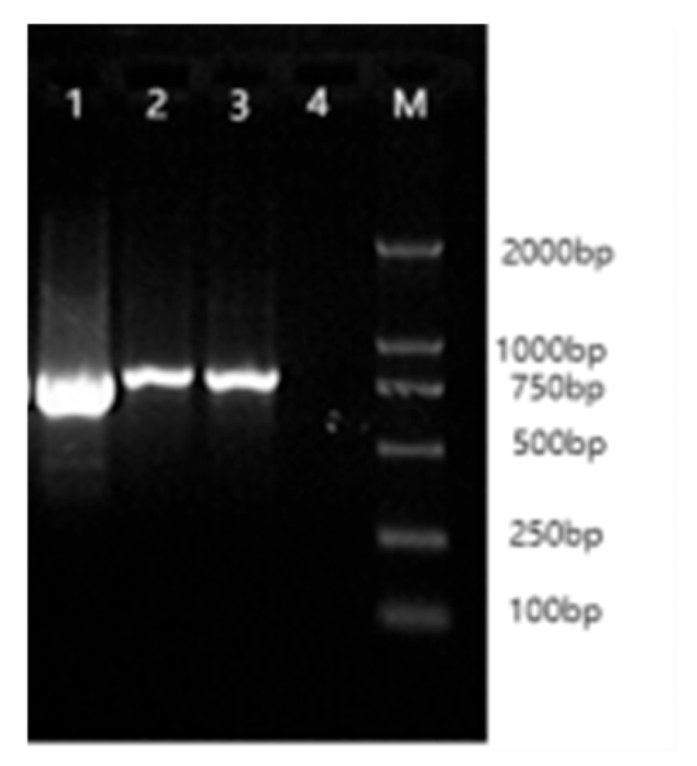
3.1. IGF1R Gene CDS Amplification Results

Total RNA extracted from the organ tissues of Arctic foxes was assessed using Nanodrop spectrophotometry, with A260/A280 ratios ranging between 1.8 and 2.0, confirming that RNA integrity met the requirements for molecular cloning. PCR amplification of cDNA templates using primers RIGF1R1-3 generated specific bands of 850 bp, 896 bp, and 727 bp, as verified by 1.5% agarose gel electrophoresis (Figure 1).

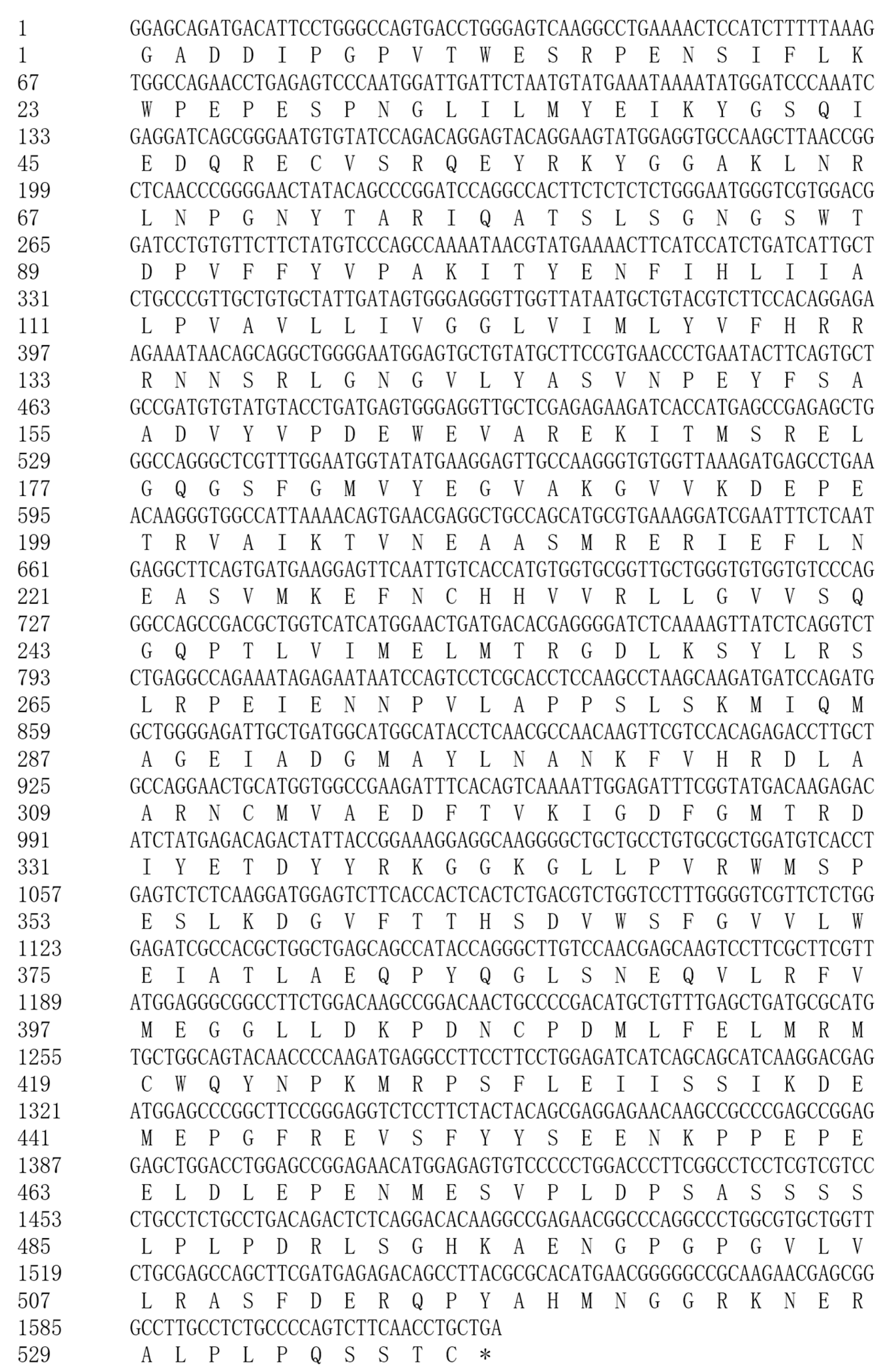
3.2. Sequencing Results and Sequence Analysis

Sequencing data assembly yielded a 2411 bp cDNA fragment of the Arctic fox IGF1R gene, consistent with the predicted structure of canine IGF1R. NCBI BLAST analysis demonstrated cross-species homology: 98% with Canis, 93% with Equus caballus, 91% with Homo sapiens, Macaca mulatta, Sus scrofa domesticus, and Ovis aries, 90% with Ailuropoda melanoleuca and Bos taurus, and 88% with Mus musculus. Phylogenetic analysis confirmed its conservation among mammalian IGF1R genes. Partial nucleotide and amino acid sequence alignments are detailed in Figure 2.

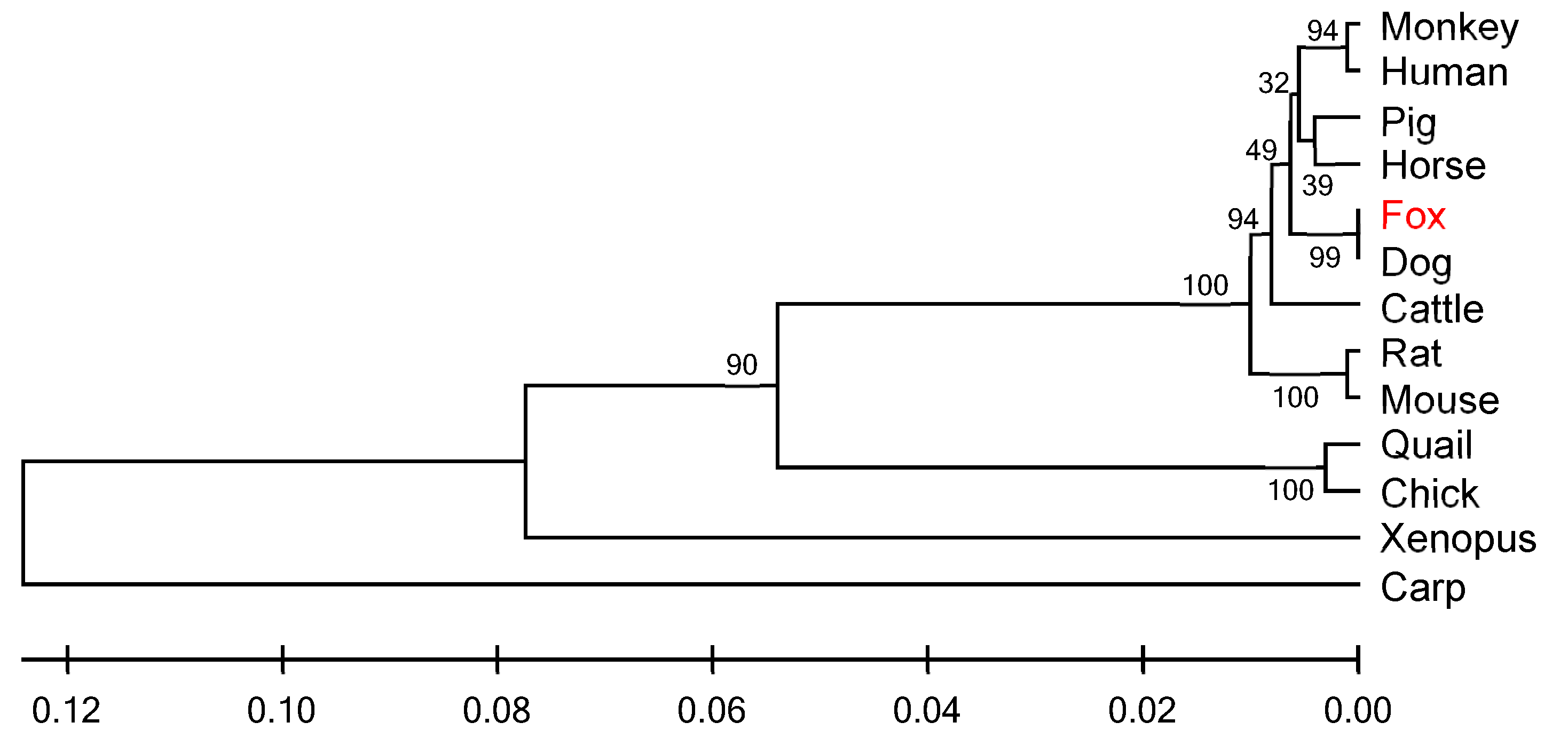
3.3. Similarity Analysis and Phylogenetic Tree Construction of IGF1R Gene

Comparative analysis of IGF1R amino acid sequences from Arctic foxes and 12 vertebrates (including Canis, Felis catus, and Bos taurus) revealed cross-class conservation: nucleotide/amino acid similarities ranged from 71% to 99%, with the highest homology to canids (99%) and the lowest to Cyprinus carpio (71%) (Table 3). A UPGMA phylogenetic tree constructed using MEGA 4.0 (Figure 3) showed Arctic foxes forming a monophyletic clade with canids, while primates exhibited topological anomalies, suggesting potential lineage-specific evolution of IGF1R in primates. Genetic distance analysis via the Kimura 2-parameter method indicated zero net distance between Arctic foxes and canids, and a maximum distance of 0.271 with Cyprinus carpio (Table 4), aligning with vertebrate phylogenetic hierarchy.

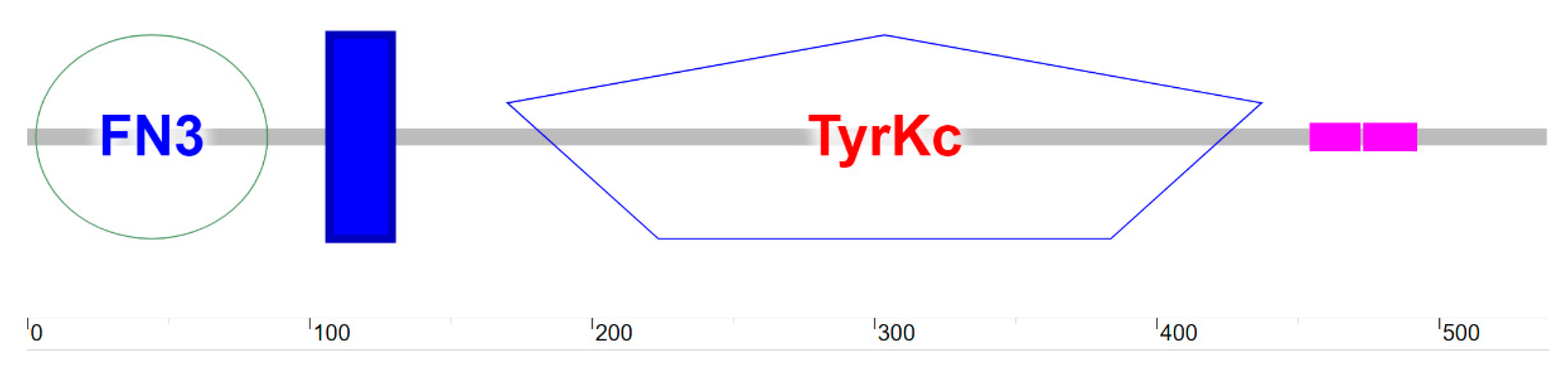
3.4. Structural and Functional Analysis of Arctic Fox IGF1R Protein

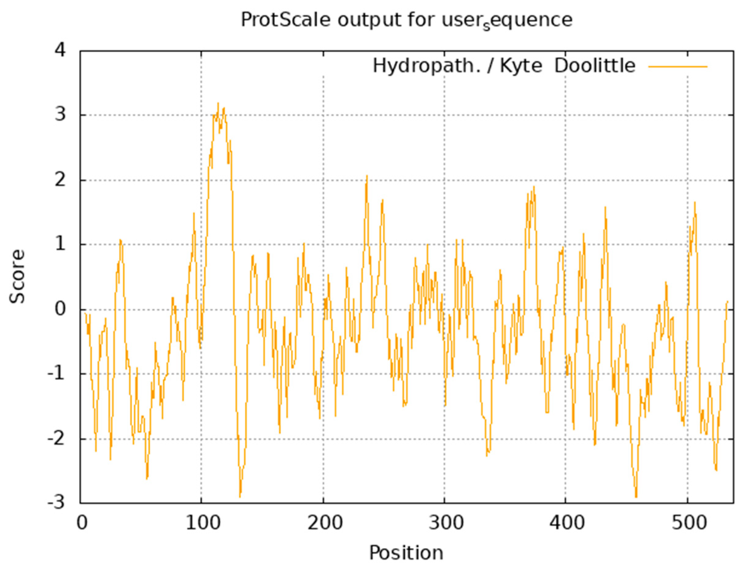
3.4.1. Physicochemical Properties and Hydrophobicity

ExPASy ProtParam analysis revealed the Arctic fox IGF1R protein (molecular formula, C2702H4203N725O805S28) has a molecular weight of 60.62 kDa, comprising 538 amino acids. Dominant residues included glutamate (Glu, 9.1%) and leucine (Leu, 9.1%), while histidine (His, 1.5%) and tryptophan (Trp, 1.5%) were least abundant. Acidic residues (Asp + Glu = 73) exceeded basic residues (Arg + Lys = 56) (Table 5), with a theoretical pI of 5.15, confirming acidic properties. The protein exhibited hydrophilicity (average GRAVY = −0.337), thermolability (instability index = 48.59), and moderate lipophilicity (aliphatic index = 81.51). Kyte–Doolittle hydrophobicity analysis identified a maximum hydrophobic peak (+3.189) at Ala114 and a minimum (−2.911) at Asp457 (Figure 4).

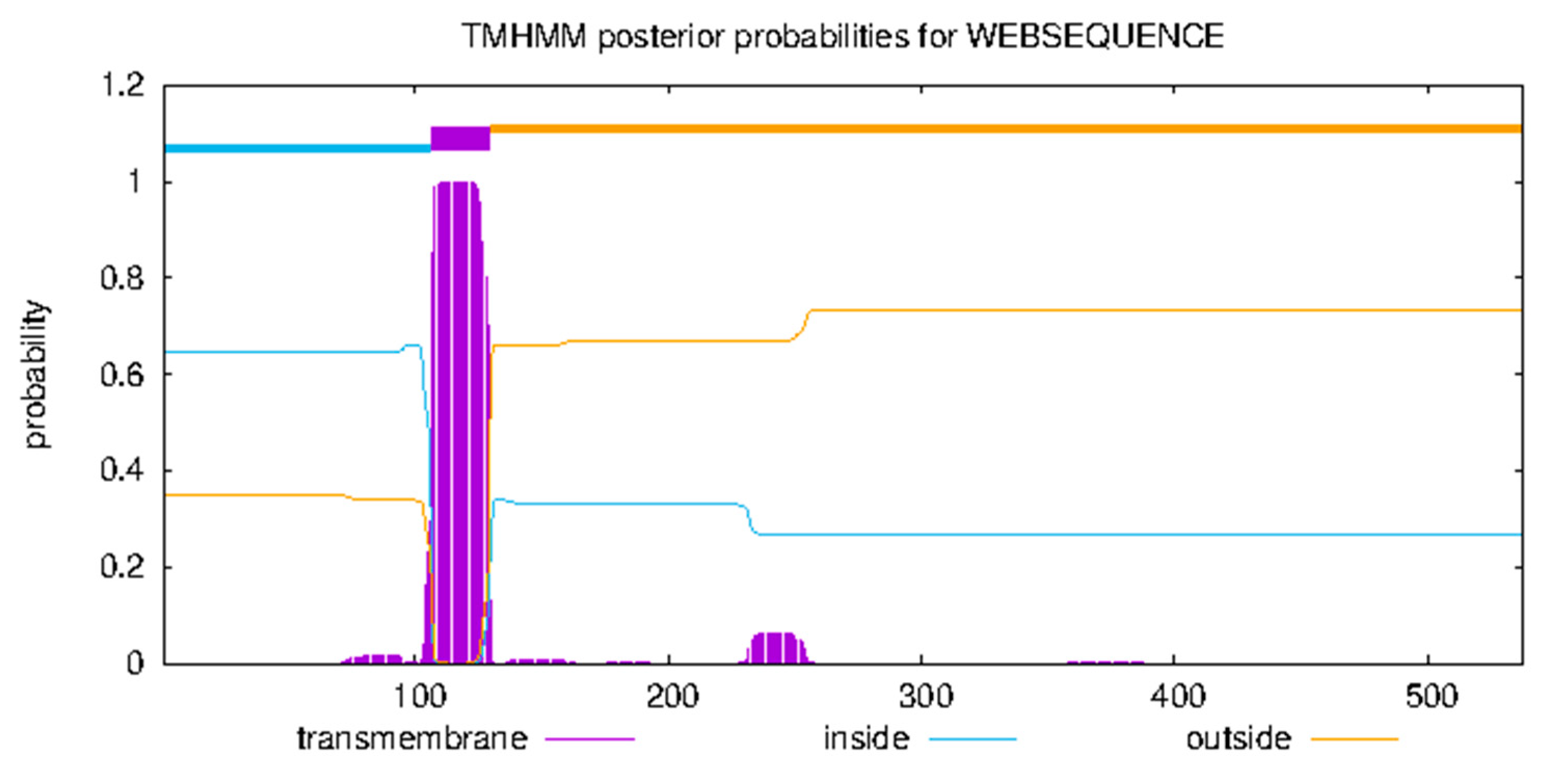
3.4.2. Transmembrane Domain and Subcellular Localization

Prediction indicated a single α-helix transmembrane domain (107–129 aa), with the N-terminus (1–106 aa) being cytoplasmic and the C-terminus (130–538 aa) being extracellular (Figure 5), consistent with type-II transmembrane receptors. PSORT II predicted nuclear (34.8%), cytosolic (21.7%), mitochondrial (21.7%), and secretory vesicle (8.7%) localization, suggesting dynamic involvement in transmembrane signaling and vesicle trafficking, aligning with IGF1R-mediated PI3K/AKT and MAPK pathway activation.

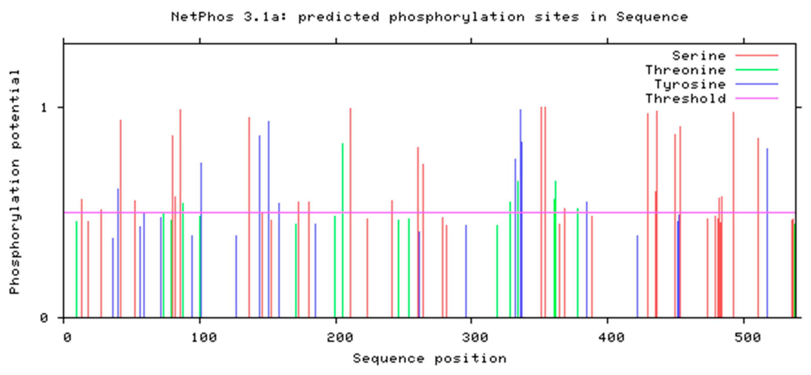
3.4.3. Phosphorylation Site Prediction

NetPhos 3.1 (threshold > 0.5) predicted 27 potential phosphorylation sites: 18 serine (66.7%), 6 threonine (22.2%), and 3 tyrosine (11.1%) residues (Figure 6).

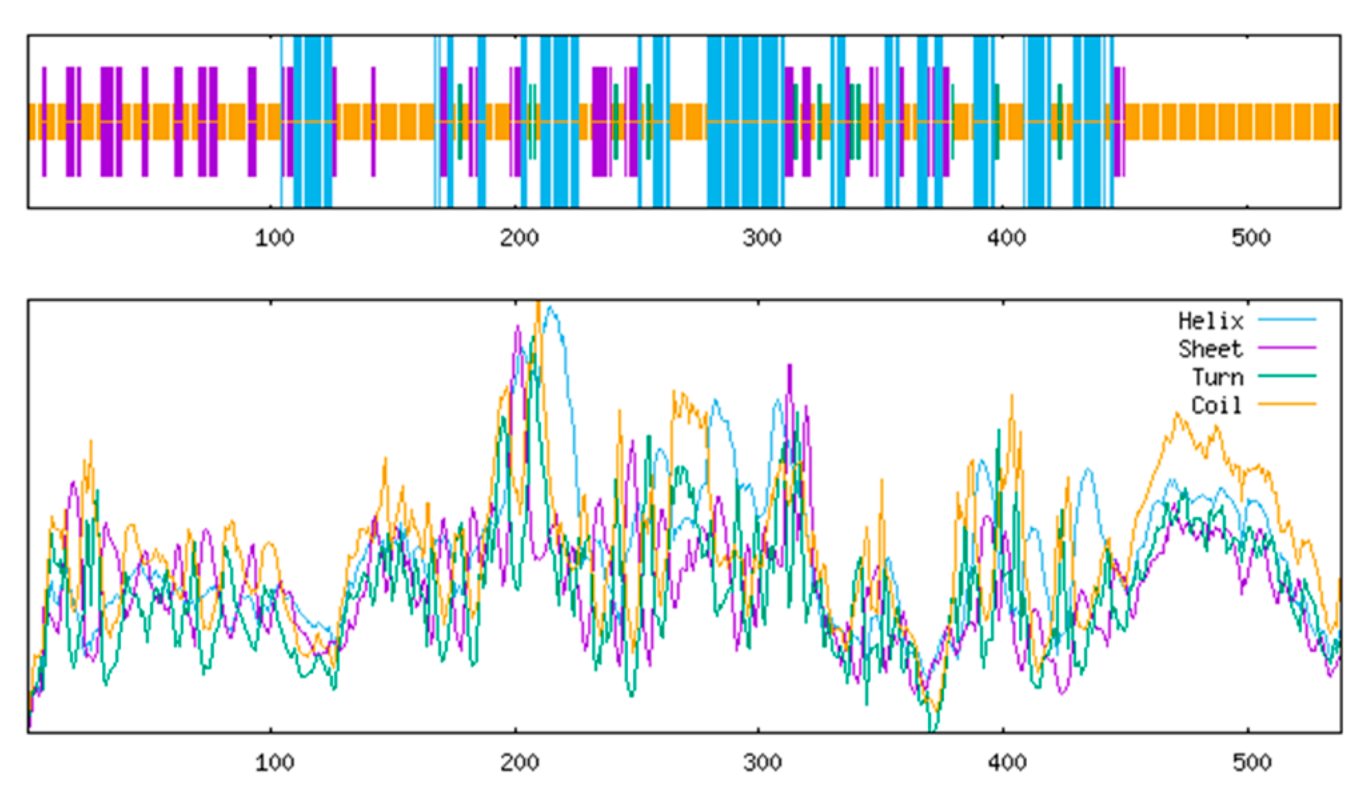
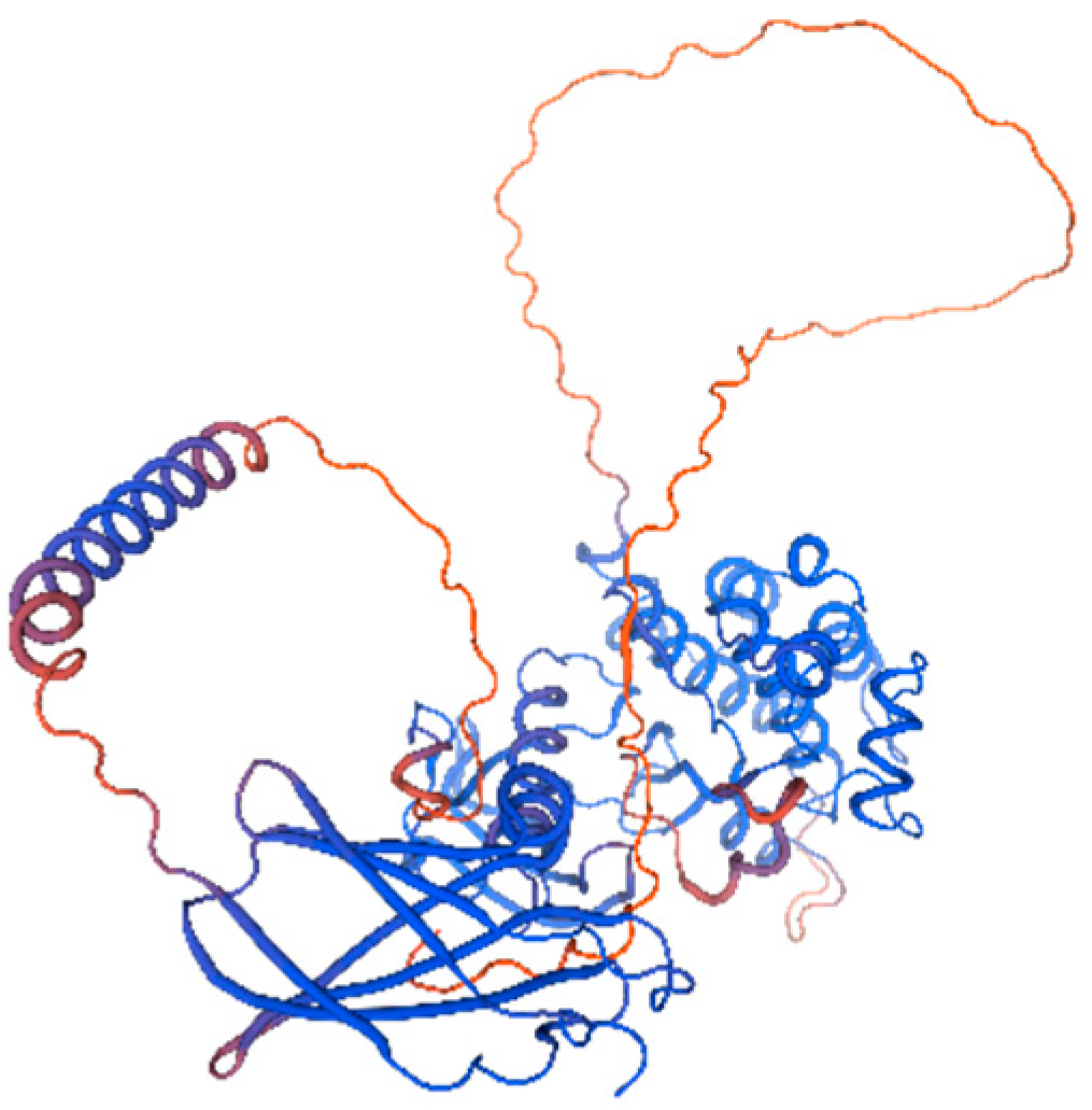
3.4.4. Secondary and Tertiary Structure Prediction

SOPMA analysis indicated dominance of random coils (52.79%) and α-helices (26.39%), with fewer extended strands (16.91%) and β-turns (3.90%) (Figure 7). SMART identified three domains: an N-terminal fibronectin type-III domain (3–85 aa) for ligand binding, a transmembrane helix (107–129 aa), and a C-terminal tyrosine kinase catalytic domain (170–437 aa) (Figure 8). SWISS-MODEL tertiary structure prediction (Figure 9) corroborated these features.

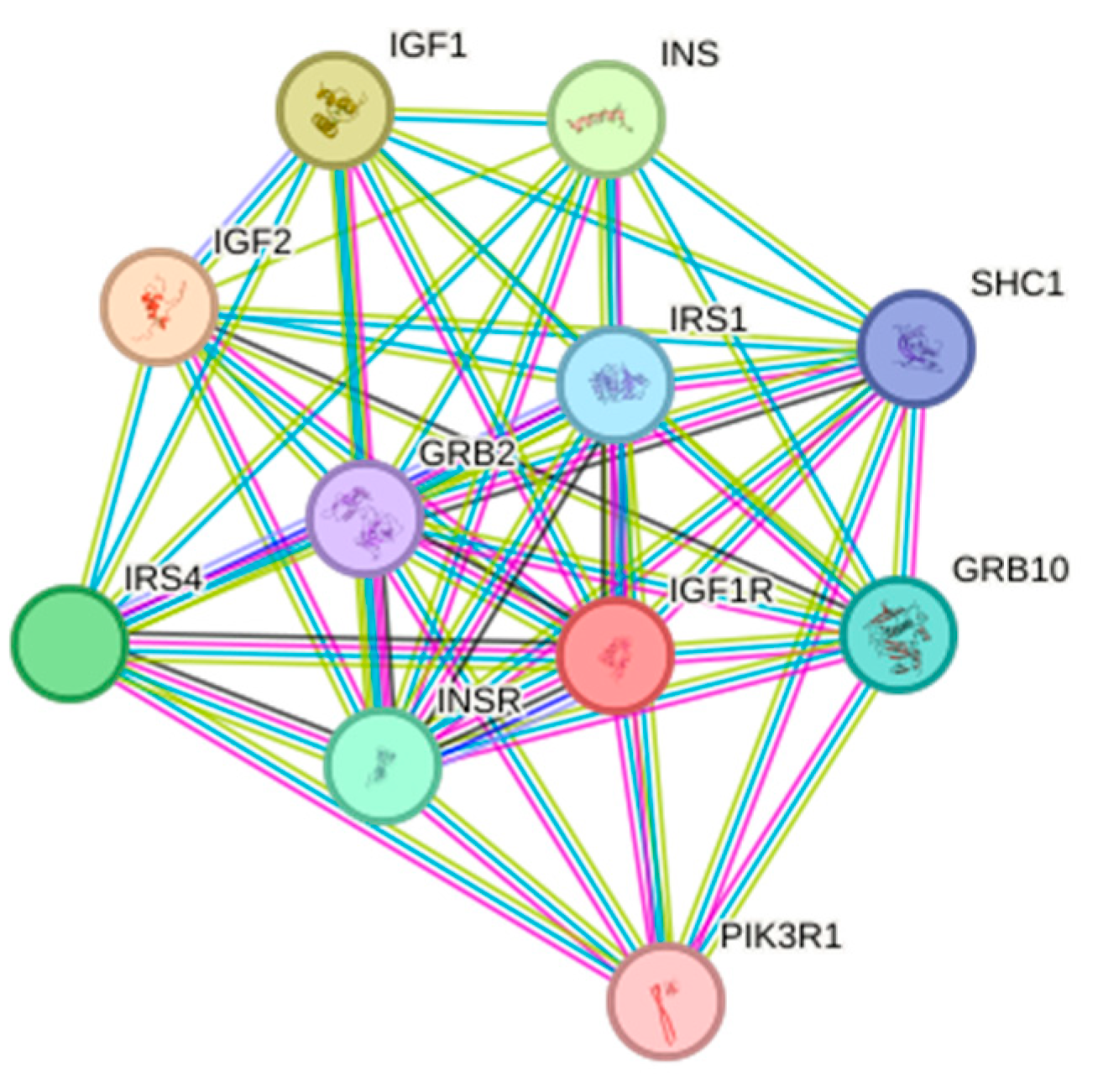
3.4.5. Protein Interaction Analysis

STRING database analysis revealed an 11-node interaction network with high connectivity (average node degree = 9.09) and clustering (local clustering coefficient = 0.911). Core interactors included insulin signaling components (INS/IGF1/IGF2), adaptors (SHC1/GRB2/GRB10), and PI3K-AKT regulators (PIK3R1/IRS1/IRS4), enriched in cell proliferation and organ development processes (Figure 10).

3.5. Tissue-Specific and Spatiotemporal Expression Patterns of IGF1R Gene in the Arctic Fox

qRT-PCR showed IGF1R expression in 11 of 12 organs (excluding duodenum) in 6-month-old Arctic foxes, with the highest levels in the testis and the lowest in the stomach (Table 6). Temporal analysis indicated age-dependent increases in liver expression, peak testis expression at 6 months, and highest muscle expression at 4 months (Table 7).

4. Discussion

Insulin-like growth factor I receptor (IGF1R), as a core effector molecule in the growth hormone axis cascade, coordinates organismal development by maintaining cellular homeostasis and driving proliferation–differentiation programs. As a key mediator of the GH-IGFs signaling axis, it exerts pleiotropic effects in metabolic regulation [1,14,15]. Loss-of-function studies demonstrate that IGF1R knockout leads to embryonic muscle-developmental defects in mice [16], hepatic regeneration disorders, and pulmonary developmental abnormalities [17], while muscle-specific overexpression induces abnormal muscle hypertrophy [5]. Evolutionary analyses reveal that structural variations in and transcriptional–translational efficiency attenuation of the IGF1R gene constitute critical mechanisms underlying dwarfism phenotypes, validated in both murine models and human genetic cases. Compared to extensive research on pigs [8], cattle [18], sheep [19], poultry [20], and aquatic species [21], functional characterization of IGF1R in Arctic foxes remains largely unexplored. This study systematically elucidates its molecular features and expression regulatory patterns for the first time, providing novel perspectives on growth axis research in polar mammals.
Based on the domestic dog IGF1R gene (Accession No. XM_545828), primers were designed to successfully clone a partial cDNA sequence (2411 bp) of Arctic fox IGF1R using RT-PCR. This sequence showed high homology with mammalian IGF1R, with the complete CDS region (1617 bp) encoding 538 amino acids. Phylogenetic analysis revealed 99% nucleotide sequence homology between Arctic fox IGF1R and domestic dogs, significantly higher than homology with pigs (98%), mice (96%), and poultry (84%), consistent with mammalian taxonomic relationships. Notably, IGF1R evolutionary trees exhibited deviations from traditional classifications in primates, suggesting potential functional divergence of the IGF family in primates. Phosphorylation prediction identified 27 potential phosphorylation sites, with structural domain remodeling induced by site-specific phosphorylation potentially regulating multi-pathway signal transduction networks [22]. Six Ser sites localized in the tyrosine kinase catalytic domain may mediate intracellular signaling through autophosphorylation [23]. Functional domain analysis demonstrated that Arctic fox IGF1R contains an 83-amino-acid fibronectin type-III (FN3) domain mediating DNA/heparin binding [24], while its transmembrane region (107–129 aa) confirms its identity as a transmembrane receptor. The tyrosine kinase (TyrKc) domain harbors multiple binding sites, enabling diverse catalytic reactions [25]. Protein interaction network analysis revealed strong interactions between IGF1R and 11 proteins, including IGF1/IGF2/INS. IGF1, as a key member of the insulin-like growth factor family, plays vital roles in growth regulation, skeletal muscle development, cellular proliferation/differentiation, tissue repair, and metabolic modulation. Importantly, IGF1R not only mediates IGF ligand signaling but also critically regulates embryonic development, cell growth/differentiation, mitosis, embryonic survival, and metabolic adaptation [26]. IGF1 drives evolutionary processes by modulating growth rates, body size differentiation, and physiological adaptability. IGF1R-IGF2 binding triggers tyrosine kinase domain autophosphorylation, activating PI3K/AKT and MAPK/ERK signaling cascades to promote metabolism, proliferation, and differentiation [26]. Genomic analyses identify numerous SNPs in porcine and ovine IGF1R genes, with specific variants significantly influencing growth traits through receptor conformation and expression levels, contributing to breed-specific size variations [27,28]. Notably, bovine IGF1R polymorphisms correlate strongly with milk-production traits (milk fat/protein content), highlighting its potential as a molecular marker in dairy cattle breeding [29]. These findings collectively underscore the central role of IGF signaling in animal growth regulation.
Using qRT-PCR (with GAPDH as reference), tissue-specific expression profiling of IGF1R in 6-month-old Arctic foxes revealed differential expression across the 12 organ tissues, except duodenum. Testicular IGF1R mRNA levels significantly exceeded other organ tissues, followed by kidney, muscle, liver, pituitary, brain, spleen, lung, heart, pancreas, and stomach. Notably, the absence of IGF1R expression in duodenal tissue may indicate its limited physiological role in this organ [30]; suggest predominant reliance on alternative signaling pathways for growth regulation [31]; or reflect technical limitations, such as undetectable expression levels (below qPCR detection threshold) or RNA degradation during preservation, affecting assay sensitivity.
Developmental expression analysis in liver, testis, and muscle tissues at 2-, 4-, and 6-month-old stages demonstrated age-dependent expression patterns. Hepatic expression showed progressive upregulation (2-month-old levels significantly lower than subsequent stages), aligning with developmental patterns observed in Nanjiang yellow goats [27]. Testicular expression peaked at 6 months (6-month-old > 2-month-old > 4-month-old), while muscular expression reached maximum at 4 months (4-month-old > 6-month-old > 2-month-old). The inclusion of testicular tissue in developmental studies stemmed from its highest expression among 12 organ tissues at 6 months, suggesting IGF1R involvement in Arctic fox reproductive regulation—a hypothesis reinforced by sustained high expression across 2- and 4-month-old testicular samples.
The spatiotemporal expression of IGF1R across tissues carries significant biological implications: In liver tissue, IGF1R expression increased with age, reflecting its core role in energy metabolism and growth substrate synthesis. As the primary site of IGF-1 synthesis, upregulated receptor expression in liver may enhance responsiveness to circulating IGF-1, promoting glycogen synthesis and protein accumulation to support rapid growth in juvenile foxes [32]. Testicular tissue expression peaked at 6 months old, suggesting IGF1R’s potential involvement in Arctic fox sexual maturation by regulating spermatogonial proliferation and testosterone synthesis, thereby influencing reproductive organ development and breeding performance [33]. The muscular expression peak at 4 months old coincided with the rapid growth phase of skeletal muscle, demonstrating that IGF1R directly regulates muscle growth efficiency through satellite cell differentiation and myofiber hypertrophy—consistent with its established role in mammalian myogenic differentiation [17].
As a transmembrane tyrosine kinase receptor, IGF1R ubiquitously localizes on cell membranes, mediating proliferation, differentiation, and anti-apoptotic functions through IGF1/IGF2 binding [34]. Current research predominantly focuses on its roles in tumorigenesis and therapeutic resistance, whereas tissue-specific functions in Arctic foxes require further exploration via proteomic interactome mapping and conditional knockout models. This study provides foundational insights into Arctic fox IGF1R expression profiles and structural characteristics, facilitating future investigations into molecular mechanisms underlying growth trait regulation.

5. Conclusions

This study successfully cloned the full CDS (1617 bp) of IGF1R in the Arctic fox, with phylogenetic analysis confirming its high conservation (99% homology) within canines. The receptor exhibited characteristic tyrosine kinase features, including a fibronectin type-III ligand-binding domain, transmembrane region, and catalytic domain, with acidic hydrophilic properties (pI = 5.15). Spatiotemporal expression analysis revealed tissue-specific and developmental stage-specific regulation: IGF1R expression was highest in testicular tissue, while dynamic patterns were observed across ages—liver expression increased progressively with postnatal development, aligning with enhanced metabolic demands; testicular expression peaked in 6-month-old individuals, suggesting roles in sexual maturation; and muscular expression maxima at 4 months of age corresponded to rapid skeletal muscle growth.

Author Contributions

W.X. analyzed the data, prepared the figures and tables, and wrote the manuscript; H.F. assisted with experiments, helped analyze data, and reviewed the manuscript; X.M. assisted in analyzing data; Y.S. assisted in the preparation of the figures and tables; F.N. provided data and helped design experiments; Z.D. conceptualized the design of the experiment and provided financial support. All authors have read and agreed to the published version of the manuscript.

Funding

This research was funded by the National Key R&D Program of China, Grant No. 2023YFD1302000.

Institutional Review Board Statement

Our research was conducted by the guidelines of the Animal Welfare and Ethics Committee of Northeast Agricultural University. Northeast Agricultural University Animal Care and Use Committee approved all the protocols (NEAU-20210240, 8 June 2023).

Informed Consent Statement

All participants in the study provided their informed consent.

Data Availability Statement

Should you require any further data, please do not hesitate to contact us. For the sake of data privacy, this information is not available to the public.

Acknowledgments

The author assert that the handling and treatment of captured animals strictly adhered to the animal welfare guidelines and have undergone thorough ethical review by Northeast Agricultural University.

Conflicts of Interest

The authors have no conflicts of interest to declare.

Abbreviations

Key Symbol Comparison Table
IGFIRInsulin-like growth factor 1 receptor
cDNAComplementary DNA
IGF1Insulin like growth factor 1
IGF2Insulin like growth factor 2
MC1RMelanocortin 1 receptor
GAPDHGlyceraldehyde-3-phosphate dehydrogenase
CDSCoding sequences
ORFOpen reading frame
DEPCDiethylpyrocarbonate
UTRUntranslated regions
PIIsoelectric point
LSMLeast squares method
SNPSingle-nucleotide polymorphism
FSHFollicle-stimulating hormone
GHGrowth hormone

References

  1. Alex, P.; Kanakkaparambil, R.; Gopalakrishnan, R.; Ramasamy, C.; Thazhathuveettil, A. The effect of insulin-like growth factor 1 receptor gene single nucleotide polymorphism on growth and milk production traits in two native Indian tropical goat breeds. Anim. Biotechnol. 2023, 34, 4828–4836. [Google Scholar] [CrossRef] [PubMed]
  2. Baker, J.; Liu, J.P.; Robertson, E.J.; Efstratiadis, A. Role of insulin-like growth factors in embryonic and postnatal growth. Cell 1993, 75, 73–82. [Google Scholar] [CrossRef]
  3. Constância, M.; Hemberger, M.; Hughes, J.; Dean, W.; Ferguson-Smith, A.; Fundele, R.; Stewart, F.; Kelsey, G.; Fowden, A.; Sibley, C.; et al. Placental-specific IGF-II is a major modulator of placental and fetal growth. Nature 2002, 417, 945–948. [Google Scholar] [CrossRef] [PubMed]
  4. Woods, K.A.; Camacho-Hübner, C.; Savage, M.O.; Clark, A.J. Intrauterine growth retardation and postnatal growth failure associated with deletion of the insulin-like growth factor I gene. N. Engl. J. Med. 1996, 335, 1363–1367. [Google Scholar] [CrossRef] [PubMed]
  5. Liu, J.P.; Baker, J.; Perkins, A.S.; Robertson, E.J.; Efstratiadis, A. Mice carrying null mutations of the genes encoding insulin-like growth factor I (Igf-1) and type 1 IGF receptor (IGF1R). Cell 1993, 75, 59–72. [Google Scholar] [CrossRef]
  6. Galifi, C.A.; Wood, T.L. Insulin-like growth factor-1 receptor crosstalk with integrins, cadherins, and the tumor microenvironment: Sticking points in understanding IGF1R function in cancer. Endocr.-Relat. Cancer 2023, 30, e230031. [Google Scholar] [CrossRef]
  7. James, H.A.; O’Neill, B.T.; Nair, K.S. Insulin Regulation of Proteostasis and Clinical Implications. Cell Metab. 2017, 26, 310–323. [Google Scholar] [CrossRef]
  8. Pierzchała, M.; Pareek, C.S.; Urbański, P.; Goluch, D.; Kamyczek, M.; Różycki, M.; Smoczynski, R.; Horbańczuk, J.O.; Kurył, J. Study of the differential transcription in liver of growth hormone receptor (GHR), insulin-like growth factors (IGF1, IGF2) and insulin-like growth factor receptor (IGF1R) genes at different postnatal developmental ages in pig breeds. Mol. Biol. Rep. 2012, 39, 3055–3066. [Google Scholar] [CrossRef]
  9. Zheng, W.; Leng, X.; Vinsky, M.; Li, C.; Jiang, H. Association of body weight gain with muscle, fat, and liver expression levels of growth hormone receptor, insulin-like growth factor I, and beta-adrenergic receptor mRNAs in steers. Domest. Anim. Endocrinol. 2018, 64, 31–37. [Google Scholar] [CrossRef]
  10. Harumi, T.; Maruyama, K.; Kagami, H.; Sano, A.; Matsubara, Y.; Tagami, T.; Naito, M. Cloning of porcine IGF1 receptor cDNA and detection of sequence polymorphisms using RT-PCR. Anim. Genet. 2001, 32, 386–389. [Google Scholar] [CrossRef]
  11. Hong, Z.; Wang, D.; Qiao, X.; Xie, Y.; Yang, S.; Hao, K.; Han, C.; Liu, H.; Liu, Z. Wnt5a negatively regulates melanogenesis in primary Arctic fox epidermal melanocytes. Gene 2025, 934, 149045. [Google Scholar] [CrossRef]
  12. Våge, D.I.; Fuglei, E.; Snipstad, K.; Beheim, J.; Landsem, V.M.; Klungland, H. Two cysteine substitutions in the MC1R generate the blue variant of the Arctic fox (Alopex lagopus) and prevent expression of the white winter coat. Peptides 2005, 26, 1814–1817. [Google Scholar] [CrossRef]
  13. Jaillardon, L.; Abadie, J.; Godard, T.; Campone, M.; Loussouarn, D.; Siliart, B.; Nguyen, F. The dog as a naturally-occurring model for insulin-like growth factor type 1 receptor-overexpressing breast cancer: An observational cohort study. BMC Cancer 2015, 15, 664. [Google Scholar] [CrossRef] [PubMed]
  14. Forbes, B.E. Molecular mechanisms underlying insulin-like growth factor action: How mutations in the GH: IGF axis lead to short stature. Pediatr. Endocrinol. Rev. 2011, 8, 374–381. [Google Scholar]
  15. Holzenberger, M.; Dupont, J.; Ducos, B.; Leneuve, P.; Géloën, A.; Even, P.C.; Cervera, P.; Le Bouc, Y. IGF-1 receptor regulates lifespan and resistance to oxidative stress in mice. Nature 2003, 421, 182–187. [Google Scholar] [CrossRef] [PubMed]
  16. López, I.P.; Rodriguez-de la Rosa, L.; Pais, R.S.; Piñeiro-Hermida, S.; Torrens, R.; Contreras, J.; Varela-Nieto, I.; Pichel, J.G. Differential organ phenotypes after postnatal IGF1R gene conditional deletion induced by tamoxifen in UBC-CreERT2; IGF1R fl/fl double transgenic mice. Transgenic Res. 2015, 24, 279–294. [Google Scholar] [CrossRef]
  17. Epaud, R.; Aubey, F.; Xu, J.; Chaker, Z.; Clemessy, M.; Dautin, A.; Ahamed, K.; Bonora, M.; Hoyeau, N.; Fléjou, J.F.; et al. Knockout of insulin-like growth factor-1 receptor impairs distal lung morphogenesis. PLoS ONE 2012, 7, e48071. [Google Scholar] [CrossRef] [PubMed]
  18. Ma, Y.L.; Wen, Y.F.; Cao, X.K.; Cheng, J.; Huang, Y.Z.; Ma, Y.; Hu, L.Y.; Lei, C.Z.; Qi, X.L.; Cao, H.; et al. Copy number variation (CNV) in the IGF1R gene across four cattle breeds and its association with economic traits. Arch. Anim. Breed. 2019, 62, 171–179. [Google Scholar] [CrossRef]
  19. Ding, N.; Tian, D.; Li, X.; Zhang, Z.; Tian, F.; Liu, S.; Han, B.; Liu, D.; Zhao, K. Genetic Polymorphisms of IGF1 and IGF1R Genes and Their Effects on Growth Traits in Hulun Buir Sheep. Genes 2022, 13, 666. [Google Scholar] [CrossRef]
  20. Jin, S.; Chen, S.; Li, H.; Lu, Y.; Xu, G.; Yang, N. Associations of polymorphisms in GHRL, GHSR, and IGF1R genes with feed efficiency in chickens. Mol. Biol. Rep. 2014, 41, 3973–3979. [Google Scholar] [CrossRef]
  21. Shah, A.; Ahmad, I.; Ahmad, I.; Amin, A.; Rather, M.A. Gene Characterization, Molecular Docking and Dynamic Simulations of Insulin-Like Growth Factor Receptor (IGF-1Ra) in Common Carp, Cyprinus carpio. Proc. Zool. Soc. 2023, 76, 382–396. [Google Scholar] [CrossRef]
  22. Niu, S.; Wang, Z.; Ge, D.; Zhang, G.; Li, Y. Prediction of functional phosphorylation sites by incorporating evolutionary information. Protein Cell 2012, 3, 675–690. [Google Scholar] [CrossRef] [PubMed]
  23. Wang, D.; Zeng, S.; Xu, C.; Qiu, W.; Liang, Y.; Joshi, T.; Xu, D. MusiteDeep: A deep-learning framework for general and kinase-specific phosphorylation site prediction. Bioinformatics 2017, 33, 3909–3916. [Google Scholar] [CrossRef] [PubMed]
  24. Skorstengaard, K.; Jensen, M.S.; Sahl, P.; Petersen, T.E.; Magnusson, S. Complete primary structure of bovine plasma fibronectin. Eur. J. Biochem. 1986, 161, 441–453. [Google Scholar] [CrossRef]
  25. Hou, Q.; Gong, R.; Liu, X.; Mao, H.; Xu, X.; Liu, D.; Dai, Z.; Wang, H.; Wang, B.; Hu, C. Poly I:C facilitates the phosphorylation of Ctenopharyngodon idellus type I IFN receptor subunits and JAK kinase. Fish Shellfish. Immunol. 2017, 60, 13–20. [Google Scholar] [CrossRef]
  26. Blyth, A.J.; Kirk, N.S.; Forbes, B.E. Understanding IGF-II Action through Insights into Receptor Binding and Activation. Cells 2020, 9, 2276. [Google Scholar] [CrossRef]
  27. Grochowska, E.; Lisiak, D.; Akram, M.Z.; Adeniyi, O.O.; Lühken, G.; Borys, B. Association of a polymorphism in exon 3 of the IGF1R gene with growth, body size, slaughter and meat quality traits in Colored Polish Merino sheep. Meat Sci. 2021, 172, 108314. [Google Scholar] [CrossRef]
  28. Wang, C.; Liu, S.; Wu, Q.; Cheng, Y.; Feng, T.; Song, J.; Yang, R.; Geng, H.; Lu, G.; Wang, S.; et al. Porcine IGF-1R synonymous mutations in the intracellular domain affect cell proliferation and alter kinase activity. Int. J. Biol. Macromol. 2020, 152, 147–153. [Google Scholar] [CrossRef]
  29. Szewczuk, M.; Zych, S.; Wójcik, J.; Czerniawska-Piątkowska, E. Association of two SNPs in the coding region of the insulin-like growth factor 1 receptor (IGF1R) gene with growth-related traits in Angus cattle. J. Appl. Genet. 2013, 54, 305–308. [Google Scholar] [CrossRef]
  30. Yakar, S.; Adamo, M.L. Insulin-like growth factor 1 physiology: Lessons from mouse models. Endocrinol. Metab. Clin. N. Am. 2012, 41, 231–247. [Google Scholar] [CrossRef]
  31. Hoyt, J.A.; Cozzi, E.; D’Alessio, D.A.; Thompson, C.C.; Aroda, V.R. A look at duodenal mucosal resurfacing: Rationale for targeting the duodenum in type 2 diabetes. Diabetes Obes. Metab. 2024, 26, 2017–2028. [Google Scholar] [CrossRef] [PubMed]
  32. Serrat, M.A.; Lovejoy, C.O.; King, D. Age- and site-specific decline in insulin-like growth factor-I receptor expression is correlated with differential growth plate activity in the mouse hindlimb. Anat. Rec. 2007, 290, 375–381. [Google Scholar] [CrossRef] [PubMed]
  33. Yoon, M.J.; Berger, T.; Roser, J.F. Localization of insulin-like growth factor-I (IGF-I) and IGF-I receptor (IGF-IR) in equine testes. Reprod. Domest. Anim. = Zuchthyg. 2011, 46, 221–228. [Google Scholar] [CrossRef]
  34. Li, R.; Pourpak, A.; Morris, S.W. Inhibition of the insulin-like growth factor-1 receptor (IGF1R) tyrosine kinase as a novel cancer therapy approach. J. Med. Chem. 2009, 52, 4981–5004. [Google Scholar] [CrossRef] [PubMed]
Figure 1. The PCR amplifies results of primers. Lanes 1, 2, and 3: PCR products of primers RIGF1R3, RIGF1R2, and RIGF1R1. Lane 4: control. M: DL 2000 marker.
Life 15 00796 g001
Figure 2. cDNA sequence and putative amino acid sequence of IGF1R from the Arctic fox.
Life 15 00796 g002
Figure 3. UPGMA phylogenetic tree analysis based on amino acid sequence of IGF1R from different species.
Life 15 00796 g003
Figure 4. Prediction of hydrophobic structure of IGF1R in the Arctic fox.
Life 15 00796 g004
Figure 5. Transmembrane structure prediction of IGF1R protein in the Arctic fox.
Life 15 00796 g005
Figure 6. Analysis of phosphorylation potential of IGF1R in the Arctic fox.
Life 15 00796 g006
Figure 7. IGF1R secondary structure prediction. The longest vertical line is the alpha helix; the second-longest vertical line is the extended chain; the secondary short vertical line is the Beta turn; and the shortest vertical line is the random coil.
Life 15 00796 g007
Figure 8. Functional domain prediction of IGF1R protein in the Arctic fox.
Life 15 00796 g008
Figure 9. Predicted tertiary structure of Arctic fox IGF1R protein.
Life 15 00796 g009
Figure 10. Protein interaction analysis of IGF1R in the Arctic fox.
Life 15 00796 g010
Table 1. Primer information.
PrimersSequencesAnnealing
Temperature
Application
IGF1R-1F: GCAGAAGGAGCAGATGACATTC
R: GAGGTGCGAGGACTGGATTAT
54PCR amplification
IGF1R-2F: AATCCAGTCCTCGCACCTC
R: AGGAGGCTGTGGCTAGATTG
60PCR amplification
IGF1R-3F: GGAGTTCTAACAATCTAGCC
R: ACCTGCGTGAATGTAAGAC
40PCR amplification
IGF1RF: GTCCTTCGCTTCGTTATG
R: CTTGTTCTCCTCGCTGTAG
60qRT-PCR
GAPDHF: GGGTGATGCTGGTGCTG
R: ACGGTCTTCTGGGTGGC
60qRT-PCR
Table 2. Bioinformatics analysis software and function.
Analysis ToolsWebsitesFunction Prediction
ExPASyhttps://web.expasy.org/protscale/ (accessed on 24 February 2025)Physicochemical properties
ProtScahttps://web.expasy.org/protscale (accessed on 24 February 2025)Hydrophilicity/hydrophobicity
Novoprohttps://www.novopro.cn/tools/tmhmm.html (accessed on 24 February 2025)Transmembrane domain
SMARThttp://smart.embl-heidelberg.de/ (accessed on 24 February 2025)Functional domain
NetPhos-3.1https://services.healthtech.dtu.dk/services/NetPhos-3.1/ (accessed on 25 February 2025)Phosphorylation sites
SOPMAhttps://npsa.lyon.inserm.fr/cgi-bin/ (accessed on 25 February 2025)Secondary structure
SWISS-MODELhttps://swissmodel.expasy.org/ (accessed on 25 February 2025)Tertiary structure
STRINGhttps://cn.string-db.org/ (accessed on 25 February 2025)Protein–protein interaction
Table 3. Amino acid homology between Arctic fox IGF1R and IGF1Rs of other species.
SpeciesSimilarity of MatureAmino Acid NumberGenBank Accession No.
Canis familiaris99%538XP_545828
Sus scrofa98%538NP_999337
Equus caballus98%538XP_001489815
Bos Taurus97%538XP_606794
Homo sapiens97%538EAX02222
Macaca mulatta97%538XP_001100407
Rattus norvegicus93%538AAA41392
Mus musculus96%538NP_034643
Gallus gallus84%538NP_990363
Coturnix japonica84%538BAF73401
Xenopus laevis79%538CAA90517
Cyprinus carpio71%538AAN52152
Table 4. Interspecific genetic distances of the IGF1R gene calculated using the Kimura 2-parameter method.
QuailChickMonkeyHumanPigCattleHorseFoxDogRatMouse Xenopus
Chick0.006
Monkey0.1340.132
Human0.1370.1340.002
Pig0.1280.1250.0080.010
Cattle 0.1340.1320.0180.0200.014
Horse0.1340.1320.0120.0140.0080.016
Fox0.1370.1340.0180.0200.0140.0240.010
Dog0.1370.1340.0180.0200.0140.0240.0100.000
Rat0.1460.1440.0320.0350.0280.0350.0280.0320.032
Mouse 0.1480.1460.0370.0390.0320.0390.0320.0370.0370.004
Xenopus0.1650.1570.2030.2050.1980.2050.2030.2050.2050.2080.210
Carp0.2480.2400.2760.2740.2710.2710.2740.2710.2710.2870.2840.266
Table 5. Physicochemical properties of IGF1R protein in Alopex.
Amino AcidsNumberFrequency (%)Amino AcidsNumberFrequency (%)
Ala (A)315.8Lys (K)234.3
Arg (R)336.1Met (M)224.1
Asn (N)275.0Phe (F)203.7
Asp (D)244.5Pro (P)397.2
Cys (C)61.1Ser (S)407.4
Gln (Q)142.6Thr (T)173.2
Glu (E)499.1Trp (W)81.5
Gly (G)407.4Tyr (Y)224.1
His (H)81.5Val (V)417.6
Ile (I)254.6Pyl (O)00
Leu (L)499.1Sec (U)00
Table 6. Expression characteristics of Arctic fox IGF1R gene in 11 organ tissues.
HistophysiologyGAPDH
(Ct1)
IGF1R
(Ct2)
Relative Expression (2Ct1-Ct2)Brain Expression-Normalized (Set to 1)
Liver22.12323.0600.522 ± 0.1091.906 ± 0.398
Spleen23.05724.9600.267 ± 0.0850.976 ± 0.309
Kidney22.87323.5200.639 ± 0.2302.331 ± 0.840
Lung19.74522.1500.189 ± 0.0780.690 ± 0.286
Stomach20.32326.1600.018 ± 0.0030.064 ± 0.011
Pancreas18.15721.1370.127 ± 0.1070.463 ± 0.389
Brain19.76721.6330.274 ± 0.1551.000 ± 0.565
Pituitary gland20.79321.8530.480 ± 0.1541.750 ± 0.561
Testis22.33320.8832.732 ± 0.4869.971 ± 1.773
Heart21.73724.1970.182 ± 0.0520.663 ± 0.190
Muscle20.44321.2730.563 ± 0.0572.053 ± 0.208
Table 7. Spatiotemporal expression characteristics of the IGF1R gene in Arctic fox organ tissues.
HistophysiologyGAPDH
(Ct1)
IGF1R
(Ct2)
Relative Expression (2Ct1-Ct2)Muscle Expression in 4-Month-Old Male Foxes-Normalized (Set to 1)
2-month-old male fox liver22.03026.5270.044 ± 0.0170.043 ± 0.016
4-month-old male fox liver18.35319.4500.467 ± 0.0950.451 ± 0.090
6-month-old male fox liver22.12323.0600.522 ± 0.1270.504 ± 0.120
2-month-old male fox testis21.12019.7602.567 ± 0.2642.480 ± 0.250
4-month-old male fox testis21.10720.0202.124 ± 0.3232.052 ± 0.306
6-month-old male fox testis22.33320.8832.732 ± 0.2252.640 ± 0.213
2-month-old male fox muscle20.22022.9930.146 ± 0.0080.141 ± 0.007
4-month-old male fox muscle18.46018.4101.035 ± 0.2641.000 ± 0.250
6-month-old male fox muscle20.44321.2730.563 ± 0.0660.544 ± 0.063
Disclaimer/Publisher’s Note: The statements, opinions and data contained in all publications are solely those of the individual author(s) and contributor(s) and not of MDPI and/or the editor(s). MDPI and/or the editor(s) disclaim responsibility for any injury to people or property resulting from any ideas, methods, instructions or products referred to in the content.

