
Sciatic Nerve Compression after a Chronic Proximal Hamstring Tear: A Report of Two Cases and a Narrative Review of the Literature

 ,
,
Abstract
:1. Introduction
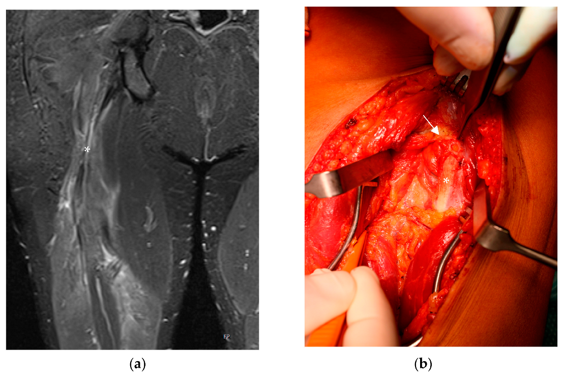
2. Case Report One
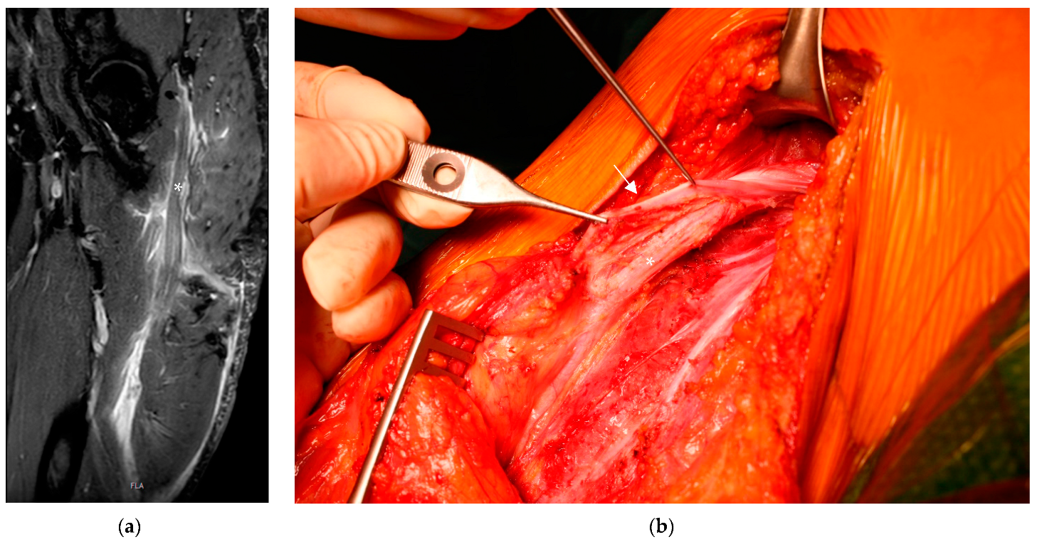
3. Case Report Two
4. Discussion
5. Conclusions
Author Contributions
Funding
Institutional Review Board Statement
Informed Consent Statement
Data Availability Statement
Conflicts of Interest
References
- Lempainen, L.; Banke, I.J.; Johansson, K.; Brucker, P.U.; Sarimo, J.; Orava, S.; Imhoff, A.B. Clinical principles in the management of hamstring injuries. Knee Surg. Sports Traumatol. Arthrosc. 2015, 23, 2449–2456. [Google Scholar] [CrossRef] [PubMed]
- Maniar, N.; Carmichael, D.S.; Hickey, J.T.; Timmins, R.G.; San Jose, A.J.; Dickson, J.; Opar, D. Incidence and prevalence of hamstring injuries in field-based team sports: A systematic review and meta-analysis of 5952 injuries from over 7 million exposure hours. Br. J. Sports Med. 2023, 57, 109–116. Available online: https://pubmed.ncbi.nlm.nih.gov/36455927/ (accessed on 18 June 2023). [CrossRef] [PubMed]
- Irger, M.; Willinger, L.; Lacheta, L.; Pogorzelski, J.; Imhoff, A.B.; Feucht, M.J. Proximal hamstring tendon avulsion injuries occur predominately in middle-aged patients with distinct gender differences: Epidemiologic analysis of 263 surgically treated cases. Knee Surg. Sports Traumatol. Arthrosc. 2020, 28, 1221–1229. [Google Scholar] [CrossRef] [PubMed]
- Miller, S.L.; Gill, J.; Webb, G.R. The proximal origin of the hamstrings and surrounding anatomy encountered during repair. A cadaveric study. J. Bone Jt. Surg. Am. 2007, 89, 44–48. [Google Scholar] [CrossRef] [PubMed]
- Wilson, T.J.; Spinner, R.J.; Mohan, R.; Gibbs, C.M.; Krych, A.J. Sciatic Nerve Injury After Proximal Hamstring Avulsion and Repair. Orthop. J. Sports Med. 2017, 5, 2325967117713685. [Google Scholar] [CrossRef] [PubMed]
- Aggen, P.D.; Reuteman, P. Conservative rehabilitation of sciatic nerve injury following hamstring tear. North Am. J. Sports Phys. Ther. NAJSPT 2010, 5, 143–154. [Google Scholar]
- Macdonald, J.; McMahon, S.E.; O’Longain, D.; Acton, J.D. Delayed sciatic nerve compression following hamstring injury. Eur. J. Orthop. Surg. Traumatol. 2018, 28, 305–308. [Google Scholar] [CrossRef] [PubMed]
- Green, B.; Bourne, M.N.; van Dyk, N.; Pizzari, T. Recalibrating the risk of hamstring strain injury (HSI): A 2020 systematic review and meta-analysis of risk factors for index and recurrent hamstring strain injury in sport. Br. J. Sports Med. 2020, 54, 1081–1088. [Google Scholar] [CrossRef] [PubMed]
- van der Made, A.D.; Tol, J.L.; Reurink, G.; Peters, R.W.; Kerkhoffs, G.M. Potential hamstring injury blind spot: We need to raise awareness of proximal hamstring tendon avulsion injuries. Br. J. Sports Med. 2019, 53, 390–392. [Google Scholar] [CrossRef] [PubMed]
- Ahmad, C.S.; Redler, L.H.; Ciccotti, M.G.; Maffulli, N.; Longo, U.G.; Bradley, J. Evaluation and management of hamstring injuries. Am. J. Sports Med. 2013, 41, 2933–2947. [Google Scholar] [CrossRef] [PubMed]
- Barnett, A.J.; Negus, J.J.; Barton, T.; Wood, D.G. Reattachment of the proximal hamstring origin: Outcome in patients with partial and complete tears. Knee Surg. Sports Traumatol. Arthrosc. 2015, 23, 2130–2135. [Google Scholar] [CrossRef]
- Wood, D.G.; Packham, I.; Trikha, S.P.; Linklater, J. Avulsion of the proximal hamstring origin. J. Bone Jt. Surg. Am. 2008, 90, 2365–2374. [Google Scholar] [CrossRef]
- Pas, H.I.M.F.L.; Reurink, G.; Tol, J.L.; Weir, A.; Winters, M.; Moen, M.H. Efficacy of rehabilitation (lengthening) exercises, platelet-rich plasma injections, and other conservative interventions in acute hamstring injuries: An updated systematic review and meta-analysis. Br. J. Sports Med. 2015, 49, 1197–1205. [Google Scholar] [CrossRef] [PubMed]
- Bertiche, P.; Mohtadi, N.; Chan, D.; Hölmich, P. Proximal hamstring tendon avulsion: State of the art. J. ISAKOS 2021, 6, 237–246. [Google Scholar] [CrossRef] [PubMed]
- Bodendorfer, B.M.; Curley, A.J.; Kotler, J.A.; Ryan, J.M.; Jejurikar, N.S.; Kumar, A.; Postma, W.F. Outcomes After Operative and Nonoperative Treatment of Proximal Hamstring Avulsions: A Systematic Review and Meta-analysis. Am. J. Sports Med. 2018, 46, 2798–2808. [Google Scholar] [CrossRef] [PubMed]
- Harris, J.D.; Griesser, M.J.; Best, T.M.; Ellis, T.J. Treatment of proximal hamstring ruptures—A systematic review. Int. J. Sports Med. 2011, 32, 490–495. [Google Scholar] [CrossRef] [PubMed]
- van der Made, A.D.; Reurink, G.; Gouttebarge, V.; Tol, J.L.; Kerkhoffs, G.M. Outcome After Surgical Repair of Proximal Hamstring Avulsions: A Systematic Review. Am. J. Sports Med. 2015, 43, 2841–2851. [Google Scholar] [CrossRef] [PubMed]
- Pihl, E.; Kristoffersen, M.H.; Rosenlund, A.M.; Laszlo, S.; Berglöf, M.; Ribom, E.; Eriksson, K.; Frihagen, F.; Mattila, V.M.; Jonsson, K. The proximal hamstring avulsion clinical trial (PHACT)—A randomised controlled non-inferiority trial of operative versus non-operative treatment of proximal hamstrings avulsions: Study protocol. BMJ Open 2019, 9, e031607. [Google Scholar] [CrossRef] [PubMed]

| Type | Characteristics | Surgery Recommended |
|---|---|---|
| Type 1 | Bone avulsion | Only if patient is symptomatic after initial conservative therapy |
| Type 2 | Musculotendinous injury | No |
| Type 3 | Incomplete tear | Only when tendons are retracted over 2 cm or patient is symptomatic after conservative treatment |
| Type 4 | Complete tear (no retraction) | Yes |
| Type 5 | Complete tear (with retraction) | Yes |
Disclaimer/Publisher’s Note: The statements, opinions and data contained in all publications are solely those of the individual author(s) and contributor(s) and not of MDPI and/or the editor(s). MDPI and/or the editor(s) disclaim responsibility for any injury to people or property resulting from any ideas, methods, instructions or products referred to in the content. |
© 2023 by the authors. Licensee MDPI, Basel, Switzerland. This article is an open access article distributed under the terms and conditions of the Creative Commons Attribution (CC BY) license (https://creativecommons.org/licenses/by/4.0/).
Share and Cite
Gattringer, M.; Schalamon, G.; Pichler, H.; Breulmann, F.L.; Buerger, H.; Mattiassich, G.; Bischofreiter, M. Sciatic Nerve Compression after a Chronic Proximal Hamstring Tear: A Report of Two Cases and a Narrative Review of the Literature. Life 2023, 13, 1762. https://doi.org/10.3390/life13081762
Gattringer M, Schalamon G, Pichler H, Breulmann FL, Buerger H, Mattiassich G, Bischofreiter M. Sciatic Nerve Compression after a Chronic Proximal Hamstring Tear: A Report of Two Cases and a Narrative Review of the Literature. Life. 2023; 13(8):1762. https://doi.org/10.3390/life13081762
Chicago/Turabian StyleGattringer, Michael, Georg Schalamon, Hannes Pichler, Franziska Lioba Breulmann, Heinz Buerger, Georg Mattiassich, and Martin Bischofreiter. 2023. "Sciatic Nerve Compression after a Chronic Proximal Hamstring Tear: A Report of Two Cases and a Narrative Review of the Literature" Life 13, no. 8: 1762. https://doi.org/10.3390/life13081762
APA StyleGattringer, M., Schalamon, G., Pichler, H., Breulmann, F. L., Buerger, H., Mattiassich, G., & Bischofreiter, M. (2023). Sciatic Nerve Compression after a Chronic Proximal Hamstring Tear: A Report of Two Cases and a Narrative Review of the Literature. Life, 13(8), 1762. https://doi.org/10.3390/life13081762