Abstract
Disinfection of surfaces with various functional purposes is a relevant measure for the inactivation of microorganisms and viruses. This procedure is used almost universally, from water treatment facilities to medical institutions and public spaces. Some of the most common disinfectants the World Health Organization recommends are chlorine-containing compounds. Sodium and calcium hypochlorites are only used for disinfection of the internal surfaces of water treatment facilities. However, it is known that ozone is a more powerful oxidizing agent. This study compares the effectiveness of inactivating yeast-like fungi Candida albicans, Gram-positive Bacillus subtilis, and Gram-negative bacteria Escherichia coli with aqueous ozone and sodium hypochlorite solutions. This study used ozone solutions in water with a concentration of 0.5–1.5 mg/L and sodium hypochlorite solutions with an active chlorine concentration of 50–150 mg/L. Steel and polymeric plates were used as substrates. Comparison of the CT (concentration by time) criterion at the ratio of LD50 in NaClO to ozonated water shows that the smallest difference, around 100 times, was observed in the inactivation of Candida albicans. The maximum difference is up to 230 times in the inactivation of Bacillus subtilis.
1. Introduction
The main types of water sources are underground (groundwater, aquifers) and surface (rivers, lakes, reservoirs). Water is purified to provide the population with high-quality drinking water from these sources. The following significant problems in water treatment can be identified: (i) ensuring a sufficient depth of water purification to meet the requirements of standards [1,2,3,4]; (ii) processing waste generated during the water treatment process (iron-containing sediments from washing water purification [5] from underground surfaces with good sorption capacity for phosphorous removal [6,7,8,9], which helps to prevent eutrophication [10], as well as coagulation sediments for synthetic gypsum production [11] or raw materials for brick-making [12], along with lime mud [13] from surface water bodies); and (iii) disinfecting water treatment facilities. Today, the disinfection of water supply facilities is carried out using chlorine-containing disinfectants with a concentration of 25–250 mg of active chlorine per liter [14,15,16]. The most widely used disinfectants in practice are calcium and sodium hypochlorites [17,18]. However, the use of chlorine-containing substances for disinfection of water supply facilities has some disadvantages, such as its significant impact on the environment at various stages of the life cycle [19,20,21] and long exposure/treatment time (up to 24 h), requiring clean water for rinsing and neutralizing rinsing water before discharge [22]. An urgent question arises about finding a safe and effective alternative to hypochlorites. Environmentally friendly disinfectants may represent an innovative approach to preventing the spread of infections and reducing the likelihood of resistance development. It has been previously demonstrated that for developing new approaches to surface disinfection [23,24,25], the use of aqueous ozone solutions is a promising direction compared to widely used chlorine-containing agents [26,27,28]. To substantiate this direction, it is essential to confirm the advantages of ozone compared to chlorine-containing agents in terms of several technical, economic, and environmental aspects. Among the technical aspects, important considerations include comparing the corrosive impact on metallic surfaces [29,30,31] and the efficiency of microorganism inactivation on surfaces [32,33,34,35]. We have previously studied the corrosive impact of low concentrations of active chlorine (50–150 mg/L) used in water supply systems [36]. Ozone has advantages compared to chlorine-containing solutions, first in terms of technology operation (for example, on-site synthesis [37], ease of production, and residual ozone decomposition within an hour [36]), and second, from an environmental standpoint, avoiding the need to flush structures after disinfection and process spent solutions [36].
Various studies have demonstrated that ozone is an excellent alternative to widely used chlorine-containing substances [37,38,39,40]. This is confirmed by numerous practical aspects, among which the key considerations include the efficiency of microorganism inactivation, ease of use, environmental friendliness throughout the life cycle, corrosive impact on materials, and cost-effectiveness.
Ozone has a high oxidation-reduction potential of +2.07 V vs. SHE (standard hydrogen electrode) (for comparison, Cl2 has +1.36 VSHE and O2 has +1.23 VSHE), which is the main reason for its activity against various water pollutants, including a broad spectrum of viruses (SARS-CoV-1 [41], MCoV [42], HSV-1 and BoHV1 [43], HAV [44], Poliovirus Type 1 [45]), bacteria (Escherichia coli [46], Pseudomonas aeruginosa, Staphylococcus aureus, Enterococcus hirae [47], Streptococcus, Staphylococcus, Aerococcus, etc. [48]), and fungi (Microsporum canis, Microsporum gypseum, Trichophyton rubrum, Trichophyton interdigitale [49], Candida albicans, Aspergillus brasiliensis [50]). It is a potent oxidizing and disinfecting agent used in drinking water treatment [51,52].
Prior studies [53,54] demonstrated that a residual ozone level of 0.4 mg/L and exposure for 4–6 min provide sufficient assurance for the inactivation of polioviruses. Furthermore, in this work, the concept of “C∙T” was introduced—a criterion that is part of the so-called Watson’s Law [55,56,57]:
where N0 and N are the microorganism concentrations at the initial (0) and current (t) time points; C is the ozone concentration in the disinfecting solution, mg/L; t is the treatment time in minutes; and k is the rate constant of pseudo-first-order inactivation of microorganisms by ozone, in L/(mg·min).
log10 (N0/N) = k∙C∙t/2.303,
Currently, the C∙T criterion is widely recognized. Of course, ozone treatment has drawbacks, such as the lack of a preserving effect and, consequently, the risk of subsequent contamination of the water volume. However, ozone represents an excellent prospect for rapid and effective surface disinfection.
An analysis of the literature reveals that most studies are focused on investigating the inactivation of different microorganisms in the volume of liquid [58]. However, there are a limited number of experimental studies addressing the effectiveness of microbial inactivation on surfaces, especially comparative analyses of inactivation on surfaces of different materials. Therefore, the aim of this research is to perform a comparative analysis of the disinfection effectiveness of aqueous ozone and sodium hypochlorite concerning yeast-like fungi Candida albicans, Gram-negative bacteria Escherichia coli, and Gram-positive bacteria Bacillus subtilis immobilized on polymer and metal plates. Within the defined goal, the following objectives are addressed: (i) development of a methodology for determining the effectiveness of surface disinfection; and (ii) comparative analysis of the disinfection effectiveness against various microorganisms using activated ozone and sodium hypochlorite solution.
2. Materials and Methods
2.1. Materials, Solutions, Nutrient Media
The study used cultures of conditionally pathogenic and non-pathogenic bacteria and yeast-like fungi from the collection of microorganisms, including yeast-like fungi Candida albicans, Gram-positive Bacillus subtilis, and Gram-negative Escherichia coli from the Department of Biotechnology at BSTU. Deionized water with ozone concentrations of 0.5, 1.0, and 1.5 mg/L was used as a disinfectant. A portable ozone generator, Pinuslongaeva F1 (Beijing, China), was used to generate aqueous ozone. Sodium hypochlorite solutions were prepared at concentrations of 50, 100, and 150 mg/L of active chlorine from a concentrated sodium hypochlorite solution (5% by weight). Plastic (polypropylene) and metal (steel St 3) plates with an area of 5 cm2 were used as the material for immobilizing microorganisms.
To make the physiological solution, 8.5 g of sodium chloride was dissolved in 1 L of distilled water. The solution was poured into 300 mL bottles and sealed with cotton-wool stoppers. Sterilization was carried out by autoclaving at 0.15 MPa for 30 min (temperature was about 103 °C).
To make the nutrient broth, 20 g of a dry mixture was dissolved in 1 L of distilled water. The pH was adjusted to approximately 7.4 by NaOH. The solution was poured into 300 mL bottles, sealed with cotton-wool stoppers, and sterilized by autoclaving at 0.2 MPa for 15 min.
The nutrient agar was created by dissolving 20 g of a dry mixture in 1 L of distilled water. The pH was approximately 7.4, and then 2–3% agar was added. The solution was poured into 300 mL bottles, sealed with cotton-wool stoppers, and sterilized by autoclaving at 0.1 MPa for 30 min.
To create the Saburo broth, 50 g of a dry mixture was dissolved in 1 L of distilled water. The pH was approximately 5.6. The solution was poured into 300 mL bottles, sealed with cotton-wool stoppers, and sterilized by autoclaving at 0.2 MPa for 15 min.
The Saburo agar was made by dissolving 50 g of a dry mixture in 1 L of distilled water. The pH was ensured to be approximately 5.6, then 2–3% agar was added. The solution was poured into 300 mL bottles, sealed with cotton-wool stoppers, and sterilized by autoclaving at 0.1 MPa for 30 min.
2.2. Method for Determining Ozone Concentration in Water
The iodometric method was used to determine the residual ozone concentration in water according to GOST 18301-72 [59]. In a conical flask with a ground stopper, 10 mL of a 2.0% solution of potassium iodate, 20 mL of 0.5 M sulfuric acid solution, and 200–250 mL of the water sample were introduced. Titration was carried out with a 0.0025 M sodium thiosulfate solution until a straw-yellow color appeared. Then, 2 mL of starch solution was added, and titration continued until the blue color disappeared.
In the presence of nitrites, iron, or other compounds capable of releasing iodine from potassium iodate, the following modifications were made to the ozone content determination method: From the water sample, 800 mL was taken from the gas wash bottle with a capacity of 1 L. Ozone was subsequently displaced with air or nitrogen, passing it through a porous plate at a rate of 0.1–0.2 L/min for at least 5 min. The displaced ozone was absorbed in the second wash bottle containing 400 mL of potassium iodate solution and connected to the first one through glass or short plastic tubes. After the displacement of ozone was complete, the contents of the second wash bottle were transferred to a flask, 20 mL of 0.5 M sulfuric acid solution (pH about 2.0) was added, and titration with sodium thiosulfate solution was carried out as described above.
Simultaneously with ozone determination, a blank test was conducted on distilled water to detect possible contamination of reagents. For this, 200 mL of distilled water was mixed with 10 mL of potassium iodate solution, 20 mL of sulfuric acid solution, and 2 mL of starch solution. When the blue color appeared, titration with a 0.0025 M sodium thiosulfate solution was performed until the solution became colorless.
The ozone content (X), mg/L, is calculated using the formula:
where a is the volume of sodium thiosulfate solution consumed for titration of the sample in mL; b is the volume of sodium thiosulfate solution consumed for titration of the blank sample in mL; K is the correction factor to the normality of the sodium thiosulfate solution; H is the normality of the sodium thiosulfate solution; 24 is the ozone content in mg corresponding to 1 mL of 0.5 M sodium thiosulfate solution; and V is the volume of the sample taken for determination in mL.
2.3. Methodology for Studying the Effectiveness of Inactivation
To obtain a 24 h culture, 2 mL of nutrient broth was added to two sterile test tubes (control and experiment) using a sterile pipette. A bacterial loop was used to inoculate an isolated colony of the microorganism into the experimental test tube. The inoculated cultures were incubated for 20–24 h in a thermostat at 37 °C for E. coli, 33 °C for B. subtilis, and 35 °C for C. albicans (nutrient broth for bacteria and Sabouraud broth for yeast-like fungi).
To obtain a culture of microorganisms in the active growth phase, 150 mL of the required nutrient medium and the 24-h culture of microorganisms were added to a chemical beaker. The culture was incubated for 1 h at 37 °C for E. coli, 33 °C for B. subtilis, and 35 °C for C. albicans with aeration at 150 rpm.
To prepare plates for immobilization and immobilization of microorganisms, plastic (polypropylene) and metal (steel St 3) plates (15 × 15 × 1 mm) were treated with a 96% solution of ethyl alcohol. The plates were introduced into a nutrient medium and incubated for 24 h at optimal temperatures with aeration at 150 rpm.
After immobilization, plates were rinsed by immersing them in ozone water or a sodium hypochlorite solution with a specified concentration for 30, 60, and 90 s. The plates were removed and rinsed with a cotton swab soaked in physiological saline. The swab was transferred to a test tube with 5 mL of physiological saline and thoroughly resuspended.
To enable culture seeding, 0.1 mL of physiological saline was added to a Petri dish with a solid nutrient medium (nutrient agar for bacteria, Sabouraud agar for yeast-like fungi). The cultures were incubated in a thermostat for 24 h at the optimal temperature for each microorganism.
Subsequently, the effectiveness of water ozone action was determined. After 24 h, the colonies of microorganisms on the Petri dish were counted. The number of colony-forming units per 1 cm2 of treated plates was determined and compared with the number of colony-forming units per 1 cm2 of control plates.
where M is the number of colony-forming bacteria per 1 cm2 in CFU/cm2; N is the number of colonies in 0.1 mL, i.e., the number of colonies on the Petri dish; 5 is the volume of physiological saline taken for the study in mL; and S is the area of the plate in cm2.
2.4. Mathematical Processing of the Results
Mathematical processing of the results was carried out using the MATLAB 8.6 software. A polynomial function was employed for fitting an adequate model. The coefficient of determination served as a criterion for the adequacy of the regression equation fit. A student’s t-test of unpaired data with unequal variance was used, with p < 0.05 as a statistically significant difference.
3. Results
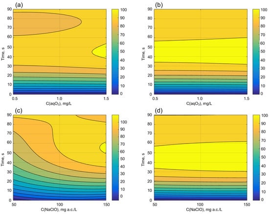
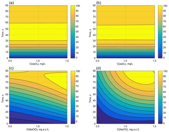
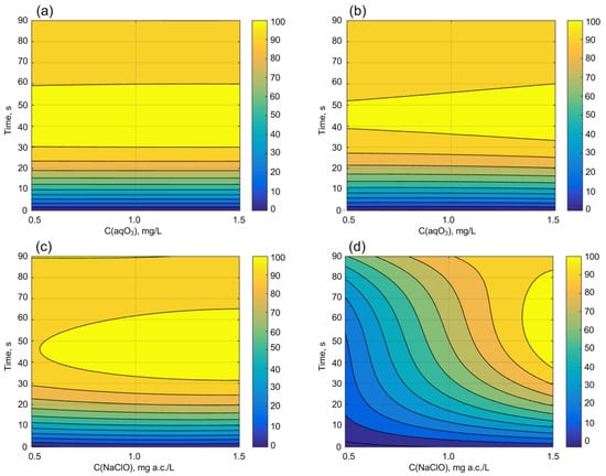
The results of a comparative analysis of the effectiveness of inactivating yeast-like fungi Candida albicans, Gram-positive Bacillus subtilis, and Gram-negative bacteria Escherichia coli by aqueous ozone and sodium hypochlorite solutions, immobilized on metal and polymer plates, are presented in the Supplementary Materials in Figures S1–S9. These data were used for mathematical treatment by defining regression equations and visualization of dependence between the variables (time of exposure and concentration) on the effectiveness of microorganisms inactivation (Figure 1, Figure 2 and Figure 3). Also, this data were used for further comparative analysis of the effectiveness of the inactivation of selected groups of microorganisms by the C·T criterion, which is widely used for comparative analysis of the microorganism’s inactivation efficiency (as further explored in the Section 4).

Figure 1.
Effectiveness of Candida albicans inactivation (Ef) by aqueous ozone solution (a,b) and sodium hypochlorite (c,d), immobilized on metal plates (a,c) and polymer plates (b,d).

Figure 2.
Effectiveness of Bacillus subtilis inactivation by aqueous ozone solution (a,b) and sodium hypochlorite (c,d), immobilized on metal plates (a,c) and polymer plates (b,d).

Figure 3.
Effectiveness of Escherichia coli inactivation by aqueous ozone solution (a,b) and sodium hypochlorite (c,d), immobilized on metal plates (a,c) and polymer plates (b,d).
The results evaluating the effectiveness of aqueous ozone against yeast-like fungi Candida albicans, immobilized on metal and polymer plates, are presented in the Supplementary Materials in Figures S1–S3. The differences between the experimental and control groups were statistically significant in all experiments (p < 0.05). The processed data in MATLAB are shown in Figure 1. Regression Equations (3)–(6) correspond to Figure 1a–d and are provided below.
where C is the concentration of ozonated water or active chlorine in mg/L, and t is the treatment time in seconds.
Ef(aqO3, metal) = 0.03954 + 0.4157·C + 5.36·t − 0.3911·C2 + 0.006039·C·t − 0.09622·t2 + 0.103·C2·t + 0.001525·C·t2 + 0.0005395·t3,
Ef(aqO3, polym) = 1.334 − 3.793·C + 5.761·t + 2.112·C2 + 0.1306·C·t − 0.1038·t2 − 0.02688·C2·t − 0.0009339·C·t2 + 0.0005769·t3,
Ef(NaClO, metal) = − 33.61 + 0.6972·C + 3.071·t − 0.003102·C2 + 0.007932·C·t − 0.04144·t2 + 2.282·10−5·C2·t − 0.0001568·C·t2 + 0.0002616·t3,
Ef(NaClO, polym) = − 3.916 + 0.08282·C + 5.838·t − 0.000375·C2 + 0.0005618·C·t − 0.1046·t2 + 3.033·10−6·C2·t − 1.489·10−5·C·t2 + 0.0005841·t3,
The determination coefficients of the obtained models are 0.9999, 0.9998, 0.9600, and 0.9996, respectively, indicating a high functional relationship between the measured and modeled results. The observed slight decrease in efficiency with increasing time, as seen in the graphs (Figure 1), is because the values of microorganism inactivation efficiency quickly rise from 0 to almost 100%, and the polynomial model cannot precisely fit the measured points, leading to its slight bending.
Results assessing the effectiveness of aqueous ozone against Gram-positive bacteria Bacillus subtilis immobilized on metal and polymer plates are presented in the Supplementary Materials in Figures S4–S6. The processed data in MATLAB are shown in Figure 2. Regression Equations (7)–(10) correspond to Figure 2a–d and are provided below.
where C is the concentration of ozonated water or active chlorine in mg/L, and t is the treatment time in seconds.
Ef(aqO3, metal) = 0.00322 − 0.02615·C + 6.1·t + 0.01971·C2 + 0.002947·C·t − 0.1109·t2 − 0.0003311·C2·t − 2.496·10−5·C·t2 + 0.0006158·t3,
Ef(aqO3, polym) = −0.01188 + 0.1032·C + 6.039·t − 0.07844·C2 − 0.00146·C·t − 0.1102·t2 + 0.007278·C2·t − 8.281·10−5·C·t2 + 0.0006141·t3,
Ef(NaClO, metal) = 10.64 − 0.3556·C − 1.156·t + 0.002142·C2 + 0.02196·C·t + 0.04386·t2 − 1.934·10−5·C2·t − 0.0002174·C·t2 − 0.00021·t3,
Ef(NaClO, polym) = − 11.26 + 0.2772·C − 2.055·t − 0.00141·C2 + 0.04388·C·t + 0.02568·t2 − 0.000113·C2·t − 0.0001751·C·t2 − 0.000102·t3,
The determination coefficients of the obtained models are 1.0000, 1.0000, 0.9651, and 0.9940, respectively, indicating a high functional relationship between the measured and modeled results. The observed slight decrease in efficiency with increasing time, as seen in the graphs (Figure 2), is why the values of microorganism inactivation efficiency quickly rise from 0 to almost 100%. The polynomial model may not precisely fit the measured points, leading to its slight bending.
The results evaluating the effectiveness of aqueous ozone against Gram-negative bacteria Escherichia coli, immobilized on metal and polymer plates, are presented in the Supplementary Materials in Figures S7–S9. The processed data in MATLAB are shown in Figure 3. Regression Equations (11)–(14) correspond to Figure 3a–d and are provided below.
where C is the concentration of ozonated water or active chlorine in mg/L, and t is the treatment time in seconds.
Ef(aqO3, metal) = − 0.01536 + 0.05664·C + 6.089·t − 0.03549·C2 + 0.0321·C·t − 0.1111·t2 − 0.01087·C2·t − 4.735·10−5·C·t2 + 0.0006177·t3,
Ef(aqO3, polym) = 2.294 − 6.527·C + 5.507·t + 3.634·C2 + 0.2255·C·t − 0.09852·t2 − 0.04645·C2·t − 0.001612·C·t2 + 0.0005476·t3,
Ef(NaClO, metal) = − 10.45 + 0.2225·C + 5.449·t − 0.001013·C2 + 0.001159·C·t − 0.09505·t2 + 8.404·10−6·C2·t − 3.638·10−5·C·t2 + 0.0005346·t3,
Ef(NaClO, polym) = − 7.5 + 0.09877·C − 0.7683·t − 0.0002043·C2 + 0.04261·C·t − 0.01206·t2 − 4.739·10−5·C2·t − 0.0003285·C·t2 + 0.000248·t3,
The determination coefficients of the obtained models are 1.0000, 0.9995, 0.9970, and 0.9792, respectively, indicating a high functional relationship between the measured and modeled results. The observed slight decrease in efficiency with increasing time, as seen in the graphs (Figure 3), is why the values of microorganism inactivation efficiency quickly rise from 0 to almost 100%. The polynomial model may not precisely fit the measured points, leading to its slight bending.
4. Discussion
4.1. Comparison of the Effectiveness of Microorganism Inactivation
The obtained results show that the number of colony-forming units on the experimental plates decreases compared to the control samples, both for bacteria and yeast-like fungi, regardless of the plate material. The inactivation efficiency was assessed by comparing the LD50 value, at which a 50% inactivation of the investigated microorganisms was observed [60,61,62]. This indicator was chosen because the effectiveness of sodium hypochlorite is not always above 90% within the selected maximum exposure time. The C·T criterion values at a 50% inactivation efficiency, obtained at different treatment times and various doses of disinfectants, are presented in Table 1.

Table 1.
The average value of the C·T criterion for a 50% inactivation efficiency obtained at different treatment times and various doses of disinfectants.
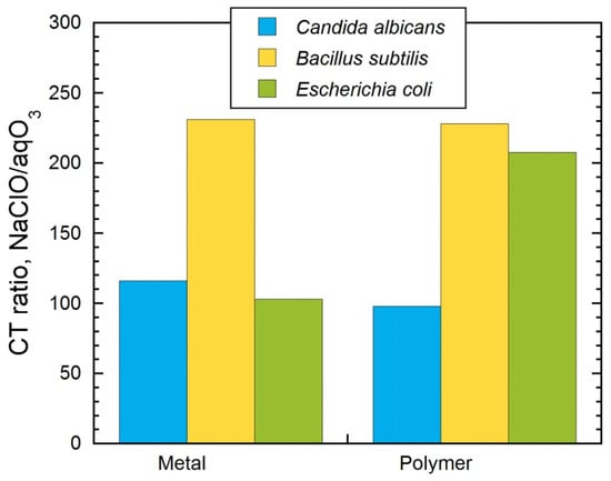
According to the obtained data in this experiment, the aqueous ozone solution is equally effective for the inactivation of yeast-like fungi Candida albicans, as well as Gram-positive Bacillus subtilis and Gram-negative bacteria Escherichia coli, regardless of the substrate (metal or polymer). Also, when using sodium hypochlorite, there was no significant difference in using different substrates for the inactivation of yeast-like fungi Candida albicans, as well as Gram-positive bacteria Bacillus subtilis. Yeast-like fungi proved to be the most easily inactivated, while Gram-positive bacteria Bacillus subtilis showed the highest resistance. Based on the presented results (Figure 4), we can conclude that sodium hypochlorite and aqueous ozone did not affect either the metal or polymer substrates during a short period of aqueous ozone or sodium hypochlorite exposure, like polymer degradation or any reasonable active corrosion of the metal surface. At the same time, the type of substrate had the most significant impact on the inactivation of Escherichia coli. Here, the C·T criterion on a metal plate is 2.04 times lower than during inactivation on a polymer plate. This means that Escherichia coli has a good connection with the polymer substrate and possibly forms the kind of biofilm during the incubation in a thermostat for 24 h at the optimal temperature.

Figure 4.
Comparison of the C·T criterion for ozonated water and sodium hypochlorite solution.
Comparing the C·T criterion at the NaClO to ozonated water ratio at LD50 shows (Figure 4) that the smallest difference of about 100 times is observed during the inactivation of Candida albicans, and the maximum difference is up to 230 times during the inactivation of Bacillus subtilis.
If we compare the inactivation of various types of microorganisms on the surfaces in this experiment with available literature data, a similar order of difference in the C·T criterion for sodium hypochlorite and ozonated water can be noted. However, most studies present results on the inactivation of microorganisms in bulk volume, while there is very little research on the inactivation of microorganisms on surfaces. From published works, it is known that ozone inactivates pathogenic microorganisms 15–20 times faster, and bacterial spores form 300–600 times faster than chlorine [14]. For disinfecting 99% of viruses and Giardia microorganisms, the CT value for ozone is 0.4 mg/(L·min) and 0.48 mg/(L·min), respectively, while for chlorine (chlorine dioxide), the CT value is 37 mg/(L·min) (10 mg/(L·min)) and 1 mg/(L·min) (2.1 mg/(L·min)), respectively [14]. The ozone dose required for water disinfection varies depending on its organic substance content, water temperature, and pH [23]. The ozone added to water can range from 0.7 to 5 mg/L, depending on the initial water quality. The ozone dose and the optimal ozonation scheme are determined based on preliminary technological studies. For the reliable destruction of microorganisms and deactivation of viruses in water, a certain minimum residual ozone concentration of 0.4 mg/L should be present [23,25], with a minimum exposure time of 4 min [43]. Thereby, we can confirm that aqueous ozone is considered a promising eco-friendly reagent instead of hypochlorites and other chlorine-containing disinfectants [62,63].
4.2. The Mechanism of Aqueous Ozone Action
Ozone has a high oxidation-reduction potential, which is the main reason for its activity against various water pollutants, including microorganisms. The oxidizing action of ozone can manifest in the following forms: direct oxidation, radical oxidation (indirect oxidation), ozonolysis, and catalysis [14]. The mechanism of ozone action on microorganisms is as follows: Ozone reacts with the lipids and lipoproteins constituting the microorganisms’ cell walls. These elements have multiple chemical bonds between atoms, and oxidation results in the formation of new angular configurations incompatible with cell viability. Additionally, ozone penetrates the cell membrane of microorganisms and reacts with cytoplasmic substances, causing the transformation of circulating DNA into freely circulating DNA. Thus, the following stages of the action of aqueous ozone can be distinguished [63,64,65]:
- –
- Upon contact with the cell wall of microorganisms, the ozone molecule causes its rupture, as phospholipids and lipoproteins undergo oxidation, leading to the formation of peroxides.
- –
- The resulting ruptures in the cell wall induce stress in the cell, gradually causing it to lose shape, while ozone molecules continue to break down the cell wall further.
- –
- If ozone exposure continues, within a few seconds, the cell wall of microorganisms loses the ability to maintain its shape, and the cell dies.
- –
- Ozone suppresses the activity of microorganisms by partially breaking down their membrane, halting the reproduction process and disrupting their ability to connect with the organism’s cells. It has been demonstrated that Gram-positive bacteria are more sensitive to ozone than Gram-negative bacteria, which is likely related to differences in the structure of their membranes, and bacteria are more sensitive than molds and yeast.
5. Conclusions
As a result of the conducted research, the following conclusions can be drawn:
- –
- Ozonated water is 100–230 times more effective as a disinfectant than sodium hypochlorite solutions, depending on the type of microorganism.
- –
- The efficiency of inactivation depends on the microorganism species, not on the substrate materials, for yeast-like fungi Candida albicans or Gram-positive Bacillus subtilis, but these materials do influence the inactivation of Gram-negative bacteria Escherichia coli, which is more than two fold lower on the polymer surface compared with the metal surface.
- –
- Inactivation efficiency with sodium hypochlorite solutions strongly depends on the substrate material and microorganism species.
This article and others published have made great strides in the investigation of the basic technical aspects of using ozone instead of the commonly used hypochlorites. Further research should be aimed at field experiments and the creation of mobile units for the quick and effective disinfection of surfaces with aqueous ozone instead of chlorine-containing disinfectants.
Supplementary Materials
The following supporting information can be downloaded at https://www.mdpi.com/article/10.3390/w16050793/s1: Figure S1: Petri dishes after inactivation of Candida albicans with an aqueous solution of ozone and sodium hypochlorite, immobilized on metal plates; Figure S2: Petri dishes after inactivation of Candida albicans with an aqueous solution of ozone and sodium hypochlorite, immobilized on polymer plates; Figure S3: Results of inactivation efficiency of Candida albicans vs concentration of ozone and sodium hypochlorite in aqueous solution: (a) in aqO3 on metal surface; (b) in aqO3 on polymer surface; (a) in NaClO on metal surface; (b) in NaClO on polymer surface; Figure S4: Petri dishes after inactivation of Bacillus subtilis with an aqueous solution of ozone and sodium hypochlorite, immobilized on metal plates; Figure S5: Petri dishes after inactivation of Bacillus subtilis with an aqueous solution of ozone and sodium hypochlorite, immobilized on polymer plates; Figure S6: Results of inactivation efficiency of Bacillus subtilis vs concentration of ozone and sodium hypochlorite in aqueous solution: (a) in aqO3 on metal surface; (b) in aqO3 on polymer surface; (a) in NaClO on metal surface; (b) in NaClO on polymer surface; Figure S7: Petri dishes after inactivation of Escherichia coli with an aqueous solution of ozone and sodium hypochlorite, immobilized on metal plates; Figure S8: Petri dishes after inactivation of Escherichia coli with an aqueous solution of ozone and sodium hypochlorite, immobilized on polymer plates; Figure S9: Results of inactivation efficiency of Escherichia coli vs concentration of ozone and sodium hypochlorite in aqueous solution: (a) in aqO3 on metal surface; (b) in aqO3 on polymer surface; (a) in NaClO on metal surface; (b) in NaClO on polymer surface.
Author Contributions
V.R.: supervision, conceptualization, validation, investigation, formal analysis, data curation, visualization, writing—original draft, writing—review and editing. A.P.: investigation, formal analysis, data curation. M.K.: formal analysis, data curation. V.L.: investigation, formal analysis, data curation. N.K.: formal analysis, data curation. E.R.: formal analysis, data curation, writing—review and editing. All authors have read and agreed to the published version of the manuscript.
Funding
The work was carried out with the support of the State Program of Scientific Research “Chemical Processes, Reagents and Technologies, Bioregulators, and Bioorganic Chemistry”, task 2.1.02 “Sorption, Catalytic, and Membrane Materials for Water Purification and Treatment”, Research Project 5 “Physico-Chemical Foundations of Material Corrosion in Disinfecting Environments and Development of Eco-Friendly and Highly Effective Disinfection Methods” (2021–2023).
Data Availability Statement
All data, models, and code generated or used during the study appear in the submitted article.
Conflicts of Interest
Author Vitaly Likhavitski was employed by the company UE QUALITY. The remaining authors declare that the research was conducted in the absence of any commercial or financial relationships that could be construed as a potential conflict of interest.
References
- Hurynovich, A.; Romanovski, V. Artificial replenishment of the deep aquifers. E3S Web Conf. 2018, 45, 00025. [Google Scholar] [CrossRef]
- Yushchenko, V.; Velyugo, E.; Romanovski, V. Development of a new design of deironing granulated filter for joint removal of iron and ammonium nitrogen from underground water. Environ. Technol. 2023. [Google Scholar] [CrossRef] [PubMed]
- Yushchenko, V.; Velyugo, E.; Romanovski, V. Influence of ammonium nitrogen on the treatment efficiency of underground water at iron removal stations. Groundw. Sustain. Dev. 2023, 22, 100943. [Google Scholar] [CrossRef]
- Gurgenidze, D.; Romanovski, V. The Pharmaceutical Pollution of Water Resources Using the Example of the Kura River (Tbilisi, Georgia). Water 2023, 15, 2574. [Google Scholar] [CrossRef]
- Ramanouski, V.I.; Andreeva, N.A. Purification of washing waters of iron removal stations. Proc. BSTU Chem. Technol. Inorg. Subst. 2012, 3, 62–65. [Google Scholar]
- Wang, C.; Bai, L.; Pei, Y. Assessing the stability of phosphorus in lake sediments amended with water treatment residuals. J. Environ. Manag. 2013, 122, 31–36. [Google Scholar] [CrossRef]
- Wang, C.; Qi, Y.; Pei, Y. Laboratory investigation of phosphorus immobilization in lake sediments using water treatment residuals. Chem. Eng. J. 2012, 209, 379–385. [Google Scholar] [CrossRef]
- Wang, C.; Liang, J.; Pei, Y.; Wendling, L.A. A method for determining the treatment dosage of drinking water treatment residuals for effective phosphorus immobilization in sediments. Ecol. Eng. 2013, 60, 421–427. [Google Scholar] [CrossRef]
- Wang, C.; Gao, S.; Pei, Y.; Zhao, Y. Use of drinking water treatment residuals to control the internal phosphorus loading from lake sediments: Laboratory scale investigation. Chem. Eng. J. 2013, 225, 93–99. [Google Scholar] [CrossRef]
- Yuan, N.; Wang, C.; Pei, Y. Investigation on the eco-toxicity of lake sediments with the addition of drinking water treatment residuals. J. Environ. Sci. 2016, 46, 5–15. [Google Scholar] [CrossRef]
- Romanovski, V.; Zhang, L.; Su, X.; Smorokov, A.; Kamarou, M. Gypsum and high quality binders derived from water treatment sediments and spent sulfuric acid: Chemical engineering and environmental aspects. Chem. Eng. Res. Des. 2022, 184, 224–232. [Google Scholar] [CrossRef]
- Huang, C.; Pan, J.R.; Sun, K.D.; Liaw, C.T. Reuse of water treatment plant sludge and dam sediment in brick-making. Water Sci. Technol. 2001, 44, 273–277. [Google Scholar] [CrossRef]
- Kamarou, M.; Moskovskikh, D.; Chan, H.L.; Wang, H.; Li, T.; Akinwande, A.A.; Romanovski, V. Low energy synthesis of anhydrite cement from waste lime mud. J. Chem. Technol. Biotechnol. 2023, 98, 789–796. [Google Scholar] [CrossRef]
- United States Environmental Protection Agency. Disinfection Profiling and Benchmarking Guidance Manual EPA 815-R-99-013; United States Environmental Protection Agency: Washington, DC, USA, 2020; 162p. Available online: https://www.epa.gov/system/files/documents/2022-02/disprof_bench_3rules_final_508.pdf (accessed on 13 November 2023).
- ECHA 2016 Biocidal Products Committee Opinions on Active Substance Approval: Active Chlorine Released from Sodium Hypochlorite. Available online: https://echa.europa.eu/regulations/biocidal-products-regulation/approval-of-active-substances/bpc-opinions-on-active-substance-approval (accessed on 13 November 2023).
- ECHA 2016 Biocidal Products Committee Opinions on Active Substance Approval: Active Chlorine Released from Calcium Hypochlorite. Available online: https://echa.europa.eu/regulations/biocidal-products-regulation/approval-of-active-substances/bpc-opinions-on-active-substance-approval (accessed on 13 November 2023).
- AWWA C653-13; Disinfection of Water Treatment Plants. American Water Works Association: Denver, CO, USA, 2013; ISBN 9781613002513. [CrossRef]
- Bayo, J.; Angosto, J.M.; Gómez-López, M.D. Ecotoxicological screening of reclaimed disinfected wastewater by Vibrio fischeri bioassay after a chlorination–dechlorination process. J. Hazard. Mater 2009, 172, 166–171. [Google Scholar] [CrossRef]
- Bull, R.J. Health effects of drinking water disinfectants and disinfectant by-products. Environ. Sci. Technol. 1982, 16, 554A–559A. [Google Scholar] [CrossRef] [PubMed]
- Jeong, C.H.; Wagner, E.D.; Siebert, V.R.; Anduri, S.; Richardson, S.D.; Daiber, E.J.; McKague, A.B.; Kogevinas, M.; Villanueva, C.M.; Goslan, E.H. Occurrence and toxicity of disinfection byproducts in European drinking waters in relation with the HIWATE epidemiology study. Environ. Sci. Technol. 2012, 46, 12120–12128. [Google Scholar] [CrossRef] [PubMed]
- Villanueva, C.M.; Fernandez, F.; Malats, N.; Grimalt, J.O.; Kogevinas, M. Meta-analysis of studies on individual consumption of chlorinated drinking water and bladder cancer. J. Epidemiol. Community Health 2003, 57, 166–173. [Google Scholar] [CrossRef] [PubMed]
- Rymovskaia, M.V.; Ramanouski, V.I. Effect of used solutions for drinking water supply facilities disinfection to soil. Proc. BSTU Chem. Technol. Inorg. Subst. 2016, 4, 151–155. [Google Scholar]
- Romanovski, V.I.; Gurinovich, A.D.; Chaika, Y.N.; Wawzhenyuk, P. Ozone disinfection of water intake wells and pipelines of drinking water supply systems. Proc. BSTU Chem. Technol. Inorg. Subst. 2013, 3, 51–56. [Google Scholar]
- Hurynovich, A.D.; Romanovski, V.I.; Wawrzeniuk, P. Analiza efektywności kaskadowego generator ozonu. Ekon. I Srodowisko 2013, 1, 156–164. [Google Scholar]
- Ramanouski, V.I.; Likhavitski, V.V.; Hurynovich, A.D. Investigation of ozone solubility in water in height of the liquid. Proc. BSTU Chem. Technol. Inorg. Subst. 2015, 3, 68–72. [Google Scholar]
- Bhuvaneshwari, M.; Eltzov, E.; Veltman, B.; Shapiro, O.; Sadhasivam, G.; Borisover, M. Toxicity of chlorinated and ozonated wastewater effluents probed by genetically modified bioluminescent bacteria and cyanobacteria Spirulina sp. Water Res. 2019, 164, 114910. [Google Scholar] [CrossRef]
- Dong, S.; Massalha, N.; Plewa, M.J.; Nguyen, T.H. The impact of disinfection Ct values on cytotoxicity of agricultural wastewaters: Ozonation vs. chlorination. Water Res. 2018, 144, 482–490. [Google Scholar] [CrossRef] [PubMed]
- Sun, H.; Liu, H.; Han, J.; Zhang, X.; Cheng, F.; Liu, Y. Chemical cleaning-associated generation of dissolved organic matter and halogenated byproducts in ceramic MBR: Ozone versus hypochlorite. Water Res. 2018, 140, 243–250. [Google Scholar] [CrossRef] [PubMed]
- Romanovski, V.I.; Chaika, Y.N. Carbon steels corrosion resistance to disinfectants. Proc. BSTU Chem. Technol. Inorg. Subst. 2014, 3, 40–43. [Google Scholar]
- Ramanouski, V.I.; Zhilinski, V.V. Steel C15E corrosion resistance to disinfectants. Proc. BSTU Chem. Technol. Inorg. Subst. 2015, 3, 24–28. [Google Scholar]
- Schulz, C.; Lohman, S. Method and Apparatus for Ozone Disinfection of Water Supply Pipelines. U.S. Patent App. 11/065,768, 10 November 2005. [Google Scholar]
- Morrison, C.M.; Hogard, S.; Pearce, R.; Gerrity, D.; von Gunten, U.; Wert, E.C. Ozone disinfection of waterborne pathogens and their surrogates: A critical review. Water Res. 2022, 214, 118206. [Google Scholar] [CrossRef]
- Seridou, P.; Kalogerakis, N. Disinfection applications of ozone micro-and nanobubbles. Environ. Sci. Nano 2021, 8, 3493–3510. [Google Scholar] [CrossRef]
- Irie, M.S.; Dietrich, L.; Souza, G.L.D.; Soares, P.B.F.; Moura, C.C.G.; Silva, G.R.D.; Paranhos, L.R. Ozone disinfection for viruses with applications in healthcare environments: A scoping review. Braz. Oral Res. 2022, 36, e006. [Google Scholar] [CrossRef]
- Moccia, G.; De Caro, F.; Pironti, C.; Boccia, G.; Capunzo, M.; Borrelli, A.; Motta, O. Development and improvement of an effective method for air and surfaces disinfection with ozone gas as a decontaminating agent. Medicina 2020, 56, 578. [Google Scholar] [CrossRef]
- Romanovski, V.; Claesson, P.M.; Hedberg, Y.S. Comparison of different surface disinfection treatments of drinking water facilities from a corrosion and environmental perspective. Environ. Sci. Pollut. Res. 2020, 27, 12704–12716. [Google Scholar] [CrossRef]
- Epelle, E.I.; Macfarlane, A.; Cusack, M.; Burns, A.; Okolie, J.A.; Mackay, W.; Rateb, M.; Yaseen, M. Ozone application in different industries: A review of recent developments. Chem. Eng. J. 2023, 15, 140188. [Google Scholar] [CrossRef]
- Kong, J.; Lu, Y.; Ren, Y.; Chen, Z.; Chen, M. The virus removal in UV irradiation, ozonation and chlorination. Water Cycle 2021, 2, 23–31. [Google Scholar] [CrossRef]
- Li, Y.; Li, J.; Ding, J.; Song, Z.; Yang, B.; Zhang, C.; Guan, B. Degradation of nano-sized polystyrene plastics by ozonation or chlorination in drinking water disinfection processes. Chem. Eng. J. 2022, 427, 131690. [Google Scholar] [CrossRef]
- Stange, C.; Sidhu, J.P.S.; Toze, S.; Tiehm, A. Comparative removal of antibiotic resistance genes during chlorination, ozonation, and UV treatment. Int. J. Hyg. Environ. Health 2019, 222, 541–548. [Google Scholar] [CrossRef] [PubMed]
- Zhang, J.M.; Zheng, C.Y.; Xiao, G.F.; Zhou, Y.Q.; Gao, R. Examination of the efficacy of ozone solution disinfectant in inactivating SARS virus. Chin. J. Disinfect. 2004, 21, 27–28. [Google Scholar]
- Hudson, J.B.; Sharma, M.; Vimalanathan, S. Development of a Practical Method for Using Ozone Gas as a Virus Decontaminating Agent. Ozone Sci. Eng. 2009, 31, 216–223. [Google Scholar] [CrossRef]
- Petry, G.; Rossato, L.G.; Nespolo, J.; Kreutz, L.C.; Bertol, C.D. In Vitro Inactivation of Herpes Virus by Ozone. Ozone Sci. Eng. 2014, 36, 249–252. [Google Scholar] [CrossRef]
- Brié, A.; Boudaud, N.; Mssihid, A.; Loutreul, J.; Bertrand, I.; Gantzer, C. Inactivation of murine norovirus and hepatitis A virus on fresh raspberries by gaseous ozone treatment. Food Microbiol. 2018, 70, 1–6. [Google Scholar] [CrossRef]
- Jiang, H.J.; Chen, N.; Shen, Z.Q.; Yin, J.; Qiu, Z.G.; Miao, J.; Yang, Z.W.; Shi, D.Y.; Wang, H.R.; Wang, X.W.; et al. Inactivation of Poliovirus by Ozone and the Impact of Ozone on the Viral Genome. Biomed. Environ. Sci. 2019, 32, 324–333. [Google Scholar] [PubMed]
- Breidablik, H.J.; Lysebo, D.E.; Johannessen, L.; Skare, Å.; Andersen, J.R.; Kleiven, O.T. Ozonized water as an alternative to alcohol-based hand disinfection. J. Hosp. Infect. 2019, 102, 419–424. [Google Scholar] [CrossRef]
- Białoszewski, D.; Bocian, E.; Bukowska, B.; Czajkowska, M.; Sokół-Leszczyńska, B.; Tyski, S. Antimicrobial activity of ozonated water. Med. Sci. Monit. 2010, 16, MT71–MT75. [Google Scholar]
- Bezirtzoglou, E.; Cretoiu, S.-M.; Moldoveanu, M.; Alexopoulos, A.; Lazar, V.; Nakou, M. A quantitative approach to the effectiveness of ozone against microbiota organisms colonizing toothbrushes. J. Dent. 2008, 36, 600–605. [Google Scholar] [CrossRef]
- Ouf, S.A.; Moussa, T.A.; Abd-Elmegeed, A.M.; Eltahlawy, S.R. Anti-fungal potential of ozone against some dermatophytes. Braz. J. Microbiol. 2016, 47, 697–702. [Google Scholar] [CrossRef] [PubMed]
- Hubbezoglu, I.; Zan, R.; Tunç, T.; Sumer, Z.; Hurmuzlu, F. Antifungal Efficacy of Aqueous and Gaseous Ozone in Root Canals Infected by Candida albicans. Jundishapur J. Microbiol. 2013, 6, 1–3. [Google Scholar] [CrossRef]
- Gottschalk, C.; Libra, J.A.; Saupe, A. Ozonation of Water and Waste Water. A Practical Guide to Understanding Ozone and Its Application; Straws Offsetdruck GmbH: Berlin, Germany, 2000; 202p. [Google Scholar]
- Xie, Y. Disinfection Byproducts in Drinking Water: Formation, Analysis, and Control; Lewis Publishers: Boca Raton, FL, USA, 2004; 176p. [Google Scholar]
- Chuwa, C.; Vaidya, D.; Kathuria, D.; Gautam, S.; Sharma, S.; Sharma, B. Ozone (O3): An Emerging Technology in the Food Industry. Food Nutr. J. 2020, 5, 224. [Google Scholar]
- Clark, R.M.; Sivaganesan, M.; Rice, E.W.; Chen, J. Development of a Ct equation for the inactivation of Cryptosporidium oocysts with chlorine dioxide. Water Res. 2003, 37, 2773–2783. [Google Scholar] [CrossRef] [PubMed]
- Clark, R.M.; Sivagenesan, M.; Rice, E.W.; Chen, J. Development of a Ct equation for the inactivation of Cryptosporidium oocysts with ozone. Water Res. 2002, 36, 3141–3149. [Google Scholar] [CrossRef]
- Peleg, M. Modeling the dynamic kinetics of microbial disinfection with dissipating chemical agents—A theoretical investigation. Appl. Microbiol. Biotechnol. 2021, 105, 539–549. [Google Scholar] [CrossRef] [PubMed]
- Sahulka, S.Q.; Bhattarai, B.; Bhattacharjee, A.S.; Tanner, W.; Mahar, R.B.; Goel, R. Differences in chlorine and peracetic acid disinfection kinetics of Enterococcus faecalis and Escherichia fergusonii and their susceptible strains based on gene expressions and genomics. Water Res. 2021, 203, 117480. [Google Scholar] [CrossRef]
- Li, H.; Feng, M.; Yu, X. Qualitative and quantitative analysis of the effects of drinking water disinfection processes on eukaryotic microorganisms: A meta-analysis. Chemosphere 2023, 332, 138839. [Google Scholar] [CrossRef]
- GOST 18301-72; Drinking Water. Methods of Determination of Ozone Residual Content. 1 January 1974; 4p. Available online: https://files.stroyinf.ru/Data2/1/4294850/4294850599.pdf (accessed on 13 November 2023).
- Srivastava, A.K.; Singh, D.; Yadav, P.; Singh, M.; Singh, S.K.; Kumar, A. Paradigm of Well-Orchestrated Pharmacokinetic Properties of Curcuminoids Relative to Conventional Drugs for the Inactivation of SARS-CoV-2 Receptors: An In Silico Approach. Stresses 2023, 3, 615–628. [Google Scholar] [CrossRef]
- Sakudo, A.; Sugiura, K.; Haritani, M.; Furusaki, K.; Kirisawa, R. Antiviral agents and disinfectants for foot-and-mouth disease. Biomed. Rep. 2023, 19, 57. [Google Scholar]
- Radzevičiūtė, E.; Malyško-Ptašinskė, V.; Kulbacka, J.; Rembiałkowska, N.; Novickij, J.; Girkontaitė, I.; Novickij, V. Nanosecond electrochemotherapy using bleomycin or doxorubicin: Influence of pulse amplitude, duration and burst frequency. Bioelectrochemistry 2022, 148, 108251. [Google Scholar] [CrossRef] [PubMed]
- Panebianco, F.; Rubiola, S.; Di Ciccio, P.A. The Use of Ozone as an Eco-Friendly Strategy against Microbial Biofilm in Dairy Manufacturing Plants: A Review. Microorganisms 2022, 10, 162. [Google Scholar] [CrossRef] [PubMed]
- Brodowska, A.J.; Nowak, A.; Śmigielski, K.B. Ozone in the food industry: Principles of ozone treatment, mechanisms of action, and applications: An overview. Crit. Rev. Food Sci. Nutr. 2018, 58, 2176–2201. [Google Scholar] [CrossRef]
- Margalit, M.; Attias, E.; Attias, D.; Elstein, D.; Zimran, A.; Matzner, Y. Effect of ozone on neutrophil function in vitro. Int. J. Lab. Hematol. 2001, 23, 243–247. [Google Scholar] [CrossRef]
Disclaimer/Publisher’s Note: The statements, opinions and data contained in all publications are solely those of the individual author(s) and contributor(s) and not of MDPI and/or the editor(s). MDPI and/or the editor(s) disclaim responsibility for any injury to people or property resulting from any ideas, methods, instructions or products referred to in the content. |
© 2024 by the authors. Licensee MDPI, Basel, Switzerland. This article is an open access article distributed under the terms and conditions of the Creative Commons Attribution (CC BY) license (https://creativecommons.org/licenses/by/4.0/).