Abstract
Background/Objectives: In this study, we conducted molecular identification of R.microplus and explored the genetic diversity of R. microplus for the first time in Mizoram, a Northeastern Hill (NEH) state of India bordering Myanmar. Methods: To assess genetic variation and evolutionary relationships, we employed phylogenetic analyses, genetic divergence metrics, and haplotype network construction based on mitochondrial (COX1 and 16S rDNA) and nuclear (ITS-2 and 18S rDNA) markers. Additionally, multivariate Principal Coordinate Analysis (PCoA) was used to visualize genetic differentiation among R. microplus populations. Results: Our analyses indicated that populations of R. microplus sensu lato from India, Bangladesh, and Pakistan form a closely related matrilineal lineage distinct from R. microplus sensu stricto, clustering within clade C of the COX1-based phylogeny. Globally, 24 COX1 haplotypes were recovered, with 1 haplotype identified in India. The Mizoram population exhibited a single 16S rDNA haplotype; however, intraspecific divergence was evident across India, with seven matrilineal haplotypes detected and nineteen globally. Further, five haplotypes were identified within R. microplus using the ITS-2 marker, while five haplotypes were observed within the Rhipicephalus genus using the 18S rDNA marker. Moreover, this study revealed the presence of Coxiella-like endosymbionts in 95% of the tick specimens analyzed. Conclusions: This study fills a critical knowledge gap by providing the first molecular documentation of tick diversity in Mizoram, a strategic region along the Indo–Myanmar border, and offers novel insights into the phylogeography and symbiotic associations of R. microplus and related tick taxa.
Keywords:
Rhipicephalus microplus; Northeast Hills of India; COX1; 16S rDNA; ITS-2; 18S rDNA; phylogeny 1. Introduction
Ticks are known to serve as vectors for highly pathogenic microorganisms that adversely affect both humans and animals. Moreover, tick infestations induce stress and anemia in cattle, significantly reducing overall productivity [,]. In India, the economic cost for managing tick and tick-borne diseases (TTBDs) in the dairy sector is estimated to be approximately USD 787.63 million annually. Increasing global connectivity has heightened the risk of introducing new diseases and their vectors into previously unaffected areas, posing a serious threat to regional livestock health [].
Mizoram, located in north-eastern India (between 21°56′ N to 24°31′ N latitude and 92°16′ E to 93°26′ E longitude), shares international borders with Myanmar to the east and south and Bangladesh to the west. Importation of cattle from Myanmar into Mizoram was halted after October 2022 (Northeast Today 22 October 2022) []. With reference to this, molecular phylogenetic analysis revealed that the Porcine Reproductive and Respiratory Syndrome (PRRS) virus affecting pigs in Mizoram is genetically closer to the strains found in China and Vietnam than in other regions of India, hinting at possible trans-boundary transmission of the disease [].
The Indo–Myanmar–Bangladesh region is recognized as one of India’s four biodiversity hotspots. Although Brahma et al. (2014) [] have examined tick fauna in Assam, their study did not include samples from other north-eastern states of India. In Arunachal Pradesh, five tick species, R. microplus, Amblyomma integrum, Haemaphysalis longicornis, H. bispinosa and Ixodes ovatus, were identified from three districts based on morphological and molecular approaches, and R. microplus was found to be the most prevalent [].
In contrast, research on tick diversity in Mizoram remains limited. Apart from the morphological identification of A. testudinarium [] and reports of haemoprotozoan pathogens transmitted by Rhipicephalus spp. [], there has been no systematic investigation into tick species composition, genetic variation, or phylogenetic relationships in the state.
The invasive tick species R. microplus is particularly concerning due to its short life cycle and increasing resistance to acaricides, which is attributed to its high adaptability and rapid evolution []. Previous studies based on the Cytochrome C Oxidase subunit I (COX1) gene have identified three major clades of R. microplus: clade A, closely related to R. australis, is found in parts of Southeast Asia, Africa, and the Americas; clade B is exclusively found in China; clade C is prevalent in India, Pakistan, Bangladesh, Myanmar, Thailand, and Malaysia and is related to R. annulatus [,,].
Similarly, analysis of the mitochondrial 16S rDNA gene bisects R. microplus into two clades: clade A (isolates from India and China), which is closely related to R. annulatus, and clade B (isolates from Africa, America, and Southeast Asia) which is closely related to R. australis [,]. Furthermore, the proposition that R. microplus populations from India and China represent cryptic species [], along with the notable diversity of haplotypes within R. microplus s.l. [], underscores the need for more robust phylogenetic analyses. Therefore, this study aims to comprehensively characterize tick species infesting bovines using both newly collected data and previously published genetic sequences from the Northeastern Hill (NEH) region. Given that northern Indian isolates appear cryptic and southern Indian isolates belong to clade C, this research focuses on assessing genetic diversity using mitochondrial (COX1 and 16S rDNA) and nuclear (ITS-2 and 18S rDNA) markers. Additionally, the study seeks to identify and describe endosymbionts present in tick vectors. These endosymbionts may play important roles in the biology and ecology of the host species. Understanding both the genetic diversity and endosymbiont composition of ticks contributes to a more comprehensive understanding of the tick population in this region.
2. Materials and Methods
2.1. Sampling
A total of 949 ticks were collected from Mizoram, India, which experiences a humid mild subtropical climate to humid temperate sub-alpine zones (Figure 1). The average temperature ranges from 20 °C to 29 °C in summer and 11 °C to 21 °C in winter with 2500 mm of rainfall. Tick samples were randomly collected from Zebu cattle (Bos indicus), Taurine cattle (Bos taurus), and Mithun (Bos frontalis) irrespective of age, sex, or breed. Ticks were carefully collected with thumb forceps from the body surfaces of animal and kept immediately in 15 mL centrifuge tube containing 70% alcohol. The collected tick samples were carried to the Department of Veterinary Parasitology, College of Veterinary Sciences and Animal Husbandry (CAU-I), Selesih, Mizoram, for examination and processing. These ticks were sorted a priori based on morphological features []. Before collection of tick samples from the animal, prior approval for a non-invasive mode of sample collection was obtained from the Institutional Animal Ethics Committee (IAEC) of the college.

Figure 1.
Digital elevation map showing the sample locations in Mizoram, India: 1. Sihphir, 2. Durtlang, 3. Muthi, 4. Govt. Complex, 5. Bilkhawthlir, 6. Kolasib, 7. Bairabi, 8. Hnahlan, 9. Champhai, 10. Samthang.
2.2. DNA Isolation and PCR Amplification
DNA isolation from ticks was performed using the Genomic DNA Extraction kit (Thermo Fisher Scientific, Waltham, MA, USA) following the manufacturer’s protocol. To minimize the contamination of host DNA during its extraction, DNA was extracted from an un-engorged tick sample. Four genetic markers were selected for this study such as mitochondrial COX1 and 16S rDNA, nuclear ITS-2 (spanning between the 5.8S and 28S rDNA), and 18S rDNA with the oligonucleotide primers listed in Table 1. The PCR amplification conditions of each gene and the amplicon sizes are also mentioned in Table 1.

Table 1.
List of oligonucleotide primer pairs, the target genes, PCR amplification conditions, amplicon length for the genetic characterization of various ticks, screening of haemoparasites of bovines and endosymbiont.
The PCR products were visualized on 1% (w/v) agarose gel with the addition of ethidium bromide. The PCR amplicons were subsequently purified using GeneJET Gel Extraction Kit (Thermo Fisher Scientific, Waltham, MA, USA). Following purification, the PCR products were ligated into the pTZ57R/T plasmid vector Thermo Fisher Scientific, Waltham, MA, USA and transformed into DH5α Escherichia coli cells in LB agar containing ampicillin. Positive clones, upon confirmation by colony PCR, and the stab cultures of positive clones in LB Agar in microcentrifuge tubes were sent for custom sequencing at the Department of Biochemistry, University of Delhi South Campus.
2.3. Detection of the Endosymbiont in Tick
To examine and characterize the endosymbionts harvested in ticks, 20 randomly selected tick DNA samples were amplified with the oligonucleotide primer pairs listed in Table 1. The samples were subjected to PCR for amplification with the following conditions, as mentioned in Table 2. One representative sample from R. microplus along with samples from H. bispinosa and Amblyomma spp. were selected for custom DNA sequencing. The 405 bp amplicons were purified from agarose gel and cloned into the pTZ57R/T cloning vector. Subsequently, the above-mentioned cloned amplicons were also subject to Sanger DNA sequencing.

Table 2.
The number of haplotypes recovered with each of the genetic markers among the ingroup and among R. microplus and their haplotype diversities.
2.4. Systematics and Molecular Analyses
The newly sequenced DNA fragments were preliminarily checked for their sequence quality and similarity using BioEdit software (version 7.7) and BLASTn (version 2.17.0). Separate datasets were compiled for each of the four markers in the ticks (COX1, 16S rDNA, ITS-2, and 18S rDNA) as well as for 16S rDNA for endosymbionts by combining our newly generated sequences with the published sequences retrieved from the GenBank database. Multiple sequence alignments for each DNA marker were performed using MUSCLE algorithm [] with default parameters in MEGA 11 []. The DNA sequences generated in this study were submitted to GenBank and obtained accession numbers.
To refine the datasets, ambiguously aligned sites were removed using the heuristic method in trimAL software (version 1.4) []. Uncorrected p-distances were estimated in MEGA 11 []. The p-distance matrices from each dataset were standardized and utilized for Principal Coordinate Analysis (PCoA) []. Optimal nucleotide substitution models were determined using PartitionFinder v2.1 [] through the Bayesian Information Criterion (BIC). Bayesian inference (BI) phylogenies were reconstructed separately for each gene using the selected nucleotide substitution models in MrBayes v3.2.5 []. The MCMC was run with four chains (one cold and three hot chains) for 20 million generations and sampled every 5000 generations. The first 25% of trees were discarded as burn-in and the Bayesian posterior probability (PP) values represented the nodal support for the BI tree. The maximum likelihood (ML) phylogenetic tree was also reconstructed in IQ-TREE [] incorporating FreeRate heterogeneity []. The ML tree was run at 10,000 Ultrafast Bootstrap (UFB) replicates [] using partitions determined by PartitionFinder v2.1 [] and models selected based on BIC values by ModelFinder [] integrated into the IQ-TREE []. The best nucleotide substitution models for the BI phylogenetic analyses selected by PartitionFinder v2.1 [] were K81UF+G for 16S, GTR+G for ITS-2, K80+G for 18S, and K80+I for 16S of CLEs, while the best nucleotide substitution models selected for the ML phylogenetic analysis by ModelFinder [] were K3Pu+F+G4 for 16S, TIM3+F+G4 for ITS-2, and K2P+G4 for 18S of ticks, and K2P+I for 16S of CLEs in the dataset. The values of estimated sample size (ESS) for the BI phylogenetic analyses in the four datasets were more than 200. The phylogenetic trees were further illustrated in iTOL software v5 []. The three aligned datasets of ticks were utilized for haplotype diversity assessment for determining the status of the study tick population in DnaSP v.6 []. The haplotype networks were plotted in PopArt v.1.7 [] using the Median-Joining method [].
3. Results
3.1. Systematics and Molecular Phylogeny of R. microplus
In this study, ticks infesting cattle from Mizoram were sampled, which comprised R. microplus and H. bispinosa; Amblyomma spp. were also collected from a species of semi-domesticated bovine, mithun. The generated DNA sequences of this study, such as COX1 (643 bp), 16S rDNA (455 bp), ITS-2 (1500 bp for R. microplus and 1700 bp for H. bispinosa), and 18S rDNA (780 bp), were submitted to GenBank (Supplementary document sheet S1).
After trimming low-quality sequences from both ends, the aligned datasets of COX1, 16S rDNA, ITS-2, and 18S rDNA of ticks and 16S rDNA of endosymbionts (identified as Coxiella-like endosymbionts (CLEs) based on sequence similarity) consist of 849 bp, 363 bp, 729 bp, 1839 bp, and 375 bp aligned sites, respectively. Both the types of phylogenetic inferences (BI and ML) are largely concorded with each other in their topologies (Figure 2A, Figure 3A, Figure 4B and Figure 5A). For most of the cases, we employed the ordination of standardized p-distances (Supplementary Material sheets S2–S5) along the first two principal coordinate axes to further visualize the genetic divergence.

Figure 2.
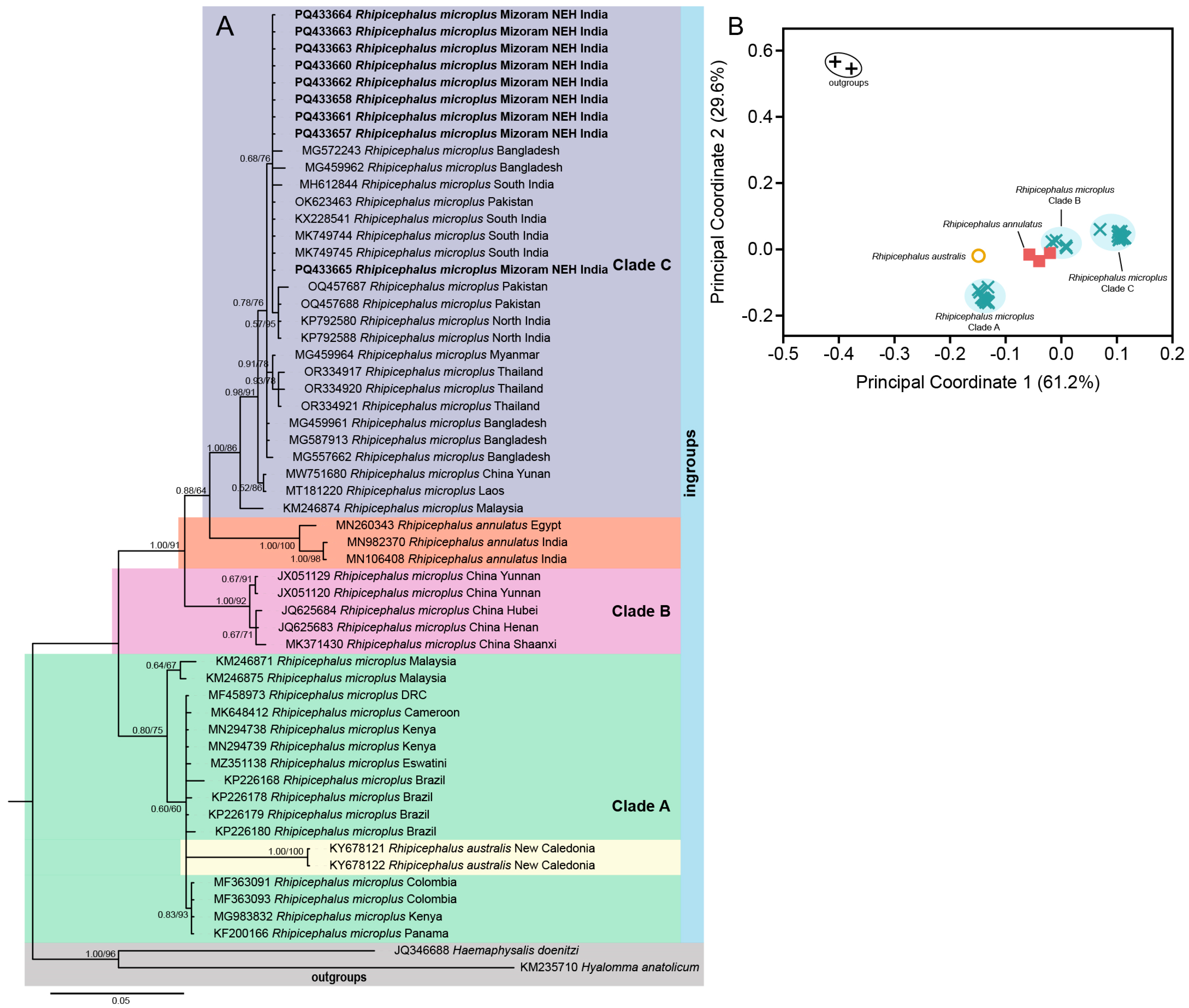
(A) Bayesian inference (BI) phylogenetic tree of the mitochondrial COX1 marker in assigning R. microplus as the ingroup while keeping H. doenitzi and Hyalomma anatolicum as outgroups. The posterior probability (PP) support values from the BI tree are given at each branch, and the ultrafast bootstrap (UFB) support for the corresponding branch from the maximum likelihood (ML) inference tree is also given beside the PP values as PP/UFB. (B) Ordination of standardized p-distance (COX1) among the ingroup and outgroup taxa along the principal coordinate (PCo) axes where a total of 61.2% and 29.6% of the variance are captured by PCo1 and PCo2, respectively.

Figure 3.
(A) Bayesian inference (BI) phylogenetic tree of the mitochondrial 16S rDNA marker in ixodid ticks with assigning R. microplus and R. annulatus as ingroups while keeping H. bispinosa, Dermacentor and other Rhipicephalus species as outgroups. The posterior probability (PP) support values from the BI tree are given at each branch, and the ultrafast bootstrap (UFB) support for the corresponding branch from the maximum likelihood (ML) inference tree is also given beside the PP values as PP/UFB. The unsupported branching from the ML tree is denoted as dash (PP/–). (B) Ordination of standardized p-distance (16S rDNA) among the ingroup and outgroup taxa along the first and second principal coordinate (PCo) axes, where a total of 70% and 21% of the variance are captured by PCo1 and PCo2, respectively. (C) Ordination of standardized p-distance among the ingroup taxa along the first and second principal coordinate (PCo) axes, where a total of 59% and 29% of the variance are captured by PCo1 and PCo2, respectively.

Figure 4.
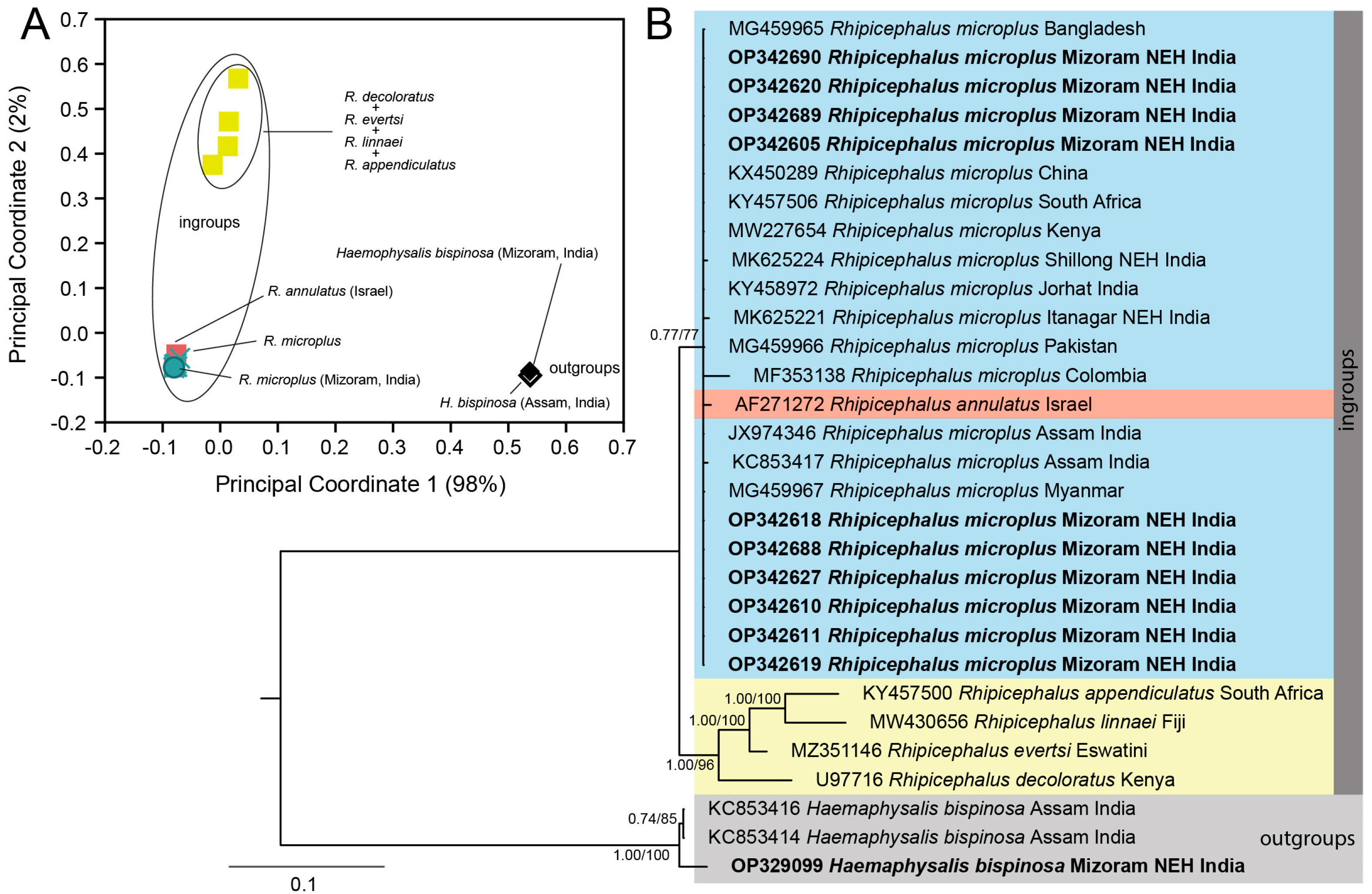
(A) Ordination of standardized p-distance (ITS-2) among the ingroup and outgroup taxa along the first and second principal coordinate (PCo) axes where a total of 98% and 2% of the variance are captured by PCo1 and PCo2, respectively. (B) Bayesian inference (BI) phylogenetic tree of the nuclear ITS-2 marker in ixodid ticks with Rhipicephalus species assigned as ingroups while keeping Haemophysalis bispinosa as an outgroup taxon. The posterior probability (PP) support values from the BI tree are given at each branch, and the ultrafast bootstrap (UFB) support for the corresponding branch from the maximum likelihood inference tree is also given beside the PP values as PP/UFB.

Figure 5.
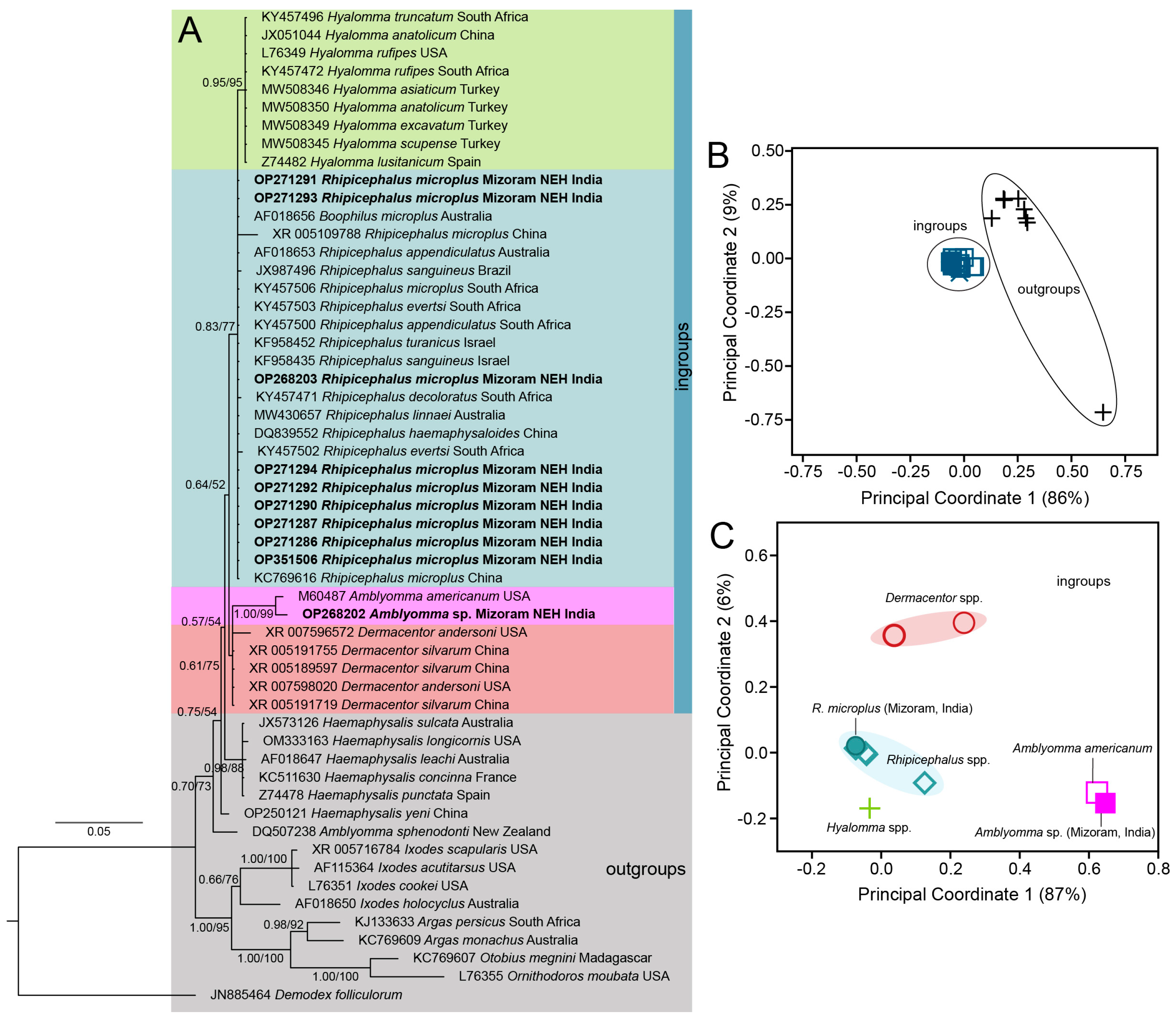
(A) Bayesian inference (BI) phylogenetic tree of the nuclear 18S rDNA marker in ixodid ticks with the genera Rhipicephalus, Hyalomma, Amblyomma, and Dermacentor assigned as ingroups while keeping A. sphenodonti and the genera Haemophysalis, Ixodes, Argas, Otobius, Ornithodorus, and Demodex as outgroups. The posterior probability (PP) support values from the BI tree are given at each branch, and the ultrafast bootstrap (UFB) support for the corresponding branch from the maximum likelihood inference tree is also given beside the PP values as PP/UFB. (B) Ordination of standardized p-distance (18S rDNA) among the ingroup and outgroup taxa along the first and second principal coordinate (PCo) axes, where a total of 86% and 9% of the variance are captured by PCo1 and PCo2, respectively. (C) Ordination of standardized p-distance among the ingroup taxa along the first and second principal coordinate (PCo) axes, where a total of 87% and 6% of the variance are captured by PCo1 and PCo2, respectively.
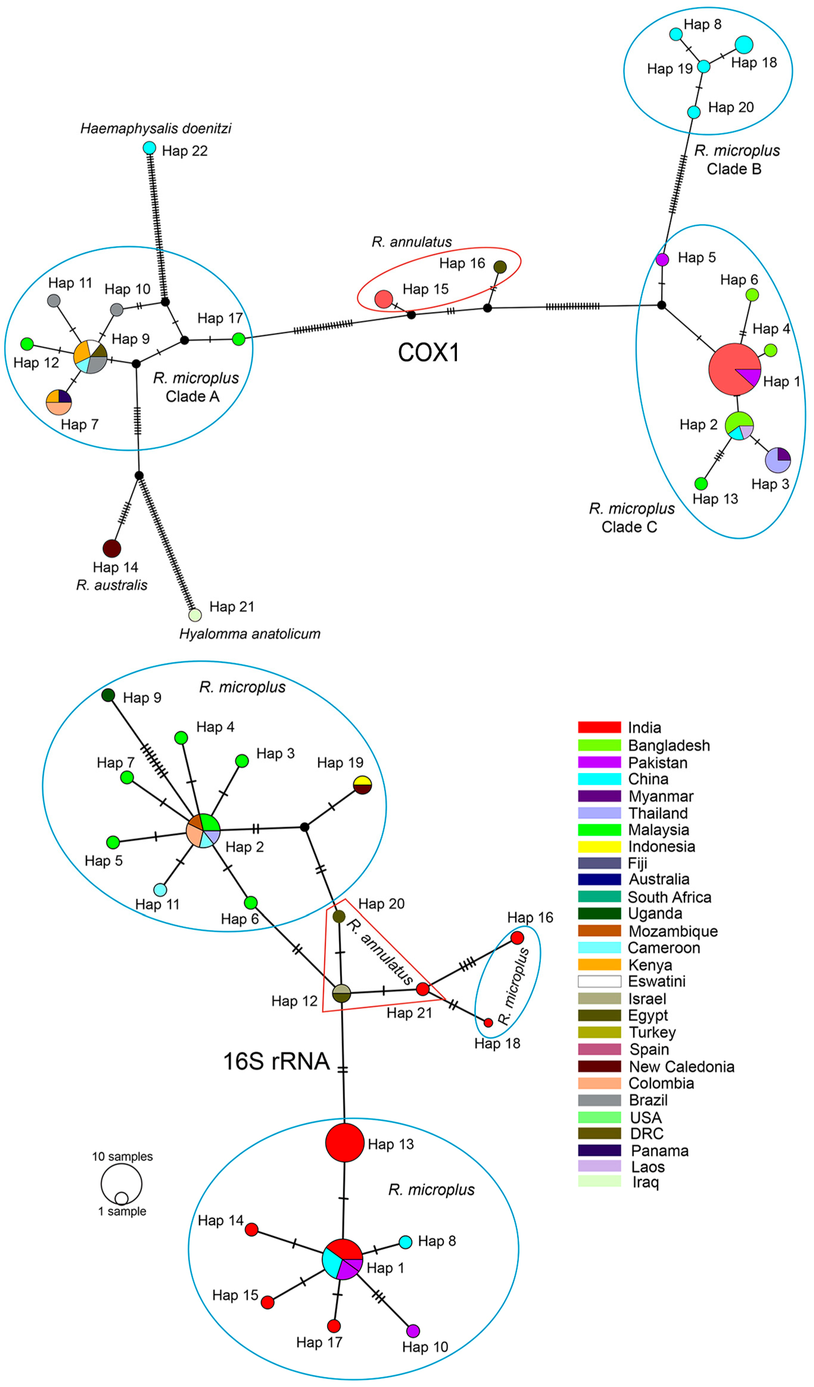
In the mitochondrial COX1 datasets, R. microplus of NEH (Mizoram) was found to be genetically close with other isolates from north and south India, as well as neighboring countries such as Pakistan, Bangladesh, Myanmar, Thailand, Laos, China, and Malaysia. The PCoA plot revealed three clusters of R. microplus, corresponding to the well-supported three distinct clades in the phylogenetic tree, and the Mizoram samples clustered with respect to the other isolates (PP = 1.00; UFB = 91) in clade C (Figure 2A). Further, the clade C formed a sister clade with R. annulatus (PP = 0.88; UFB = 64), as opposed to the other parasites in clade A and B. In this gene fragment, we are able to identify 24 haplotypes with a haplotype diversity (hd) of 0.8774 across ingroups and 0.8531 among R. microplus (Table 2). The Indian isolates, including those in this investigation, are clustered into a single haplotype with Pakistani isolates (Accession No. KP792580) (Figure 3). Analyzing with the neighboring countries, three haplotypes were retrieved from Bangladesh (hap 2, 4, and 6), one haplotype from Myanmar (hap 3), and two from Pakistan (hap 3 and 5) (Figure 6).

Figure 6.
Median-joining haplotype networks based on the mitochondrial COX1 and 16S rDNA. Numbers at the branch represent mutational steps found between haplotypes, and black dots at the branch are either inferred missing or unsampled steps. The different color codes denote the different countries where the samples originate from.
Secondly, using PCoA, the mitochondrial 16S rDNA dataset of R. microplus of this study, isolates from India, certain isolates from China, and R. annulatus were grouped into clade A, while we assigned R. microplus from Malaysia, Cameroon, Thailand, Columbia, and Mozambique together with R. australis into clade B; and the other Rhipicephalus spp., H. bispinosa and Dermacentor atrosignatus as outgroups (Figure 3A). The constructed phylogenetic tree exhibited a well-supported clade that encompassed both R. microplus and R. annulatus with respect to the outgroups (PP = 0.99; UFB = 78). Notably, all of the tick samples from Mizoram (India) were clustered together, forming a sub-clade (PP = 0.70; UFB = 97). Interestingly, R. annulatus appeared to be cohesively clustered among R. microplus (Figure 3B,C). Considering this clustering pattern from PCoA, we estimated the haplotype diversity among the ingroup samples (Table 2), disclosing the existence of 21 distinct haplotypes with a haplotype diversity value (hd) of 0.8995; within R. microplus, 18 distinct haplotypes with an hd of 0.8804 were recovered. Intriguingly, all Mizoram samples were contained within a single haplotype (Hap 13) (Figure 6). This study retrieved seven (7) haplotypes from India, four (4) of which were from the NEH region (Hap 13, 16, 17, and 18). The haplotypes of R. annulatus were nested and interconnecting with R. microplus haplotypes from India (Hap 21), Israel (Hap 12), and Egypt (Hap 12, 20).
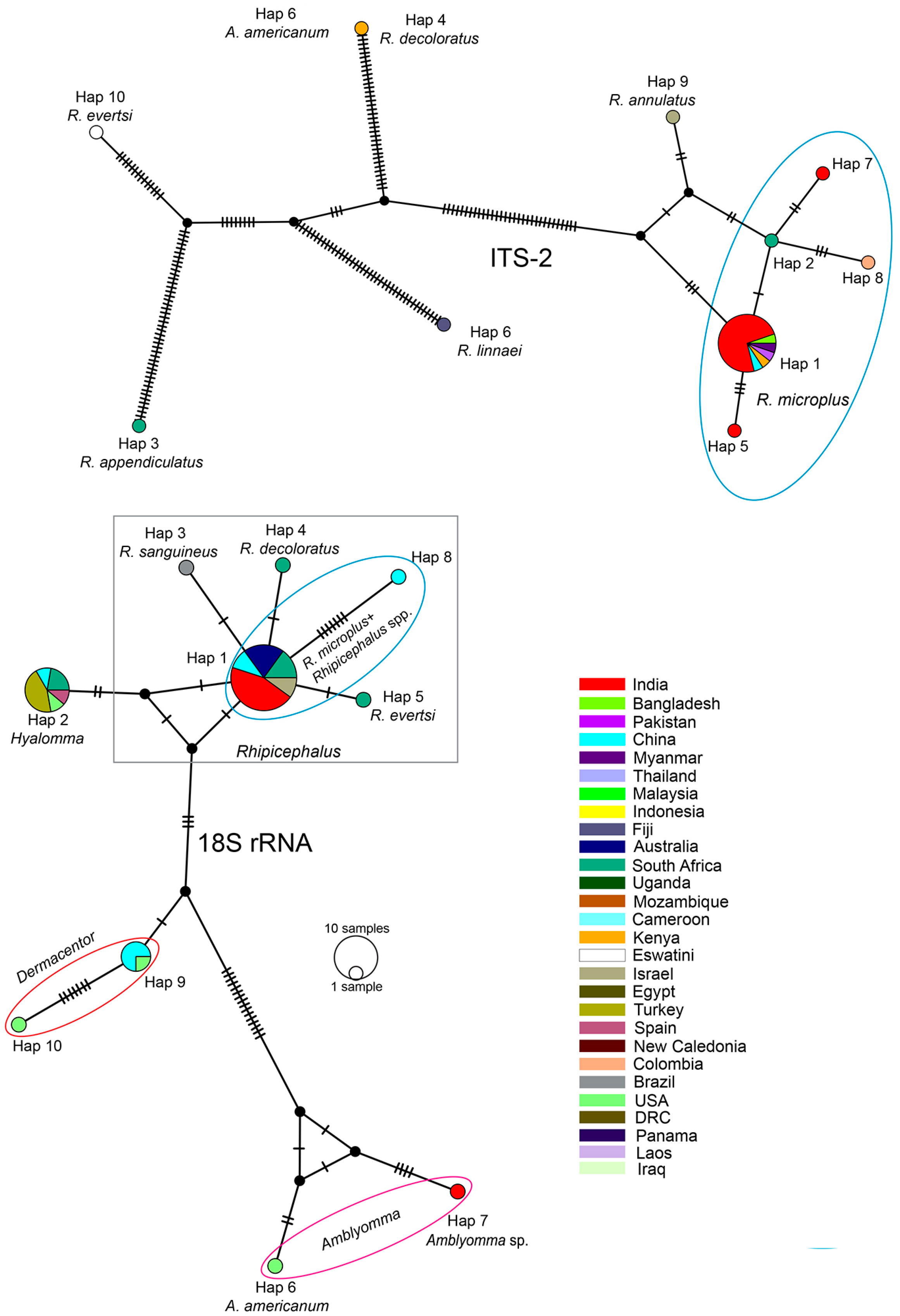
In the nuclear ITS-2 analysis, we assigned all the members of Rhipicephalus as ingroups while assigning H. bispinosa as an outgroup taxon considering the topology of the phylogenetic tree. The insights gained from both phylogenetic inferences and PCoA ordination showed that a cohesive cluster comprising all R. microplus samples, along with a single R. annulatus sample (Israel, Accession No. AF271272), formed a sister clade (PP = 0.77/UFB = 77) distinct from other Rhipicephalus species (Figure 4A,B). Correspondingly, our H. bispinosa sample from Bairabi, sample location no. 7 (Accession No. OP329099), clustered among the two conspecific samples from the neighboring state of India Assam, with robust branch support (PP = 1.00; UFB = 100). Among the ingroups, we determined a total of 10 distinct haplotypes with an hd of 0.5476; within R. microplus, 5 distinct haplotypes with an hd of 0.3377 were recovered (Table 2). Notably, all the R. microplus samples from Mizoram (India), along with the populations from Bangladesh (Accession No. MG459965), Pakistan (Accession No. MG459966), Myanmar (Accession No. MG459967), China (Accession No. KX450289), Kenya (Accession No. MW227654), and India (Jorhat, Accession No. KY458972; Tezpur, Accession No. JX974346; Assam, Accession No. KC853417), formed a single haplotype (Hap 1). In contrast, two Indian samples from Shillong (Hap 5, Accession No. MK625224) and Itanagar (Hap 7, Accession No. MK625221), the South African sample (Hap 2, Accession No. KY457506), and the Colombian sample (Hap 8, Accession No. MF353138) constituted separate haplotypes (Figure 4).
Regarding the nuclear 18S rDNA, based on the PCoA clusters, we assigned the genera Hyalomma, Rhipicephalus, Amblyomma (except A. sphenodonti), and Dermacentor as ingroups, while Haemophysalis, Ixodes, Argas, Otobius, Ornithodorus, Demodex, and A. sphenodonti were considered as outgroups. The resulting phylogenetic tree depicted clustering of Amblyomma spp. samples from Hnahlan {Figure 1 (8)} (Mizoram Accession No. OP268202) with A. americanum from the USA with a well-supported branch (PP = 1.00; UFB = 99) and their genetic divergence was estimated as 0.9%. Notably, Rhipicephalus and Hyalomma formed a distinct clade (PP = 0.83; UFB = 77) in contrast to Amblyomma and Dermacentor. The presence of extensive polytomy, particularly among Rhipicephalus species, complicated the resolution of phylogenetic relationships within this group from the available dataset (Figure 5A).
However, when observing the ordination of the ingroup taxa on the first and second principal coordinate axes, clear clustering emerged among different ingroup taxa. For instance, the R. microplus population from Mizoram (India) displayed cohesive clustering with conspecific sequences. Likewise, the Amblyomma spp. population from Mizoram (India, Accession No. OP268202) clustered alongside A. americanum (Figure 5A,C). Moreover, within the ingroup taxa, a total of 10 distinct haplotypes were identified with an overall hd of 0.7026; within R. microplus, 2 distinct haplotypes with an hd of 0.1538 were recovered (Table 2). Except for the R. microplus sample from China (Hap 8, Accession No. XR_005109788), all the R. microplus samples from different countries and congeneric species such as R. haemophysaloides (China, Accession No. DQ839552), R. appendiculatus (Australia, Accession No. AF018653; South Africa, Accession No. KY457500), R. sanguineus (Israel, Accession No. KF958435), R. turanicus (Israel, Accession No. KF958452), R. evertsi (South Africa, Accession No. KY457503), and R. linnaei (Australia, Accession No. MW430657) were accommodated in a single haplotype (Hap 1). However, distinct haplotypes were formed by other Rhipicephalus species like R. sanguineus (Hap 3), R. decoloratus (Hap 4), and R. evertsi (Hap 5). Additionally, other ingroup taxa formed distant haplotypes like Hyalomma (Hap 2), Dermacentor (Hap 9, 10), A. americanum (Hap 6), and Amblyomma spp. from Mizoram, India (Hap 7) (Figure 7).

Figure 7.
Median-joining haplotype networks based on the nuclear 18S rDNA and ITS-2 markers in ixodid ticks. Numbers at the branch represent mutational steps found between haplotypes, and black dots at the branches are either inferred missing or unsampled steps. The different color codes denote the different countries where the samples originate from.
3.2. Detection of the Endosymbiont in Tick
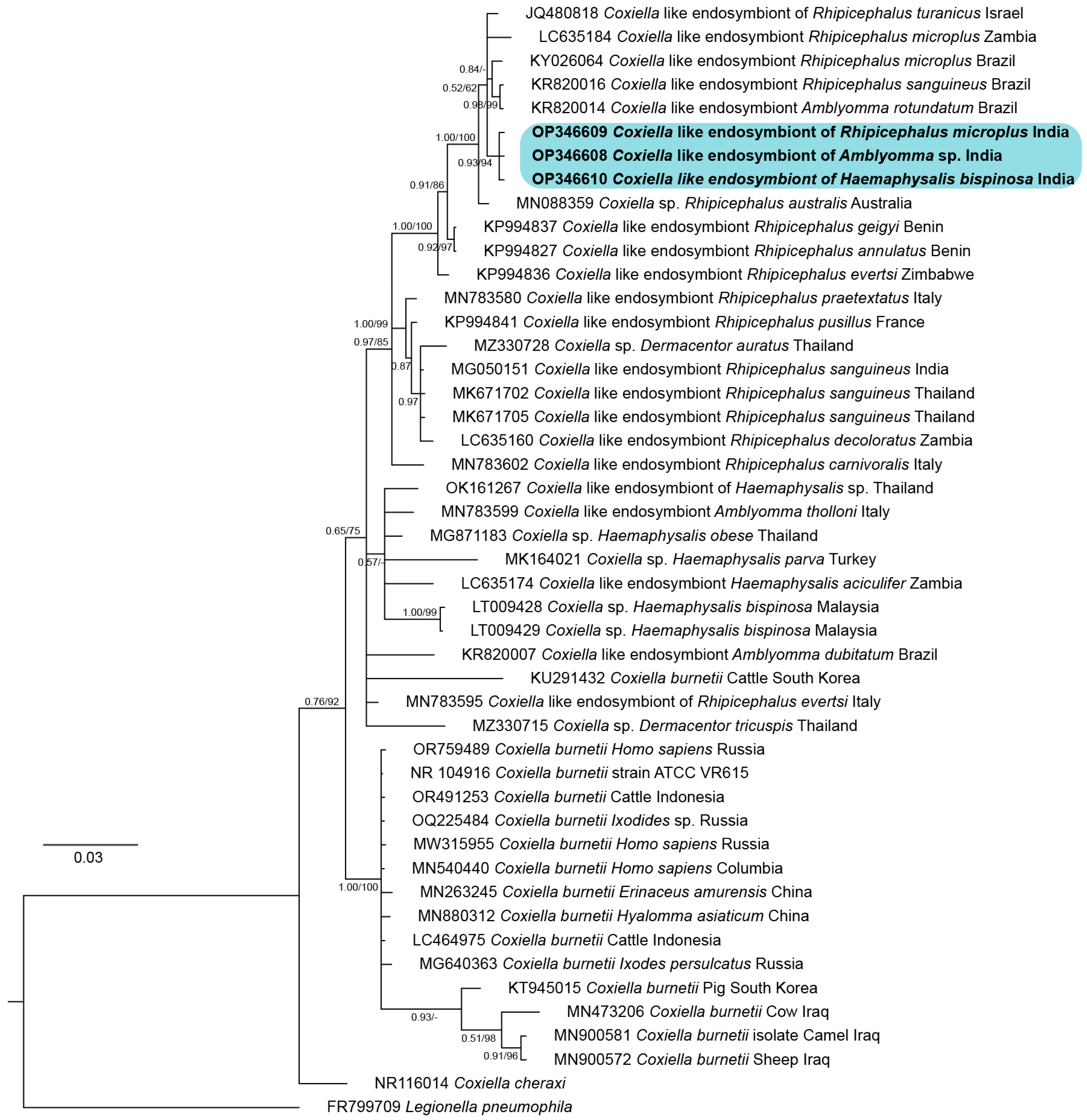
Twenty randomly selected tick samples representing three species of ticks (R. microplus, H. bispinosa, and Amblyomma spp.) were subject to screening for the presence of endosymbionts using the 16S rDNA universal primer for prokaryotes (Table 1). Out of the 20 randomly selected tick samples, 95% (n = 19) showed amplification of the desired fragment size, at ~405 bp. From this pool, one representative amplicon from each of the three tick species was selected for subsequent custom DNA sequencing. Analyses of the DNA sequences from these representative amplicons showed 100% identity to each other. Additionally, the sequence showed a similarity of 93–99% with various Coxiella sp. sequences available in the public database. Notably, the DNA sequence generated in the present study showed a similarity of 95.9% with C. burnetti.
The BI and ML trees of the Coxiella spp. based on the 16S rDNA fragment showed that the tick samples from Mizoram (Accession No. OP346608-10) formed a distinct cluster with a well-supported branch (PP = 0.93; UFB = 94) and were forming a subclade together with JQ480818 (Coxiella-like endosymbiont of R. turanicus from Israel), LC635184 (Coxiella-like endosymbiont of R. microplus from Zambia), KY026064 (Coxiella-like endosymbiont of R. microplus from Brazil), KR820014 (Coxiella-like endosymbiont of R. rotundatum from Brazil), and KR820016 (Coxiella-like endosymbiont of R. sanguineus from Brazil) (Figure 8). This subclade was forming a sister lineage to MN088359 (Coxiella sp. of R. australis) with a highly supported branch (PP = 1.00; UFB = 100).

Figure 8.
Bayesian inference (BI) phylogenetic tree of the mitochondrial 16S rDNA marker in Coxiella with Legionella pneumophila assigned as the outgroup. The posterior probability (PP) support values from the BI tree are given at each branch, and the ultrafast bootstrap (UFB) support for the corresponding branch from the maximum likelihood (ML) inference tree is also given beside the PP values as PP/UFB. The unsupported branching from the ML tree is denoted as dash (PP/–). The color shading corresponds to the sequences generated in this study.
4. Discussion
The cattle tick, R. microplus, is the most widely distributed tick in the world infesting bovine livestock [,]. This well-known parasite not only sucks blood from animals but also transmits some of the most important diseases of livestock such as babesiosis, oriental theileriosis, and anaplasmosis [,]. The parasite has complex taxonomic characteristics where the R. microplus complex includes R. annulatus, R. australis, and R. microplus clades A-C based on COX1 [,]. The COX1 marker helps in the reinstatement of R. australis and investigation of intraspecies variation as well as cryptic diversity which may arise from admixture of the parasite species []. In India, R. microplus clade C is found in the northern and southern states [], and it is considered to be a cryptic species []. Morphological differentiation from its sister species, R. annulatus, can be reliably made by observing the caudal appendage in males; however, caution is advised as this feature may occasionally be absent []. Clade A of R. microplus spread to Africa and America through cattle importation [,]. In certain areas of southeast Asia, such as northeast Thailand and Malaysia, clade A is seen along with clade C [,], demonstrating the admixture of the parasite. Therefore, understanding its distribution [,], the diversity of various subspecies, and the pathogens and the endosymbionts it harbors will help in gaining insights into tick biology for effective tick control [].
In the present study, tick samples were collected from Mizoram, the NEH state of India bordering Myanmar. Three types of hard ticks were identified as R. microplus, H. bispinosa and Amblyomma spp. based on COX1, 16S rDNA, ITS-2, and 18S rDNA; and we studied their molecular phylogeny and their haplotype diversity as well. By molecular analyses, we reaffirm that R. microplus in the Indian subcontinent belongs to clade C based on COX1 corroborating earlier findings from South India and neighboring countries like Bangladesh, Myanmar, and Pakistan [,,,,,]. Upon analysis of the partial DNA sequence of R. microplus based on COX1, this study revealed 23 haplotypes across the world. An integrative taxonomic study is imperative to disclose the precise species boundaries within the R. microplus complex. Comprehensive studies encompassing morphology, molecular analysis, and crossbreeding are of paramount importance to uncover the potential biological mechanisms that might underlie conflicting phylogenetic signals among different loci of the genome (e.g., hybridization and/or introgression, incomplete lineage sorting, etc.).
Our analysis of 16S rDNA also revealed the presence of 18 haplotypes across the world and 7 unique haplotypes specific to India, with 2 of them (hap 16 and 18) displaying closer proximity to R. annulatus than R. microplus. Moreover, the study population of R. microplus in this work formed a single haplotype, while the overall Indian R. microplus samples showed a noticeable intraspecies divergence, with a total of seven distinct matrilineal haplotypes in the 16S rDNA marker, while a single and three distinct haplotypes were seen in ITS-2 and 18S rDNA markers, respectively. These findings align with prior observations of similar polytomies identified within specific Rhipicephalus species groups, as highlighted by Bakkes et al. (2021) []. By superimposing the evidence from our phylogenetic inferences, genetic divergence, and haplotype estimation, we are convinced of the presence of cryptic diversity within R. microplus in India, as previously elucidated by Burger et al. (2014) [].
Previous research has established that the evolution of the tick species experiencing pronounced diversification rates, such as the R. sanguineus group, is shaped by climatic conditions marked by wide annual and seasonal temperature variations []. Additionally, niche divergence driven by climatic conditions during dispersion episodes in off-host habitats can also contribute to limiting tick distribution and fostering speciation [,]. The haplotype estimation analysis conducted in the present study using 16S rDNA gene sequence aligns with this assertion by revealing a closely related matrilineal lineage between tick populations in China and India [,,]. Conversely, upon analyses with the COX1 marker, the Indian (including Pakistan, Bangladesh, and Myanmar) and Malaysian isolates formed a discrete clade C [,], indicating a convergence among the Indian isolates based on the marker selection. The imprecise demarcation particularly notable in the Indian isolates could potentially be addressed through morpho-taxonomic studies, bio-geographic evaluation of their dispersal pattern, exploration of interbreeding tendencies with other species, expansion of sample size, and utilization of more comprehensive inferential approaches.
Systematic studies using phylogeny and haplotype networks using ITS-2 unveiled a distinct clustering of R. microplus and R. annulatus apart from other Rhipicephalus spp. However, this genetic marker was previously criticized for its ineffectiveness in differentiating R. microplus and R. annulatus in southern India and neighboring countries [,]. Burger et al. (2014) [] recommended the exploration of an alternative nuclear marker to ITS-2. The present research found a low genetic divergence between R. microplus and R. annulatus using ITS-2 when compared to other ticks under the genus Rhipicephalus spp. Despite these previous studies, our research also asserts that the haplotype network of ITS-2 distinctively demarcates these sister species, demonstrating its potential for use in tick genetic diversity studies. In contrast, analysis of the 18S rDNA sequences failed to provide resolution beyond the generic level of tick in the phylogenetic inference, aligning with the observations of Mangold et al. (1998) []. Nevertheless, the haplotype network exhibited significant demarcation at the generic level of the ixodid ticks. This outcome also suggests the amalgamation of the Boophilus and Rhipicephalus genera (Murrell et al. 2000; Murrell & Barker 2003) [,].
This study also unveiled that 95% of the ticks harvested had CLEs. Instances of C. burnetti infection have been reported in goat-derived ixodid ticks in the north-eastern parts of India []. The BI tree depicted a sister lineage (putative species B) to a distinct clade comprising CLEs derived from various ticks and the pathogenic C. burnetti. An earlier study had shown that Dermacentor variabilis Kunitz-type serine protease inhibitor has a static effect against Rickettsia montanensis []. Consequently, the pathogenicity of these uncultured organisms identified as tick endosymbionts is still debatable, necessitating further comprehensive investigations.
5. Conclusions
This study genetically identified three tick species, R. microplus, H. bispinosa, and Amblyomma spp., for the first time from Mizoram, a Northeastern Hill (NEH) state in India. Based on the data we generated and recent systematic frameworks by previous researchers, we speculate that R. microplus s.l. dispersed across various parts of Asia through complex networks. This study also confirms that various haplotypes of the parasites circulating within India could be a cryptic species, as earlier suggested. Consequently, thorough elucidation of population genetic diversity through this study underscores the necessity of comprehensive investigations into vectors and vector-borne diseases in the north-eastern part of India. Such efforts are vital for implementing effective measures to mitigate economic losses in the livestock sector. The haplotype dispersal among R. microplus elucidated in this study further underscores the influence of animal transportation as a key factor driving intraspecific genetic divergence. Given that ticks are often found attached to bovine livestock, their potential to facilitate transboundary movement is significant, which could influence the transmission of ticks and tick-borne diseases (TTBDs) in the region, affecting the livestock business in the country.
Supplementary Materials
The following supporting information can be downloaded at https://www.mdpi.com/article/10.3390/genes16101216/s1.
Author Contributions
Conceptualization, K.L. (Khawlhring Lalawmpuii), P.B., L.B. and H.L.; methodology and supervision, K.L. (Khawlhring Lalrintluanga), T.C.T., J.L., C.L. and H.L.; software, L.B. and H.L.; writing—original draft preparation, K.L. (Khawlhring Lalawmpuii), S.S.J., L.B. and H.L.; writing—review and editing, H.T.L., K.L. (Khawlhring Lalawmpuii) and H.L. All authors have read and agreed to the published version of the manuscript.
Funding
This research was carried out as part of MVSc Research in the College of Veterinary Sciences & Animal Husbandry, Aizawl, Mizoram Central Agricultural University, Imphal, India.
Institutional Review Board Statement
The study was conducted after being approved by the Institutional Animal Ethics Committee (IAEC), 2020 (CVSC/CAU/IAEC/19-20/p-6).
Informed Consent Statement
Not applicable.
Data Availability Statement
The GenBank accession numbers are provided in each of the DNA sequences generated in this study.
Acknowledgments
The authors truly acknowledge Dean, College of Veterinary Sciences & Animal Husbandry, CAU, Selesih, Mizoram for providing the necessary facility and funds for DNA sequencing. The authors also would like to acknowledge the veterinarians who helped in tick sample collection from various parts of Mizoram: Engkunga Chhangte, Lalruatfela, Zohlimpuia, Malsawmdawngliana, Zomuana, and Zomawia. The authors have edited and reviewed the output and take full responsibility for the content of the publication. During the preparation of this manuscript/study, the author(s) used QuillBot (free version) and Copilot (free version) for the purpose of grammar editing and paraphrasing. The authors have reviewed and edited the output and take full responsibility for the content of this publication.
Conflicts of Interest
The authors declare no conflicts of interest. The funders had no role in the design of the study; in the collection, analyses, or interpretation of data; in the writing of the manuscript; or in the decision to publish the results.
Abbreviations
The following abbreviations are used in this manuscript:
NEH | Northeastern Hill |
COX1 | Cytochrome C Oxidase sub-unit 1 |
PCoA | Principal Coordinate Analysis |
rDNA | Ribosomal Deoxyribonucleic acid |
TTBDs | Ticks and tick-borne diseases |
IAEC | Institutional Animal Ethics Committee |
ITS-2 | Internal Transcribed Spacer-2 |
BIC | Bayesian Information Criterion |
UFB | Ultrafast bootstrap |
CLEs | Coxiella-like endosymbionts |
References
- Johansson, M.; Mysterud, A.; Flykt, A. Livestock owners’ worry and fear of tick-borne diseases. Parasit. Vectors 2020, 13, 331. [Google Scholar] [CrossRef]
- Singh, K.; Kumar, S.; Sharma, A.K.; Jacob, S.S.; RamVerma, M.; Singh, N.K.; Shakya, M.; Sankar, M.; Ghosh, S. Economic impact of predominant ticks and tick-borne diseases on Indian dairy production systems. Exp. Parasitol. 2022, 243, 108408. [Google Scholar] [CrossRef] [PubMed]
- Bishop, R.P.; Githaka, N.W.; Bazarusanga, T.; Bhushan, C.; Biguezoton, A.; Vudriko, P.; Muhanguzi, D.; Tumwebaze, M.; Bosco, T.J.; Shacklock, C.; et al. Control of ticks and tick-borne diseases in Africa through improved diagnosis and utilisation of data on acaricide resistance. Parasit. Vectors 2023, 16, 224. [Google Scholar] [CrossRef] [PubMed]
- Mizoram Government Bans Import of Cattle from Myanmar. Northeast Today. 22 October 2022. Available online: https://www.indiatodayne.in/mizoram/story/mizoram-government-bans-import-cattle-myanmar-461713-2022-10-22 (accessed on 21 October 2022).
- Rajkhowa, T.K.; Mohanarao, J.G.; Gogoi, A.; Hauhnar, L. Indian porcine reproductive and respiratory syndrome virus bears discontinuous deletion of 30 amino acids in nonstructural protein 2. Virusdisease 2016, 27, 287–293. [Google Scholar] [CrossRef] [PubMed]
- Brahma, R.K.; Dixit, V.; Sangwan, A.K.; Doley, R. Identification and characterization of Rhipicephalus (Boophilus) microplus and Haemaphysalis bispinosa ticks (Acari: Ixodidae) of North East India by ITS2 and 16S rDNA sequences and morphological analysis. Exp. Appl. Acarol. 2014, 62, 253–265. [Google Scholar] [CrossRef]
- Bagra, N.; Roy, P.; Elango, A.; Basumatary, M.J.; Mize, D.; Doley, R. Morpho-molecular characterization of tick species prevalent in domesticated and semi-domesticated bovine species in three districts of Arunachal Pradesh. Acta Trop. 2025, 268, 107720. [Google Scholar] [CrossRef]
- Chamuah, J.K.; Bhattacharjee, K.; Sarmah, P.C.; Raina, O.K.; Mukherjee, S.; Rajkhowa, C. Report of Amblyomma testudinarium in mithuns (Bos frontalis) from eastern Mizoram (India). J. Parasit. Dis. 2016, 40, 1217–1220. [Google Scholar] [CrossRef]
- Ghosh, S.; Patra, G.; Borthakur, S.K.; Behera, P.; Tolenkhomba, T.C.; Deka, A.; Khare, R.K.; Biswas, P. Prevalence of haemoprotozoa in cattle of Mizoram, India. Biol. Rhythm Res. 2020, 51, 76–87. [Google Scholar] [CrossRef]
- Muhanguzi, D.; Byaruhanga, J.; Amanyire, W.; Ndekezi, C.; Ochwo, S.; Nkamwesiga, J.; Mwiine, F.N.; Tweyongyere, R.; Fourie, J.; Madder, M.; et al. Invasive cattle ticks in East Africa: Morphological and molecular confirmation of the presence of Rhipicephalus microplus in south-eastern Uganda. Parasit. Vectors 2020, 13, 165. [Google Scholar] [CrossRef]
- Low, V.L.; Tay, S.T.; Kho, K.L.; Koh, F.X.; Tan, T.K.; Lim, Y.A.; Ong, B.L.; Panchadcharam, C.; Norma-Rashid, Y.; Sofian-Azirun, M. Molecular characterisation of the tick Rhipicephalus microplus in Malaysia: New insights into the cryptic diversity and distinct genetic assemblages throughout the world. Parasit. Vectors 2015, 8, 341. [Google Scholar] [CrossRef]
- Tantrawatpan, C.; Vaisusuk, K.; Chatan, W.; Pilap, W.; Suksavate, W.; Andrews, R.H.; Petney, T.N.; Saijuntha, W. Genetic diversity and phylogenetic analyses of ixodid ticks infesting cattle in northeast Thailand: The discovery of Rhipicephalus microplus clade C and the rarely detected R. haemaphysaloides. Exp. Appl. Acarol. 2022, 86, 535–548. [Google Scholar] [CrossRef] [PubMed]
- Barker, S.C.; Barker, D. Ticks of Australasia: 125 species of ticks in and around Australia. Zootaxa 2023, 5253, 1–670. [Google Scholar] [CrossRef] [PubMed]
- Li, L.H.; Zhang, Y.; Wang, J.Z.; Li, X.S.; Yin, S.Q.; Zhu, D.; Xue, J.B.; Li, S.G. High genetic diversity in hard ticks from a China-Myanmar border county. Parasit. Vectors 2018, 11, 469. [Google Scholar] [CrossRef] [PubMed]
- Ali, A.; Khan, M.A.; Zahid, H.; Yaseen, P.M.; Khan, Q.M.; Nawab, J.; Rehman, Z.; Ateeq, M.; Khan, S.; Ibrahim, M. Seasonal dynamics, record of ticks infesting humans, wild and domestic animals and molecular phylogeny of Rhipicephalus microplus in Khyber Pakhtunkhwa Pakistan. Front. Physiol. 2019, 10, 793. [Google Scholar] [CrossRef] [PubMed]
- Burger, T.D.; Shao, R.; Barker, S.C. Phylogenetic analysis of mitochondrial genome sequences indicates that the cattle tick, Rhipicephalus (Boophilus) microplus, contains a cryptic species. Mol. Phylogenet. Evol. 2014, 76, 241–253. [Google Scholar] [CrossRef]
- Thinnabut, K.; Rodpai, R.; Sanpool, O.; Maleewong, W.; Tangkawanit, U. Genetic diversity of tick (Acari: Ixodidae) populations and molecular detection of Anaplasma and Ehrlichia infesting beef cattle from upper-northeastern Thailand. Infect. Genet. Evol. 2023, 107, 105394. [Google Scholar] [CrossRef]
- Barker, S.C.; Walker, A.R. Ticks of Australia. The species that infest domestic animals and humans. Zootaxa 2014, 3816, 1–144. [Google Scholar] [CrossRef]
- Csordas, B.G.; Garcia, M.V.; Cunha, R.C.; Giachetto, P.F.; Blecha, I.M.; Andreotti, R. New insights from molecular characterization of the tick Rhipicephalus (Boophilus) microplus in Brazil. Rev. Bras. Parasitol. Vet. 2016, 25, 317–326. [Google Scholar] [CrossRef]
- Chitimia, L.; Lin, R.Q.; Cosoroaba, I.; Wu, X.Y.; Song, H.Q.; Yuan, Z.G.; Zhu, X.Q. Genetic characterization of ticks from southwestern Romania by sequences of mitochondrial cox1 and nad5 genes. Exp. Appl. Acarol. 2010, 52, 305–311. [Google Scholar] [CrossRef]
- Lv, J.; Wu, S.; Zhang, Y.; Zhang, T.; Feng, C.; Jia, G.; Lin, X. Development of a DNA barcoding system for the Ixodida (Acari: Ixodida). Mitochondrial DNA 2014, 25, 142–149. [Google Scholar] [CrossRef]
- Ceraul, S.M.; Dreher-Lesnick, S.M.; Mulenga, A.; Rahman, M.S.; Azad, A.F. Functional characterization and novel rickettsiostatic effects of a Kunitz-type serine protease inhibitor from the tick Dermacentor variabilis. Infect. Immun. 2008, 76, 5429–5435. [Google Scholar] [CrossRef]
- Edgar, R.C. MUSCLE: Multiple sequence alignment with high accuracy and high throughput. Nucleic Acids Res. 2004, 32, 1792–1797. [Google Scholar] [CrossRef]
- Tamura, K.; Stecher, G.; Kumar, S. MEGA11: Molecular evolutionary genetics analysis version 11. Mol. Biol. Evol. 2021, 38, 3022–3027. [Google Scholar] [CrossRef] [PubMed]
- Capella-Gutiérrez, S.; Silla-Martínez, J.M.; Gabaldón, T. trimAl: A tool for automated alignment trimming in large-scale phylogenetic analyses. Bioinformatics 2009, 25, 1972–1973. [Google Scholar] [CrossRef]
- Gower, J.C. Some distance properties of latent root and vector methods used in multivariate analysis. Biometrika 1966, 53, 325–338. [Google Scholar] [CrossRef]
- Lanfear, R.; Frandsen, P.B.; Wright, A.M.; Senfeld, T.; Calcott, B. PartitionFinder 2: New methods for selecting partitioned models of evolution for molecular and morphological phylogenetic analyses. Mol. Biol. Evol. 2017, 34, 772–773. [Google Scholar] [CrossRef]
- Ronquist, F.; Teslenko, M.; van der Mark, P.; Ayres, D.L.; Darling, A.; Hohna, S.; Larget, B.; Liu, L.; Suchard, M.A.; Huelsenbeck, J.P. MrBayes 3.2: Efficient Bayesian phylogenetic inference and model choice across a large model space. Syst. Biol. 2012, 61, 539–542. [Google Scholar] [CrossRef]
- Nguyen, L.T.; Schmidt, H.A.; von Haeseler, A.; Minh, B.Q. IQ-TREE: A fast and effective stochastic algorithm for estimating maximum-likelihood phylogenies. Mol. Biol. Evol. 2015, 32, 268–274. [Google Scholar] [CrossRef] [PubMed]
- Soubrier, J.; Steel, M.; Lee, M.S.; Sarkissian, C.; Guindon, S.; Ho, S.Y.; Cooper, A. The influence of rate heterogeneity among sites on the time dependence of molecular rates. Mol. Biol. Evol. 2012, 29, 3345–3358. [Google Scholar] [CrossRef] [PubMed]
- Minh, B.Q.; Nguyen, M.A.; von Haeseler, A. Ultrafast approximation for phylogenetic bootstrap. Mol. Biol. Evol. 2013, 30, 1188–1195. [Google Scholar] [CrossRef]
- Kalyaanamoorthy, S.; Minh, B.Q.; Wong, T.K.F.; von Haeseler, A.; Jermiin, L.S. ModelFinder: Fast model selection for accurate phylogenetic estimates. Nat. Methods 2017, 14, 587–589. [Google Scholar] [CrossRef]
- Letunic, I.; Bork, P. Interactive Tree Of Life (iTOL) v5: An online tool for phylogenetic tree display and annotation. Nucleic Acids Res. 2021, 49, 293–296. [Google Scholar] [CrossRef]
- Rozas, J.; Ferrer-Mata, A.; Sánchez-DelBarrio, J.C.; Guirao-Rico, S.; Librado, P.; Ramos-Onsins, S.E.; Sánchez-Gracia, A. DnaSP 6: DNA sequence polymorphism analysis of large data sets. Mol. Biol. Evol. 2017, 34, 3299–3302. [Google Scholar] [CrossRef]
- Leigh, J.W.; Bryant, D. POPART: Full-feature software for haplotype network construction. Methods Ecol. Evol. 2015, 6, 110–1116. [Google Scholar] [CrossRef]
- Bandelt, H.J.; Forster, P.; Rohl, A. Median-joining networks for inferring intraspecific phylogenies. Mol. Biol. Evol. 1999, 16, 37–48. [Google Scholar] [CrossRef]
- Petney, T.N.; Saijuntha, W.; Boulanger, N.; Chitimia-Dobler, L.; Pfeffer, M.; Eamudomkarn, C.; Andrews, R.H.; Ahamad, M.; Putthasorn, N.; Muders, S.V.; et al. Ticks (Argasidae, Ixodidae) and tick-borne diseases of continental Southeast Asia. Zootaxa 2019, 4558, 1–89. [Google Scholar] [CrossRef]
- Kasaija, P.D.; Estrada-Peña, A.; Contreras, M.; Kirunda, H.; de la Fuente, J. Cattle ticks and tick-borne diseases: A review of Uganda’s situation. Ticks Tick Borne Dis. 2021, 12, 101756. [Google Scholar] [CrossRef] [PubMed]
- Kakati, P.; Sarmah, P.C.; Ray, D.; Bhattacharjee, K.; Sharma, R.K.; Barkalita, L.M.; Sarma, D.K.; Baishya, B.C.; Borah, P.; Stanley, B. Emergence of oriental theileriosis in cattle and its transmission through Rhipicephalus (Boophilus) microplus in Assam, India. Vet. World 2015, 8, 1099–1104. [Google Scholar] [CrossRef] [PubMed]
- Martins, K.R.; Garcia, M.V.; Bonatte-Junior, P.; Duarte, P.O.; Csordas, B.G.; Higa, L.O.S.; Zimmermann, N.P.; Barros, J.C.; Andreotti, R. Seasonal fluctuations of Babesia bigemina and Rhipicephalus microplus in Brangus and Nellore cattle reared in the Cerrado biome, Brazil. Parasit. Vectors 2022, 15, 395. [Google Scholar] [CrossRef]
- Amrutha, B.M.; Kumar, K.G.A.; Kurbet, P.S.; Varghese, A.; Deepa, C.K.; Pradeep, R.K.; Nimisha, M.; Asaf, M.; Juliet, S.; Ravindran, R.; et al. Morphological and molecular characterization of Rhipicephalus microplus and Rhipicephalus annulatus from selected states of southern India. Ticks Tick Borne Dis. 2023, 14, 102086. [Google Scholar] [CrossRef]
- Uilenberg, G. Boophilus (Uroboophilus) fallax Minning, 1934, synonyme de Boophilus microplus (Canestrini, 1887) (Ixodidae). Rev. Elev. Méd. Vét. Pays Trop. 1962, 15, 387–398. [Google Scholar] [CrossRef]
- Madder, M.; Thys, E.; Achi, L.; Toure, A.; De Deken, R. Rhipicephalus (Boophilus) microplus: A most successful invasive tick species in West-Africa. Exp. Appl. Acarol. 2011, 53, 139–145. [Google Scholar] [CrossRef]
- Silatsa, B.A.; Kuiate, J.R.; Njiokou, F.; Simo, G.; Feussom, J.K.; Tunrayo, A.; Amzati, G.S.; Bett, B.; Bishop, R.; Githaka, N.; et al. A countrywide molecular survey leads to a seminal identification of the invasive cattle tick Rhipicephalus (Boophilus) microplus in Cameroon, a decade after it was reported in Cote d’Ivoire. Ticks Tick Borne Dis. 2019, 10, 585–593. [Google Scholar] [CrossRef]
- Segura, J.A.; Isaza, J.P.; Botero, L.E.; Alzate, J.F.; Gutierrez, L.A. Assessment of bacterial diversity of Rhipicephalus microplus ticks from two livestock agroecosystems in Antioquia, Colombia. PLoS ONE 2020, 15, e0234005. [Google Scholar] [CrossRef]
- Roy, B.C.; Estrada-Peña, A.; Krucken, J.; Rehman, A.; Nijhof, A.M. Morphological and phylogenetic analyses of Rhipicephalus microplus ticks from Bangladesh, Pakistan and Myanmar. Ticks Tick Borne Dis. 2018, 9, 1069–1079. [Google Scholar] [CrossRef]
- Bakkes, D.K.; Ropiquet, A.; Chitimia-Dobler, L.; Matloa, D.E.; Apanaskevich, D.A.; Horak, I.G.; Mans, B.J.; Matthee, C.A. Adaptive radiation and speciation in Rhipicephalus ticks: A medley of novel hosts, nested predator-prey food webs, off-host periods and dispersal along temperature variation gradients. Mol. Phylogenet. Evol. 2021, 162, 107178. [Google Scholar] [CrossRef]
- Klompen, J.S.; Black, W.C.; Keirans, J.E.; Oliver, J.H. Evolution of ticks. Annu. Rev. Entomol. 1996, 41, 141–161. [Google Scholar] [CrossRef] [PubMed]
- Kozak, K.H.; Wiens, J.J. Accelerated rates of climatic-niche evolution underlie rapid species diversification. Ecol. Lett. 2010, 13, 1378–1389. [Google Scholar] [CrossRef]
- Mangold, A.J.; Bargues, M.D.; Mas-Coma, S. 18S rDNA gene sequences and phylogenetic relationships of European hard-tick species (Acari: Ixodidae). Parasitol. Res. 1998, 84, 31–37. [Google Scholar] [CrossRef] [PubMed]
- Murrell, A.; Campbell, N.J.; Barker, S.C. Phylogenetic analyses of the Rhipicephaline ticks indicate that the genus Rhipicephalus is paraphyletic. Mol. Phylogenet. Evol. 2000, 16, 1–7. [Google Scholar] [CrossRef]
- Murrell, A.; Barker, S.C. Synonymy of Boophilus Curtice, 1891 with Rhipicephalus Koch, 1844 (Acari: Ixodidae). Syst. Parasitol. 2003, 56, 169–172. [Google Scholar] [CrossRef] [PubMed]
- Patra, G.; Ghosh, S.; Priyanka; Efimova, M.A.; Sahara, A.; Al-Awsi, G.R.L.; Polley, S.; Debbarma, A. Molecular detection of Coxiella burnetti and Borrelia burgdorferi in ticks infesting goats in North-Eastern states of India. Int. J. Acarol. 2020, 46, 431–438. [Google Scholar] [CrossRef]
Disclaimer/Publisher’s Note: The statements, opinions and data contained in all publications are solely those of the individual author(s) and contributor(s) and not of MDPI and/or the editor(s). MDPI and/or the editor(s) disclaim responsibility for any injury to people or property resulting from any ideas, methods, instructions or products referred to in the content. |
© 2025 by the authors. Licensee MDPI, Basel, Switzerland. This article is an open access article distributed under the terms and conditions of the Creative Commons Attribution (CC BY) license (https://creativecommons.org/licenses/by/4.0/).