
Ocular Involvement in Hereditary Transthyretin Amyloidosis: A Case Series Describing Novel Potential Biomarkers

 ,
,  ,
, 
Abstract
1. Introduction
2. Materials and Methods
2.1. Subjects
2.2. Data Acquisition
2.3. OCT Assessment
2.4. Electroretinogram Assessment
2.5. Statistical Analyses
3. Results
3.1. Demographic, Genetic and Systemic Findings
3.2. Ocular Findings
- (a)
- Best Corrected Visual Acuity
- (b)
- Anterior Segment
- (c)
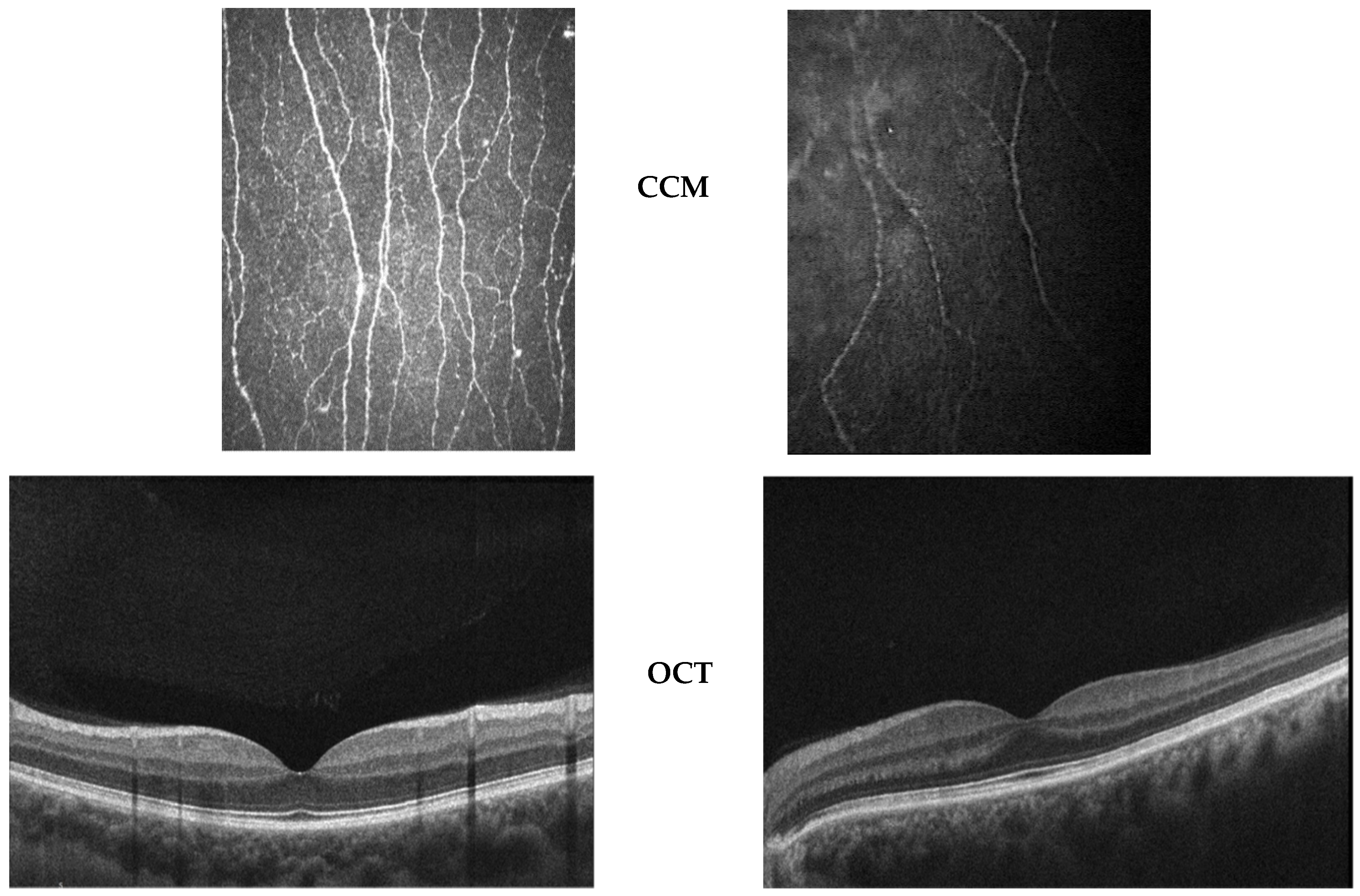
- Posterior Segment
- (d)
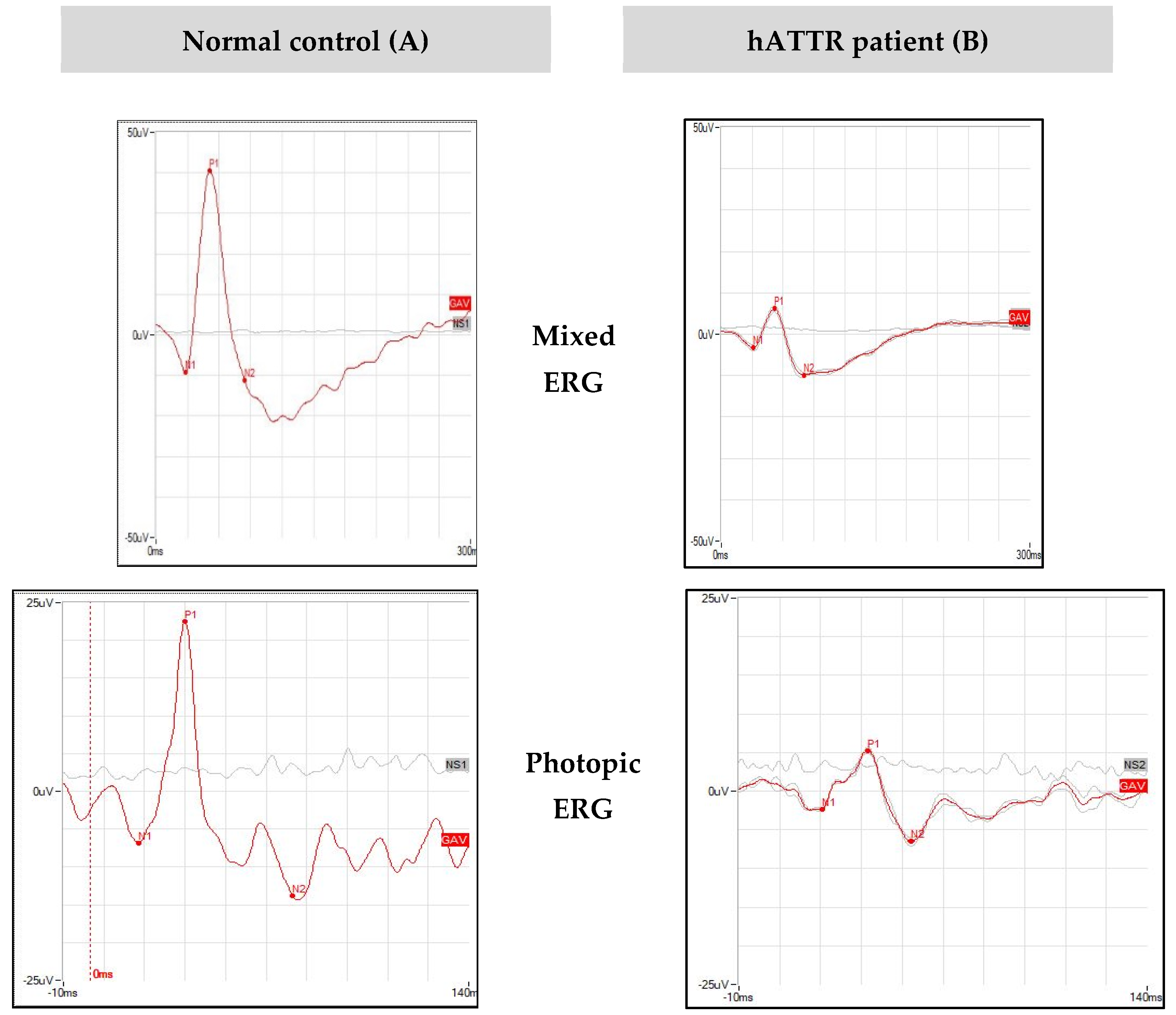
- Functional Studies
4. Discussion
5. Conclusions
Author Contributions
Funding
Institutional Review Board Statement
Informed Consent Statement
Data Availability Statement
Conflicts of Interest
References
- Planté-Bordeneuve, V.; Said, G. Familial amyloid polyneuropathy. Lancet Neurol. 2011, 10, 1086–1097. [Google Scholar] [CrossRef]
- Adams, D.; Koike, H.; Slama, M.; Coelho, T. Hereditary transthyretin amyloidosis: A model of medical progress for a fatal disease. Nat. Rev. Neurol. 2019, 15, 387–404. [Google Scholar] [CrossRef]
- Saraiva, M.J.; Birken, S.; Costa, P.P.; Goodman, D.S. Family studies of the genetic abnormality in transthyretin (prealbumin) in Portuguese patients with familial amyloidotic polyneuropathy. Ann. N. Y. Acad. Sci. 1984, 435, 86–100. [Google Scholar] [CrossRef]
- Richardson, S.J. Cell and molecular biology of transthyretin and thyroid hormones. Int. Rev. Cytol. 2007, 258, 137–193. [Google Scholar]
- Martins, A.C.; Rosa, A.M.; Costa, E.; Tavares, C.; Quadrado, M.J.; Murta, J.N. Ocular Manifestations and Therapeutic Options in Patients with Familial Amyloid Polyneuropathy: A Systematic Review. Biomed. Res. Int. 2015. [Google Scholar] [CrossRef]
- Sekijima, Y. Transthyretin (ATTR) amyloidosis: Clinical spectrum, molecular pathogenesis and disease-modifying treatments. J. Neurol. Neurosurg. Psychiatry 2015, 86, 1036–1043. [Google Scholar] [CrossRef]
- Adams, D.; Lozeron, P.; Lacroix, C. Amyloid neuropathies. Curr. Opin. Neurol. 2012, 25, 564–572. [Google Scholar] [CrossRef]
- Planté-Bordeneuve, V.; Ferreira, A.; Lalu, T.; Zaros, C.; Lacroix, C.; Adams, D.; Said, G. Diagnostic pitfalls in sporadic transthyretin familial amyloid polyneuropathy (TTR-FAP). Neurology 2007, 69, 693–698. [Google Scholar] [CrossRef]
- Luigetti, M.; Conte, A.; Del Grande, A.; Bisogni, G.; Madia, F.; Lo Monaco, M.; Laurenti, L.; Obici, L.; Merlini, G.; Sabatelli, M. TTR-related amyloid neuropathy: Clinical, electrophysiological and pathological findings in 15 unrelated patients. Neurol. Sci. 2013, 34, 1057–1063. [Google Scholar] [CrossRef]
- Ando, E.; Ando, Y.; Okamura, R.; Uchino, M.; Ando, M.; Negi, A. Ocular manifestations of familial amyloidotic polyneuropathy type I: Long-term follow up. Br. J. Ophthalmol. 1997, 81, 295–298. [Google Scholar] [CrossRef]
- Haraoka, K.; Ando, Y.; Ando, E.; Sandgren, O.; Hirata, A.; Nakamura, M.; Terazaki, H.; Tajiri, T.; Tanoue, Y.; Sun, X.; et al. Amyloid deposition in ocular tissues of patients with familial amyloidotic polyneuropathy (FAP). Amyloid 2002, 9, 183–189. [Google Scholar] [CrossRef]
- Beirão, J.M.; Malheiro, J.; Lemos, C.; Matos, E.; Beirão, I.; Pinho-Costa, P.; Torres, P. Impact of liver transplantation on the natural history of oculopathy in Portuguese patients with transthyretin (V30M) amyloidosis. Amyloid 2015, 22, 31–35. [Google Scholar] [CrossRef]
- Hara, R.; Kawaji, T.; Ando, E.; Ohya, Y.; Ando, Y.; Tanihara, H. Impact of liver transplantation on transthyretin-related ocular amyloidosis in Japanese patients. Arch. Ophthalmol. 2010, 128, 206–210. [Google Scholar] [CrossRef]
- Galli-Resta, L.; Falsini, B.; Rossi, G.; Piccardi, M.; Ziccardi, L.; Fadda, A.; Minnella, A.M.; Marangoni, D.; Placidi, G.; Campagna, F.; et al. Bilateral Symmetry of Visual Function Loss in Cone–Rod Dystrophies. Investig. Ophthalmol. Vis. Sci. 2016, 57, 3759–3768. [Google Scholar] [CrossRef][Green Version]
- Abed, E.; Piccardi, M.; Rizzo, D.; Chiaretti, A.; Ambrosio, L.; Petroni, S.; Parrilla, R.; Dickmann, A.; Riccardi, R.; Falsini, B. Functional Loss of the Inner Retina in Childhood Optic Gliomas Detected by Photopic Negative Response. Investig. Ophthalmol. Vis. Sci. 2015, 56, 2469–2474. [Google Scholar] [CrossRef]
- Abed, E.; Placidi, G.; Campagna, F.; Federici, M.; Minnella, A.; Guerri, G.; Bertelli, M.; Piccardi, M.; Galli-Resta, L.; Falsini, B. Early impairment of the full-field photopic negative response in patients with Stargardt disease and pathogenic variants of the ABCA4 gene. Clin. Exp. Ophthalmol. 2018, 46, 519–530. [Google Scholar] [CrossRef]
- Benson, M.D.; Buxbaum, J.N.; Eisenberg, D.S.; Merlini, G.; Saraiva, M.J.M.; Sekijima, Y.; Sipe, J.D.; Westermark, P. Amyloid nomenclature 2020: Update and recommendations by the International Society of Amyloidosis (ISA) nomenclature committee. Amyloid 2020, 27, 217–222. [Google Scholar] [CrossRef]
- Russo, M.; Obici, L.; Bartolomei, I.; Cappelli, F.; Luigetti, M.; Fenu, S.; Cavallaro, T.; Chiappini, M.G.; Gemelli, C.; Pradotto, L.G.; et al. ATTRv amyloidosis Italian Registry: Clinical and epidemiological data. Amyloid 2020, 27, 259–265. [Google Scholar] [CrossRef]
- Luigetti, M.; Guglielmino, V.; Antonini, G.; Casali, C.; Ceccanti, M.; Chiappini, M.; De Giglio, L.; Di Lazzaro, V.; Di Muzio, A.; Goglia, M.; et al. ATTRv in Lazio-Italy: A High-Prevalence Region in a Non-Endemic Country. Genes 2021, 12, 829. [Google Scholar] [CrossRef] [PubMed]
- Frishman, L.; Sustar, M.; Kremers, J.; McAnany, J.J.; Sarossy, M.; Tzekov, R.; Viswanathan, S. ISCEV extended protocol for the photopic negative response (PhNR) of the full-field electroretinogram. Doc. Ophthalmol. 2018, 136, 207–211. [Google Scholar] [CrossRef]
- Falsini, B.; Anselmi, G.M.; Marangoni, D.; D’Esposito, F.; Fadda, A.; Di Renzo, A.; Campos, E.C.; Riva, C.E. Subfoveal choroidal blood flow and central retinal function in retinitis pigmentosa. Investig. Ophthalmol. Vis. Sci. 2011, 52, 1064–1069. [Google Scholar] [CrossRef] [PubMed]
- Chen, X.; Graham, J.; Dabbah, M.A.; Petropoulos, I.N.; Ponirakis, G.; Asghar, O.; Alam, U.; Marshall, A.; Fadavi, H.; Ferdousi, M.; et al. Small nerve fiber quantification in the diagnosis of diabetic sensorimotor polyneuropathy: Comparing corneal confocal microscopy with intraepidermal nerve fiber density. Diabetes Care 2015, 38, 1138–1144. [Google Scholar] [CrossRef]
- Tavakoli, M.; Marshall, A.; Thompson, L.; Kenny, M.; Waldek, S.; Efron, N.; Malik, R.A. Corneal confocal microscopy: A novel noninvasive means to diagnose neuropathy in patients with Fabry disease. Muscle Nerve 2009, 40, 976–984. [Google Scholar] [CrossRef] [PubMed]
- Rousseau, A.; Cauquil, C.; Dupas, B.; Labbé, A.; Baudouin, C.; Barreau, E.; Théaudin, M.; Lacroix, C.; Guiochon-Mantel, A.; Benmalek, A.; et al. Potential Role of In Vivo Confocal Microscopy for Imaging Corneal Nerves in Transthyretin Familial Amyloid Polyneuropathy. JAMA Ophthalmol. 2016, 134, 983–989. [Google Scholar] [CrossRef] [PubMed]


| Patient | Sex | Age | Age of Onset | Age at Diagnosis | Follow-Up (Months) | TTR Pathogenic Variant | FAP Stage | NIS (0–244) | Kumamoto sco–re (0–120) | Inheritance | Systemic invo–lvement | Treatment (Months Since Starting) | ||
|---|---|---|---|---|---|---|---|---|---|---|---|---|---|---|
| PN | CM | GI | ||||||||||||
| #1 | F | 87 | 79 | 81 | 84 | V30M | III | 140.25 | 30 | U | Yes | Yes | Yes | Diflunisal (12) |
| #2 | M | 64 | 58 | 59 | 76 | V30M | II | 73.75 | 30 | U | Yes | Yes | Yes | Patisiran (48) |
| #3 | M | 79 | 77 | 79 | 11 | V122I | I | 21 | 36 | U | Yes | Yes | No | Tafamidis (10) |
| #4 | M | 76 | 65 | 70 | 19 | A109S | II | 58.7 | 28 | U | Yes | No | Yes | Patisiran (16) |
| #5 | F | 52 | - | - | 10 | V30M | 0 | 0 | 0 | Maternal | No | No | No | None |
| #6 | F | 58 | 58 | 58 | 19 | F64L | I | 4 | 3 | Paternal | Yes | No | No | Tafamidis (8) |
| #7 | M | 51 | 51 | 51 | 19 | F64L | I | 12.50 | 0 | Paternal | Yes | Yes | No | Tafamidis (8) |
| #8 | M | 56 | 47 | 48 | 20 | T59K | II | 123 | 35 | Maternal | Yes | Yes | Yes | Tafamidis (77) + Inotersen (7) |
| #9 | M | 78 | 71 | 71 | 8 | F64L | II | 88 | 28 | U | Yes | No | No | Tafamidis (72) + Patisiran (8) |
| Patient | Eye | BCVA (ETDRS Letters) | IOP (mmHg) | Lens Condition | Vitreous Opacities on Ophthalmos Copy | OCT Finding | ||||
|---|---|---|---|---|---|---|---|---|---|---|
| Vitreous Opacity (0–5) | V-R Interface Alterations (0–5) | OR Alterations (0–5) | CMT and SFCT (µ) | ONL (µ) | ||||||
| #1 | RE | 77 | 18 | Pseudofakia, PCO | No | 3 | 4 | 1 | 245 and 320 | 69.6 |
| LE | 56 | 15 | Pseudofakia, PCO | No | 3 | 3 | 4 | 290 and 330 | 52.8 | |
| #2 | RE | 90 | 14 | PPC | No | 1 | 1 | 0 | 270 and 330 | 70.4 |
| LE | 87 | 12 | PPC | No | 1 | 1 | 1 | 280 and 350 | 61.8 | |
| #3 | RE | 85 | 11 | Normal | No | 0 | 0 | 0 | 244 and 180 | 82.2 |
| LE | 85 | 13 | Normal | No | 0 | 0 | 0 | 231 and 160 | 84.8 | |
| #4 | RE | 88 | 10 | CNC | No | 3 | 1 | 0 | 279 and 300 | 79.4 |
| LE | 87 | 10 | CNC | No | 2 | 1 | 0 | 287 and 270 | 74.2 | |
| #5 | RE | 90 | 13 | Normal | No | 2 | 1 | 0 | 211 and 300 | 70.6 |
| LE | 90 | 16 | Normal | No | 1 | 0 | 0 | 207 and 310 | 71.8 | |
| #6 | RE | 85 | 15 | Normal | No | 2 | 2 | 0 | 281 and 330 | 73.2 |
| LE | 85 | 16 | Normal | No | 2 | 2 | 0 | 276 and 244 | 71.2 | |
| #7 | RE | 88 | 12 | Normal | No | 0 | 1 | 0 | 262 and 250 | 75.4 |
| LE | 87 | 12 | Normal | No | 0 | 1 | 0 | 266 and 250 | 77.8 | |
| #8 | RE | 88 | 10 | Pseudofakia | No | 2 | 2 | 0 | 237 and 248 | 74 |
| LE | 88 | 11 | Normal | No | 1 | 1 | 0 | 237 and 240 | 82.8 | |
| #9 | RE | 86 | 16 | Pseudofakia | No | 0 | 0 | 1 | 269 and 238 | 61.2 |
| LE | 85 | 16 | Pseudofakia | No | 1 | 0 | 0 | 260 and 150 | 73.2 | |
| Patients | Eye | CCM | Other Remarks | ||
|---|---|---|---|---|---|
| Absent/Rarefied Subepithelial NP (Extension and Density) | Nerve Segmentation and/or Fragmentation | Thinning of Stromal Nerves | Deposits between Bowman and Stroma | ||
| #1 | RE | yes | no | no | yes |
| LE | yes | no | no | yes | |
| #2 | RE | yes | no | no | yes |
| LE | yes | no | no | yes | |
| #3 | RE | yes | yes | no | no |
| LE | yes | yes | no | no | |
| #4 | RE | yes | yes | no | no |
| LE | no | yes | no | no | |
| #5 | RE | no | yes | no | no |
| LE | no | yes | no | no | |
| #6 | RE | no | no | yes | no |
| LE | no | no | yes | no | |
| #7 | RE | no | yes | yes | yes |
| LE | no | yes | yes | yes | |
| #8 | RE | yes | yes | no | no |
| LE | yes | yes | no | no | |
| #9 | RE | yes | yes | no | no |
| LE | yes | yes | no | no | |
| Patient | Eye | Mixed ERG | Photopic ERG | PhNR | |||
|---|---|---|---|---|---|---|---|
| B Wave Amplitude (µV) | B Wave Peak Time (ms) | B Wave Amplitude (µV) | B Wave Peak Time (ms) | Amplitude (µV) | Peak Time (ms) | ||
| #1 | RE | 53.22 | 53.91 | 29.84 | 37.2 * | 7.19 * | 55.08 * |
| LE | 47.47 | 52.73 | 33.63 | 36.62 * | 4.35 * | 51.86 * | |
| #2 | RE | 25.73 | 50.39 | 18.60 | 33.40 * | 7.67 * | 51.86 * |
| LE | 24.76 * | 50.39 | 19.46 | 33.40 * | 5.83 * | 50.39 * | |
| #3 | RE | 34.97 | 60.35 * | 15.97 | 39.84 * | 8.43 * | 58.59* |
| LE | 42.33 | 59.18 | 17.70 | 41.31 * | 7.04 * | 50.98 * | |
| #4 | RE | 14.06 * | 50.98 | 10.67 | 37.21 * | 4.62 * | 54.79 * |
| LE | 9.39 * | 51.56 | 7.63 * | 37.21 * | 7.03 * | 53.32 * | |
| #5 | RE | 23.38 | 46.29 | 22.12 | 32.81 | 7.66 * | 47.75 |
| LE | 23.44 * | 46.88 | 23.00 | 32.52 | 9.44 | 47.17 | |
| #6 | RE | 34.45 | 49.80 | 29.91 | 33.40 * | 8.12 * | 50.10 * |
| LE | 35.78 | 49.22 | 28.06 | 33.69 * | 9.70 | 50.39* | |
| #7 | RE | 25.46 | 50.98 | 22.06 | 34.57 * | 5.94 * | 50.39 * |
| LE | 30.53 | 52.73 | 21.83 | 34.57 * | 5.55 * | 50.98* | |
| #8 | RE | 30.22 | 48.63 | 28.75 | 34.28 * | 7.28 * | 49.51 * |
| LE | 19.03 * | 49.22 | 17.61 | 34.57 * | 6.40 * | 50.39 * | |
| #9 | RE | 22.34 | 50.98 | 21.83 | 34.86 * | 9.99 | 73.24* |
| LE | 24.75 * | 52.15 | 18.70 | 35.45 * | 5.68 * | 74.12 * | |
| Mixed ERG | Photopic ERG | PhNR | ||||||||||
|---|---|---|---|---|---|---|---|---|---|---|---|---|
| Amplitude (μV) | Peak Time (ms) | Amplitude (μV) | Peak Time (ms) | Amplitude (μV) | Peak Time (ms) | |||||||
| RE | LE | RE | LE | RE | LE | RE | LE | RE | LE | RE | LE | |
| hATTR patients | ||||||||||||
| Mean | 29.31 | 28.60 | 51.36 | 51.56 | 22.19 | 20.84 | 35.28 | 35.48 | 7.43 | 6.78 | 54.59 | 53.28 |
| Std. deviation | 10.40 | 11.13 | 3.71 | 3.25 | 6.19 | 6.85 | 2.18 | 2.49 | 1.42 | 1.68 | 7.3 | 7.5 |
| N of patients | 9 | 9 | 9 | |||||||||
| Normal controls | ||||||||||||
| Mean | 50.55 | 49.28 | 52 | 52 | 25.70 | 25.76 | 29 | 29 | 19.17 | 19.17 | 40 | 40 |
| Std. deviation | 16.36 | 10.86 | 4 | 4 | 9.23 | 8.35 | 2 | 2 | 5 | 5 | 4 | 4 |
| N of patients | 40 | 40 | 40 | |||||||||
Publisher’s Note: MDPI stays neutral with regard to jurisdictional claims in published maps and institutional affiliations. |
© 2021 by the authors. Licensee MDPI, Basel, Switzerland. This article is an open access article distributed under the terms and conditions of the Creative Commons Attribution (CC BY) license (https://creativecommons.org/licenses/by/4.0/).
Share and Cite
Minnella, A.M.; Rissotto, R.; Maceroni, M.; Romano, A.; Fasciani, R.; Luigetti, M.; Sabatelli, M.; Rizzo, S.; Falsini, B. Ocular Involvement in Hereditary Transthyretin Amyloidosis: A Case Series Describing Novel Potential Biomarkers. Genes 2021, 12, 927. https://doi.org/10.3390/genes12060927
Minnella AM, Rissotto R, Maceroni M, Romano A, Fasciani R, Luigetti M, Sabatelli M, Rizzo S, Falsini B. Ocular Involvement in Hereditary Transthyretin Amyloidosis: A Case Series Describing Novel Potential Biomarkers. Genes. 2021; 12(6):927. https://doi.org/10.3390/genes12060927
Chicago/Turabian StyleMinnella, Angelo Maria, Roberta Rissotto, Martina Maceroni, Angela Romano, Romina Fasciani, Marco Luigetti, Mario Sabatelli, Stanislao Rizzo, and Benedetto Falsini. 2021. "Ocular Involvement in Hereditary Transthyretin Amyloidosis: A Case Series Describing Novel Potential Biomarkers" Genes 12, no. 6: 927. https://doi.org/10.3390/genes12060927
APA StyleMinnella, A. M., Rissotto, R., Maceroni, M., Romano, A., Fasciani, R., Luigetti, M., Sabatelli, M., Rizzo, S., & Falsini, B. (2021). Ocular Involvement in Hereditary Transthyretin Amyloidosis: A Case Series Describing Novel Potential Biomarkers. Genes, 12(6), 927. https://doi.org/10.3390/genes12060927