Abstract
During brain development, neurons need to form the correct connections with one another in order to give rise to a functional neuronal circuitry. Mistakes during this process, leading to the formation of improper neuronal connectivity, can result in a number of brain abnormalities and impairments collectively referred to as neurodevelopmental disorders. Cell adhesion molecules (CAMs), present on the cell surface, take part in the neurodevelopmental process regulating migration and recognition of specific cells to form functional neuronal assemblies. Among CAMs, the members of the protocadherin (PCDH) group stand out because they are involved in cell adhesion, neurite initiation and outgrowth, axon pathfinding and fasciculation, and synapse formation and stabilization. Given the critical role of these macromolecules in the major neurodevelopmental processes, it is not surprising that clinical and basic research in the past two decades has identified several PCDH genes as responsible for a large fraction of neurodevelopmental disorders. In the present article, we review these findings with a focus on the non-clustered PCDH sub-group, discussing the proteins implicated in the main neurodevelopmental disorders.
1. Introduction
Neurodevelopmental disorders are a group of diseases occurring in early life and characterized by a significant alteration of the central nervous system (CNS) functioning, resulting in the failure to meet the typical developmental milestones. Brain dysfunction can manifest in several different ways ranging from intellectual alterations and problems of learning and communication to motor function impairment. Although the different disorders have a heterogeneous etiology, there is often overlap of symptomatology between the numerous neurodevelopmental disorders [1,2], and association with many co-morbidities including epilepsy, mood disorders, impairment in vision and hearing, sleep disturbances, and gastrointestinal and breathing problems. Many studies have suggested that shared molecular pathways could account for the multiple clinical signs that characterize these diseases [3]. The altered sociability, often observed in individuals with neurodevelopmental disorders, makes such diseases a significant and expanding public health problem. The therapeutic options available to treat the symptoms are limited. Thus, there is a great need to understand the core neuropathology and the underlying molecular mechanisms to identify new therapies.
Many neurodevelopmental disorders are thought to result from an interaction between genetic and environmental factors [4]. However, in some cases, the disorders can be traced to genetic abnormalities ranging from single nucleotide changes to loss or gain of up to thousands of nucleotides and chromosomal rearrangements. A growing body of literature suggests that mutations or deficits in genes that regulate synapse and circuit development and/or function lead to neurodevelopmental disorders [5]. Regarding the genes encoding synaptic proteins, the prevalence of mutations has been found in the synaptic cell adhesion molecules (CAMs), including protocadherins (PCDHs). In this review, we will summarize the current knowledge about PCDHs, a group of proteins regulating multiple aspects of the synapse development, function and plasticity. We will focus on selected non-clustered PCDHs discussing their implication in neurodevelopmental disorders.
2. Protocadherins in the Central Nervous System
2.1. Overall Structure and Classification
PCDHs are single membrane-spanning glycoproteins and constitute the largest sub-group of the cadherin superfamily, cell-surface molecules implicated in the regulation of the cell-cell contacts.
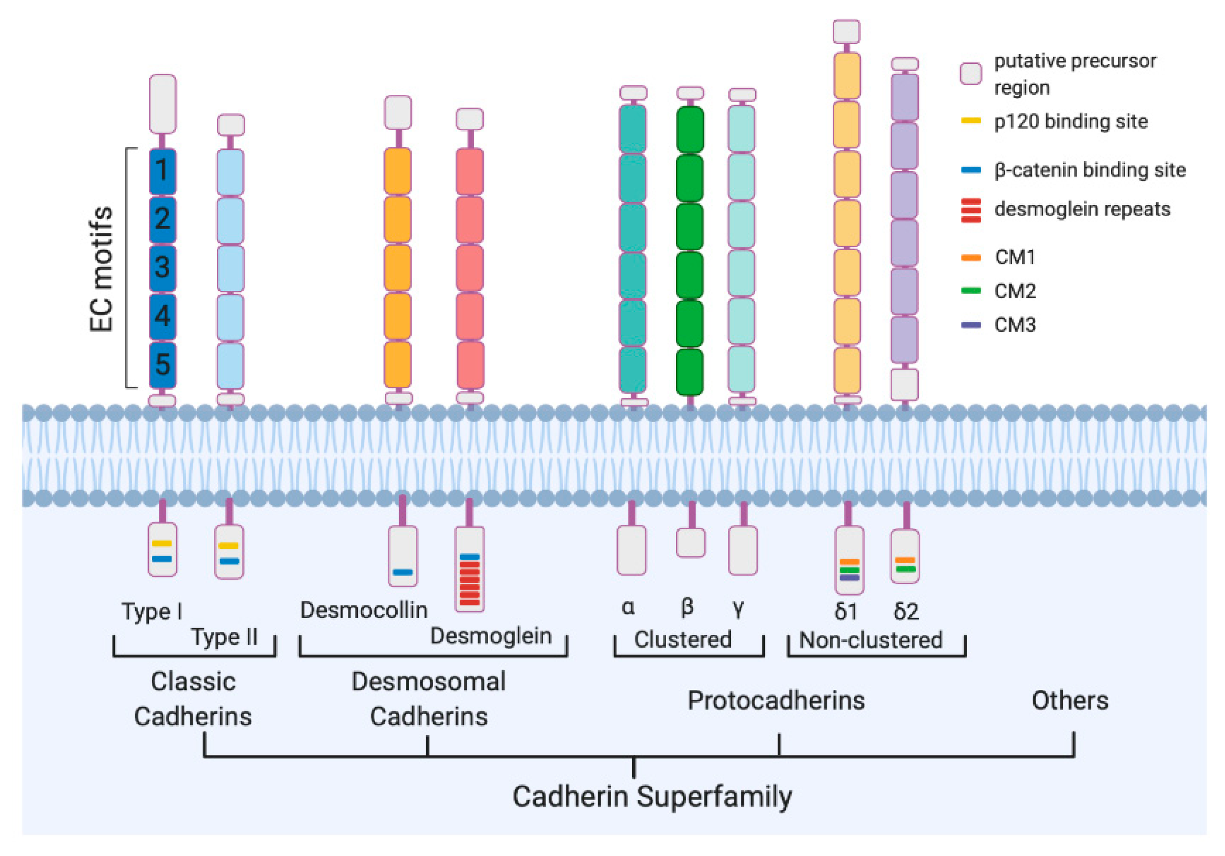
Cadherins consist of an extracellular region subdivided into extracellular cadherins (EC) domains, each including a repetitive sequence named cadherin motif, and a cytoplasmic domain (Figure 1) [6]. The cadherin motif contains conserved Ca2+-binding sequences that are required for protein functioning. The cytoplasmic domain interacts with the armadillo repeat proteins p120 catenin and β-catenin [7,8,9], forming the cadherin–catenin complex that is crucial for the mechanical adhesion between cells. Based on shared properties and sequence similarity cadherins are organized in sub-groups: the classical type I and the related type II, desmosomal cadherins, and PCDHs (Figure 1).
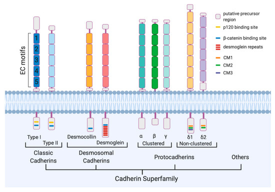
Figure 1.
Cadherin superfamily. Schematic drawing of representative members of Classic Cadherins, Desmosomal Cadherins, and protocadherins. All the proteins share “Cadherin motifs” or extracellular cadherin (EC) motifs at their extracellular domain. The number of EC motifs varies from one subfamily to another. The cytoplasmic sequences are conserved only among the members of each family or subfamily.
In humans, the cadherin superfamily includes over 100 members, more than half of which are PCDHs [10]. Although PCDHs are very similar to the classical cadherins for the presence of a single transmembrane domain and the conserved cadherin motifs, they present some differences. Unlike cadherins, which present 5 EC repeats, PCDHs have 6 or 7 EC repeats (in few cases more) that show low sequence similarities to the EC domains of the classical cadherins (Figure 1). Differences in the cytoplasmic domain, where the catenin-binding sites result absent, have also been highlighted from the functional point of view.
PCDHs are encoded by more than 70 different genes (PCDH genes). According to the genomic organization, PCDHs are divided into clustered and non-clustered PCDHs (Figure 1) [11]. Two additional sub-groups, known as atypical PCDHs, have been identified and include the seven-transmembrane PCDHs and the giant Fat PCDHs.
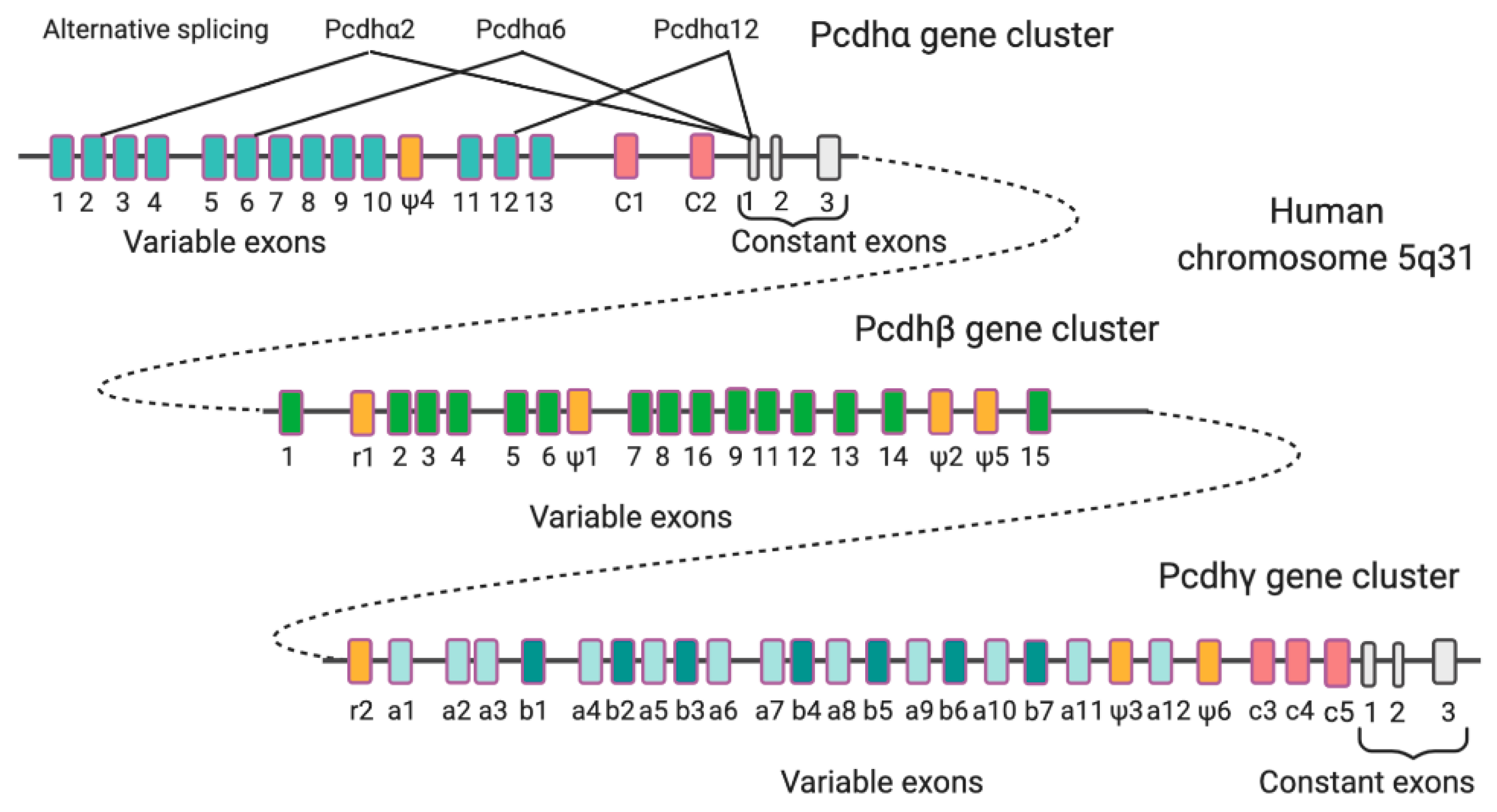
In clustered PCDHs, which include the three families PCDHα, PCDHβ, and PCDHγ, the gene clusters are arranged in tandem in a small genome locus on a single chromosome (human chromosome 5q31; mouse chromosome 18) [12]. Every gene cluster presents several variable exons, each involved in the generation of an extracellular domain, a transmembrane domain, and a variable portion of the cytoplasmic domain. Downstream to the variable exons, only in PCDHα and γ cluster genes, it is possible to find three constant exons coding for a shared portion of the cytoplasmic domain (Figure 2). The presence of variable exons and this gene organization allows the production of more than 50 PCDH isoforms from the three gene clusters through alternative splicing or through the choice of alternative promoters.
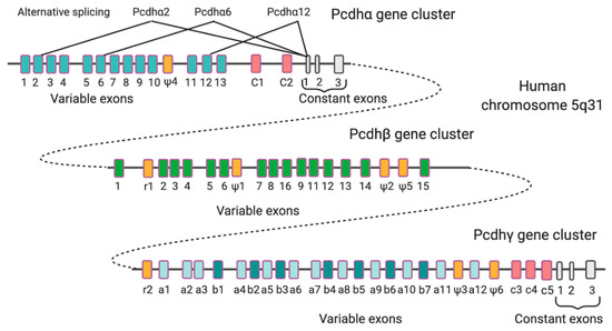
Figure 2.
Genomic organization of human PCDH gene clusters. Shown are PCDHα, PCDHβ, and PCDHγ gene clusters. Each gene family contains multiple tandem variable exons indicated in a different color; only PCDHα and PCDHγ share common constant region exons (grey) that form the cytoplasmic tail. In all the three gene clusters are present relic (r) or pseudogene (ψ) variable region sequences (orange). The generation of PCDHa2, PCDHa6, and PCDHa12 through alternative splicing is indicated as an example of exon selection that produces various isoforms.
Conversely, non-clustered PCDHs do not present a clustered genome locus but the genes are dispersed on multiple chromosomes. Although the genes in non-clustered PCDHs produce alternative splicing variants, they do not encode variable extracellular domains; this results in the possibility to generate only small variations in the protein product.
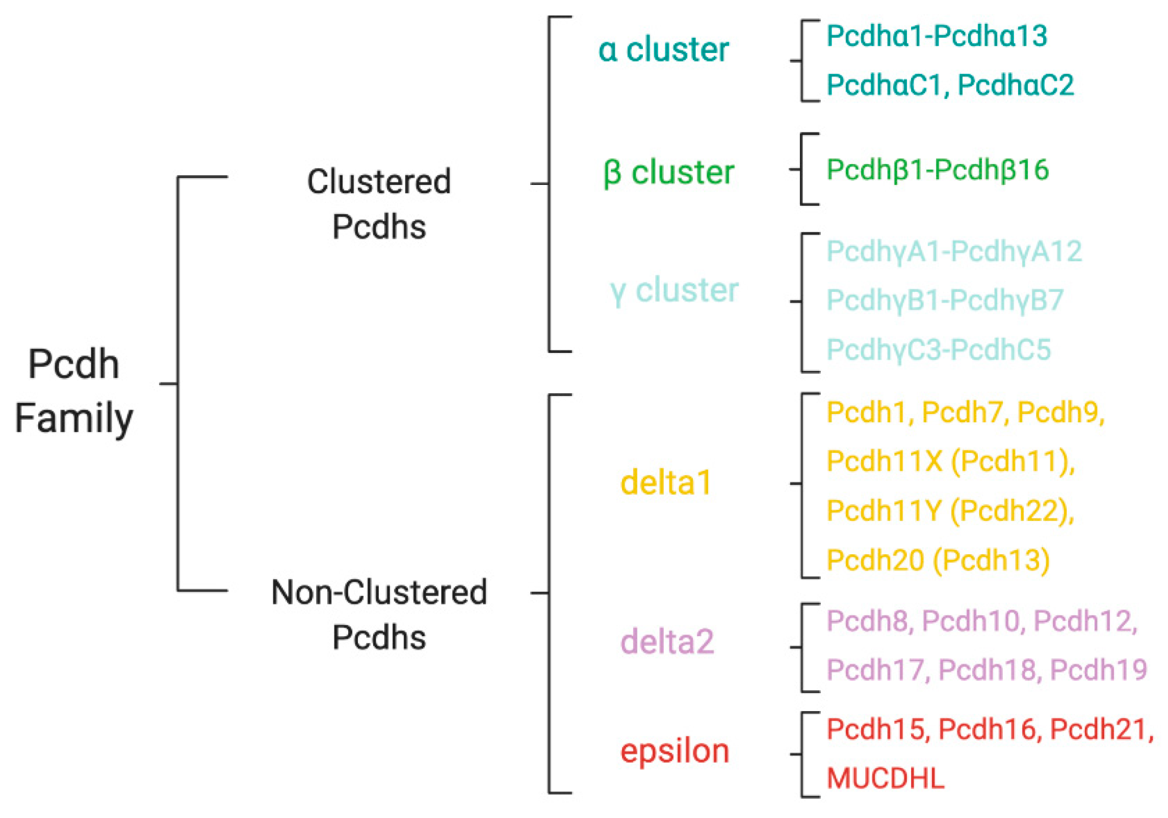
Non-clustered PCDHs are organized into two sub-groups: PCDHδ and solitary PCDHs, also known as PCDHε [11]. On the basis of the homology, the number of EC repeats, and the amino acid sequence of the cytoplasmic domain, PCDHδ family can be further divided into two sub-groups, PCDHδ1, and PCDHδ2. While PCDHδ1 has 7 EC domains and 3 conserved cytoplasmic motifs, CM1, CM2, and CM3, PCDHδ2 has 6 EC domains and only 2 cytoplasmic motifs, CM1 and CM2 (Figure 1 and Figure 3). δ1 sub-group includes PCDH-1, 7, 9 and 11; δ2 sub-group contains PCDH-8, 10, 17, 18 and 19. In addition to the nine canonical family members, in δ2 and δ1 subfamily there are PCDH12 and PCDH20, respectively, that diverge in their intracellular regions characterized by the absence of the CM1, CM2, and CM3 motifs (Figure 3).
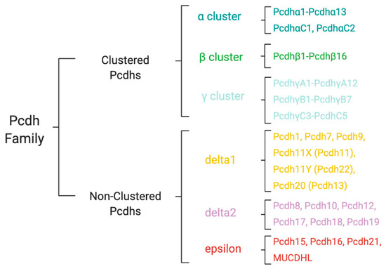
Figure 3.
Classification of human protocadherin (PCDH) family. The members of PCDH family are organized in Clustered (alpha, beta, and gamma subfamilies) and Non-Clustered (delta1 and delta2) PCDHs. The Non-Clustered PCDH subfamily also includes PCDHs of the epsilon group.
PCDHε sub-group includes PCDH-15, 16, 21, and MUCDHL (Figure 3). Except PCDH21, the other PCDHs in this sub-group are characterized by the presence of higher or lower number of EC repeats compared to PCDHδ; in fact, PCDH15, PCDH16, and MUCDHL have 11, 27 and 4 cadherin repeats, respectively.
The two families of large atypical PCDHs include those typified by Drosophila Fat, a giant protein with 34 EC repeats in its extracellular domain, and those typified by Drosophila Flamingo, a 7-transmembrane (7TM) domain protein with 8 EC repeats, along with their many vertebrate homologues [13].
2.2. Tissue Distribution
PCDHs are primarily expressed in the CNS and regulate multiple aspects of cell interaction, synapse maturation, synapse function and plasticity, essential for the proper brain development and correct functioning. Like classical cadherins, PCDH expression is spatiotemporally regulated during brain development in several vertebrate species. PCDHs are constitutively distributed in the cortex, hippocampus, amygdala, cerebellum, thalamus, hypothalamus, basal ganglia nuclei, limbic system structures, olfactory system, and visual system (Table 1) [14,15], but some of them show prominent gradients or regionalized expression [15,16,17] reflecting a functional differentiation. Moreover, due to the projections to different functionally related areas, for example from the cortex to the striatum, the presence of selective PCDHs strongly impacts the activity of the whole circuitry.

Table 1.
Summary of PCDH mRNA expression in human tissues. Shown are the major PCDHs implicated in neurodevelopmental disorders. Level of expression is indicated: +++ strong; ++ moderate; + weak; - negative.
3. Functional Roles of Neuronal Protocadherins
3.1. Cell-Cell Adhesion
Like cadherins, PCDHs are synaptic calcium-dependent adhesion glycoproteins that take part in the process of cell aggregation and regulate cell–cell interactions. They have been detected in synaptic and extrasynaptic membranes as well as in the intracellular compartments [12,18,19]. Cells can vary the number of PCDHs expressed, the level of surface expression, and the kind of PCDHs exposed. The combination of expression of different PCDHs on the neuronal surface seems to strongly influence the cell functions, as different members possess distinct apparent adhesive affinities [20].
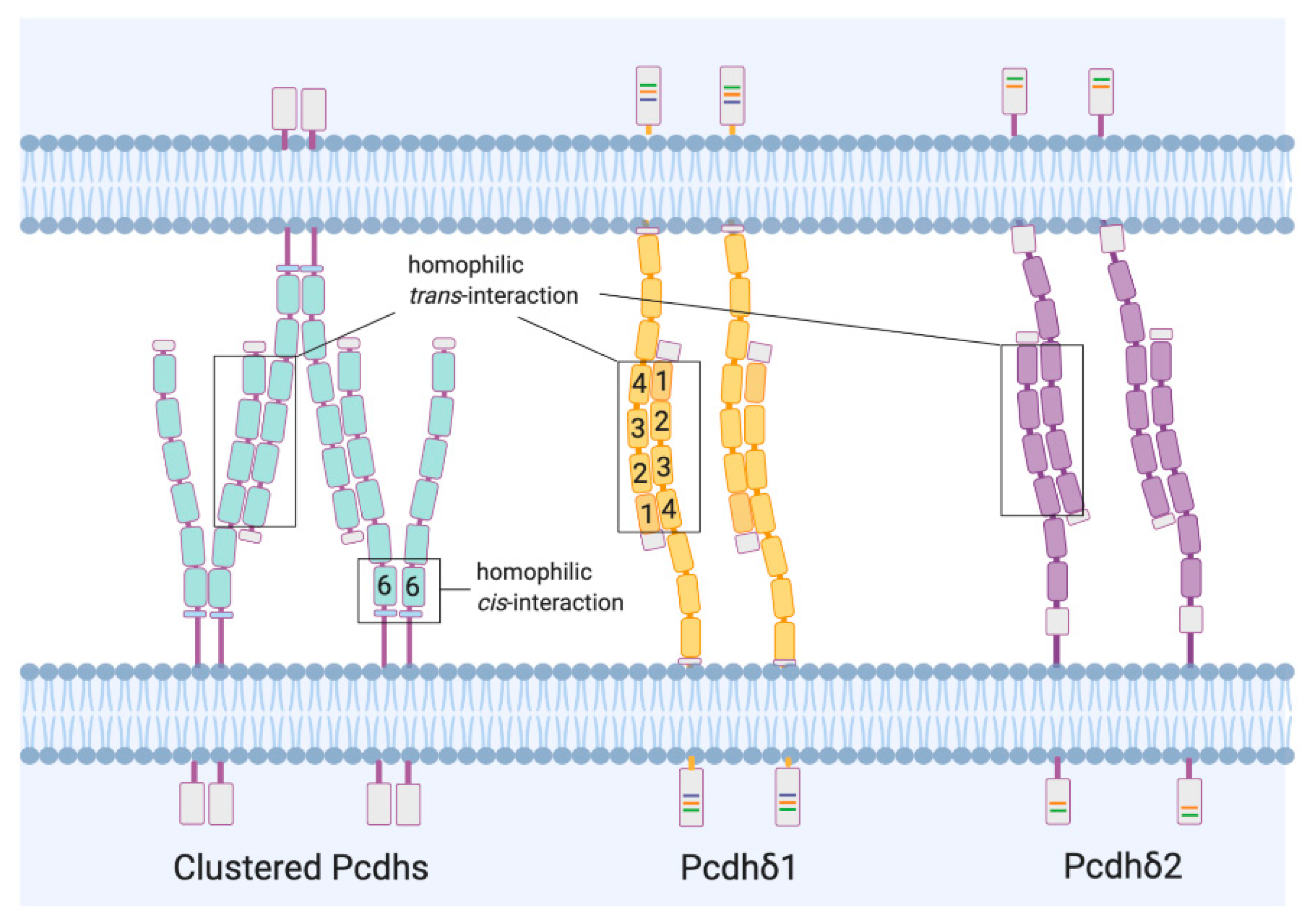
The interaction between pre- and post-synaptic sites occurs through connections of the extracellular domains of PCDH isoforms. The engagement of PCDH isoform-specific dimers is mediated by the EC domains. For both the clustered and non-clustered PCDHs of the δ family, the EC1-EC4 domains, combined in an antiparallel orientation (head-to-tail), mediate the formation of the trans (cell-cell) dimers (Figure 4) [21,22]. Cis (same-cell) interactions have also been shown in clustered PCDHs (Figure 4) [23] and are nonselective between different isoforms. This aspect further contributes to create an enormous combinatorial diversity. Whether PCDHs of the δ family are involved in similar cis interactions is not clear yet, although a recent investigation seems to exclude such possibility [24].
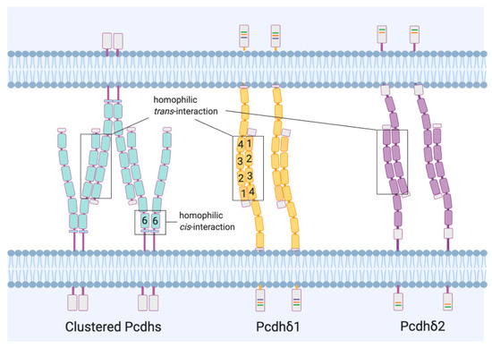
Figure 4.
PCDH-mediated cell interaction. Clustered and Non-clustered PCDH isoforms, from different neurites of two cells, recognize each other in an anti-parallel fashion through strict homophilic trans-interactions of the EC1-EC4 domains, to mediate the adhesion between cells. In Clustered PCDH isoforms it is also possible to observe promiscuous cis-interactions between EC6 domains.
Non-clustered PCDH-1, 7, 8, 10, 18, and 19 exhibit homophilic trans binding activity that is preferred to the heterophilic interactions [25]. However, for some of these molecules (PCDH-8, 10, and 19), the strength of the homophilic binding is weak [26,27,28]. In the case of PCDH8, any cell aggregation has been observed; by contrast, PCDH8 has been shown to interfere with the classic cadherin-mediated adhesion [29,30] that, however, can be restored when the cytoplasmic tail of PCDH8 is removed [31]. Similarly, in the case of PCDH19, the strength of the cell-cell adhesion can be enhanced after removal of the cytoplasmic domain, suggesting that this region may not stabilize the interaction to facilitate adhesion. Conversely, it negatively regulates the adhesion mediated by the extracellular domain [28].
Although less frequent, heterophilic interactions have been reported between PCDH15 and cadherin 23 [32], PCDHγ-C5 and GABA(A) receptor (GABA(A)R [33] and between the clustered PCDHα4 and β1 integrin [34]. This latter is due to the presence of the RGD motif, a peptide sequence of three amino acids (Arg-Gly-Asp) that is recognized by integrins. The RGD motif has been seen not only in the extracellular domain of clustered, but also non-clustered PCDHs (PCDH-17, 19 and MUCDHL), raising the intriguing possibility that such proteins may be ligands for integrins through heterophilic bindings.
3.2. Synapse Maturation and Circuit Formation
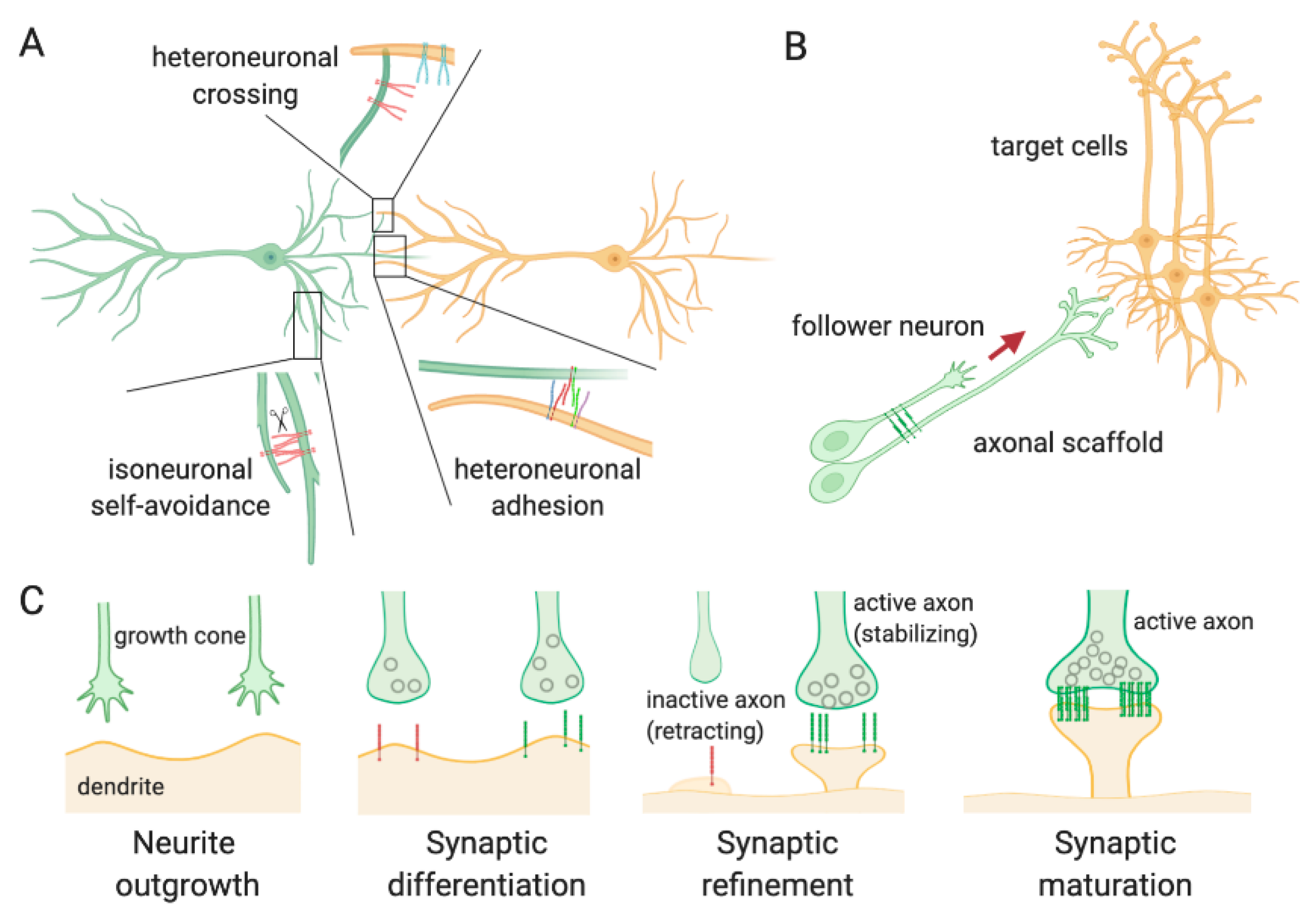
The neuronal specificity of wiring in the CNS is achieved at different levels during the development. In large part, this is due to the action of CAMs, including PCDHs, and recognition proteins, which allow cells to sense the environment and establish selective interactions between the enormous number of other neurons and glial cells. It has been proposed that the combination of PCDHs expressed on the neuronal surface generates a recognition unit [23,25,35] that prevents the interaction at synapses expressing matching PCDHs (self-avoidance) [36,37], or facilitates the adhesion between synapses from different cells (Figure 5A). This makes PCDHs crucial during neurodevelopment and implicates them in both synaptic destabilization/pruning and synaptic stabilization and maturation.
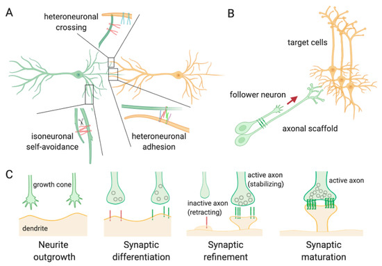
Figure 5.
Schematic overview of PCDH functions in neuronal development and circuit formation. (A) PCDH-mediated synapse self-recognition and self-avoidance. The expression of identical combinatorial profiles of PCDHs on the same neuron determines repulsion between sister neurites (isoneuronal self-avoidance) following PCDH cleavage and loss of interaction ability. Conversely, when matching PCDHs are expressed on neurites from different cells, the interactions are favored (heteroneuronal adhesion). Lastly, when two neurites from different neurons express distinct combinations of PCDHs, the assembly is interrupted by the mismatched isoforms, and the neurites cross each other (heteroneuronal crossing); (B) PCDH regulation of axon-axon interactions through homo- or heterophilic binding between isoforms. The interaction between the growing axon (follower) with a preexisting axon tract (scaffold) via PCDHs mediates the extension of the axon in the appropriate direction, favoring the interaction with other cells; (C) Different stages of synapse formation. At an early stage, the dendritic spines are elongated to seek the synaptic partner (neurite outgrowth) and assemble a synapse (synaptic differentiation). The recruitment of PCDHs stabilizes the contacts or determines the retraction of the axon (synaptic refinement), according to the isoforms of PCDHs expressed and to the synaptic activity/plasticity. PCDH complexes promote the expansion of the dendritic spine head and the maturation of the synapse.
PCDHs participate both in early and in late neurodevelopmental events. During the embryonic stages, PCDHs finely process the dendritic arbors of the post-mitotic neurons in the correct spatial orientation and control the axons outgrowth along the right paths (Figure 5B) [38,39,40,41,42,43].
The importance of PCDHs in dendritic arbor formation has been demonstrated by Garrett and colleagues [44] in cortically restricted PCDHγ mutant mice. Analogously, Suo, and colleagues [45] have found that PCDHα mutant hippocampal neurons have simplified dendritic arbors and a reduction in dendritic spine density. A similar phenotype has been observed in serotoninergic neurons of the rostral raphe nuclei from the same mutant mice [42]. Together, these findings provide evidence that both PCDHγ and α proteins promote dendrite arborization.
Defects in PCDHs, have also been correlated to defects in the formation and the extension of axonal tracts [46,47]. For example, while the reduction of PCDH10 seems responsible for defects in the outgrowth of the striatal axons [46,48], the absence of PCDH17 has been considered determinant in causing defects in the extension of axons from specific subdivisions of the amygdala. In vitro experiments of migration, performed using amygdala neurons from PCDH17 KO mice, have in fact shown that the growth cones of the axons stop moving at the contact points with other encountered axons instead of cross them [47], clearly supporting the hypothesis that PCDH17 serves to aid the axonal sorting. Other PCDHs involved in axon outgrowth and guidance are PCDH7 and PCDH18b, as shown following interference experiments [49,50,51].
Interestingly, also PCDHγ proteins, in addition to the dendritic arbor formation, have been implicated in the correct axonal development. Although studies on PCDHγ mutant mice show normal axonal outgrowth, targeting, and formation [44,52], alterations in the patterning of the axonal terminals have been observed in PCDHγ mutant mice. In mice where all 22 PCDHγ genes were deleted, it has been observed a loss of synapses in the late embryonic period with a reduction of the excitatory and inhibitory synaptic puncta by 40–50% [52] that has been ascribed to the absence of ubiquitiously-expressed three C-type variable exons C3, C4 and C5 rather than to the absence of the sparsely-expressed A1, A2 and A3 exons of the clustered PCDHs [53].
As neurodevelopment proceeds, PCDHs play a crucial role in the interaction between axons and dendrites to regulate the formation of excitatory synapses (Figure 5C) [54]. The presence of PCDHs at synaptic level has been confirmed in synaptosome preparations and post-synaptic density protein fractions [55,56,57]. Similarly, immunohistochemistry and electron microscopy experiments have confirmed PCDHs distribution at some, but not all, synapses; however, these technical approaches, have highlighted the presence of much of the proteins at perisynaptic level or in other compartments of dendrites and axons [56]. The differential expression of PCDHs and their compartmental distribution define the number of synapses generated in a given region. In some cases, the formation of new synapses is favored, whereas, in other cases, there is a downregulation in the number of synapses. For example, PCDH-γs KO mice show increased synapses in cortical neurons [58] but a reduced number of synapses in spinal cord neurons [52]. Furthermore, the number of synapses has been found to be decreased not only in vivo but also in cultured hippocampal neurons where PCDH-γs had been knocked down using shRNA [45]. These differences seem to be mainly correlated to the cell type.
Changes in the number of the dendritic spines have been observed also in arcadlin/PCDH8 KO mice [59] and in PCDH10-heterozygous mice [60]. In both cases, the absence or even the reduction of these proteins determines an increase in the spines available, suggesting that these molecules are involved in the elimination of inappropriate or unused synapses during the normal development process.
In addition to the formation of new spines, PCDHs seem to be implicated also in the progressive accumulation of synaptic vesicles at the presynaptic level, another distinctive feature of the synaptic maturation (Figure 5C). A study performed on PCDH17 KO mice has reported an enhanced presynaptic vesicle accumulation in both excitatory and inhibitory synapses of corticobasal ganglia [61]. The investigation of the functional synaptic maturation in the absence of PCDH17 has demonstrated altered synaptic transmission efficacy that has an impact on the circuit formation and activity.
The study on PCDH17 by Hoshina and colleagues [61], together with investigations on PCDH10 and other non-clustered PCDHs [14,26,62], have highlighted the importance of PCDHs in determining a precise functional topography in the brain and a pathway-specific synapse development necessary for the neural circuit formation. Only neurons of a given functional network present the same repertoire of PCDHs. For example, the expression of PCDH17 in the cortex, basal ganglia, and thalamus [61], is opposite to that of PCDH10 [14,26,62]. Moreover, the expression pattern can be distinctive of a specific phase of the neurodevelopment. This makes PCDHs rarely overlapping in a specific brain region; conversely, they complement each other in their functions and in the proper circuit development.
4. Protocadherins in Neurodevelopmental Disorders
Given the wide distribution of PCDHs in the vertebrate CNS, and the fact that they are involved not only in the developmental steps but also in synapse maturation and functioning, as well as in circuit formation, it is perhaps not wholly surprising that this class of molecules is involved in a large number of CNS disorders.
To date, PCDHs have been implicated in several neurodevelopmental disorders such as intellectual disability (ID), epilepsy, Fragile-X syndrome (FXS), and Autism spectrum disorders (ASDs) and have been demonstrated to fulfill roles in disease states conceptualized as neurodevelopmental disorders including schizophrenia (SZ) and bipolar disorder (BD) (Table 2).

Table 2.
PCDHs associated with neurodevelopmental disorders. Shown are the members of the PCDH family, their physiological functions in neuronal circuitry development, the disorders associated with their alterations and the main brain areas involved.
Although certain PCDH genes have been assigned as major genes causative of a specific neurodevelopmental disorder, the advancement in genome-wide analysis studies have identified many PCDHs genes as susceptibility genes or risk factors for a particular disorder. Below we have summarized recent progress in the study of some non-clustered PCDHs that have been implicated in several neurodevelopmental diseases.
4.1. PCDH19
PCDH19, a member of the PCDHδ2 sub-group, has attracted the attention of the scientific community since 2008. This year represented a turned point for PCDH19 research, as PCDH19 was recognized as the gene responsible for the neurodevelopmental syndrome known as Developmental and Epileptic Encephalopathy 9; DEE9 (OMIM # 300088) [63]. This syndrome, also known as Epilepsy, Female-restricted, with Mental Retardation (EFMR) [64], was first reported in 1971 as a convulsive disorder limited to females [65] (Table 2).
DEE9 peculiar feature is the recurrence of seizures clusters with childhood onset and commonly observed remission during adolescence, often resistant to drug treatments. Frequent comorbid symptoms are ID of variable degree and a wide spectrum of behavioral and psychiatric symptoms, ranging from ASDs to schizophrenia [63,65,66,67,68].
More than 175 mutations have been reported so far in PCDH19 [69]. A significant mutations proportion consists of missense point mutations affecting the PCDH extracellular domain. However, alternative mutation types (i.e., nonsense point mutations, small insertions/deletions, whole-gene deletion) and locations (i.e., the intracellular domain) have been reported [69,70]. Since no clear correlation has been observed between the different mutations and phenotype severity, pathogenic variants might ultimately lead to protein loss of function [67,69].
Notably, PCDH19 mutations cause DEE9 only when expressed in heterozygosis. PCDH19 is located on the chromosome X (Xq22) and females with a mutant allele and a wild type allele develop DEE9, while hemizygous males with a unique mutant allele do not. In females, one of the two X chromosomes randomly undergoes inactivation, which implies that cells expressing a functional and a mutated PCDH19 allele coexist in DEE9 female patients. It has been hypothesized that this mosaic condition might cause cellular interference [71]. How these two cell populations, which diverge in PCDH19 expression, would interfere with each other in practice remains unknown. We can speculate that this condition might scramble cell-cell communication. In fact, the specific set of adhesion molecules that each cell expresses on its surface defines its identity. Adhesion molecules allow cells to recognize themselves and trigger bidirectional intracellular signaling cascades that ultimately affect the assembly of neuronal circuits. An altered expression of PCDH19 might confer fake identities to neurons and convey altered messages that might affect circuit wiring. In support of the cellular interference hypothesis came the DEE9 diagnosis for some male patients with a mosaic expression of PCDH19, due to a post-zygotic somatic mutation in their unique PCDH19 allele [68,71,72,73,74,75,76,77] or to the mutation in one of the two PCDH19 alleles in the context of Klinefelter syndrome (XXY) [78].
PCDH19 expression in rodent brain extends from the embryonic development [14,79] to adulthood [17,80]. In the cortex and hippocampus, PCDH19 expression peaks in the first postnatal period [81,82], suggesting PCDH19 involvement in brain maturation. In the first postnatal week, PCDH19 is highly expressed in the limbic structures [80,83] where patients’ seizures originate [84], including the amygdala, hippocampal regions CA1-CA3, and subiculum, and some hypothalamic areas. While PCDH19 is not detectable in dentate gyrus (DG) hippocampal neurons at this stage, its expression becomes evident in adulthood [17,80]. In addition to neurons, PCDH19 expression was observed also in endothelial cells [80], which is consistent with a putative role of PCDH19 in maintaining the blood-brain barrier integrity [85].
During the embryonic period, PCDH19 is highly expressed in neuronal progenitors, and an increasing body of evidence highlights a role of PCDH19 in regulating progenitor cells polarity, proliferation and fate [82,86,87,88]. In particular, PCDH19 expression was shown to be important for the proliferation of radial glia, from which all cerebral cortex neurons originate [82]. In a recent paper, Xiaohui et al. demonstrated that PCDH19 downregulation in mouse brain decreased the number of excitatory neurons, but not glial cells, arising from individual radial glial progenitors [88], thus supporting the role of PCDH19 in neurogenesis during cortical embryonic development. PCDH19 expression was also important to preserve the physiological lateral clustering of clonally related excitatory neurons (i.e., neurons arising from a common progenitor) and their preferential intra-clone synaptic connections [88]. These and additional findings indicate that PCDH19 role goes beyond the regulation of neuronal number and includes neuronal spatial distribution, maturation, and connectivity. Altered sorting pattern of neuroprogenitors was observed in heterozygous knock-out (KO) mice with PCDH19 mosaic expression [89,90]. Abnormal migration and morphological maturation were observed in rat hippocampal neurons in which PCDH19 expression was downregulated [81]. In Zebrafish, pcdh19 loss impaired the columnar organization of optic tectum neurons [91] and affected the developmental trajectories of neuronal network functional properties [92].
Cell-cell recognition mechanisms during migration and circuit formation likely rely on PCDH19 adhesive properties, both in trans with itself and in cis with N-cadherin [93,94,95]. Furthermore, PCDH19 has been shown to associate with both the WASP-family verprolin homologous protein (WAVE) regulatory complex, which controls actin cytoskeleton dynamics, and with regulators of Rho GTPases and of the microtubule cytoskeleton [94,96,97] and this likely explains its role in cell proliferation and neuronal maturation.
PCDH19 role in neuronal proliferation, migration, and maturation provides an explanation for the structural defects recently described in DEE9 patients’ brain. Radiological analyses revealed focal cortical malformations [98] and quantitative magnetic resonance imaging (MRI) allowed in-depth study of morphological abnormalities in the limbic cortex. The cortical surface morphology and the underlying white-matter bundles were altered in DEE9 patients, as indicated by local gyrification reduction and bundles abnormal directional diffusion, respectively. These defects are the likely structural counterpart of functional connectivity defects [99].
Mouse models were able to recapitulate DEE9 core symptoms, such as cognitive and social behavior impairment and seizure susceptibility. Hippocampal and amygdala-dependent deficits have been observed in heterozygous female KO mice but not in hemizygous KO males tested in the fear conditioning paradigm [89], in agreement with the cellular interference hypothesis. By contrast, both KO groups, i.e., heterozygous females and hemizygous males, showed autism-like behaviors, evaluated as reduced sociality in the 3-chamber test [100]. This is consistent with inflexible personality and obsessive traits observed in some male patients with PCDH19 mutations [67]. Even though spontaneous epilepsy has not been reported, increased seizure susceptibility has been observed both in rats, in which PCDH19 had been downregulated in a subset of hippocampal neurons [81], and in PCDH19 KO mice [101]. Surprisingly, both homozygous and heterozygous KO female mice displayed enhanced susceptibility to induced seizures, contrary to KO males [101]. These data challenge the cellular interference hypothesis and suggest that (i) PCDH19 expression is not totally dispensable, since its complete loss can associate with autistic traits and (ii) other determinants such as gender, in addition to PCDH19 mosaic expression, might be at play.
With this regard, it has been observed that seizures in DEE9 female patients occur in a time-window in which sex hormones are low. Furthermore, DEE9 female patients display altered expression of neurosteroid hormones metabolizing enzymes in their skin fibroblasts and reduced level of allopregnanolone in blood [102]. Allopregnanolone is a potent GABA(A)R modulator [103]. Notably, PCDH19 has been shown to interact with the GABA(A)R and to modulate its surface expression and kinetics in hippocampal neurons, with important consequences for both tonic and phasic inhibition and ultimately neuronal excitability [81,104]. The clinical efficacy of an allopregnanolone analog, ganaxolone, is currently under evaluation [105,106] and represents a promising therapeutic option to face DEE9-related epilepsy, for which current treatments are unsatisfactory.
4.2. PCDH10
PCDH10, also known as OL-protocadherin, is a member of the PCDHδ2 sub-group with a unique cytoplasmic domain. This protein, endowed with homophilic adhesion activity, mediates target recognition, and synapse formation [48]. It is largely expressed in several brain regions during development. Certain local circuits, including the olfactory and visual system, as well as the olivocerebellar projections, are particularly enriched in PCDH10 [26,62,107]; however, the cerebral districts where PCDH10 is very abundant are the basal ganglia system and the amygdala.
The studies on PCDH10 in the basal ganglia have stressed its role of regulator of the axonal growth and of the synaptic connection development, as well as its implication in cognition, motor activity and emotion, three behavioral domains supported by the corticostriatal-thalamic circuits. Defects at the cellular level and abnormal connectivity in the subcortical structures (caudate, dorsal striatum, and thalamus) are considered responsible for the atypical development of these networks and for the onset of repetitive behaviors, cognitive inflexibility, and abnormalities in the motivated behaviors observed in ASD [108] (Table 2). At a striatal level, PCDH10 has been found essential for the outgrowth of the striatal axons that, in turn, is necessary for the formation of the thalamocortical projections [46,48]. The process of elongation of the striatal axons has been hypothesized to be correlated to the ability of PCDH10 to modify the cytoskeleton dynamics and promote the rearrangement of actin filaments that, destabilizing the cell adhesion, could promote the axonal outgrowth [109]. Although this mechanism still needs to be tested, it could be conjectured that it is similar to the mechanism used for the cell movement, consisting in the binding of the cytoplasmic domain of PCDH10 with Nap-1, a component of the WAVE complex that regulates actin polymerization [110].
In homozygous mice, KO for PCDH10 gene, the absence of PCDH10 determines defects in the axon pathways through the ventral telencephalon; in particular, both striatal and thalamocortical projections cannot cross this area [46] showing abnormal wirings.
The crucial role of PCDH10 in synaptic connectivity and circuit formation has been also confirmed during investigations performed on PCDH10-heterozygous mice (PCDH10+/−) where it has been demonstrated that even a partial reduction in functional PCDH10 causes biological alterations at amygdala level that are associated with neurodevelopmental disorders [60]. Amygdala is linked to the social and emotional deficits observed in ASD. The altered expression of PCDH10 at amygdala level has been implicated in the alterations in communication and social behaviors. Recently, it has been observed that the haploinsufficiency of PCDH10 in PCDH10+/− mice reduces the social approach in a gender-sensitive fashion [60] and that this phenotype strongly correlates with electrophysiological and morphological alterations. PCDH10+/− mice show both in vitro and in vivo altered electrophysiological responses. In particular, experiments of voltage sensitive dye imaging showed changes in amygdala circuit synchronization following high stimulation of the synaptic inputs with gamma frequency (40 Hz) [60], supporting the idea that PCDH10 is necessary for the optimal synchrony of the amygdala circuits during the development stages. Analogously, the band responses following in vivo high frequency activity (30–100 Hz, Gamma), that reflect a large-scale brain network functioning fundamental for many cognitive and behavioral functions, result altered [111] like in ASD patients [112] where copy number variation at the PCDH10 locus has been found [113,114].
Moreover, in the same animals, neuronal morphological alterations have been reported. An increase in the density of the dendritic spines [60], that is an index for the number of excitatory synapses, confirms that PCDH10 is implicated in the developmental synapse elimination. In PCDH10+/− mice, the loss of the ability to refine or to eliminate excitatory synapses is perhaps responsible for the overconnectivity/hyperexcitability observed in the amygdala and contributes to the changes in the power of the gamma band activity and the social behavior deficits.
The increase in dendritic spines and in immature spine number occurs also in a genetic model of ASD, the FXS [115], suggesting that such disorder may result from a deficit in synapse elimination involving PCDH10. Fragile X Syndrome is caused by transcriptional silencing or loss-of-function mutations in the FMRP gene, which encodes for the Fragile X mental retardation protein (FMRP) that is required for the activity-dependent synapse elimination by PCDH10, triggered following the activation of the transcription factor myocyte enhancer factor 2 (mef2) [116]. It has been demonstrated that both FMRP and mef2 cooperatively regulate the expression of PCDH10 [117] and its binding to PSD-95, a post-synaptic scaffold protein that has a role in anchoring synaptic proteins. In the synapse elimination process, PSD-95 ubiquitinylated binds PCDH10, which links it to the proteasome for degradation. Noteworthy, PSD-95 interacts with GluN2A/2B, two subunits of the NMDA receptor, regulating the development, localization and signaling of this kind of receptor. Thus, the alteration in PCDH10 may indirectly determine the changes in the levels of NMDA receptor subunits, as observed by Schoch and colleagues [60], which would concur with the aberrant oscillatory activity measured in ASD.
4.3. Other Protocadherins
4.3.1. PCDH9
PCDH9 is a member of the PCDHδ1 sub-group. It is implicated in the formation of functional neuronal circuits with a specific and restricted spatiotemporal expression pattern. It has been found in different regions of the CNS such as olfactory bulb, hippocampus, caudate putamen and cerebral cortex, since the early developmental stages. However, its expression levels are not constant for the whole life but decrease in adulthood [118,119].
Studies on PCDH9 KO mice give insight in the possible roles of this protein in neurodevelopmental disorders including ASD (Table 2). The absence of PCDH9, in fact, strongly influences the cortical thickness. This anatomical abnormality is due to an altered synapse development that causes an increased cell density reducing the processing of sensory information relevant for social adaptation [119]. Behavioral analysis of these mice also shows that social long-term memory is impaired [119], clearly suggesting that PCDH9 may participate in the clinical features of ASD, likely through a reduction in the thickness of the cortical areas.
Interestingly, PCDH9 has recently been identified as a risk gene for depressive disorder [120], often occurring in individuals with neurodevelopmental disorders. The analysis of post-mortem brains has shown a decrease in PCDH9 transcript levels in cortex and hippocampus, suggesting that this protein is a key regulator of circuit formation and functioning.
4.3.2. PCDH8
PCDH8 belongs to the δ2 sub-group of PCDHs. It is localized at synapses and neuronal somas of hippocampus, amygdala, and anterior thalamic nuclei of the limbic system where, in addition to the homophilic adhesion activity, modulates synaptic plasticity phenomena that are essential to the process of learning and memory [27] and impaired in neurodevelopmental disorders such as ASD (Table 2).
PCDH8 seems to be implicated in the homeostatic control of the neural complexity. If on one side the disruption of the homophilic interactions mediated by PCDH8, using blocking antibodies, causes a reduction of the amplitude of the excitatory post-synaptic potentials (EPSPs) and a loss of long-term potentiation (LTP) in hippocampal slices [27], on the other side the absence of PCDH8 in hippocampal cultures from PCDH8 KO mice determines an increase in the dendritic spine density [30]. Although these observations appear to be contradictory, it can be speculated that the control of the spine number by PCDH8 is activated following a synaptic plasticity event and is essential to avoid an excessive neuronal complexity. The mechanism of control of the spine density seems to be dependent on the ability of PCDH8 to interact with N-cadherin and promote its endocytosis via the activation of intracellular signaling involving TAO2β and p38MAPK [30]. It is possible that the loss of spines observed in epilepsy, may be explained through this mechanism based on the shedding of N-cadherin [121] dependent on PCDH8 activity. Similarly, based on the fact that PCDH8 is an ASD-linked gene [122], it could be speculated that the impairments in synaptic plasticity, which is a causative factor underlying ASD pathology, can be dependent on alterations in PCDH8 functions.
4.3.3. PCDH17
PCDH17 is one of the PCDHδ2 sub-group proteins that plays an important role in cell migration and in axon outgrowth and arborization. Its function has been highlighted in investigations on amygdala neurons where the absence of PCDH17 determines the impossibility of axons to grow normally and their tendency to misroute during the extension [47]. In fact, through the interaction with actin polymerization regulators, via its cytoplasmic domain, PCDH17 creates a kind of machinery at cell-cell contact sites that sustains cell motility. This process allows the migration of growth cones that contact with other axons supporting their collective extension as well as the fascicle formation, which is crucial for a proper neuronal wiring [47].
PCDH17 is predominantly expressed in prefrontal cortex, thalamus, striatum, anterior cingulate and amygdala [60,122,123]. In particular, the enrichment along the amygdala neurons and the basal ganglia synapses has made PCDH17 particularly interesting for its potential implications in mood disorders (Table 2).
In amygdala, the enhanced expression of PCDH17 has been associated to an increased risk to develop major mood disorders, including bipolar disorder, as observed in postmortem brains of patients with higher PCDH17 mRNA levels [124]. The analysis of PCDH17 in primary amygdala cultures has revealed a decrease in the spine density and an aberrant dendritic morphology [124].
The study of PCDH17 in the basal ganglia circuits has highlighted that PCDH17 KO mice show an antidepressant-like phenotype when challenged with a battery of behavioral tests aimed at evaluating cognition, anxiety, and depression [61], suggesting that this protein could play an important role not only in disease states conceptualized as neurodevelopmental disorders, such as the bipolar disorder, but also in depression that is a comorbid condition in people with neurodevelopmental disorders. Accordingly, single-nucleotide polymorphisms (SNPs) in PCDH17 gene were found associated with mood disorders [124]. Interestingly, in PCDH17 KO animals, PCDH17 has also been found implicated in the control of vesicle assembly at the basal ganglia circuits where its absence seems to induce an increased number of docked and total synaptic vesicles at presynaptic terminals [61]. Accordingly, when overexpressed in primary neuronal cultures, PCDH17 determines an enhancement in the mobility of synaptic vesicle clusters along the axons [61], suggesting that it inhibits the accumulation of synaptic vesicles.
Collectively, these studies not only reveal that alterations in the levels of PCDH17 cause changes in dendritic spine morphology and abnormal synapse development, which underlie mood disorders, but also that the levels of PCDH17 are crucial for the risk to develop a disease state. Moreover, considering the widespread distribution of PCDH17, it is possible that the alteration in the levels of this protein occurs within an extended brain network that includes not only basal ganglia and amygdala, but also other areas that play an essential role in the control of emotions and social behaviors including cortex, hypothalamus and mesolimbic district.
4.3.4. PCDH15
PCDH15, a member of the PCDHε, is implicated in the regulation of neuronal projections and in synaptic connectivity. It is expressed in brain, kidney, sensory epithelia of the cochlea, and various epithelia during embryogenesis [125].
Deeply investigated for its role in the maintenance of the normal retinal and cochlear function and for its involvement in causing hearing loss and Usher Syndrome Type IF (USH1F) in the presence of mutations in PCDH15 gene [126], PCDH15 has recently gained attention for its potential implication also in neurodevelopmental disorders (Table 2).
Polymorphisms in PCDH15 gene have been found in association with schizophrenia [127] and clinically significant copy number variations (CNVs) in PCDH15 locus have been identified as risk factor for bipolar disorder, ASD, and schizophrenia [128,129,130,131,132,133].
The possible contribution of PCDH15 mutations in causing neurodevelopmental impairment through synaptic alterations has been substantiated in a recent study performed using iPSC lines derived from bipolar patients carrying a CNV in PCDH15 locus. Exonic deletions in PCDH15 gene determine a reduction in dendrite length and in the number of both glutamatergic and GABAergic synapses [133]. These data are consistent with studies performed on postmortem human brains of bipolar patients where reduced spine density and dendrite length were observed [134]. Collectively, these observations further confirm the importance of PCDH15 for normal neuronal functions and strongly support the hypothesis that PCDH15 plays a pathogenetic role in neurodevelopmental disorders.
5. Conclusions
The proper brain function is critically dependent on the underlying neural architecture and connectivity. The ability of cells to recognize their partners through cell surface receptors, expressed in a given moment, is essential for the formation of a functional neuronal circuitry during development. PCDHs are increasingly recognized as molecules playing important roles in cell adhesion. However, in the last two decades, the research has highlighted that these molecules, in addition to a mechanical role, have other biological functions highly relevant for the neural circuitry development. The expression of PCDHs is tightly regulated during development, and each tissue or cell type shows a characteristic pattern of PCDH expression. This differential expression in the developing nervous system has revealed that PCDHs are implicated in every step of the synapse maturation and circuit formation. This idea has been supported by their involvement in a range of neurodevelopmental disorders and disease states conceptualized as neurodevelopmental disorders (Table 2). Downregulation of PCDHs or their functional alterations have been observed in many human neurodevelopmental diseases. Loss of function in PCDH19, responsible for Developmental and Epileptic Encephalopathy 9, has been reported. Functional alterations determining abnormal synaptic connectivity or plasticity have been found in PCDH-10, -9, and -8, three ASD-linked genes. Changes in PCDH17 and PCDH15 have been implicated in mood disorder onset. These observations clearly demonstrate the relevance of these molecules from a clinical point of view, although the role of many PCDHs in disease etiology still needs to be clarified.
Many advancements in defining PCDH/neurodevelopment relationship have been done. However, the understanding of the mechanisms regulating the activity of PCDHs is still at the beginning due to the high number of molecules expressed, the complexity of their interactions, and the fact that the same PCDHs can mediate opposite functions in different brain districts and at different times. Either the introduction of genetic mutations or the deletion of given PCDHs in animal models have strongly helped to elucidate the physiological significance of these molecules in cell connectivity, synapse maturation, and circuit formation. At the same time, the use of genome-wide association (GWA) studies as an approach to dissect complex human diseases has identified several genes of the PCDH family as possible candidates for neurodevelopmental disorders. However, the limited statistical power has made sure that only few candidate genes of the PCDH family have reached the conventional significance thresholds and have been confirmed as implicated in specific pathological states. Considering the high variety of existing PCDHs, an improvement in GWA analyses will likely uncover further links between PCDHs and neurodevelopmental disorders, boosting the research in this field. Therefore, several important challenges remain to elucidate the PCDHs associated with the neurodevelopmental processes and the molecular mechanisms by which PCDH dysfunctions impact on brain development. This understanding will be critical for the development of effective therapies for these complex conditions.
Author Contributions
Conceptualization, M.M. and M.P.; writing-original draft preparation, M.M. and S.B.; writing-review and editing, M.M., S.B., M.M. Figure preparation, M.M. All authors have read and agreed to the published version of the manuscript.
Funding
This work was supported by Telethon Foundation, grant number GGP17283, to M.P. and grant number GGP17260 to S.B.; PRIN 2017-20172C9HLW, Fondazione Le Jeune, Fondazione Cariplo 2019-3438 to M.P.
Acknowledgments
Figures were created with BioRender.com.
Conflicts of Interest
The authors declare no conflict of interest.
References
- Niemi, M.E.K.; Martin, H.C.; Rice, D.L.; Gallone, G.; Gordon, S.; Kelemen, M.; MaAloney, K.; McRae, J.; Radford, E.J.; Yu, S.; et al. Common genetic variants contribute to risk of rare severe neurodevelopmental disorders. Nature 2018, 562, 268–271. [Google Scholar] [CrossRef] [PubMed]
- Tărlungeanu, D.C.; Novarino, G. Genomics in neurodevelopmental disorders: An avenue to personalized medicine. Exp. Mol. Med. 2018, 50, 100. [Google Scholar] [CrossRef] [PubMed]
- Hormozdiari, F.; Kichaev, G.; Yang, W.Y.; Pasaniuc, B.; Eskin, E. Identification of causal genes for complex traits. Bioinformatics 2015, 31, i206–i213. [Google Scholar] [CrossRef] [PubMed]
- Van Loo, K.M.; Martens, G.J. Genetic and environmental factors in complex neurodevelopmental disorders. Curr. Genom. 2007, 8, 429–444. [Google Scholar]
- Peek, S.L.; Mah, K.M.; Weiner, J.A. Regulation of neural circuit formation by protocadherins. Cell Mol. Life Sci. 2017, 74, 4133–4157. [Google Scholar] [CrossRef]
- Tsukasaki, Y.; Miyazaki, N.; Matsumoto, A.; Nagae, S.; Yonemura, S.; Tanoue, T.; Iwasaki, K.; Takeichi, M. Giant cadherins and Dachsous self-bond to organize properly spaced intercellular junctions. Proc. Natl. Acad. Sci. USA 2014, 111, 16011–16016. [Google Scholar] [CrossRef]
- Gumbiner, B.M. Regulation of cadherin-mediated adhesion in morphogenesis. Nat. Rev. Mol. Cell Biol. 2005, 6, 622–634. [Google Scholar] [CrossRef]
- Takeichi, M. The cadherin superfamily in neuronal connections and interactions. Nat. Rev. Neurosci. 2007, 8, 11–20. [Google Scholar] [CrossRef]
- Nelson, W.J. Regulation of cell-cell adhesion by the cadherin-catenin complex. Biochem. Soc. Trans. 2008, 36, 149–155. [Google Scholar] [CrossRef]
- Nollet, F.; Kools, P.; van Roy, F. Phylogenetic analysis of the cadherin superfamily allows identification of six major subfamilies besides several solitary members. J. Mol. Biol. 2000, 299, 551–572. [Google Scholar] [CrossRef]
- Redies, C.; Vanhalst, K.; Roy, F. Delta-protocadherins: Unique structures and functions. Cell Mol. Life Sci. 2005, 62, 2840–2852. [Google Scholar] [CrossRef] [PubMed]
- Wu, Q.; Maniatis, T. A striking organization of a large family of human neural cadherin-like cell adhesion genes. Cell 1999, 97, 779–790. [Google Scholar] [CrossRef]
- Halbleib, J.M.; Nelson, W.J. Cadherins in development: Cell adhesion, sorting, and tissue morphogenesis. Genes Dev. 2006, 20, 3199–3214. [Google Scholar] [CrossRef] [PubMed]
- Kim, S.Y.; Chung, H.S.; Sun, W.; Kim, H. Spatiotemporal expression pattern of non-clustered protocadherin family members in the developing rat brain. Neuroscience 2007, 147, 996–1021. [Google Scholar] [CrossRef]
- Kim, S.Y.; Yasuda, S.; Takana, H.; Yamagata, K.; Kim, H. Non-clustered protocadherin. Cell Adh. Migr. 2011, 5, 97–105. [Google Scholar] [CrossRef]
- Hertel, N.; Krishna, K.; Nuernberger, M.; Redies, C. A cadherin-based code for the divisions of the mouse basal ganglia. J. Comp. Neurol. 2008, 508, 511–528. [Google Scholar] [CrossRef]
- Kim, S.Y.; Mo, J.W.; Choi, S.Y.; Han, S.B.; Moon, B.H.; Rhyu, I.J.; Sun, W.; Kim, H. The expression of non-clustered protocadherins in adult rat hippocampal formation and the connecting brain regions. Neuroscience 2010, 170, 189–199. [Google Scholar] [CrossRef]
- Morishita, H.; Yagi, T. Protocadherin family: Diversity, structure, and function. Curr. Opin. Cell Biol. 2007, 19, 584–592. [Google Scholar] [CrossRef]
- Li, Y.; Serwanski, D.R.; Miralles, C.P.; Fiondella, C.G.; Loturco, J.J.; Rubio, M.E.; De Blas, A.L. Synaptic and nonsynaptic localization of protocadherin-gammaC5 in the rat brain. J. Comp. Neurol. 2010, 518, 3439–3463. [Google Scholar] [CrossRef]
- Bisogni, A.J.; Ghazanfar, S.; Williams, E.O.; Marsh, H.M.; Yang, J.Y.; Lin, D.M. Tuning of delta-protocadherin adhesion through combinatorial diversity. Elife 2018, 7, e41050. [Google Scholar] [CrossRef]
- Goodman, K.M.; Rubinstein, R.; Thu, C.A.; Bahna, F.; Mannepalli, S.; Ahlsén, G.; Rittenhouse, C.; Maniatis, T.; Honig, B.; Shapiro, L. Structural basis of diverse homophilic recognition by clustered α- and β-protocadherins. Neuron 2016, 90, 709–723. [Google Scholar] [CrossRef] [PubMed]
- Goodman, K.M.; Rubinstein, R.; Thu, C.A.; Mannepalli, S.; Bahna, F.; Ahlsén, G.; Rittenhouse, C.; Maniatis, T.; Honig, B.; Shapiro, L. γ-Protocadherin structural diversity and functional implications. Elife 2016, 5, e20930. [Google Scholar] [CrossRef] [PubMed]
- Goodman, K.M.; Rubinstein, R.; Dan, H.; Bahna, F.; Mannepalli, S.; Ahlsén, G.; Aye Thu, C.; Sampogna, R.V.; Maniatis, T.; Honig, B.; et al. Protocadherin cis-dimer architecture and recognition unit diversity. Proc. Natl. Acad. Sci. USA 2017, 114, E9829–E9837. [Google Scholar] [CrossRef] [PubMed]
- Harrison, O.J.; Brasch, J.; Katsamba, P.S.; Ahlsen, G.; Noble, A.J.; Dan, H.; Sampogna, R.V.; Potter, C.S.; Carregher, B.; Honig, B.; et al. Family-wide structural and biophysical analysis of binding interactions among non-clustered δ-protocadherins. Cell Rep. 2020, 30, 2655–2671.e7. [Google Scholar] [CrossRef] [PubMed]
- Schreiner, D.; Weiner, J.A. Combinatorial hemophilic interaction between gamma-protocadherin multimers greatly expands the molecular diversity of cell adhesion. Proc. Natl. Acad. Sci. USA 2010, 107, 14893–14898. [Google Scholar] [CrossRef]
- Hirano, S.; Yan, Q.; Suzuki, S.T. Expression of novel protocadherin, OL-protocadherin, in a subset of functional systems of the developing mouse brain. J. Neurosci. 1999, 19, 995–1005. [Google Scholar] [CrossRef]
- Yamagata, K.; Andreasson, K.I.; Sugiura, H.; Maru, E.; Dominique, M.; Irie, Y.; Miki, N.; Hayashi, Y.; Yoshioka, M.; Kaneko, K.; et al. Arcadlin is a neural activity-regulated cadherin involved in long term potentiation. J. Biol. Chem. 1999, 274, 19437–19479. [Google Scholar] [CrossRef]
- Tai, K.; Kubota, M.; Shiono, K.; Tokutsu, H.; Suzuki, S.T. Adhesion properties and retinofugal expression of chicken protocadherin-19. Brain Res. 2010, 1344, 13–24. [Google Scholar] [CrossRef]
- Chen, X.; Gumbiner, B.M. Crosstalk between different adhesion molecules. Curr. Opin. Cell Biol. 2006, 18, 572–578. [Google Scholar] [CrossRef]
- Yasuda, S.; Tanaka, H.; Sugiura, H.; Okamura, K.; Sakaguchi, T.; Tran, U.; Takemiya, T.; Mizoguchi, A.; Yagita, Y.; Sakurai, T.; et al. Activity-induced protocadherin arcadlin regulates dendritic spine number by triggering N-cadherin endocytosis via TAO2beta and p38 MAP kinases. Neuron 2007, 56, 456–471. [Google Scholar] [CrossRef]
- Kim, S.H.; Yamamoto, A.; Bouwmeester, T.; Agius, E.; Robertis, E.M. The role of paraxial protocadherin in selective adhesion and cell movements of the mesoderm during Xenopus gastrulation. Development 1998, 125, 4681–4690. [Google Scholar] [PubMed]
- Kazmierczak, P.; Sakaguchi, H.; Tokita, J.; Wilson-Kubalek, E.M.; Milligan, R.A.; Müller, U.; Kachar, B. Cadherin 23 and protocadherin 15 interact to form tip-link filaments in sensory hair cells. Nature 2007, 449, 87–91. [Google Scholar] [CrossRef] [PubMed]
- Li, Y.; Xiao, H.; Chiou, T.T.; Jin, H.; Bonhomme, B.; Miralles, C.P.; Pinal, N.; Ali, R.; Chen, W.V.; Maniatis, T.; et al. Molecular and functional interaction between protocadherin-gammaC5 and GABAA receptors. J. Neurosci. 2012, 32, 11780–11797. [Google Scholar] [CrossRef] [PubMed]
- Mutoh, T.; Hamada, S.; Senzaki, K.; Murata, Y.; Yagi, T. Cadherin-related neuronal receptor1 (CNR1) has cell adhesion activity with beta1 integrin mediated through the RGD site of CNR1. Exp. Cell Res. 2004, 294, 494–508. [Google Scholar] [CrossRef] [PubMed]
- Brasch, J.; Goodman, K.M.; Noble, A.J.; Rapp, M.; Mannepalli, S.; Bahna, F.; Dandey, V.P.; Bepler, T.; Berger, B.; Maniatis, T.; et al. Visualization of clustered protocadherin neuronal self-recognition complexes. Nature 2019, 569, 280–283. [Google Scholar] [CrossRef] [PubMed]
- Lefevbre, J.L.; Kostadinov, D.; Chen, W.V.; Maniatis, T.; Sanes, J.R. Protocadherins mediate dendritic self-avoidance in the mammalian nervous system. Nature 2012, 488, 517–521. [Google Scholar]
- Ing-Esteves, S.; Kostadinov, D.; Marocha, J.; Sing, A.D.; Joseph, K.S.; Laboulaye, M.A.; Sanes, J.R.; Lefevbre, J.L. Combinatorial effects of alpha- and gamma-protocadherins on neuronal survival and dendritic self-avoidance. J. Neurosci. 2018, 38, 2713–2729. [Google Scholar] [CrossRef]
- Garrett, A.M.; Weiner, J.A. Control of CNS synapse development by {gamma}-protocadherin-mediated astrocyte-neuron contact. J. Neurosci. 2009, 29, 11723–11731. [Google Scholar] [CrossRef]
- Molumby, M.J.; Keeler, A.B.; Weiner, J.A. Homophilic protocadherin cell-cell interactions promote dendrite complexity. Cell Rep. 2016, 15, 1037–1050. [Google Scholar] [CrossRef]
- Hasegawa, S.; Hamada, S.; Kumode, Y.; Esumi, S.; Katori, S.; Fukuda, E.; Uchiyama, Y.; Hirabayashi, T.; Mombaerts, P.; Yagi, T. The protocadherin-alpha family is involved in axonal coalescence of olfactory sensory neurons into glomeruli of the olfactory bulb in mouse. Mol. Cell Neurosci. 2008, 38, 66–79. [Google Scholar] [CrossRef]
- Prasad, T.; Weiner, J.A. Direct and indirect regulation of spinal cord Ia afferent terminal formation by the γ-protocadherins. Front. Mol. Neurosci. 2011, 4, 54. [Google Scholar] [CrossRef] [PubMed]
- Katori, S.; Noguchi-Katori, Y.; Okayama, A.; Kawamura, Y.; Luo, W.; Sakimura, K.; Hirabayashi, T.; Iwasato, T.; Yagi, T. Protocadherin-αC2 is required for diffuse projections of serotoninergic axons. Sci. Rep. 2017, 7, 15908. [Google Scholar] [CrossRef] [PubMed]
- Lu, W.C.; Zhou, Y.X.; Qiao, P.; Zheng, J.; Wu, Q.; Shen, Q. The protocadherin alpha cluster is required for axon extension and myelination in the developing central nervous system. Neural. Regen Res. 2018, 13, 427–433. [Google Scholar]
- Garrett, A.M.; Schreiner, D.; Lobas, M.A.; Weiner, J.A. γ-protocadherins control cortical dendrite arborization by regulating the activity of a FAK/PKC/MARCKS signaling pathway. Neuron 2012, 74, 269–276. [Google Scholar] [CrossRef] [PubMed]
- Suo, L.; Lu, H.; Ying, G.; Capecchi, M.R.; Wu, Q. Protocadherin clusters and cell adhesion kinase regulate dendrite complexity through Rho GTPase. J. Mol. Cell Biol. 2012, 4, 362–376. [Google Scholar] [CrossRef]
- Uemura, M.; Nakao, S.; Suzuki, S.T.; Takeichi, M.; Hirano, S. OL-Protocadherin is essential for growth of striatal axons and thalamocortical projections. Nat. Neurosci. 2007, 10, 1151–1159. [Google Scholar] [CrossRef]
- Hayashi, S.; Inoue, Y.; Kiyonari, H.; Abe, T.; Misaki, K.; Moriguchi, H.; Tanaka, Y.; Takeichi, M. Protocadherin-17 mediates collective axon extension by recruiting actin regulator complexes to interaxonal contacts. Dev. Cell 2014, 30, 673–687. [Google Scholar] [CrossRef]
- Hirano, S. Pioneers in the ventral telencephalon: The role of OL-protocadherin-dependent striatal axon growth in neural circuit formation. Cell Adh. Migr. 2007, 1, 176–178. [Google Scholar] [CrossRef]
- Piper, M.; Dwivedy, A.; Leung, L.; Bradley, R.S.; Holt, C.E. NF-protocadherin and TAF1 regulate retinal axon initiation and elongation in vivo. J. Neurosci. 2008, 28, 100–105. [Google Scholar] [CrossRef]
- Leung, L.C.; Urbančič, V.; Baudet, M.L.; Dwivedy, A.; Bayley, T.G.; Lee, A.C.; Harris, W.A.; Holt, C.E. Coupling of NF-protocadherin signaling to axon guidance by cue-induced translation. Nat. Neurosci. 2013, 16, 166–173. [Google Scholar] [CrossRef]
- Biswas, S.; Emond, M.R.; Duy, P.Q.; Hao, L.T.; Beattie, C.E.; Jontes, J.D. Protocadherin-18b interacts with Nap1 to control motor axon growth and arborization in zebrafish. Mol. Biol. Cell 2014, 25, 633–642. [Google Scholar] [CrossRef]
- Weiner, J.A.; Wang, X.; Tapia, J.C.; Sanes, J.R. Gamma protocadherins are required for synaptic development in the spinal cord. Proc. Natl. Acad. Sci. USA 2005, 102, 8–14. [Google Scholar] [CrossRef] [PubMed]
- Chen, W.V.; Alvarez, F.J.; Lefebvre, J.L.; Friedman, B.; Nwakeze, C.; Geiman, E.; Smith, C.; Thu, C.A.; Tapia, J.C.; Tasic, B.; et al. Functional significance of isoform diversification in the protocadherin gamma gene cluster. Neuron 2012, 75, 402–409. [Google Scholar] [CrossRef] [PubMed]
- Jontes, J.D.; Phillips, G.R. Selective stabilization and synaptic specificity: A new cell-biological model. Trends Neurosci. 2006, 29, 186–191. [Google Scholar] [CrossRef] [PubMed]
- Kohmura, N.; Senzaki, S.; Hamada, S.; Kai, N.; Yasuda, R.; Watanable, M.; Ishii, H.; Yasuda, M.; Mishina, M.; Yagi, T. Diversity revealed by a novel family of cadherins expressed in neurons at a synaptic complex. Neuron 1998, 20, 1137–1151. [Google Scholar] [CrossRef]
- Phillips, G.R.; Huang, J.K.; Wang, Y.; Tanaka, H.; Shapiro, L.; Zhang, W.; Shan, W.S.; Arndt, K.; Frank, M.; Gordon, R.E.; et al. The presynaptic particle web: Ultrastructure, composition, dissolution, and reconstitution. Neuron 2001, 32, 63–77. [Google Scholar] [CrossRef]
- Phillips, G.R.; Tanaka, H.; Frank, M.; Elste, A.; Fidler, L.; Benson, D.L.; Colman, R. Gamma-protocadherins are target to subsets of synapses and intracellular organelles in neurons. J. Neurosci. 2003, 23, 5096–5104. [Google Scholar] [CrossRef]
- Molumby, M.J.; Anderson, R.M.; Newbold, D.J.; Koblesky, N.K.; Garrett, A.M.; Schreiner, D.; Radley, J.J.; Weiner, J.A. γ-protocadherins interact with neuroligin-1 and negatively regulate dendritic spine morphogenesis. Cell Rep. 2017, 18, 2702–2714. [Google Scholar] [CrossRef]
- Takeichi, M.; Abe, K. Synaptic contact dynamics controlled by cadherin and catenins. Trends Cell Biol. 2005, 15, 216–221. [Google Scholar] [CrossRef]
- Schoch, H.; Kreibich, A.S.; Ferri, S.L.; White, R.S.; Bohorquez, D.; Banerjee, A.; Port, R.G.; Dow, H.C.; Cordero, L.; Pallathra, A.A.; et al. Sociability deficits and altered amygdala circuits in mice lacking Pcdh10, an autism associated gene. Biol. Psychiatry 2017, 81, 193–202. [Google Scholar] [CrossRef]
- Hoshina, N.; Tanimura, A.; Yamasaki, M.; Inoue, T.; Fukabori, R.; Kuroda, T.; Yokoyama, K.; Tezuka, T.; Sagara, H.; Hirano, S.; et al. Protocadherin 17 regulates presynaptic assembly in topographic corticobasal ganglia circuits. Neuron 2013, 78, 839–854. [Google Scholar] [CrossRef] [PubMed]
- Aoki, E.; Kimura, R.; Sukuzi, S.T.; Hirano, S. Distribution of OL-protocadherin protein in correlation with specific neural compartments and local circuits in the postnatal mouse brain. Neuroscience 2003, 117, 593–614. [Google Scholar] [CrossRef]
- Dibbens, L.M.; Tarpey, P.S.; Hynes, K.; Bayly, M.A.; Scheffer, I.E.; Smith, R.; Bomar, J.; Sutton, E.; Vandeleur, L.; Shoubridge, C.; et al. X-linked protocadherin 19 mutations cause female-limited epilepsy and cognitive impairment. Nat. Genet. 2008, 40, 776–781. [Google Scholar] [CrossRef] [PubMed]
- Ryan, S.G.; Chance, P.F.; Zou, C.H.; Spinner, N.B.; Golden, J.A.; Smietana, S. Epilepsy and mental retardation limited to females: An X-linked dominant disorder with male sparing. Nat. Genet. 1997, 17, 92–95. [Google Scholar] [CrossRef]
- Juberg, R.C.; Hellman, C.D. A new familial form of convulsive disorder and mental retardation limited to females. J. Pediatr. 1971, 79, 726–732. [Google Scholar] [CrossRef]
- Vlaskamp, D.R.M.; Bassett, A.S.; Sullivan, J.E.; Robblee, J.; Sadleir, L.G.; Scheffer, I.E.; Andrade, D.M. Schizophrenia is a later-onset feature of pcdh19 girls clustering epilepsy. Epilepsia 2019, 60, 429–440. [Google Scholar] [CrossRef] [PubMed]
- Kolc, K.L.; Sadleir, L.G.; Scheffer, I.E.; Ivancevic, A.; Roberts, R.; Pham, D.H.; Gecz, J. A systematic review and meta-analysis of 271 pcdh19-variant individuals identifies psychiatric comorbidities, and association of seizure onset and disease severity. Mol. Psychiatry 2018, 24, 241–251. [Google Scholar] [CrossRef]
- Kolc, K.L.; Sadleir, L.G.; Depienne, C.; Marini, C.; Scheffer, I.E.; Møller, R.S.; Trivisano, M.; Specchio, N.; Pham, D.; Kumar, R.; et al. A standardized patient-centered characterization of the phenotypic spectrum of pcdh19 girls clustering epilepsy. Transl. Psychiatry 2020, 10, 127. [Google Scholar] [CrossRef]
- Niazi, R.; Fanning, E.A.; Depienne, C.; Sarmady, M.; Tayoun, A.N.A. A mutation update for the pcdh19 gene causing early-onset epilepsy in females with an unusual expression pattern. Hum. Mutat. 2019, 40, 243–257. [Google Scholar] [CrossRef]
- Depienne, C.; LeGuern, E. Pcdh19-related infantile epileptic encephalopathy: An unusual x-linked inheritance disorder. Hum. Mutat. 2012, 33, 627–634. [Google Scholar] [CrossRef]
- Depienne, C.; Bouteiller, D.; Keren, B.; Cheuret, E.; Poirier, K.; Trouillard, O.; Benyahia, B.; Quelin, C.; Carpentier, W.; Julia, S.; et al. Sporadic infantile epileptic encephalopathy caused by mutations in pcdh19 resembles Dravet syndrome but mainly affects females. PLoS Genet. 2009, 5, e1000381. [Google Scholar] [CrossRef]
- van Harssel, J.J.; Weckhuysen, S.; van Kempen, M.J.; Hardies, K.; Verbeek, N.E.; de Kovel, C.G.; Gunning, W.B.; van Daalen, E.; de Jonge, M.V.; Jansen, A.C.; et al. Clinical and genetic aspects of pcdh19-related epilepsy syndromes and the possible role of pcdh19 mutations in males with autism spectrum disorders. Neurogenetics 2013, 14, 23–34. [Google Scholar] [CrossRef] [PubMed]
- Terracciano, A.; Trivisano, M.; Cusmai, R.; De Palma, L.; Fusco, L.; Compagnucci, C.; Bertini, E.; Vigevano, F.; Specchio, N. Pcdh19-related epilepsy in two mosaic male patients. Epilepsia 2016, 57, e51–e55. [Google Scholar] [CrossRef] [PubMed]
- Thiffault, I.; Farrow, E.; Smith, L.; Lowry, J.; Zellmer, L.; Black, B.; Abdelmoity, A.; Miller, N.; Soden, S.; Saunders, C. Pcdh19-related epileptic encephalopathy in a male mosaic for a truncating variant. Am. J. Med. Genet. A 2016, 170, 1585–1589. [Google Scholar] [CrossRef] [PubMed]
- de Lange, I.M.; Rump, P.; Neuteboom, R.F.; Augustijn, P.B.; Hodges, K.; Kistemaker, A.I.; Brouwer, O.F.; Mancini, G.M.S.; Newman, H.A.; Vos, Y.J.; et al. Male patients affected by mosaic pcdh19 mutations: Five new cases. Neurogenetics 2017, 18, 147–153. [Google Scholar] [CrossRef]
- Perez, D.; Hsieh, D.T.; Rohena, L. Somatic mosaicism of pcdh19 in a male with early infantile epileptic encephalopathy and review of the literature. Am. J. Med. Genet. A 2017, 173, 1625–1630. [Google Scholar] [CrossRef]
- Tan, Y.; Hou, M.; Ma, S.; Liu, P.; Xia, S.; Wang, Y.; Chen, L.; Chen, Z. Chinese cases of early infantile epileptic encephalopathy: A novel mutation in the pcdh19 gene was proved in a mosaic male- case report. BMC Med. Genet. 2018, 19, 92. [Google Scholar] [CrossRef]
- Romasko, E.J.; DeChene, E.T.; Balciuniene, J.; Akgumus, G.T.; Helbig, I.; Tarpinian, J.M.; Keena, B.A.; Vogiatzi, M.G.; Zackai, E.H.; Izumi, K.; et al. Pcdh19-related epilepsy in a male with klinefelter syndrome: Additional evidence supporting pcdh19 cellular interference disease mechanism. Epilepsy Res. 2018, 145, 89–92. [Google Scholar] [CrossRef]
- Gaitan, Y.; Bouchard, M. Expression of the delta-protocadherin gene pcdh19 in the developing mouse embryo. Gene. Expr. Patterns 2006, 6, 893–899. [Google Scholar] [CrossRef]
- Schaarschuch, A.; Hertel, N. Expression profile of N-cadherin and protocadherin-19 in postnatal mouse limbic structures. J. Comp. Neurol. 2018, 526, 663–680. [Google Scholar] [CrossRef]
- Bassani, S.; Cwetsch, A.W.; Gerosa, L.; Serratto, G.M.; Folci, A.; Hall, I.F.; Mazzanti, M.; Cancedda, L.; Passafaro, M. The female epilepsy protein pcdh19 is a new GABAAR-binding partner that regulates GABAergic transmission as well as migration and morphological maturation of hippocampal neurons. Hum. Mol. Genet. 2018, 27, 1027–1038. [Google Scholar] [CrossRef] [PubMed]
- Fujitani, M.; Zhang, S.; Fujiki, R.; Fujihara, Y.; Yamashita, T. A chromosome 16p13.11 microduplication causes hyperactivity through dysregulation of mir-484/protocadherin-19 signaling. Mol. Psychiatry 2017, 22, 364–374. [Google Scholar] [CrossRef] [PubMed]
- Hertel, N.; Redies, C.; Medina, L. Cadherin expression delineates the divisions of the postnatal and adult mouse amygdala. J. Comp. Neurol. 2012, 520, 3982–4012. [Google Scholar] [CrossRef] [PubMed]
- Marini, C.; Darra, F.; Specchio, N.; Mei, D.; Terracciano, A.; Parmeggiani, L.; Ferrari, A.; Sicca, F.; Mastrangelo, M.; Spaccini, L.; et al. Focal seizures with affective symptoms are a major feature of pcdh19 gene-related epilepsy. Epilepsia 2012, 53, 2111–2119. [Google Scholar] [CrossRef]
- Higurashi, N.; Takahashi, Y.; Kashimada, A.; Sugawara, Y.; Sakuma, H.; Tomonoh, Y.; Inoue, T.; Hoshina, M.; Satomi, R.; Ohfu, M.; et al. Immediate suppression of seizure clusters by corticosteroids in pcdh19 female epilepsy. Seizure 2015, 27, 1–5. [Google Scholar] [CrossRef]
- Compagnucci, C.; Petrini, S.; Higuraschi, N.; Trivisano, M.; Specchio, N.; Hirose, S.; Bertini, E.; Terracciano, A. Characterizing pcdh19 in human induced pluripotent stem cells (ipscs) and ipsc-derived developing neurons: Emerging role of a protein involved in controlling polarity during neurogenesis. Oncotarget 2015, 6, 26804–26813. [Google Scholar] [CrossRef]
- Homan, C.C.; Pederson, S.; To, T.H.; Tan, C.; Piltz, S.; Corbett, M.A.; Wolvetang, E.; Thomas, P.Q.; Jolly, L.A.; Gecz, J. Pcdh19 regulation of neural progenitor cell differentiation suggests asynchrony of neurogenesis as a mechanism contributing to pcdh19 girls clustering epilepsy. Neurobiol. Dis. 2018, 116, 106–119. [Google Scholar] [CrossRef]
- Lv, X.; Ren, S.Q.; Zhang, X.J.; Shen, Z.; Ghosh, T.; Xianyu, A.; Gao, P.; Li, Z.; Lin, S.; Yu, Y.; et al. Tbr2 coordinates neurogenesis expansion and precise microcircuit organization via protocadherin 19 in the mammalian cortex. Nat. Commun. 2019, 10, 3946. [Google Scholar] [CrossRef]
- Hayashi, S.; Inoue, Y.; Hattori, S.; Kaneko, M.; Shioi, G.; Miyakawa, T.; Takeichi, M. Loss of X-linked Protocadherin-19 differentially affects the behavior of heterozygous female and hemizygous male mice. Sci. Rep. 2017, 7, 5801. [Google Scholar] [CrossRef]
- Pederick, D.T.; Richards, K.L.; Piltz, S.G.; Kumar, R.; Mincheva-Tasheva, S.; Mandelstam, S.A.; Dale, R.C.; Scheffer, I.E.; Gecz, J.; Petrou, S.; et al. Abnormal cell sorting underlies the unique x-linked inheritance of pcdh19 epilepsy. Neuron 2018, 97, 59–66.e55. [Google Scholar] [CrossRef]
- Cooper, S.R.; Emond, M.R.; Duy, P.Q.; Liebau, B.G.; Wolman, M.A.; Jontes, J.D. Protocadherins control the modular assembly of neuronal columns in the zebrafish optic tectum. J. Cell Biol. 2015, 211, 807–814. [Google Scholar] [CrossRef] [PubMed]
- Light, S.E.W.; Jontes, J.D. Multiplane calcium imaging reveals disrupted development of network topology in zebrafish. eNeuro 2019, 6. [Google Scholar] [CrossRef] [PubMed]
- Biswas, S.; Emond, M.R.; Jontes, J.D. Protocadherin-19 and N-cadherin interact to control cell movements during anterior neurulation. J. Cell Biol. 2010, 191, 1029–1041. [Google Scholar] [CrossRef] [PubMed]
- Emond, M.R.; Biswas, S.; Blevins, C.J.; Jontes, J.D. A complex of protocadherin-19 and N-cadherin mediates a novel mechanism of cell adhesion. J. Cell Biol. 2011, 195, 1115–1121. [Google Scholar] [CrossRef]
- Cooper, S.R.; Jontes, J.D.; Sotomayor, M. Structural determinants of adhesion by protocadherin-19 and implications for its role in epilepsy. Elife 2016, 5, e18529. [Google Scholar] [CrossRef]
- Chen, B.; Brinkmann, K.; Chen, Z.; Pak, C.W.; Liao, Y.; Shi, S.; Henry, L.; Grishin, N.V.; Bogdan, S.; Rosen, M.K. The wave regulatory complex links diverse receptors to the actin cytoskeleton. Cell 2014, 156, 195–207. [Google Scholar] [CrossRef]
- Emon, M.R.; Biswas, S.; Morrow, M.L.; Jontes, J.D. Proximity-dependent proteomics reveals extensive interactions of protocadherin-19 with regulators of rho GTPases and the microtubule cytoskeleton. Neuroscience 2020, 452, 26–36. [Google Scholar] [CrossRef]
- Kurian, M.; Korff, C.M.; Ranza, E.; Bernasconi, A.; Lübbig, A.; Nangia, S.; Ramelli, G.P.; Wohlrab, G.; Nordli, D.R.; Bast, T. Focal cortical malformations in children with early infantile epilepsy and pcdh19 mutations: Case report. Dev. Med. Child Neurol. 2018, 60, 100–105. [Google Scholar] [CrossRef]
- Lenge, M.; Marini, C.; Canale, E.; Napolitano, A.; De Masi, S.; Trivisano, M.; Mei, D.; Longo, D.; Rossi Espagnet, M.C.; Lucenteforte, E.; et al. Quantitative MRI-based analysis identifies developmental limbic abnormalities in pcdh19 encephalopathy. Cereb. Cortex 2020, 30, 6039–6050. [Google Scholar] [CrossRef]
- Lim, J.; Ryu, J.; Kang, S.; Noh, H.J.; Kim, C.H. Autism-like behaviors in male mice with a pcdh19 deletion. Mol. Brain 2019, 12, 95. [Google Scholar] [CrossRef]
- Rakotomamonjy, J.; Sabetfakhri, N.P.; McDermott, S.L.; Guemez-Gamboa, A. Characterization of seizure susceptibility in pcdh19 mice. Epilepsia 2020, 61, 2313–2320. [Google Scholar] [CrossRef] [PubMed]
- Tan, C.; Shard, C.; Ranieri, E.; Hynes, K.; Pham, D.H.; Leach, D.; Buchanan, G.; Corbett, M.; Shoubridge, C.; Kumar, R.; et al. Mutations of protocadherin 19 in female epilepsy (pcdh19-fe) lead to allopregnanolone deficiency. Hum. Mol. Genet. 2015, 24, 5250–5259. [Google Scholar] [CrossRef] [PubMed]
- Belelli, D.; Lambert, J.J. Neurosteroids: Endogenous regulators of the GABA(A) receptor. Nat. Rev. Neurosci. 2005, 6, 565–575. [Google Scholar] [CrossRef] [PubMed]
- Serratto, G.M.; Pizzi, E.; Murru, L.; Mazzoleni, S.; Pelucchi, S.; Marcello, E.; Mazzanti, M.; Passafaro, M.; Bassani, S. The epilepsy-related protein pcdh19 regulates tonic inhibition, GABA. Mol. Neurobiol. 2020, 57, 5336–5351. [Google Scholar] [CrossRef]
- Sullivan, J.; Specchio, N.; Chez, M.; Pinna, G.; Locci, A.; Jarrar, R.; Poduri, A.; Ridel, K.; Masuoka, L.; Gasior, M.; et al. Preliminary evidence of a predictive clinical biomarker in PCDH19-related epilepsy: Significant treatment effect of ganaxolone in biomarker-positive patients. In Proceedings of the American Epilepsy Society Annual Meeting, New Orleans Ernest, N. Morial Convention Center, New Orleans, LA, USA, 30 November–4 December 2018. [Google Scholar]
- ClinicalTrials.gov [Internet]. Bethesda (MD): National Library of Medicine (US). Study of Adjunctive Ganaxolone Treatment in Female Children with Protocadherin 19 (PCDH19)-Related Epilepsy (Violet Study). Available online: https://clinicaltrials.gov/ct2/show/NCT03865732 (accessed on 16 December 2019).
- Nakao, S.; Uemura, M.; Aoki, E.; Suzuki, S.T.; Takeichi, M.; Hirano, S. Distribution of OL-protocadherin in axon fibers in the developing chick nervous system. Mol. Brain Res. 2005, 134, 294–308. [Google Scholar] [CrossRef]
- Schuetze, M.; Park, M.T.; Choi, I.Y.; MacMaster, F.P.; Chakravarty, M.M.; Bray, S.L. Morphological alterations in the thalamus, striatum, and pallidum in Autism Spectrum Disorder. Neuropsychopharmacology 2016, 41, 2627–2637. [Google Scholar] [CrossRef]
- Hirano, S.; Takeichi, M. Cadherins in brain morphogenesis and wiring. Physiol. Rev. 2012, 92, 597–634. [Google Scholar] [CrossRef]
- Nakao, S.; Platek, A.; Hirano, S.; Takeichi, M. Contact-dependent promotion of cell migration by the OL-protocadherin-Nap1 interaction. J. Cell Biol. 2008, 182, 395–410. [Google Scholar] [CrossRef]
- Port, R.G.; Gajewski, C.; Krizman, E.; Dow, H.C.; Hirano, S.; Brodkin, E.S.; Carlson, G.C.; Robinson, M.B.; Roberts, T.P.L.; Siegel, S.J. Protocadherin 10 alters δ-oscillations, amino acid levels, and their coupling; baclofen partially restores these oscillatory deficits. Nerobiol. Dis. 2017, 108, 324–338. [Google Scholar] [CrossRef]
- Rojas, D.C.; Wilson, L.B. γ-band abnormalities as markers of autism spectrum disorders. Biomark Med. 2014, 8, 353–368. [Google Scholar] [CrossRef]
- Morrow, E.M.; Yoo, S.Y.; Flavell, S.W.; Kim, T.L.; Lin, Y.; Hill, R.S.; Mukaddes, N.M.; Balkhy, S.; Gascon, G.; Hashmi, A.; et al. Identifying autism loci and genes by tracing recent shared ancestry. Science 2008, 321, 218–223. [Google Scholar] [CrossRef] [PubMed]
- Bucan, M.; Abrahams, B.S.; Wang, K.; Glessner, J.T.; Herman, E.I.; Sonnenblick, L.I.; Alvarez Retuerto, A.I.; Imielinski, M.; Hadley, D.; Bradfield, J.P.; et al. Genome-wide analysis of exonic copy number variants in a family—Based study point to novel autism susceptibility genes. PLoS Genet. 2009, 5, e1000536. [Google Scholar] [CrossRef] [PubMed]
- Bagni, C.; Greenough, W.T. From mRNP trafficking to spine dysmorphogenesis: The roots of Fragile X syndrome. Nat. Rev. Neurosci. 2005, 6, 376–387. [Google Scholar] [CrossRef] [PubMed]
- Pfeiffer, B.E.; Zang, T.; Wilkerson, J.R.; Taniguchi, M.; Maksimova, M.A.; Smith, L.N.; Cowan, C.W.; Huber, K.M. Fragile X mental retardation protein is required for synapse elimination by the activity-dependent transcription factor MEF2. Neuron 2010, 66, 191–197. [Google Scholar] [CrossRef]
- Tsai, N.P.; Wilkerson, J.R.; Guo, W.; Maksimova, M.A.; DeMartino, G.N.; Cowan, C.W.; Huber, K.M. Multiple autism-linked genes mediate synapse elimination via proteasomal degradation of a synaptic scaffold PSD-95. Cell 2012, 151, 1581–1594. [Google Scholar] [CrossRef]
- Asahina, H.; Masuba, A.; Hirano, S.; Yuri, K. Distribution of protocadherin 9 protein in the developing mouse nervous system. Neuroscience 2012, 225, 88–104. [Google Scholar] [CrossRef]
- Bruining, H.; Matsui, A.; Oguro-Ando, A.; Kahn, R.S.; Van’t Spijker, H.M.; Akkermans, G.; Stiedl, O.; van Engeland, H.; Koopmans, B.; van Lith, H.A.; et al. Genetic mapping in mice reveals the involvement of Pcdh9 in long-term social and object recognition and sensorimotor development. Biol. Psychiatry 2015, 78, 485–495. [Google Scholar] [CrossRef]
- Xiao, X.; Zheng, F.; Chang, H.; Ma, Y.; Yao, Y.G.; Luo, X.J.; Li, M. The gene encoding protocadherin 9 (PCDH9), a novel risk factor for major depressive disorder. Neuropsychopharmacology 2018, 43, 1128–1137. [Google Scholar] [CrossRef]
- Sugiura, H.; Tanaka, H.; Yasuda, S.; Takemiya, T.; Yamagata, K. Transducing neuronal activity into dendritic spine morphology: New roles for p38 MAP kinase and N-cadherin. Neuroscientist 2009, 15, 90–104. [Google Scholar] [CrossRef]
- Butler, M.G.; Rafi, S.K.; Hossain, W.; Stephan, D.A.; Manzardo, A.M. Whole exome sequencing in females with autism implicates novel and candidate genes. Int. J. Mol. Sci. 2015, 16, 1312–1335. [Google Scholar] [CrossRef]
- Abrahams, B.S.; Tentler, D.; Perederiy, J.V.; Oldham, M.C.; Coppola, G.; Geschwind, D.H. Genome-wide analyses of human perisylvian cerebral cortical patterning. Proc. Natl. Acad. Sci. USA 2007, 104, 17849–17854. [Google Scholar] [CrossRef] [PubMed]
- Chang, H.; Hoshina, N.; Zhang, C.; Ma, Y.; Cao, H.; Wang, Y.; Wu, D.D.; Bergen, S.E.; Landén, M.; Hultman, C.M.; et al. The protocadherin 17 gene affects cognition, personality, amygdala structure and function, synapse development and risk of major mood disorders. Mol. Psychiatry 2018, 23, 400–412. [Google Scholar] [CrossRef] [PubMed]
- Murcia, C.L.; Woychik, R.P. Expression of Pcdh15 in the inner ear, nervous system and various epithelia of the developing embryo. Mech. Dev. 2001, 105, 163–166. [Google Scholar] [CrossRef]
- Ahmed, Z.M.; Riazzudin, S.; Bernstein, S.L.; Ahmed, Z.; Khan, S.; Griffith, A.J.; Morell, R.J.; Friedman, T.B.; Riazuddin, S.; Wilcox, E.R. Mutations of the protocadherin gene PCDH15 cause Usher syndrome type 1F. Am. J. Hum. Genet. 2001, 69, 25–34. [Google Scholar] [CrossRef] [PubMed]
- Gregório, S.P.; Sallet, P.C.; Do, K.A.; Lin, E.; Gattaz, W.F.; Dias-Neto, E. Polymorphisms in genes involved in neurodevelopment may be associated with altered brain morphology in schizophrenia: Preliminary evidence. Psychiatry Res. 2009, 165, 1–9. [Google Scholar] [CrossRef] [PubMed]
- Sorte, H.S.; Gjevik, E.; Sponheim, E.; Eiklid, K.L.; Rødningen, O.K. Copy number variation findings among 50 children and adolescents with autism spectrum disorder. Psychiatr. Genet. 2013, 23, 61–69. [Google Scholar] [CrossRef]
- Fromer, M.; Pocklington, A.J.; Kavanagh, D.H.; Williams, H.J.; Dwyer, S.; Gormley, P.; Georgieva, L.; Rees, E.; Palta, P.; Ruderfer, D.M.; et al. De novo mutations mutations in schizophrenia implicate synaptic networks. Nature 2014, 506, 179–184. [Google Scholar] [CrossRef]
- Georgieva, L.; Rees, E.; Moran, J.L.; Chambert, K.D.; Milanova, V.; Craddock, N.; Purcell, S.; Sklar, P.; McCarroll, S.; Holmans, P.; et al. De novo CNVs bipolar affective disorder and schizophrenia. Hum. Mol. Genet. 2014, 23, 6677–7783. [Google Scholar] [CrossRef]
- Noor, A.; Lionel, A.C.; Cohen-Woods, S.; Moghimi, N.; Rucker, J.; Fennell, A.; Thiruvahindrapuram, B.; Kaufman, L.; Degagne, B.; Wei, J.; et al. Copy number variant study of bipolar disorder in Canadian and UK populations implicates synaptic genes. Am. J. Med. Genet. B Neuropsychiatr. Genet. 2014, 165B, 303–313. [Google Scholar] [CrossRef]
- Kushima, I.; Aleksic, B.; Nakatochi, M.; Shimamura, T.; Okada, T.; Uno, Y.; Morikawa, M.; Ishizuka, K.; Shiino, T.; Kimura, H.; et al. Comparative analyses of copy-number variation in autism spectrum disorder and schizophrenia reveal etiological overlap and biological insights. Cell Rep. 2018, 24, 2838–2856. [Google Scholar] [CrossRef]
- Ishii, T.; Ishikawa, M.; Fujimori, K.; Maeda, T.; Kushima, I.; Arioka, Y.; Mori, D.; Nakatake, Y.; Yamagata, B.; Nio, S.; et al. In Vitro modeling of the bipolar disorder and schizophrenia using patient-derived induced pluripotent stem cells with copy number variations of PCDH15 and RELN. eNeuro 2019, 6. [Google Scholar] [CrossRef] [PubMed]
- Konopaske, G.T.; Lange, N.; Coyle, J.T.; Benes, F.M. Prefrontal cortical dendritic spine pathology in schizophrenia and bipolar disorder. JAMA Psychiatry 2014, 71, 1323–1331. [Google Scholar] [CrossRef] [PubMed]
Publisher’s Note: MDPI stays neutral with regard to jurisdictional claims in published maps and institutional affiliations. |
© 2020 by the authors. Licensee MDPI, Basel, Switzerland. This article is an open access article distributed under the terms and conditions of the Creative Commons Attribution (CC BY) license (http://creativecommons.org/licenses/by/4.0/).

