Review

Targeting Epigenetic Modifiers of Tumor Plasticity and Cancer Stem Cell Behavior

1
Department of Biochemistry and Molecular Medicine, UC Davis Medical Center, Sacramento, CA 95817, USA
2
Department of Stem Cell Research and Regenerative Medicine, School of Medicine, Stanford University, Stanford, CA 94305, USA
3
Department of Obstetrics and Gynecology, UC Davis Medical Center, Sacramento, CA 95817, USA
*
Author to whom correspondence should be addressed.
These authors contributed equally to this work.
Cells 2022, 11(9), 1403; https://doi.org/10.3390/cells11091403
Submission received: 28 March 2022 / Revised: 11 April 2022 / Accepted: 15 April 2022 / Published: 21 April 2022
(This article belongs to the Special Issue Epigenetic and Metabolic Regulation of Cancer)

Abstract

:
Tumor heterogeneity poses one of the greatest challenges to a successful treatment of cancer. Tumor cell populations consist of different subpopulations that have distinct phenotypic and genotypic profiles. Such variability poses a challenge in successfully targeting all tumor subpopulations at the same time. Relapse after treatment has been previously explained using the cancer stem cell model and the clonal evolution model. Cancer stem cells are an important subpopulation of tumor cells that regulate tumor plasticity and determine therapeutic resistance. Tumor plasticity is controlled by genetic and epigenetic changes of crucial genes involved in cancer cell survival, growth and metastasis. Targeting epigenetic modulators associated with cancer stem cell survival can unlock a promising therapeutic approach in completely eradicating cancer. Here, we review various factors governing epigenetic dysregulation of cancer stem cells ranging from the role of epigenetic mediators such as histone and DNA methyltransferases, histone deacetylases, histone methyltransferases to various signaling pathways associated with cancer stem cell regulation. We also discuss current treatment regimens targeting these factors and other promising inhibitors in clinical trials.

1. Introduction

Genetic and epigenetic tumor heterogeneity play an integral role in regulating tumor development, progression, cancer cell survival and therapeutic response. While personalized therapeutics are an attractive option in precision cancer medicine, approaches are complicated by the existence of genetic and epigenetic variations found within the same tumor (intratumoral heterogeneity) [1] (see Figure 1). Moreover, clonal evolution and genetic diversity in cancer cells can be attributed to cellular heterogeneity in tumors leading to the natural selection of genetic and epigenetic profiles shaped by tumor microenvironment and cellular adaptations to new metastatic niches during the epithelial–mesenchymal (EMT) transition [2]. The genetic/mutational heterogeneity then manifests as phenotypic variations observed in tumors with alterations in classical signal transduction pathways. This genetic and resulting phenotypic heterogeneity ensures the selection of fitness traits in primary tumors and promotes adaptation in metastatic secondary tumor sites [3]. In addition to mutational heterogeneity, there are epigenetic alterations that play an integral role in modulating cancer cell behavior. Epigenetic changes play an important role in determining cell fate and distinct tumor lineages. Of the various tumor cell lineages present, a specific subset of quiescent tumor cells with enhanced chemotherapy resistance, radiotherapy resistance and metastatic potential characterized as cancer stem cells (CSCs) or tumor-initiating cells (TICs) are implicated in tumor relapse [3].
Here we review some common epigenetic regulatory pathways controlling tumor cell plasticity and cancer stem cell properties. We also focus on the EMT transition and the epigenetic factors controlling invasion and metastasis in aggressive tumor types. Finally, we discuss current therapeutics being developed and utilized to target epigenetic vulnerabilities in CSCs for the treatment of cancer.

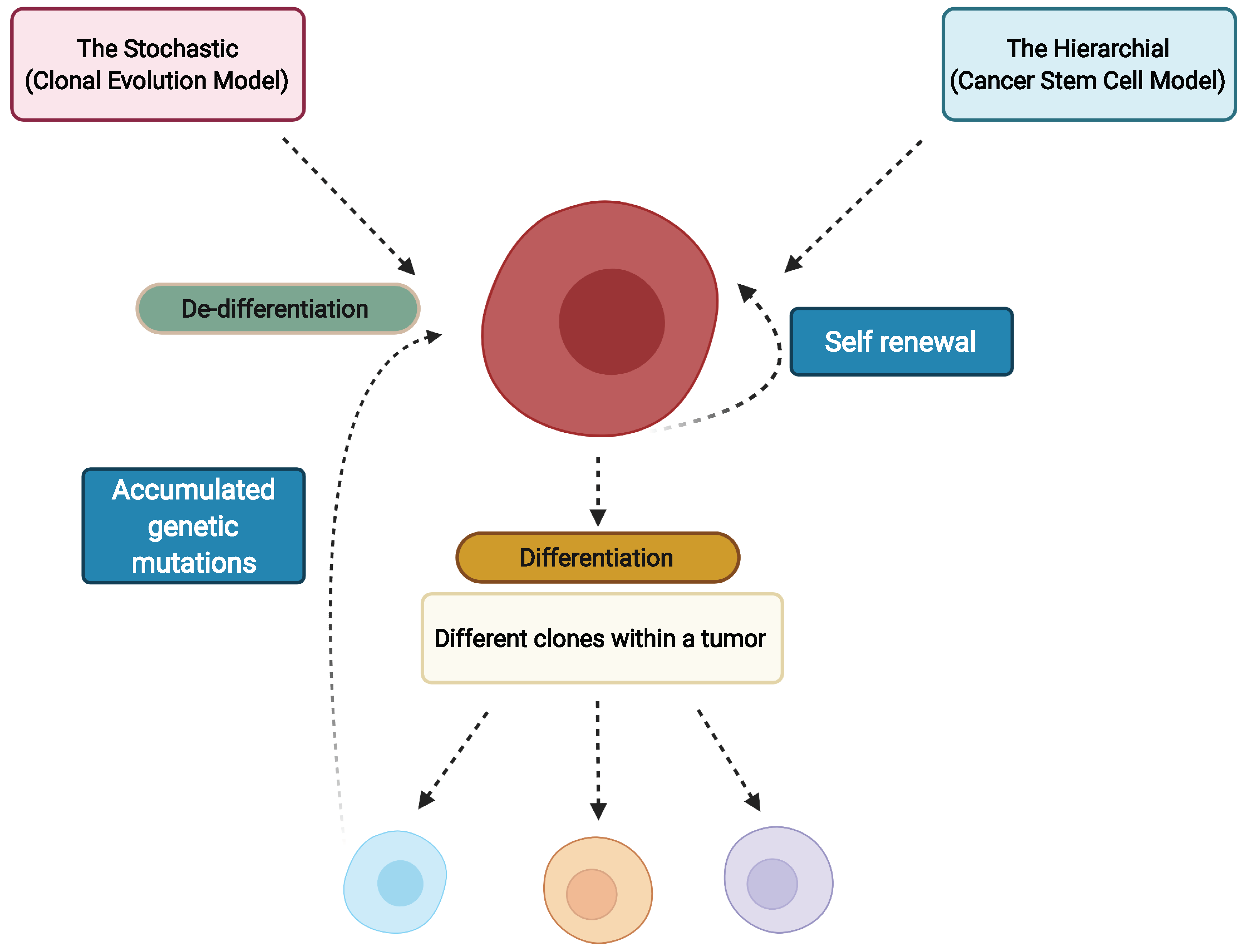
2. Cancer Stem Cells

Cancer stem cells (CSCs) are a subpopulation of tumor cells within bulk tumor tissue characterized by multipotency, enabling them to self-renew and differentiate into distinct tumor lineages [4]. Long-term self-renewal of CSCs allows proliferation of cells while maintaining an undifferentiated state, aiding indefinite persistence of quiescent CSCs in tumor tissues [5]. A CSC undergoes asymmetric division, producing two daughter cells with differential fate: one retains the stem cell identity, thus maintaining the self-renewal capacity, while the other becomes a specialized progenitor with capacity to produce mature tumor cells and populate the bulk of tumor tissue [6]. Accordingly, the genetic and epigenetic heterogeneity within the CSC subpopulation contributes to intratumoral heterogeneity, and the self-renewal capacity of the CSC subpopulation is a factor in driving tumor relapse [7]. With some exception, most of the CSCs have high resistance to chemo- and radio-therapies due to a range of factors, including a favorable tumor niche, overexpression of drug efflux pumps, pro-survival and anti-apoptotic signals, intracellular drug-inactivating enzymes and enhanced DNA repair [8]. Therefore, targeting the vulnerabilities in CSC is critical for sensitizing tumors to cancer therapeutics and preventing relapse and metastasis.

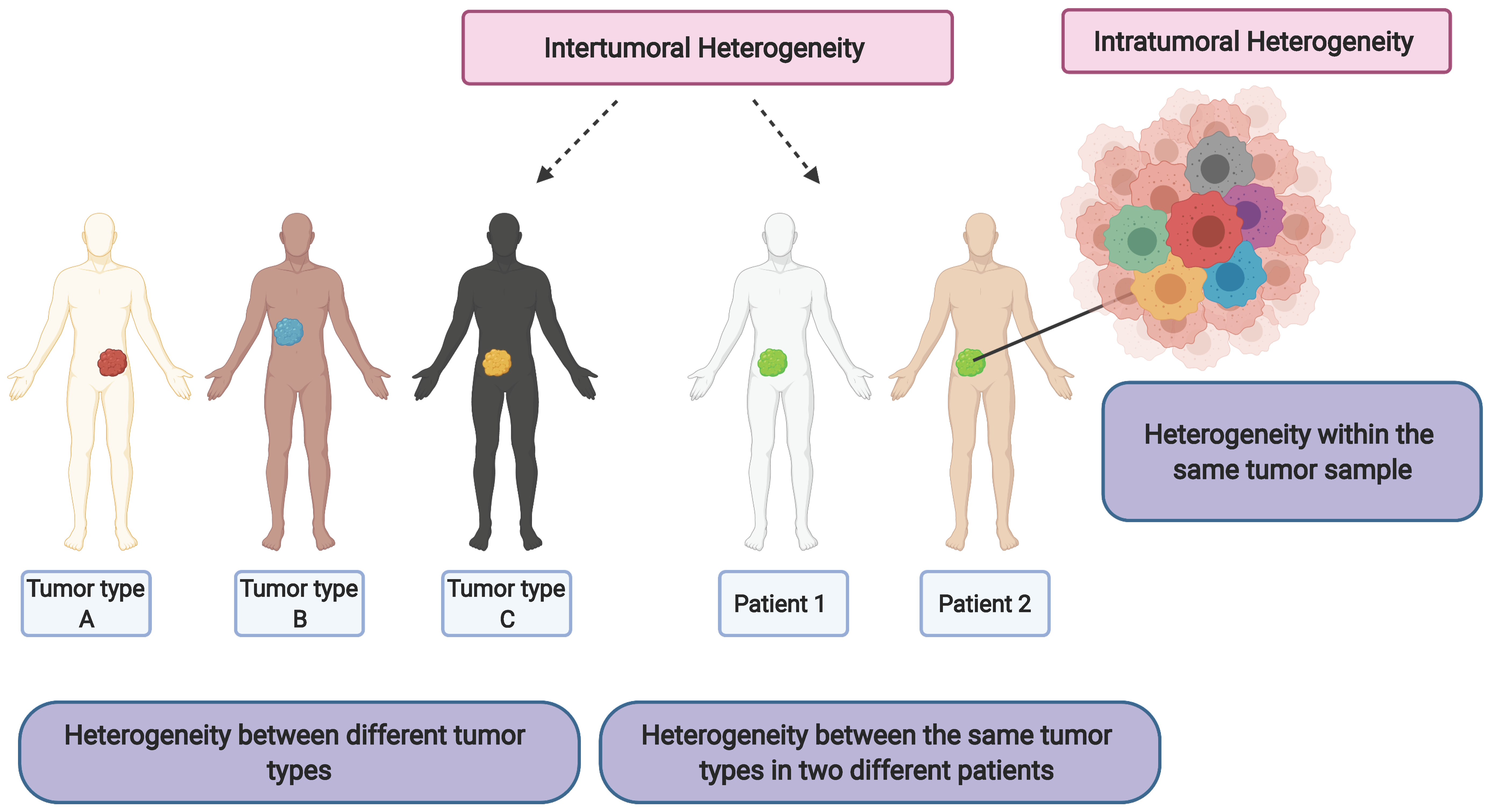
3. Cancer Stem Cells and Clonal Evolution as the Source of Intratumor Heterogeneity

The heterogeneity in cancer has been described by two models: the hierarchical cancer stem cell model and the stochastic clonal evolution model (Figure 2) [9]. In the hierarchical CSC model, it is hypothesized that a pre-existing CSC undergoes self-renewal and differentiation to produce both CSC and the non-CSC tumor cells that form the bulk of the tumor tissue. The CSCs may acquire tumorigenicity through clonal evolution and subsequent differentiation into non-CSC tumor cells via epigenetic mechanisms producing phenotypic heterogeneity [10,11]. The stochastic clonal evolution model hypothesizes that some tumor cells may accumulate genetic mutations over extended periods, and these genetic mutations contribute to phenotypic heterogeneity [9]. Studies describing tumor heterogeneity by the CSC model [10,11], stochastic evolution model [12] and non-CSC dedifferentiation into tumorigenic CSCs [13,14] have shown that CSCs are more resistant to conventional therapeutics than their non-CSC counterparts. Improved understanding of these CSC models is required since the true nature of CSCs will influence strategies for therapeutics and help lay the foundation for understanding critical properties of tumoral plasticity. The CSC model has significant clinical implications making it critical to target cancer stem cells to ensure complete elimination of tumor cells.

4. Cancer Stem Cell Plasticity

Tumor cell plasticity is defined as the property of cancer cells to exist in a dynamic stem/non-stem cell phenotype, epithelial/mesenchymal transitions or quiescent/proliferating condition [15]. This property permits CSCs to switch phenotypic states in response to environmental signals and is governed by intrinsic factors such as transactivators of EMT [16] and extrinsic factors such as signals transduced by the tumor microenvironment (TME) [17]. Tumor cell plasticity is characterized by epigenetic dysregulation caused by altered signaling from the TME, as well as variations in the pathways of metabolic glycolysis, oxidative phosphorylation (OXPHOS) and cell cycle progression [18]. The epigenetic mechanisms responsible for tumor plasticity include, but are not limited to: chromatin remodeling, histone modifications, DNA methylations or indirect control through noncoding RNAs, and mutations of cell cycle regulators such as p16 [19] and p21 [20]. Cellular plasticity is a major contributor to intratumoral heterogeneity [2]. With so many potential phenotypes within a single tumor, targeted therapeutic interventions may prove effective against some, but not all subpopulations, leaving resistant variants to grow back as relapsed tumors [21]. For example, considering that conventional chemotherapeutics target actively proliferating cells, quiescent CSCs may escape the treatment.
As described above, maintenance of cancer stem cell plasticity is essential to promote tumor cell survival. This maintenance of the stem cell state in tumor cells is governed by genomic rearrangements, DNA mutations, epigenetic modifications and microenvironmental cues [2]. As opposed to genetic changes, phenotypic variations through epigenetic switches are incorporated with each cell cycle and are thus responsive to immediate changes in the microenvironment [2]. It has been reported that during cancer initiation and growth, mutations in epigenetic regulators enhance tumor plasticity favoring adaptability and resistance to therapy thus significantly dictating cell fate [22,23]. Since most epigenetic dysregulation mechanisms are both heritable and reversible, therapies targeting epigenetic regulation are promising treatment strategies [24].

5. Epigenetic Regulation/Dysregulation in Cancer Stem Cells

Increasing evidence has shown that malignant transformation is not a consequence of genetic mutations alone, but also involves reversible epigenetic dysregulation [25]. Histones are proteins that are involved in the regulation of gene expression and silencing through interaction with DNA. For example, H3K9 trimethylation (addition of three methyl groups to lysine residue at position 9 on histone 3) together with deacetylation of histones H4 and H3 is associated with gene repression, while H4K8 (lysine residue at position 8 of histone 4) acetylation and H3K14 (lysine residue at position 14 of histone 3) acetylation along with H3S10 (serine residue at position 10 of histone 3) phosphorylation is associated with gene expression [26]. In cancer, several of these post-translational modifications can undergo dysregulation, driving intratumoral heterogeneity and leading to tumor subpopulations with novel epigenetic regulation. These epigenetic regulations are carried out mainly by histone writers, erasers and readers (Figure 3).

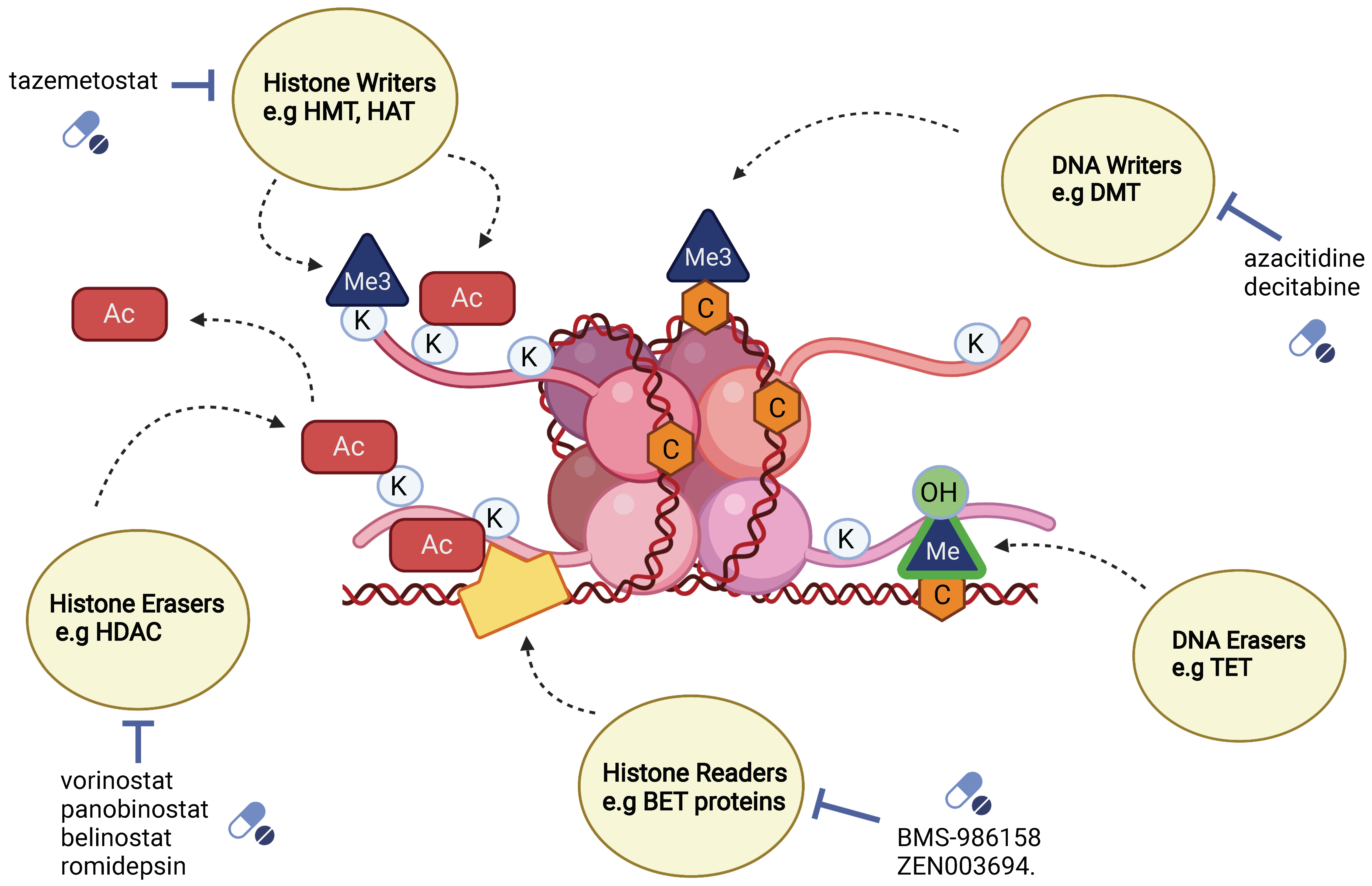
5.1. Classification of Epigenetic Mediators: Writers, Erasers and Readers

5.1.1. Writer Enzymes

Writers are enzymes that are involved in adding methyl or acetyl groups to specific amino acid residues of histones or methyl group to cytosine nucleotides of DNA [25]. Adding methyl groups to CpG islands present in DNA or histone tails helps prevent transcription by blocking transcription factors [25]. Transcription can be further prevented by the recruitment of proteins that bind to methylated sites, creating additional inaccessible binding sites on the chromatin [25].
DNA methyltransferases (DNMT) are writer enzymes that are involved in adding a methyl group to cytosine residues that are a part of CpG dinucleotides, forming 5-methylcytosine [27]. CpG dinucleotides are found in CpG islands in promoters [27]. Methylation of these regions in genes is associated with gene silencing [27]. DNMTs are divided into DNMT1 and de novo DNMT. De novo DNMTs are expressed largely during development and are important in the maintenance of methylation patterns in human embryonic stem cells, whereas during differentiation DNMT1 becomes highly expressed, with a reduction in de novo DNMTs [27]. DNMT1s are responsible for ensuring proper inheritance of epigenetic patterns during replication [27]. Several studies have shown the importance of DNMT1 in the maintenance of the CSC phenotype [28]. For example, a knockout of DNMT1 led to a reduction of cancer stem cell markers such as high expression levels of ALDH (aldehyde dehydrogenase), CD44+ and CD24+ in colon cancer cell lines [29].
Histone lysine methyltransferases (KMTs) are also a type of writer enzyme involved in the transfer of one, two or three methyl groups to specific lysine (K) positions at the tails of histones, which correspond to different biological responses [27]. Methylation at positions H3K4 (lysine residue at position 4 of histone 3), H3K35 (lysine residue at position 35 of histone 3) and H3K79 (lysine residue at position 79 of histone 3) is associated with gene expression, or open chromatin, while methylation at positions H3K27 (lysine residue at position 27 of histone 3), H3K9 (lysine residue at position 9 of histone 3) and H4K20 (lysine residue at position 20 of histone 4) is associated with gene repression [27]. A trimethylation pattern has been associated with transcriptionally active promoters, while monomethylation has been associated with active enhancers [30]. KMTs (lysine methyltransferases) can be subdivided based on the presence of the SET [Su(var)3–9 Enhancer-of-zeste and Trithorax] domain [30]. These enzymes are highly site-specific and are responsible for adding mono, di and tri methyl groups to histones [30]. A list of these enzymes is well detailed in C. Hon and Hawkins [30]. Several of the KMT enzymes are over-expressed in multiple cancers [27]. Enhancer of Zeste-Homolog 2 (EZH2), Enhancer of Zeste-Homolog 3 (EZH3) and H3K27(lysine residue at position 27 of histone 3) KMTs were found at elevated levels in glioma, breast and leukemia CSCs, and are credited with maintaining their quiescent state [31].

5.1.2. Eraser Enzymes

Erasers remove epigenetic modifications from DNA and histones. Histone demethylases remove methyl groups from histones; KDMs (histone lysine demethylases) remove methyl groups from lysine specifically. Lysine-specific demethylases are categorized into two groups: the FAD (flavin adenine dinucleotide)-dependent KDMs, KDM1A and KDM1B can remove mono and dimethyl groups but not trimethyl groups [27]. The Jumonji-C domain-containing histone demethylases, the second group of KDMs, can remove trimethyl groups from histone lysine residues, as well as di and monomethyl groups [27]. These two KDM categories are further subdivided into KDM2, KDM3, KDM4, KDM5 and KDM6, with each demethylating a specific lysine residue [27]. In embryonic stem cells, KDM1A (also known as lysine specific demethylase 1 (LSD1)) expression levels are typically elevated, but are reduced during differentiation [32]. LSD1 was also reported to maintain the balance between H3K4 di/tri methylation and H3K27 trimethylation marks (bivalent heterochromatin) at regulatory regions of several developmental genes, thus suppressing differentiation-associated genes and maintaining pluripotency in embryonic stem cells [33]. LSD1 has been shown to be an important regulator in maintaining the CSC population in treatment-resistant breast cancer cell lines. Pretreatment with LSD1 inhibitors improved treatment sensitivity to doxorubicin treatment [34]. Increased levels of KDM5A and KDM5B have been associated with chemoresistance in cancer and appear to contribute to an increase in cancer cell proliferation [35]. High levels of KDM5B are associated with the repression of tumor suppressor genes and apoptosis-related genes [36]. DICER, an enzyme involved in processing of microRNAs that are regulating the EMT pathway, showed a decrease in expression in breast cancer cell lines after hypoxic exposure, even though KDM6A and KDM6B were enriched in the promoter region, highlighting the importance of oxygen to carry out demethylating activity of KDM6A and KDM6B [37]. KDM6A was inactivated by a hypoxic environment in myoblast cell line C2C12, resulting in sustained H3K27me3, preventing myogenic differentiation [38].
Histone deacetylases (HDACs) are another group of eraser enzymes that remove acetyl groups from histones, which leads to chromatin compaction. As a result, DNA becomes inaccessible to transcription factors for gene expression. These enzymes cannot bind to DNA directly and require repressor complexes to facilitate DNA binding, such as NuRD (nucleosome remodeling and deacetylase complex), CoREST (co-repressor for element-1-silencing transcription factor) and KDM [27]. Knockdown experiments reveal that HDAC7 epigenetically modifies transcription start sites and super-enhancers of oncogenes such as C-MYC, CD44 and BMI-1 and reduces the expression of CD49f in breast cancer stem cells [39]. A knockdown of HDAC7 led to a reduction of sphere-forming ability and in vivo tumor growth of classic CSC phenotypes, and HDAC7 levels of expression in CSCs were also found to be higher than in non-stem cancer cells [40]. Being downstream of class 1 and class 2 HDACs, HDAC7 could be important in a targeting strategy for eliminating breast and ovarian CSCs [39]. Class 3 HDACs, also known as sirtuins, are dependent on NAD+ (nicotinamide adenine dinucleotide) for their activity thus playing a role in the metabolic regulation of the cell [27]. Class 4 HDACs include HDAC11, which has also been correlated with increased expression in lung cancer stem cell lines and regulates the expression of SOX2 [(sex determining region Y)-box 2], an important transcription factor to maintain CSC self-renewal [41].
DNA demethylation proteins are a third type of eraser involved in the oxidation of 5-methylcytosine (5mC), present in CG dinucleotides, into 5 hydroxymethylcytosine [27]. TET (Ten Eleven Translocation) demethylating proteins fall under this group of epigenetic regulators. TET1, TET2 and TET3 play pivotal roles in the development and maintenance of the stem cell phenotype [27]. TET2 was found to be involved in the regulation of genes associated with self-renewal and differentiation in hematopoietic stem cells [42], and mutations in TET2 have been associated with hematological malignancies [43].

5.1.3. Reader Enzymes

Readers are proteins that recognize histone and DNA modifications and can sense chromatin conformation. Bromodomain belongs to bromodomain and extra-terminal domain (BET) family members and recognizes acetylated lysine [44]. BRD4 (bromodomain-containing protein 4), which belongs to the BET family, binds to an acetylated promoter, allowing transcription of target genes [44]. C-MYC expression is regulated by BRD4 binding to the promoter and enhancer [22]. C-MYC was targeted using a BRD4 inhibitor, JQ1, in medulloblastoma. Inhibition of BRD4 was found to downregulate transcription of C-MYC, which led to a further reduction in cell growth as well as cell cycle arrest at the G1 phase. Stem cell-associated pathways were found to be downregulated upon treatment with JQ1. NANOG, Nestin and SOX2 classical stem cell markers were downregulated, while MAP2 (microtubule associated protein 2), a differentiation marker for neurons, was found to be upregulated [45]. C-MYC was validated as the target of BRD4 using luciferase-expressing plasmids controlled by C-MYC, studied in Daoy cells. This work showed a decrease in luciferase activity in the presence of JQ1 [45]. In another study, BRD4 was found to be associated with the N-termini of TWIST and WNT5A gene expression which are essential regulators of the EMT pathway [46].

6. Other Regulators of Cancer Stem Cells

6.1. Long Non-Coding RNA

A large part of the human genome contains DNA that does not code for any proteins and was long assumed to be “junk DNA”. Over the years, research has uncovered that although a large portion of transcribed RNA does not code for proteins, it serves a vital function in the regulation of genes [47]. Many of these regulatory segments fall into the category of long non-coding RNAs (lncRNA), which are greater than 200 nucleotides in length. LncRNA can be found in intergenic regions, transcribed from introns, sense RNA or antisense RNA [48]. LncRNAs can act as decoys, guides and signaling molecules. Decoys can cause gene repression by blocking the binding of proteins to RNA, inhibiting transcription [47,49]. Alternately, guides can facilitate gene expression by helping transcription factors or multiprotein complexes such as PRC (polycomb repression complex) to bind to target genes [47]. LncRNAs can also play integral roles in the regulation of CSCs. In the case of liver cancer stem cells, increased expression of lncTCF7 led to the activation of the Wnt pathway through the recruitment of SWI/SNF to the promoter of TCF [50]. In embryonic stem cells, lncRNAs were found to be important targets of the transcription factors OCT4, SOX2, C-MYC, KLF4 (Krueppel-like factor 4) and NANOG, which are involved in maintaining pluripotency [51]. Knocking down lncTCF7 expression caused a reduction in the expression of OCT4, SOX2, C-MYC, KLF4 and NANOG, along with reduced sphere-forming ability of liver cancer stem cells [50]. High XIST (LncRNA X inactive specific transcript) expression, a long non-coding RNA, was correlated with low miR-200c expression levels, and was identified as a potential target to eradicate bladder cancer stem cells [52].

6.2. ATP-Dependent Chromatin-Remodeling Complexes

Dynamic modification of chromatin is performed by ATP-dependent chromatin-remodeling complexes that utilize ATP to slide (translocate or move the histone along the DNA), evict or replace nucleosomes, thereby affecting gene expression [53]. Depending on the catalytic unit and associated subunits, the four classes of chromatin remodelers are SWI/SNF, CHD, ISWI and INO80. These four classes of remodelers possess an ATPase domain which is conserved across eukaryotes and function as multi-subunit complexes in association with tissue-specific subunits [54,55]. Epigenetic modification occurs through the binding of these complexes to specific chromatin domains such as bromo, chromo and SANT domains [56].

7. Hypoxia on Cancer Stem Cells

The hypoxic tumor microenvironment in solid tumors has been reported to play a role in CSC maintenance and reprogramming of non-CSCs to CSCs [57,58]. Hypoxia-inducible factor 1α (HIF1α) and factor 2α (HIF2α) are examples of primary mediators of hypoxia reported to be co-opted by CSCs to maintain stemness as well as evade hypoxia by enabling EMT. HIF1α-activated Notch signaling pathway was reported to be a critical regulator of CSC maintenance in hypoxic glioblastoma stem cells (GSC) [59]. In in vitro models of U251 cells, HIF1α mRNA and protein levels were found to be elevated along with the STAT3 and PI3K/Akt pathways in GSC. Inhibition of these pathways also partly reduced the hypoxia-mediated activation of Notch pathway [59]. HIF2α-induced release of VEGF was shown to promote angiogenesis in hypoxic TMEs in glioblastomas [57]. In in vitro stem cell models derived from patient biopsies and in vivo xenograft models, HIF2α was reported to be preferentially expressed in GSC vs non-GSC. Inhibition of HIF2α also reduced self-renewal and proliferation of GSC in vitro and decreased tumor initiation potential of GSC in vivo [57].
Hypoxia also regulates tumoral plasticity and EMT transitions in CSCs. Hypoxia mediated EMT plays an important role in inducing metastatic cells with CSC properties [60,61]. Hypoxia induced Jagged2 promoted metastasis in breast cancer in vitro as well as acquisition of stem cell phenotype by activating Notch signaling [60]. In in vitro and xenograft models of gastric CSCs, HIF1α induced EMT through the SNAIL pathway [61]. CSC maintenance in hypoxic conditions is also aided by glycolysis. Inhibition of glycolysis pathways has been reported to suppress CSC maintenance [62]. Pyruvate dehydrogenase kinase 1 (PDK1) is a glycolytic enzyme involved in the tricarboxylic acid cycle. PDK, a downstream target of HIF1α, is elevated in breast CSCs through the lncRNA H19/miRNA let-7/HIF1α signaling axis. siRNA mediated knockdown of PDK1 resulted in loss of colony formation and related stemness features in breast CSCs both in vitro and in vivo [62]. Given that hypoxia influences cell signaling pathways affecting CSC plasticity and maintenance, targeting HIFs and other signaling pathway factors influenced by hypoxia may be a relevant strategy to drive the loss of stem-like state in CSCs.

8. Cell Signaling Pathways Regulating Cancer Stem Cells

Various cell signaling pathways in normal stem cells that regulate cell survival, growth, differentiation and self-renewal are found to be mutated in CSCs. Commonly implicated signaling pathways include Wnt, Notch, hedgehog, NF-κB and PI3K/PTEN [3]. Regulation through these pathways is not linear, but brought about by cross-talk between the pathways with shared subunits such as Gli and GSK-3β, [63] which aid the maintenance of CSC populations [63]. In this manner, genetic and epigenetic modifications of subunits participating in these signaling pathways can influence multiple other pathways and affect CSC maintenance. Therefore, identification of the main effectors of epigenetic regulation of signaling pathways is increasingly viewed as a potential area for therapeutic research.

8.1. Wnt Signaling Pathways

The canonical wingless and integration site growth factor (Wnt/β-catenin) signaling pathway is recognized as a crucial modifier of tumor cell plasticity [64]. Gene activation through the canonical Wnt signaling pathway is mediated by the transcription factor β-catenin. In the absence of Wnt ligand, the β-catenin destruction complex consisting of adenomatous polyposis coli (APC), Axin, Dishevelled-1 (DVL-1), glycogen synthase kinase 3 β (GSK-3β) and casein kinase 1 (CK1) prevents β-catenin translocation to the nucleus [65]. GSK-3β phosphorylates β-catenin for ubiquitination and proteasomal degradation. However, upon binding of the Wnt ligand to Wnt receptor Frizzled and Wnt co-receptor Low density lipoprotein receptor-related protein (LDR5/6), DVL is activated and causes disassembly of the degradation complex [66]. Dephosphorylated β-catenin, now free, translocates into the nucleus and associates with transcription factors T-cell factor/lymphoid enhancer (TCF/LEF), inducing expression of Wnt target genes such as CCND1 and MYC [67]. Epigenetic regulation of Wnt signaling is reported in mice models of glioma, wherein activation of Wnt/TCF4 signaling causes overexpression of histone demethylase KDM4C, which removes H3K9me3 from Wnt target genes, promoting cell proliferation and tumorigenicity [68]. Meanwhile, DNA promoter methylation of secreted Frizzled receptor protein 1 (SFRP1) in hepatoblastoma was also found to correlate with mutated β-catenin [69]. Moreover, a loss of methylation at H3R8me2(arginine residue at position 8 of histone 3) and H4R3me2 (arginine residue at position 3 of histone 4) promoter regions of AXIN2 and Wnt Inhibitory Factor 1 (WIF1) caused by the inhibition of protein arginine methyltransferase 5 (PMRT5) promoted lymphoma cell survival in patient-derived NHL cell lines [70].

8.2. Notch Signaling Pathway

Notch signaling is an evolutionarily conserved pathway regulating differentiation and self-renewal in stem cells [71]. It is a contact-based signaling pathway wherein binding of ligands such as Jagged 1/2 or Delta1–4 causes the release of Notch intracellular cytoplasmic domain (NICD) into the cytoplasm through γ-secretase-induced cleavage of the transmembrane Notch receptor [72]. NICD then translocates to the nucleus which is followed by the activation of Recombination Signal Binding Protein 1 for J- Kappa (RBPJ- κ) for expression of MYC and HES1 Notch genes [73]. Depending on the tumor microenvironment, the Notch pathway is involved in oncogenic or tumor suppressive roles in a wide range of tumors. It is reported that aberrant Notch signaling promotes CSC self-renewal and metastasis in ovarian, breast and HCC (hepatocellular carcinoma) CSCs [71]. In clear cell renal cell carcinoma (CCRCC), overexpressed Jagged1 was associated with DNA demethylation of H3K4me1-associated enhancer regions [74]. Epigenetic profiling of osteosarcoma cells identified leukemia inhibitory factor (LIF) as an essential factor controlled by super enhancers. LIF activation of NOTCH1 signaling by H3K27me3 demethylation through the Ubiquitously Transcribed Tetratricopeptide Repeat on chromosome X (UTX) histone demethylase correlated with “stemness” related genes, sphere formation, self-renewal and metastasis in osteosarcoma [75]. In cutaneous T cell lymphoma (CTCL), the binding of the epigenetic reader bromodomain-containing protein 4 (BRD4) to promoter and enhancer regions of CD4+ T cells of CTCL patients resulted in tumorigenesis through increased expression of NOTCH1 and RBPJ genes [76].

8.3. Hedgehog Signaling Pathway

In CSCs, the hedgehog (Hh) signaling pathway is implicated in driving tumor growth, development and regulation of tumors after therapeutic intervention [77,78]. The hedgehog signaling pathway is mediated through (Glioma associated oncogene homologue) Gli proteins, which, in the absence of Hh signals are sequestered by Suppressor of Fused (SUFU) and Kinesin family member 7 (Kif7) [79]. However, upon the binding of Sonic hedgehog (Shh) ligand to the Patched receptor (PTCH1), Smoothened (SMO) is no longer suppressed by PTCH1 and becomes activated. Once activated, SMO causes the release of the sequestered Gli proteins, which translocate to the nucleus and regulate cell growth, proliferation and differentiation [71]. In patient-derived colorectal cancer-initiating cells, Indian hedgehog (IHH) gene was found to exist in a bivalent state with both the activating H3K4me3 and repressive H3K27me3 histone marks. The presence of such a bivalent state in the IHH gene aided stem cell maintenance of the colorectal cancer-initiating cells. Subsequent disruption of bivalency through the inhibition of EZH2 resulted in decreased self-renewal [77]. In gastric adenocarcinoma, significant DNA methylation of Shh transcription factors CDX1/2 and KLF5 correlated with decreased expression of CDX1 and KLF5 and an increased expression of CDX2. The increased expression of CDX2 was associated with formation of metastatic lymph nodes in patients [78]. Similarly, in bladder cancer, it was reported that DNA hypermethylation of the CpG shore of the SHH gene caused the loss of the SHH gene and resulted in invasive urothelial carcinoma [80].

9. Epithelial–Mesenchymal Transition Is a Critical Regulator of the CSC Phenotype

Epithelial–mesenchymal transition (EMT) is a reversible transition process wherein cells in the primary tumor site acquire mesenchymal attributes to gain motility, and upon the invasion of a secondary tumor site ultimately revert to an epithelial state through a mesenchymal–epithelial transition (MET) [81]. In the primary tumor, cells are maintained in the epithelial state through epigenetic and transcriptional control of EMT genes [82]. Signals in the tumor microenvironment (TGFβ, WNT etc.) contribute to alterations in the expression of EMT genes, and can cause tumor cells at the margin to undergo the epithelial–mesenchymal transition, causing them to lose cell adhesion and polarity, dislocate from the primary tumor, gain cellular motility, survive in circulation through the body and infiltrate secondary organs [83]. E-cadherin is an adherens junction protein encoded by the CDH1 gene that maintains the stemness of cells in the epithelial state. E-cadherin downregulation is a key step for initiating EMT and causing the detachment of cells from the bulk tumor [84]. E-cadherin expression is regulated by transcription factors Snail, Twist and Zinc finger E-box binding homeobox 1 (Zeb1/Zeb2) after activation of EMT [23]. Histone modifications that remove repressive H3K27me3 from promoter genes such as ZEB1, which are in the bivalent state (simultaneous methylations at different histone sites H3K27me3 and H3K4me3) also lead to alterations in EMT [82]. Such epigenetic alterations of transcription factors are mediated through chromatin remodeling complexes such as NURD and miRNA silencing, affecting the ZEB1 promoter gene. ZEB1 represses epithelial genes such as CDH1 that code for E-cadherin through epigenetic regulation and maintains the epithelial state of cells [85]. In NSCLC models, ZEB1 was reported to physically recruit the CHD4/NuRD complex to promoters and induce repression of miR-200c/141 and TBC1D2b, which suppress invasion and metastasis. This results in activation of Rab22, thus promoting metastasis through internalization and degradation of E-cadherin [85]. Invasive and migration properties of ZEB1 are suppressed by miR-639. Repression of miR-639 by DNMT3A-mediated hypermethylation of its promoter gene was found to promote tumorigenesis in liver cancer mouse xenograft models [86]. Similarly, transcription factor Forkhead Box protein A2 (FOXA2) binding to the CDH1 promoter caused a high expression of E-cadherin and reduced cancer cell migration in oral cancer [87]. Direct regulation of E-cadherin is also brought about by regulation of the CDH1 gene. For example, in cancer cell line models of NSCLC, transcription factor Six2′s overexpression caused promoter methylation of CDH1, inhibiting E-cadherin expression and enhancing stemness and chemosensitivity [88]. Another epigenetic regulator, KDM7B, activates gene expression of EMT genes SNAI1 and VIM by erasing repressive histone markers H3K9me1/2, H3K27me2 and H4K20me19 [89,90]. In HCC, it was reported that FIP-200-dependent autophagy caused PHF8-mediated repression of E-cadherin by SNAI1 upregulation, which promoted EMT by causing degradation of E-cadherin [91].
Signaling pathways such as TGFβ are also known to affect epigenetic regulation of EMT genes [92,93]. In murine HCC models, TGFβ-mediated SNAI2 upregulation resulted in an upregulation of H3K9 methylation and downregulation in H3K4 and H3K56 acetylation in E-cadherin promoter regions [92]. Additionally, this SNAI2 upregulation also interacted with G9a and HDACs to suppress E-cadherin transcription, resulting in migration and invasion [92]. In another study, it was reported that in response to the exogenous TGFβ activation, PMRT5-MEP50 complex affected transcriptional activation by methylating H3R2me1 and H3K4, and repressed transcriptional activation through H4R3me2s demethylation of EMT genes and invasion pathways in lung and breast cancer cells [93]. Downstream targets of TGFβ signaling are also implicated in EMT. Epigenetic regulation of RASSF10 (Ras-association domain family), a target of TGFβ, was reported to inhibit EMT by interacting with and stabilizing the ASPP2 tumor suppressor gene [92]. It was reported that depletion of RASSF10 caused SMAD2 phosphorylation and promoted tumor invasion [94].
The epigenetic regulator Polycomb Repressive Complex 2 (PRC2) is found to be altered in GBM. Recently, the miR-490-3p/TGIF2/TGFBR1 axis was reported to affect EMT in GBM cell lines, wherein the EZH2 unit of PRC2 was found to regulate this miRNA and CHRM2 host gene directly [95]. Another mechanism of EMT regulation is through the chromatin-modifying enzyme lysine-specific demethylase 1 (LSD1), which is overexpressed in colorectal cancers (CRCs). In an in vitro study of CRC cell lines, it was reported that LSD1-mediated AKT activity promoted EMT in cells containing the PIK3CA mutations [96]. Similarly, in another study on CRC cell lines, LSD1-mediated loss of H3K9me2 in the TSPAN8 promoter region reportedly caused TSPAN8 overexpression and promotion of EMT [97].

10. Integrins Alter Cancer Stem Cells Behavior

Integrins are heterodimeric cell surface receptors that can sense extracellular cues and signal intracellular processes governing proliferation, differentiation, adhesion and migration [98]. They have an extracellular portion, a transmembrane portion and a cytosolic tail [98]. Ligand binding induces a change in conformation of the integrin, leading to the recruitment of a cytoskeletal protein, talin, to the cytosolic portion of the β subunit [99], which activates focal adhesion kinases (FAK) and SRC family kinases [100]. Growth factors present in the ECM (extracellular matrix) require integrins to interact with their respective ligands as cofactors, in order to carry out their own signaling activities [100]. Unligated αVβ3 integrins can complex with KRAS to promote NFκB (nuclear factor kappa-light-chain-enhancer of activated B cells) activity, which has implications in cancer stem cell survival via self-renewal, proliferation and EMT (epithelial–mesenchymal) transition [101]. Integrins form dimers in various combinations of 18α and 8β subunits to form twenty-four sets of heterodimers [98]. Overexpression of integrins in cancer has been reported and various peptidic ligands against integrins have also been developed for targeted therapy and imaging [102,103].
αVβ3 integrins have been reported to contribute to resistance against receptor tyrosine kinase inhibitors [104]. An increase in ALDH (aldehyde dehydrogenase) activity, a marker for cancer progenitors, was observed in erlotinib-resistant lung cancer cells that displayed integrin β3+ compared to those cells that did not express integrin β3+, suggesting a contributing role of β3+ to the cancer stem cell phenotype [104]. In the same study, an increase in the expression of αVβ3 integrins was found in erlotinib-resistant lung tumor biopsies [104]. In another study, integrin α6 was highly expressed in glioblastoma cells that have a high expression of CD133, a cancer stem cell marker for glioblastoma [105]. α6(CD49f) has been used as a biomarker for identifying adult stem cells [106]. Interestingly, in the case of pluripotent stem cells, α6 expression is important to prevent FAK activation through β1 and to promote expression of genes associated with pluripotency and self-renewal [106,107].
Yes-associated protein (YAP) and transcriptional co-activator with PDZ-binding motif (TAZ) are transcriptional coactivators and have high expression levels in many cancers [108]. SRC activation was correlated with increased YAP/TAZ transcriptional activity [108]. A splice variant of α6 cytoplasmic domain (α6Bβ1) was found to activate TAZ, leading to transcription of genes associated with self-renewal [99], as well as the transcription of LMα5, which leads to the production of laminin (LM511) matrix [99,109]. The niche microenvironment surrounding cells has important functions in maintaining their stem cell properties [109]. LM511 has been shown to contribute to maintaining the stem cell phenotype upon interaction with integrins [109,110]. Autocrine VEGF signaling was found to control the α6B variant, but not α6A, and promote the breast cancer stem cell phenotype [99,111]. Overexpression of the α3 integrin in cancer cells that had acquired resistance was found to be associated with restrained metastasis through the suppression of Rho GTPase activity through Abl kinases in the Hippo tumor suppressor signaling pathway in prostate cancer [112]. However, in the case of glioblastoma, α3 was found to correlate with invasion and metastasis through ERK1/2 pathway [113].
Integrins can also mediate cell signaling events that regulate stem cell proliferation. For example, epithelial stem cells can differentiate into hair follicles and integrins β1, β4 and α6 are used to identify epidermal stem cells [114]. Other cells that support stem cells in their niche can secrete certain ligands that help to signal stem cells to remain in their quiescent state, as in the case of intestinal stem cells, where pericryptal fibroblasts secrete hedgehog ligands to inhibit proliferation [114]. Periostin plays an important role in creating a niche for metastatic cancer stem cells [115] and signaling through integrins is required for successful homing to the distant site [99]. Therefore, the survival of cancer stem cells is a result of extracellular signaling events in niches that help them maintain their quiescent state or promote their proliferative state.

11. Targeting Vulnerabilities in Cancer Stem Cells

Key pathways in the maintenance of CSCs are regulated by epigenetic mechanisms [23]. Therefore, “epigenetic reconditioning”, or inducing differentiation of CSCs via drugs that modify epigenetic changes, such as hypomethylating agents, is one of the approaches utilized for targeting CSCs [116]. Since tumoral plasticity conferred by epigenetic modulators is reversible, it is an attractive approach for therapeutic interventions [117]. Epigenetic inhibitors targeting DNA methyltransferases, histone deacetylases, non-coding RNA and chromatin remodelers are able to reverse or inhibit epigenetic regulation of tumor cells and are currently being developed and investigated in different clinical phases [118,119,120].

11.1. Histone Methyltransferase Inhibitors

The EZH2 unit of the PRC2 complex is responsible for the repression of tumor suppressor genes and is frequently overexpressed in cancers, therefore EZH2 is currently being investigated in cancer therapy research [121]. 3-Deazaneplanocin A (DZNep) is one of the most studied EZH2 inhibitors. It indirectly affects EZH2 expression by inhibiting S-adenosylhomocysteine (SAH)-hydrolase, which causes an upregulation of SAH, leading to degradation of PRC2 and subsequent EZH2 inhibition [121]. Another EZH2 inhibitor, tazemetostat (TAZVERIK) was recently FDA approved for treatment of patients with epithelioid sarcoma and is undergoing trials for diffuse large B-cell lymphoma and mesothelioma as well [122]. Tazemetostat (EPZ6438) at a dose of 800 mg twice daily showed promising results in a phase I study that included relapsed or refractory non-Hodgkin’s lymphoma patients and solid tumor patients possessing the EZH2-activating mutations, and with patients that displayed a wild type tumor phenotype [123]. In the subsequent, ongoing phase II clinical trial by Epizyme, which is estimated to be completed by 2021 (NCT01897571) patients with epithelial sarcoma were grouped into two cohorts consisting of either treatment-naïve or relapsed patients [118]. Out of twenty-four naïve patients, the objective response rate (ORR) was 6% and disease control rate was 42%, as of the preliminary press release. Out of thirty-eight relapsed or refractory patients, ORR was 8% and the disease control rate was 16% [118]. A phase I study of patients with hematological malignancies and solid tumors involving GSK126
(GSK2816126), which inhibits EZH2 directly by competing with S-adenosylmethionine (SAM), was terminated because 51% of the patients showed progressive disease, while only 34% showed stable disease (NCT02082977) [124]. A possible reason put forward for this failure was that GSK126 promoted differentiation of hematopoietic stem cells into myeloid-derived suppressor cells and reduced the anti-tumor activity of the inhibitor [125]. However, it was observed that a combination of GSK126 with gemcitabine/5-fluorouracil showed an increase in anti-tumor activity compared to the use of either agent as a monotherapy (Refer to Table 1) [125].

11.2. Histone Methylase Inhibitors

Overexpression of lysine-specific demethylase 1 (LSD1) is reported in aggressive forms of cancers [27]. LSD1 inhibitors are being investigated in solid tumors (metastatic breast cancer, NSCLC, Ewings sarcoma) post reported success in AML (acute myeloid leukemia) [126]. Tranylcypromine is an LSD1 inhibitor with known toxicities, but there are less toxic, chemically modified forms of tranylcypromine with enhanced specificity to LSD1, such as ORY-1001(RO7051790) and GSK2879552, that are undergoing investigation in clinical trials for solid tumors (Refer to Table 2) [34]. Recently, a dual class I HDAC and LSD-inhibitor Domatinostat (4SC-202) was reported to show cytotoxic and cytostatic effects on ATRT cells using in vitro 2D and 3D spheroid models [127].

11.3. DNA Methyltransferase Inhibitors

Therapy with azanucleosides such as 5-azacitidine and decitabine, which are analogues of cytidine, have been shown to activate silenced tumor suppressor genes in vitro [128]. Azanucleosides work either by being incorporated into DNA and RNA, triggering the DNA damage response pathway, or by directly inhibiting DNA methyltransferases [128] through trapping DNMTs, preventing them from further methylation activity during replication [129]. Earlier studies found azacitidine mostly being effective in the S phase of the cell cycle, and demonstrating cytotoxic effects [130]. However, at lower doses, the effect of azacitidine preferentially acts on DNMTs, thus activating silenced tumor suppressor genes and differentiation processes [131,132]. Currently, they are approved by the FDA for acute myeloid leukemia and myelodysplastic [128]. However, for solid tumors and other hematological cancers, there is no evidence to support azacitidine for use as a single agent [129]. Nonetheless, it may improve the response to immunotherapy [132]. The development of resistance to treatment in patients who achieved cancer remission has been reported, and the issue of resistance cannot be completely eliminated by using DNMT inhibitors alone [121,128]. A study found that in hematological malignancies such as MDS, the hematopoietic progenitors that did not respond to azacitidine had an upregulation of the ITGA5 gene which codes for integrin α5 and is correlated with CSC quiescence [133]. Using an anti-α5 antibody with simultaneous azacitidine treatment to block α5 signaling, an improved hematopoiesis response was observed [133].
Strategies proposed for successful long-term survival without relapse include priming cancer cells to cytotoxic therapy by treating them with decitabine before chemotherapy, as put forward in a study consisting of non-small cell lung carcinoma (NSCLC) patients with a history of prior chemotherapy [134]. The study used a combination of azacitidine and entinostat, a HDAC inhibitor. For treating solid tumors, a combination of Vidaza, the commercial name of azacitidine from Celgene, with Abraxane (nanoparticle albumin-bound paclitaxel) administered to patients with breast cancer and other advanced and metastatic solid tumors showed a 61.5% better response to 75 mg/m2 azacitidine and 100 mg/m2 of Abraxane, versus the control (NCT00748553). The ORR for a time frame of 1.5 years was 53.8%. In the case of decitabine, at lower doses, older patients suffering from AML were able to tolerate treatment [129]. Additionally, a comparison between decitabine and azacitidine showed a higher ORR (overall response rate) and PFS (progression free survival) to a five-day decitabine treatment regimen every four weeks, versus a seven-day azacitidine treatment regimen. However, there were no significant differences between OS and EFS (event free survival) (Refer to Table 3) [135].
Non-nucleosides have been also used as inhibitors due to the side effects of using azanucleosides [136] EGCG (epigallocatechin gallate) is a polyphenol found in green tea. EGCG increased the sensitivity of colon cancer cells in vitro to subsequent treatment with 5-fluorouracil [136]. However, in phase II clinical trials assessing the effects of EGCG as a maintenance therapy to prevent recurrence in ovarian cancer patients who had previously undergone chemotherapy, five of the sixteen women in the study did not have an ovarian cancer recurrence, but twelve women faced grade 1 adverse events [137].
Hydralazine, another non-nucleoside inhibitor against DNMTs, was used earlier along with valproic acid, an HDAC inhibitor, for sensitizing patients suffering from advanced-stage solid tumors [138]. The phase II single arm study noted that 80% of patients had clinical benefit in terms of partial response and stable disease (Refer to Table 3) [138]. The combination of hydralazine and valproic acid as antimetastatic agents in Ras-transformed 3T3 cells demonstrated growth inhibition in vitro and in vivo [139]. Overall, azanucleosides seem to be more effective than non-nucleosides.

11.4. Histone Deacetylase Inhibitors

HDAC inhibitors can be classified into selective, non-selective and multi-pharmacological HDAC inhibitors [140,141]. Vorinostat is a non-selective HDAC inhibitor which targets class 1 and class 2 HDACs and is used in the treatment of cutaneous T cell lymphoma (CTCL) [131]. In relapsed/refractory Hodgkin’s lymphoma, vorinostat administered as a single agent did not lead to any significant changes. One out of twenty-five patients who had previous chemotherapeutic treatment had a partial response and twelve of the patients had stable disease [140]. A meta-analysis of clinical trials consisting of three HDAC inhibitors (vorinostat, panobinostat and ricolinostat) concluded that panobinostat is the most effective of these three HDAC inhibitors, with an ORR (Overall Response Rate) of 0.64 in patients with relapsed/refractory multiple myeloma [142]. The authors of the study also concluded that patients who had previously been treated with lenalidomide can have an improved outcome upon treatment with HDAC inhibitors. An HDAC6 inhibitor, ricolinostat, had better clinical outcomes in combination with bortezomib and dexamethasone, rather than as a monotherapy for patients with multiple myeloma (Refer to Table 4) [143]. Romidepsin is a cyclic peptide that can inhibit HDAC1 and HDAC2 specifically [144]. Though HDAC inhibitors have had positive clinical outcomes in hematological malignancies, in solid tumors, such success has not been seen [145]. Curcumin has strong anti-oxidative and anti-inflammatory properties [146] and has been demonstrated to improve treatment against cancer stem cells when in combination with the FOLFOX chemotherapy regimen in patient-derived ex vivo colorectal cancer models [147]. Belinostat, a pan HDAC inhibitor, is approved by the FDA as a single agent treatment for relapsed refractory T-cell lymphoma (PTCL) [148].

11.5. BET Inhibitors

BRD4 of the BET family is an epigenetic reader that binds to acetylated lysine on histones and is reported to play a role in tumorigenesis in a variety of cancers [44]. BET inhibitors bind to bromodomains, blocking recognition of acetylated lysine on histones and inhibiting acetylation [27]. JQ1 (small molecule) and I-BET762 (synthetic mimic) are heavily utilized competitive inhibitors of BRD4 [149]. JQ1 has been reported to suppress the expression of ALDH(aldehyde dehydrogenase) by targeting the super enhancer regions of ALDH1A1 [150]. In vitro and in vivo SMARCA4/A2-deficient ovarian and lung cancer models showed BETi mediated anti-proliferative effects, showing that SMARCA4/A2 (SWI/SNF-related, matrix-associated, actin-dependent regulator of chromatin, subfamily a, member 4/2) may be used as biomarkers for BETi sensitivity [127]. There has also been an interest in proteolysis targeting chimera (PROTAC)-based BET protein inhibition, as ARV-825 has been reported to possess antileukemic activity alone or in combination with cytarabine in murine models [151]. BMS-986158 is a BET inhibitor currently investigated for treatment of pediatric cancers in Phase I clinical trials (NCT03936465). ZEN003694 is another bromodomain inhibitor currently being investigated in Phase II clinical trials with a PARP inhibitor drug talazoparib as a treatment for patients with Triple Negative Breast Cancer (TNBC) (Refer to Table 5).

11.6. Inhibitors against Signaling Pathways

Given that epigenetic regulation of signaling pathways also affects tumor plasticity, drugs affecting signaling pathways may also be used for conferring epigenetic control [70,119]. Notch pathway inhibitors usually inhibit γ-secretase that is responsible for cleaving the ICD of the Notch receptor. A γ-secretase inhibitor called nirogacestat (PF-3084014) has been investigated in twenty-four patients with desmoid tumors in phase I and II clinical trials, showing good tolerability and is currently undergoing phase III trials (NCT01981551). DAPT is another γ-secretase inhibitor reported to increase cisplatin sensitization by inhibiting proliferation, increasing apoptosis and reducing motility in osteosarcoma CSCs alone or in combination with cisplatin, which shows that a combination of a γ-secretase inhibitor with other treatment modalities can improve drug efficacy [119]. However, γ-secretase inhibitors alone are not effective, as seen in clinical trials of T cell acute lymphoblastic leukemia where a γ-secretase inhibitor showed low efficacy due to the presence of γ-secretase inhibitor-resistant cells with BRD4-dependent transcription; hence, combination treatments are required to target treatment-resistant subpopulations [22]. Approved hedgehog (Hh) signaling pathway inhibitors targeting the Smo subunits vismodegib and sonidegib have reduced efficacies in unselected patients, Smo-mutated patients and in patients showing initial treatment response due to acquired resistance and/or cross resistance to these SMO-inhibitors, thus making the use of single agent therapy unfeasible [152]. Glasdegib by Pfizer is an oral Hh inhibitor approved for AML, currently in phase II clinical trials for myelodysplastic leukemia and chronic myelomonocytic leukemia [153]. Other inhibitors of Hh pathways are drugs that prevent the binding of Gli transcription factors to DNA such as arsenic trioxide (ATO), which is in phase II clinical trials [154], and GANT61, which is under preclinical study [155]. Napabucasin (BBI608) is an example of a STAT3 inhibitor found to deplete cancer stem cells in NSCLC and prostate cancer, and is currently being investigated in phase III clinical trials for use in metastatic colorectal carcinoma (NCT02753127) [120]. Mutations in ARID1A, a component of the SWI/SNF chromatin remodeling complex, are recognized as biomarkers of single-agent ATR checkpoint inhibition, and represent a synthetic lethal approach [156]. Use of ATR inhibitors as single-agent treatments are being investigated in an ongoing phase II clinical trial involving patients with ARID1A loss in gynecological cancer treated with an ATR-inhibitor in combination with olaparib (NCT04065269) (Refer to Table 6). Tumor plasticity driven by epigenetic regulators in response to environmental cues and therapeutic interventions assists tumor evasion and relapse. Therefore, a recognition and understanding of epigenetic heterogeneity is essential to develop synthetic lethality-based treatment modalities, since targeting isolated pathway mechanisms would leave behind tumorigenic subpopulations.

12. Conclusions

In this review we discuss several key players that dictate and modulate the behavior of cancer stem cells. Current cancer therapeutic strategies still face challenges especially due to quiescent stem cell populations that promote tumor relapse. The heterogeneous nature of cancer makes it difficult to find one treatment that can kill all cancer cells. The dynamic nature of these cells, resulting from the accumulation of mutations over time causing malfunction of epigenetic factors or by tumor microenvironment allowing CSC phenotype survival, has made it challenging to find a one size fits all solution. Research on epigenetic factors affecting CSC phenotype maintenance and survival has uncovered key epigenetic signatures and related signaling pathways that are linked to CSC formation and maintenance.
FDA (US Food and Drug Administration) approved drugs targeting epigenetic factors, currently in the market, are a handful. Drug development in this area is gaining momentum with many inhibitor analogs against HATs, HDACs, BETs, TETs, HMTs and DMTs being tested as monotherapy or in combination with existing therapies in clinical trials. Currently, most of the approved drugs are indicated for hematological malignancies that tend to relapse. As epigenetic signatures involved in CSCs survival can also be found in normal cells, stem cells and tissues, targeted therapies may become necessary for improved efficacy and reduced off target effects. We provide a comprehensive list of several factors governing cancer stem cell populations in heterogeneous tumors. We also discuss the interplay between epigenetic modifiers and stem cell survival. In addition, our review focusses on therapeutics that target these epigenetic modifiers in current in vitro, in vivo and clinical studies. We provide a detailed understanding of the underlying determinants of the stem cell niche. Finally, we aim to inform the scientific and clinical community of the various therapeutic options available in clinic and point towards exciting new avenues that warrant further research.
An emerging field of epigenetics that is becoming more relevant to CSC maintenance and differentiation is bivalent chromatin biology. Initial discovery that promoter regions of developmentally regulated genes in embryonic stem cells (ESCs) contain both transcriptionally repressive mark (H3K27me3) and active mark (H3K4me3) [157] fosters the idea that chromatin bivalency is closely linked to the pluripotent state of stem cells and that stable selection of either mark allows lineage specification [158]. Subsequent discoveries of these bivalent chromatin domains in cancer suggest that this epigenetic pattern may play a role in genes regulating CSC phenotype [159] and clonal selection upon drug treatment [160]. As such, combinations of epigenetic therapies that affect chromatin bivalency may be effective in altering CSC stem properties and may be best-suited to limit heterogeneity and plasticity of CSCs [161]. Future studies in regulation and targeting of chromatin bivalency in cancer may hold the key to epigenetic targeting of cancer cell plasticity, heterogeneity and multipotency.

Author Contributions

Conceptualization, V.E.K. and R.N.; writing—original draft preparation, V.E.K. and R.N.; writing-reviewing and editing A.N., C.D.S., J.C. and K.S.L.; visualization—A.N. and C.D.S. All authors have read and agreed to the published version of the manuscript.

Funding

This study was funded by the UC Davis Department of Biochemistry and Molecular Medicine, and the California Institute of Regenerative Medicine (FA1-00611).

Acknowledgments

The authors would like to thank the University of California Medical Center, California State University, Channel Islands and the California Institute of Regenerative Medicine for the CIRM Bridges Fellowship. Figures have been generated using BioRender.com.

Conflicts of Interest

The authors declare no conflict of interest.

References

  1. Burrell, R.A.; McGranahan, N.; Bartek, J.; Swanton, C. The causes and consequences of genetic heterogeneity in cancer evolution. Nature 2013, 501, 338–345. [Google Scholar] [CrossRef] [PubMed]
  2. Wahl, G.M.; Spike, B.T. Cell state plasticity, stem cells, EMT, and the generation of intra-tumoral heterogeneity. NPJ Breast Cancer 2017, 3, 14. [Google Scholar] [CrossRef] [PubMed] [Green Version]
  3. Ayob, A.Z.; Ramasamy, T.S. Cancer stem cells as key drivers of tumour progression. J. Biomed. Sci. 2018, 25, 20. [Google Scholar] [CrossRef] [PubMed]
  4. Ailles, L.E.; Weissman, I.L. Cancer stem cells in solid tumors. Curr. Opin. Biotechnol. 2007, 18, 460–466. [Google Scholar] [CrossRef] [PubMed]
  5. O’Brien, C.A.; Kreso, A.; Jamieson, C.H. Cancer stem cells and self-renewal. Clin. Cancer Res. Off. J. Am. Assoc. Cancer Res. 2010, 16, 3113–3120. [Google Scholar] [CrossRef] [PubMed] [Green Version]
  6. Najafi, M.; Mortezaee, K.; Ahadi, R. Cancer stem cell (a)symmetry & plasticity: Tumorigenesis and therapy relevance. Life Sci. 2019, 231, 116520. [Google Scholar] [CrossRef]
  7. Eun, K.; Ham, S.W.; Kim, H. Cancer stem cell heterogeneity: Origin and new perspectives on CSC targeting. BMB Rep. 2017, 50, 117–125. [Google Scholar] [CrossRef]
  8. Zhao, J. Cancer stem cells and chemoresistance: The smartest survives the raid. Pharmacol. Ther. 2016, 160, 145–158. [Google Scholar] [CrossRef] [Green Version]
  9. Shackleton, M.; Quintana, E.; Fearon, E.R.; Morrison, S.J. Heterogeneity in cancer: Cancer stem cells versus clonal evolution. Cell 2009, 138, 822–829. [Google Scholar] [CrossRef] [Green Version]
  10. Al-Hajj, M.; Wicha, M.S.; Benito-Hernandez, A.; Morrison, S.J.; Clarke, M.F. Prospective identification of tumorigenic breast cancer cells. Proc. Natl. Acad. Sci. USA 2003, 100, 3983–3988. [Google Scholar] [CrossRef] [Green Version]
  11. Bonnet, D.; Dick, J.E. Human acute myeloid leukemia is organized as a hierarchy that originates from a primitive hematopoietic cell. Nat. Med. 1997, 3, 730–737. [Google Scholar] [CrossRef] [PubMed]
  12. Odoux, C.; Fohrer, H.; Hoppo, T.; Guzik, L.; Stolz, D.B.; Lewis, D.W.; Gollin, S.M.; Gamblin, T.C.; Geller, D.A.; Lagasse, E. A stochastic model for cancer stem cell origin in metastatic colon cancer. Cancer Res. 2008, 68, 6932–6941. [Google Scholar] [CrossRef] [PubMed] [Green Version]
  13. Gupta, P.B.; Fillmore, C.M.; Jiang, G.; Shapira, S.D.; Tao, K.; Kuperwasser, C.; Lander, E.S. Stochastic state transitions give rise to phenotypic equilibrium in populations of cancer cells. Cell 2011, 146, 633–644. [Google Scholar] [CrossRef] [PubMed] [Green Version]
  14. Chaffer, C.L.; Brueckmann, I.; Scheel, C.; Kaestli, A.J.; Wiggins, P.A.; Rodrigues, L.O.; Brooks, M.; Reinhardt, F.; Su, Y.; Polyak, K.; et al. Normal and neoplastic nonstem cells can spontaneously convert to a stem-like state. Proc. Natl. Acad. Sci. USA 2011, 108, 7950–7955. [Google Scholar] [CrossRef] [PubMed] [Green Version]
  15. da Silva-Diz, V.; Lorenzo-Sanz, L.; Bernat-Peguera, A.; Lopez-Cerda, M.; Munoz, P. Cancer cell plasticity: Impact on tumor progression and therapy response. Semin Cancer Biol. 2018, 53, 48–58. [Google Scholar] [CrossRef] [PubMed]
  16. Krebs, A.M.; Mitschke, J.; Lasierra Losada, M.; Schmalhofer, O.; Boerries, M.; Busch, H.; Boettcher, M.; Mougiakakos, D.; Reichardt, W.; Bronsert, P.; et al. The EMT-activator Zeb1 is a key factor for cell plasticity and promotes metastasis in pancreatic cancer. Nat. Cell Biol. 2017, 19, 518–529. [Google Scholar] [CrossRef] [Green Version]
  17. Kim, I.S.; Heilmann, S.; Kansler, E.R.; Zhang, Y.; Zimmer, M.; Ratnakumar, K.; Bowman, R.L.; Simon-Vermot, T.; Fennell, M.; Garippa, R.; et al. Microenvironment-derived factors driving metastatic plasticity in melanoma. Nat. Commun. 2017, 8, 14343. [Google Scholar] [CrossRef] [Green Version]
  18. Yang, X.G.; Zhu, L.C.; Wang, Y.J.; Li, Y.Y.; Wang, D. Current Advance of Therapeutic Agents in Clinical Trials Potentially Targeting Tumor Plasticity. Front. Oncol. 2019, 9, 887. [Google Scholar] [CrossRef]
  19. Merlo, A.; Herman, J.G.; Mao, L.; Lee, D.J.; Gabrielson, E.; Burger, P.C.; Baylin, S.B.; Sidransky, D. 5’ CpG island methylation is associated with transcriptional silencing of the tumour suppressor p16/CDKN2/MTS1 in human cancers. Nat. Med. 1995, 1, 686–692. [Google Scholar] [CrossRef]
  20. Ocker, M.; Bitar, S.A.; Monteiro, A.C.; Gali-Muhtasib, H.; Schneider-Stock, R. Epigenetic Regulation of p21(cip1/waf1) in Human Cancer. Cancers 2019, 11, 1343. [Google Scholar] [CrossRef] [Green Version]
  21. Han, L.; Shi, S.; Gong, T.; Zhang, Z.; Sun, X. Cancer stem cells: Therapeutic implications and perspectives in cancer therapy. Acta Pharm. Sin. B 2013, 3, 65–75. [Google Scholar] [CrossRef] [Green Version]
  22. Wainwright, E.N.; Scaffidi, P. Epigenetics and Cancer Stem Cells: Unleashing, Hijacking, and Restricting Cellular Plasticity. Trends Cancer 2017, 3, 372–386. [Google Scholar] [CrossRef] [PubMed] [Green Version]
  23. Toh, T.B.; Lim, J.J.; Chow, E.K. Epigenetics in cancer stem cells. Mol. Cancer 2017, 16, 29. [Google Scholar] [CrossRef] [Green Version]
  24. Roberti, A.; Valdes, A.F.; Torrecillas, R.; Fraga, M.F.; Fernandez, A.F. Epigenetics in cancer therapy and nanomedicine. Clin. Epigenetics 2019, 11, 81. [Google Scholar] [CrossRef] [PubMed]
  25. Asmar, F.; Søgaard, A.; Grønbæk, K. Chapter 2—DNA Methylation and Hydroxymethylation in Cancer. In Epigenetic Cancer Therapy; Gray, S.G., Ed.; Academic Press: Boston, MA, USA, 2015; pp. 9–30. [Google Scholar]
  26. Peterson, C.L.; Laniel, M.A. Histones and histone modifications. Curr. Biol. 2004, 14, R546–R551. [Google Scholar] [CrossRef] [Green Version]
  27. Nicholson, T.B.; Veland, N.; Chen, T. Chapter 3—Writers, Readers, and Erasers of Epigenetic Marks. In Epigenetic Cancer Therapy; Gray, S.G., Ed.; Academic Press: Boston, MA, USA, 2015; pp. 31–66. [Google Scholar]
  28. Li, S.; Han, Z.; Zhao, N.; Zhu, B.; Zhang, Q.; Yang, X.; Sheng, D.; Hou, J.; Guo, S.; Wei, L.; et al. Inhibition of DNMT suppresses the stemness of colorectal cancer cells through down-regulating Wnt signaling pathway. Cell Signal 2018, 47, 79–87. [Google Scholar] [CrossRef] [PubMed]
  29. Morita, R.; Hirohashi, Y.; Suzuki, H.; Takahashi, A.; Tamura, Y.; Kanaseki, T.; Asanuma, H.; Inoda, S.; Kondo, T.; Hashino, S.; et al. DNA methyltransferase 1 is essential for initiation of the colon cancers. Exp. Mol. Pathol. 2013, 94, 322–329. [Google Scholar] [CrossRef] [PubMed]
  30. Hon, G.C.; Hawkins, R.D.; Ren, B. Predictive chromatin signatures in the mammalian genome. Hum. Mol. Genet. 2009, 18, R195–R201. [Google Scholar] [CrossRef] [Green Version]
  31. Roca, M.S.; Di Gennaro, E.; Budillon, A. Implication for Cancer Stem Cells in Solid Cancer Chemo-Resistance: Promising Therapeutic Strategies Based on the Use of HDAC Inhibitors. J. Clin. Med. 2019, 8, 912. [Google Scholar] [CrossRef] [Green Version]
  32. Maiques-Diaz, A.; Somervaille, T.C. LSD1: Biologic roles and therapeutic targeting. Epigenomics 2016, 8, 1103–1116. [Google Scholar] [CrossRef] [Green Version]
  33. Adamo, A.; Sese, B.; Boue, S.; Castano, J.; Paramonov, I.; Barrero, M.J.; Izpisua Belmonte, J.C. LSD1 regulates the balance between self-renewal and differentiation in human embryonic stem cells. Nat. Cell Biol. 2011, 13, 652–659. [Google Scholar] [CrossRef] [PubMed]
  34. Verigos, J.; Karakaidos, P.; Kordias, D.; Papoudou-Bai, A.; Evangelou, Z.; Harissis, H.V.; Klinakis, A.; Magklara, A. The Histone Demethylase LSD1/ΚDM1A Mediates Chemoresistance in Breast Cancer via Regulation of a Stem Cell Program. Cancers 2019, 11, 1585. [Google Scholar] [CrossRef] [PubMed] [Green Version]
  35. Plch, J.; Hrabeta, J.; Eckschlager, T. KDM5 demethylases and their role in cancer cell chemoresistance. Int. J. Cancer. J. Int. Du Cancer 2019, 144, 221–231. [Google Scholar] [CrossRef] [PubMed] [Green Version]
  36. Xhabija, B.; Kidder, B.L. KDM5B is a master regulator of the H3K4-methylome in stem cells, development and cancer. Semin Cancer Biol. 2019, 57, 79–85. [Google Scholar] [CrossRef] [PubMed]
  37. van den Beucken, T.; Koch, E.; Chu, K.; Rupaimoole, R.; Prickaerts, P.; Adriaens, M.; Voncken, J.W.; Harris, A.L.; Buffa, F.M.; Haider, S.; et al. Hypoxia promotes stem cell phenotypes and poor prognosis through epigenetic regulation of DICER. Nat. Commun. 2014, 5, 5203. [Google Scholar] [CrossRef] [Green Version]
  38. Chakraborty, A.A.; Laukka, T.; Myllykoski, M.; Ringel, A.E.; Booker, M.A.; Tolstorukov, M.Y.; Meng, Y.J.; Meier, S.R.; Jennings, R.B.; Creech, A.L.; et al. Histone demethylase KDM6A directly senses oxygen to control chromatin and cell fate. Science 2019, 363, 1217–1222. [Google Scholar] [CrossRef] [Green Version]
  39. Caslini, C.; Hong, S.; Ban, Y.J.; Chen, X.S.; Ince, T.A. HDAC7 regulates histone 3 lysine 27 acetylation and transcriptional activity at super-enhancer-associated genes in breast cancer stem cells. Oncogene 2019, 38, 6599–6614. [Google Scholar] [CrossRef] [Green Version]
  40. Witt, A.E.; Lee, C.W.; Lee, T.I.; Azzam, D.J.; Wang, B.; Caslini, C.; Petrocca, F.; Grosso, J.; Jones, M.; Cohick, E.B.; et al. Identification of a cancer stem cell-specific function for the histone deacetylases, HDAC1 and HDAC7, in breast and ovarian cancer. Oncogene 2017, 36, 1707–1720. [Google Scholar] [CrossRef]
  41. Bora-Singhal, N.; Mohankumar, D.; Saha, B.; Colin, C.M.; Lee, J.Y.; Martin, M.W.; Zheng, X.; Coppola, D.; Chellappan, S. Novel HDAC11 inhibitors suppress lung adenocarcinoma stem cell self-renewal and overcome drug resistance by suppressing Sox2. Sci. Rep. 2020, 10, 4722. [Google Scholar] [CrossRef]
  42. Cimmino, L.; Abdel-Wahab, O.; Levine, R.L.; Aifantis, I. TET family proteins and their role in stem cell differentiation and transformation. Cell Stem Cell 2011, 9, 193–204. [Google Scholar] [CrossRef] [Green Version]
  43. Ko, M.; Bandukwala, H.S.; An, J.; Lamperti, E.D.; Thompson, E.C.; Hastie, R.; Tsangaratou, A.; Rajewsky, K.; Koralov, S.B.; Rao, A. Ten-Eleven-Translocation 2 (TET2) negatively regulates homeostasis and differentiation of hematopoietic stem cells in mice. Proc. Natl. Acad. Sci. USA 2011, 108, 14566–14571. [Google Scholar] [CrossRef] [PubMed] [Green Version]
  44. Musselman, C.A.; Lalonde, M.E.; Cote, J.; Kutateladze, T.G. Perceiving the epigenetic landscape through histone readers. Nat. Struct. Mol. Biol. 2012, 19, 1218–1227. [Google Scholar] [CrossRef] [PubMed] [Green Version]
  45. Venkataraman, S.; Alimova, I.; Balakrishnan, I.; Harris, P.; Birks, D.K.; Griesinger, A.; Amani, V.; Cristiano, B.; Remke, M.; Taylor, M.D.; et al. Inhibition of BRD4 attenuates tumor cell self-renewal and suppresses stem cell signaling in MYC driven medulloblastoma. Oncotarget 2014, 5, 2355–2371. [Google Scholar] [CrossRef] [PubMed] [Green Version]
  46. Shi, J.; Wang, Y.; Zeng, L.; Wu, Y.; Deng, J.; Zhang, Q.; Lin, Y.; Li, J.; Kang, T.; Tao, M.; et al. Disrupting the interaction of BRD4 with diacetylated Twist suppresses tumorigenesis in basal-like breast cancer. Cancer Cell 2014, 25, 210–225. [Google Scholar] [CrossRef] [PubMed] [Green Version]
  47. Huang, X.; Xiao, R.; Pan, S.; Yang, X.; Yuan, W.; Tu, Z.; Xu, M.; Zhu, Y.; Yin, Q.; Wu, Y.; et al. Uncovering the roles of long non-coding RNAs in cancer stem cells. J. Hematol. Oncol. 2017, 10, 62. [Google Scholar] [CrossRef] [PubMed] [Green Version]
  48. Wright, C.M. Chapter 5—Long Noncoding RNAs and Cancer. In Epigenetic Cancer Therapy; Gray, S.G., Ed.; Academic Press: Boston, MA, USA, 2015; pp. 91–114. [Google Scholar]
  49. Wang, K.C.; Chang, H.Y. Molecular mechanisms of long noncoding RNAs. Mol. Cell 2011, 43, 904–914. [Google Scholar] [CrossRef] [Green Version]
  50. Wang, Y.; He, L.; Du, Y.; Zhu, P.; Huang, G.; Luo, J.; Yan, X.; Ye, B.; Li, C.; Xia, P.; et al. The long noncoding RNA lncTCF7 promotes self-renewal of human liver cancer stem cells through activation of Wnt signaling. Cell Stem Cell 2015, 16, 413–425. [Google Scholar] [CrossRef] [Green Version]
  51. Guttman, M.; Donaghey, J.; Carey, B.W.; Garber, M.; Grenier, J.K.; Munson, G.; Young, G.; Lucas, A.B.; Ach, R.; Bruhn, L.; et al. lincRNAs act in the circuitry controlling pluripotency and differentiation. Nature 2011, 477, 295–300. [Google Scholar] [CrossRef] [Green Version]
  52. Xu, R.; Zhu, X.; Chen, F.; Huang, C.; Ai, K.; Wu, H.; Zhang, L.; Zhao, X. LncRNA XIST/miR-200c regulates the stemness properties and tumourigenicity of human bladder cancer stem cell-like cells. Cancer Cell Int. 2018, 18, 41. [Google Scholar] [CrossRef]
  53. Flaus, A.; Owen-Hughes, T. Mechanisms for ATP-dependent chromatin remodelling: Farewell to the tuna-can octamer? Curr. Opin. Genet. Dev. 2004, 14, 165–173. [Google Scholar] [CrossRef]
  54. Saha, A.; Wittmeyer, J.; Cairns, B.R. Chromatin remodelling: The industrial revolution of DNA around histones. Nat. Rev. Mol. Cell Biol. 2006, 7, 437–447. [Google Scholar] [CrossRef]
  55. Clapier, C.R.; Iwasa, J.; Cairns, B.R.; Peterson, C.L. Mechanisms of action and regulation of ATP-dependent chromatin-remodelling complexes. Nat. Rev. Mol. Cell Biol. 2017, 18, 407–422. [Google Scholar] [CrossRef] [PubMed]
  56. Wang, G.G.; Allis, C.D.; Chi, P. Chromatin remodeling and cancer, Part II: ATP-dependent chromatin remodeling. Trends Mol. Med. 2007, 13, 373–380. [Google Scholar] [CrossRef] [PubMed] [Green Version]
  57. Li, Z.; Bao, S.; Wu, Q.; Wang, H.; Eyler, C.; Sathornsumetee, S.; Shi, Q.; Cao, Y.; Lathia, J.; McLendon, R.E.; et al. Hypoxia-inducible factors regulate tumorigenic capacity of glioma stem cells. Cancer Cell 2009, 15, 501–513. [Google Scholar] [CrossRef] [PubMed] [Green Version]
  58. Heddleston, J.M.; Li, Z.; Lathia, J.D.; Bao, S.; Hjelmeland, A.B.; Rich, J.N. Hypoxia inducible factors in cancer stem cells. Br. J. Cancer 2010, 102, 789–795. [Google Scholar] [CrossRef] [PubMed] [Green Version]
  59. Qiang, L.; Wu, T.; Zhang, H.W.; Lu, N.; Hu, R.; Wang, Y.J.; Zhao, L.; Chen, F.H.; Wang, X.T.; You, Q.D.; et al. HIF-1alpha is critical for hypoxia-mediated maintenance of glioblastoma stem cells by activating Notch signaling pathway. Cell Death Differ. 2012, 19, 284–294. [Google Scholar] [CrossRef] [Green Version]
  60. Xing, F.; Okuda, H.; Watabe, M.; Kobayashi, A.; Pai, S.K.; Liu, W.; Pandey, P.R.; Fukuda, K.; Hirota, S.; Sugai, T.; et al. Hypoxia-induced Jagged2 promotes breast cancer metastasis and self-renewal of cancer stem-like cells. Oncogene 2011, 30, 4075–4086. [Google Scholar] [CrossRef] [Green Version]
  61. Yang, S.W.; Zhang, Z.G.; Hao, Y.X.; Zhao, Y.L.; Qian, F.; Shi, Y.; Li, P.A.; Liu, C.Y.; Yu, P.W. HIF-1alpha induces the epithelial-mesenchymal transition in gastric cancer stem cells through the Snail pathway. Oncotarget 2017, 8, 9535–9545. [Google Scholar] [CrossRef] [Green Version]
  62. Peng, F.; Wang, J.H.; Fan, W.J.; Meng, Y.T.; Li, M.M.; Li, T.T.; Cui, B.; Wang, H.F.; Zhao, Y.; An, F.; et al. Glycolysis gatekeeper PDK1 reprograms breast cancer stem cells under hypoxia. Oncogene 2018, 37, 1062–1074. [Google Scholar] [CrossRef] [Green Version]
  63. Matsui, W.H. Cancer stem cell signaling pathways. Medicine 2016, 95, S8–S19. [Google Scholar] [CrossRef]
  64. Wend, P.; Holland, J.D.; Ziebold, U.; Birchmeier, W. Wnt signaling in stem and cancer stem cells. Semin. Cell Dev. Biol. 2010, 21, 855–863. [Google Scholar] [CrossRef] [PubMed]
  65. Zhan, T.; Rindtorff, N.; Boutros, M. Wnt signaling in cancer. Oncogene 2017, 36, 1461–1473. [Google Scholar] [CrossRef] [PubMed]
  66. Metcalfe, C.; Mendoza-Topaz, C.; Mieszczanek, J.; Bienz, M. Stability elements in the LRP6 cytoplasmic tail confer efficient signalling upon DIX-dependent polymerization. J. Cell Sci. 2010, 123, 1588–1599. [Google Scholar] [CrossRef] [PubMed] [Green Version]
  67. Shang, Z.; Zhao, J.; Zhang, Q.; Cao, C.; Tian, S.; Zhang, K.; Liu, L.; Shi, L.; Yu, N.; Yang, S. USP9X-mediated deubiquitination of B-cell CLL/lymphoma 9 potentiates Wnt signaling and promotes breast carcinogenesis. J. Biol. Chem. 2019, 294, 9844–9857. [Google Scholar] [CrossRef] [PubMed]
  68. Chen, Y.; Fang, R.; Yue, C.; Chang, G.; Li, P.; Guo, Q.; Wang, J.; Zhou, A.; Zhang, S.; Fuller, G.N.; et al. Wnt-Induced Stabilization of KDM4C Is Required for Wnt/beta-Catenin Target Gene Expression and Glioblastoma Tumorigenesis. Cancer Res. 2020, 80, 1049–1063. [Google Scholar] [CrossRef]
  69. Regel, I.; Eichenmuller, M.; Mahajan, U.M.; Hagl, B.; Benitz, S.; Haberle, B.; Vokuhl, C.; von Schweinitz, D.; Kappler, R. Downregulation of SFRP1 is a protumorigenic event in hepatoblastoma and correlates with beta-catenin mutations. J. Cancer Res. Clin. Oncol. 2020, 146, 1153–1167. [Google Scholar] [CrossRef] [Green Version]
  70. Chung, J.; Karkhanis, V.; Baiocchi, R.A.; Sif, S. Protein arginine methyltransferase 5 (PRMT5) promotes survival of lymphoma cells via activation of WNT/beta-catenin and AKT/GSK3beta proliferative signaling. J. Biol. Chem. 2019, 294, 7692–7710. [Google Scholar] [CrossRef]
  71. Yang, L.; Shi, P.; Zhao, G.; Xu, J.; Peng, W.; Zhang, J.; Zhang, G.; Wang, X.; Dong, Z.; Chen, F.; et al. Targeting cancer stem cell pathways for cancer therapy. Signal Transduct. Target 2020, 5, 8. [Google Scholar] [CrossRef] [Green Version]
  72. Andersson, E.R.; Lendahl, U. Therapeutic modulation of Notch signalling--are we there yet? Nat. Rev. 2014, 13, 357–378. [Google Scholar] [CrossRef]
  73. Munoz, P.; Iliou, M.S.; Esteller, M. Epigenetic alterations involved in cancer stem cell reprogramming. Mol. Oncol. 2012, 6, 620–636. [Google Scholar] [CrossRef] [Green Version]
  74. Bhagat, T.D.; Zou, Y.; Huang, S.; Park, J.; Palmer, M.B.; Hu, C.; Li, W.; Shenoy, N.; Giricz, O.; Choudhary, G.; et al. Notch Pathway Is Activated via Genetic and Epigenetic Alterations and Is a Therapeutic Target in Clear Cell Renal Cancer. J. Biol. Chem. 2017, 292, 837–846. [Google Scholar] [CrossRef] [PubMed] [Green Version]
  75. Lu, B.; He, Y.; He, J.; Wang, L.; Liu, Z.; Yang, J.; Gao, Z.; Lu, G.; Zou, C.; Zhao, W. Epigenetic Profiling Identifies LIF as a Super-enhancer-Controlled Regulator of Stem Cell-like Properties in Osteosarcoma. Mol. Cancer Res. MCR 2020, 18, 57–67. [Google Scholar] [CrossRef] [PubMed] [Green Version]
  76. Kohnken, R.; Wen, J.; Mundy-Bosse, B.; McConnell, K.; Keiter, A.; Grinshpun, L.; Hartlage, A.; Yano, M.; McNeil, B.; Chakravarti, N.; et al. Diminished microRNA-29b level is associated with BRD4-mediated activation of oncogenes in cutaneous T-cell lymphoma. Blood 2018, 131, 771–781. [Google Scholar] [CrossRef] [PubMed] [Green Version]
  77. Lima-Fernandes, E.; Murison, A.; da Silva Medina, T.; Wang, Y.; Ma, A.; Leung, C.; Luciani, G.M.; Haynes, J.; Pollett, A.; Zeller, C.; et al. Targeting bivalency de-represses Indian Hedgehog and inhibits self-renewal of colorectal cancer-initiating cells. Nat. Commun. 2019, 10, 1436. [Google Scholar] [CrossRef] [PubMed] [Green Version]
  78. Samadani, A.A.; Nikbakhsh, N.; Taheri, H.; Shafaee, S.; Fattahi, S.; Pilehchian Langroudi, M.; Hajian, K.; Akhavan-Niaki, H. CDX1/2 and KLF5 Expression and Epigenetic Modulation of Sonic Hedgehog Signaling in Gastric Adenocarcinoma. Pathol. Oncol. Res. 2019, 25, 1215–1222. [Google Scholar] [CrossRef] [PubMed]
  79. Pak, E.; Segal, R.A. Hedgehog Signal Transduction: Key Players, Oncogenic Drivers, and Cancer Therapy. Dev. Cell 2016, 38, 333–344. [Google Scholar] [CrossRef] [Green Version]
  80. Kim, S.; Kim, Y.; Kong, J.; Kim, E.; Choi, J.H.; Yuk, H.D.; Lee, H.; Kim, H.R.; Lee, K.H.; Kang, M.; et al. Epigenetic regulation of mammalian Hedgehog signaling to the stroma determines the molecular subtype of bladder cancer. eLife 2019, 8, e43024. [Google Scholar] [CrossRef]
  81. Kalluri, R.; Weinberg, R.A. The basics of epithelial-mesenchymal transition. J. Clin. Investig. 2009, 119, 1420–1428. [Google Scholar] [CrossRef] [Green Version]
  82. Tam, W.L.; Weinberg, R.A. The epigenetics of epithelial-mesenchymal plasticity in cancer. Nat. Med. 2013, 19, 1438–1449. [Google Scholar] [CrossRef] [Green Version]
  83. Serrano-Gomez, S.J.; Maziveyi, M.; Alahari, S.K. Regulation of epithelial-mesenchymal transition through epigenetic and post-translational modifications. Mol. Cancer 2016, 15, 18. [Google Scholar] [CrossRef] [Green Version]
  84. Onder, T.T.; Gupta, P.B.; Mani, S.A.; Yang, J.; Lander, E.S.; Weinberg, R.A. Loss of E-cadherin promotes metastasis via multiple downstream transcriptional pathways. Cancer Res. 2008, 68, 3645–3654. [Google Scholar] [CrossRef] [PubMed] [Green Version]
  85. Manshouri, R.; Coyaud, E.; Kundu, S.T.; Peng, D.H.; Stratton, S.A.; Alton, K.; Bajaj, R.; Fradette, J.J.; Minelli, R.; Peoples, M.D.; et al. ZEB1/NuRD complex suppresses TBC1D2b to stimulate E-cadherin internalization and promote metastasis in lung cancer. Nat. Commun. 2019, 10, 5125. [Google Scholar] [CrossRef] [PubMed]
  86. Xiao, J.; Liu, Y.; Wu, F.; Liu, R.; Xie, Y.; Yang, Q.; Li, Y.; Liu, M.; Li, S.; Tang, H. miR-639 Expression Is Silenced by DNMT3A-Mediated Hypermethylation and Functions as a Tumor Suppressor in Liver Cancer Cells. Mol. Ther. J. Am. Soc. Gene Ther. 2020, 28, 587–598. [Google Scholar] [CrossRef]
  87. Bow, Y.D.; Wang, Y.Y.; Chen, Y.K.; Su, C.W.; Hsu, C.W.; Xiao, L.Y.; Yuan, S.S.; Li, R.N. Silencing of FOXA2 decreases E-cadherin expression and is associated with lymph node metastasis in oral cancer. Oral Dis. 2020, 26, 756–765. [Google Scholar] [CrossRef] [PubMed]
  88. Hou, H.; Yu, X.; Cong, P.; Zhou, Y.; Xu, Y.; Jiang, Y. Six2 promotes non-small cell lung cancer cell stemness via transcriptionally and epigenetically regulating E-cadherin. Cell Prolif. 2019, 52, e12617. [Google Scholar] [CrossRef] [PubMed]
  89. Qi, H.H.; Sarkissian, M.; Hu, G.Q.; Wang, Z.; Bhattacharjee, A.; Gordon, D.B.; Gonzales, M.; Lan, F.; Ongusaha, P.P.; Huarte, M.; et al. Histone H4K20/H3K9 demethylase PHF8 regulates zebrafish brain and craniofacial development. Nature 2010, 466, 503–507. [Google Scholar] [CrossRef] [PubMed] [Green Version]
  90. Liu, W.; Tanasa, B.; Tyurina, O.V.; Zhou, T.Y.; Gassmann, R.; Liu, W.T.; Ohgi, K.A.; Benner, C.; Garcia-Bassets, I.; Aggarwal, A.K.; et al. PHF8 mediates histone H4 lysine 20 demethylation events involved in cell cycle progression. Nature 2010, 466, 508–512. [Google Scholar] [CrossRef] [Green Version]
  91. Zhou, W.; Gong, L.; Wu, Q.; Xing, C.; Wei, B.; Chen, T.; Zhou, Y.; Yin, S.; Jiang, B.; Xie, H.; et al. PHF8 upregulation contributes to autophagic degradation of E-cadherin, epithelial-mesenchymal transition and metastasis in hepatocellular carcinoma. J. Exp. Clin. Cancer Res. 2018, 37, 215. [Google Scholar] [CrossRef]
  92. Hu, Y.; Zheng, Y.; Dai, M.; Wang, X.; Wu, J.; Yu, B.; Zhang, H.; Cui, Y.; Kong, W.; Wu, H.; et al. G9a and histone deacetylases are crucial for Snail2-mediated E-cadherin repression and metastasis in hepatocellular carcinoma. Cancer Sci. 2019, 110, 3442–3452. [Google Scholar] [CrossRef] [Green Version]
  93. Chen, H.; Lorton, B.; Gupta, V.; Shechter, D. A TGFbeta-PRMT5-MEP50 axis regulates cancer cell invasion through histone H3 and H4 arginine methylation coupled transcriptional activation and repression. Oncogene 2017, 36, 373–386. [Google Scholar] [CrossRef] [Green Version]
  94. Richter, A.M.; Kuster, M.M.; Woods, M.L.; Walesch, S.K.; Gokyildirim, M.Y.; Krueger, M.; Dammann, R.H. RASSF10 Is a TGFbeta-Target That Regulates ASPP2 and E-Cadherin Expression and Acts as Tumor Suppressor That Is Epigenetically Downregulated in Advanced Cancer. Cancers 2019, 11, 1976. [Google Scholar] [CrossRef] [PubMed] [Green Version]
  95. Vinchure, O.S.; Sharma, V.; Tabasum, S.; Ghosh, S.; Singh, R.P.; Sarkar, C.; Kulshreshtha, R. Polycomb complex mediated epigenetic reprogramming alters TGF-beta signaling via a novel EZH2/miR-490/TGIF2 axis thereby inducing migration and EMT potential in glioblastomas. Int. J. Cancer. J. Int. Du Cancer 2019, 145, 1254–1269. [Google Scholar] [CrossRef] [PubMed]
  96. Miller, S.A.; Policastro, R.A.; Savant, S.S.; Sriramkumar, S.; Ding, N.; Lu, X.; Mohammad, H.P.; Cao, S.; Kalin, J.H.; Cole, P.A.; et al. Lysine-Specific Demethylase 1 Mediates AKT Activity and Promotes Epithelial-to-Mesenchymal Transition in PIK3CA-Mutant Colorectal Cancer. Mol. Cancer Res. MCR 2020, 18, 264–277. [Google Scholar] [CrossRef] [PubMed] [Green Version]
  97. Zhang, H.S.; Liu, H.Y.; Zhou, Z.; Sun, H.L.; Liu, M.Y. TSPAN8 promotes colorectal cancer cell growth and migration in LSD1-dependent manner. Life Sci. 2020, 241, 117114. [Google Scholar] [CrossRef] [PubMed]
  98. Seguin, L.; Desgrosellier, J.S.; Weis, S.M.; Cheresh, D.A. Integrins and cancer: Regulators of cancer stemness, metastasis, and drug resistance. Trends Cell Biol. 2015, 25, 234–240. [Google Scholar] [CrossRef] [Green Version]
  99. Cooper, J.; Giancotti, F.G. Integrin Signaling in Cancer: Mechanotransduction, Stemness, Epithelial Plasticity, and Therapeutic Resistance. Cancer Cell 2019, 35, 347–367. [Google Scholar] [CrossRef] [PubMed]
  100. Desgrosellier, J.S.; Cheresh, D.A. Integrins in cancer: Biological implications and therapeutic opportunities. Nat. Rev. Cancer 2010, 10, 9–22. [Google Scholar] [CrossRef] [Green Version]
  101. Rinkenbaugh, A.L.; Baldwin, A.S. The NF-kappaB Pathway and Cancer Stem Cells. Cells 2016, 5, 16. [Google Scholar] [CrossRef]
  102. Xiao, W.; Ma, W.; Wei, S.; Li, Q.; Liu, R.; Carney, R.P.; Yang, K.; Lee, J.; Nyugen, A.; Yoneda, K.Y.; et al. High-affinity peptide ligand LXY30 for targeting alpha3beta1 integrin in non-small cell lung cancer. J. Hematol. Oncol. 2019, 12, 56. [Google Scholar] [CrossRef] [Green Version]
  103. Wang, Y.; Xiao, W.; Zhang, Y.; Meza, L.; Tseng, H.; Takada, Y.; Ames, J.B.; Lam, K.S. Optimization of RGD-Containing Cyclic Peptides against alphavbeta3 Integrin. Mol. Cancer Ther. 2016, 15, 232–240. [Google Scholar] [CrossRef] [Green Version]
  104. Seguin, L.; Kato, S.; Franovic, A.; Camargo, M.F.; Lesperance, J.; Elliott, K.C.; Yebra, M.; Mielgo, A.; Lowy, A.M.; Husain, H.; et al. An integrin beta(3)-KRAS-RalB complex drives tumour stemness and resistance to EGFR inhibition. Nat. Cell Biol. 2014, 16, 457–468. [Google Scholar] [CrossRef] [PubMed] [Green Version]
  105. Lathia, J.D.; Gallagher, J.; Heddleston, J.M.; Wang, J.; Eyler, C.E.; Macswords, J.; Wu, Q.; Vasanji, A.; McLendon, R.E.; Hjelmeland, A.B.; et al. Integrin alpha 6 regulates glioblastoma stem cells. Cell Stem Cell 2010, 6, 421–432. [Google Scholar] [CrossRef] [PubMed] [Green Version]
  106. Krebsbach, P.H.; Villa-Diaz, L.G. The Role of Integrin alpha6 (CD49f) in Stem Cells: More than a Conserved Biomarker. Stem Cells Dev. 2017, 26, 1090–1099. [Google Scholar] [CrossRef] [PubMed]
  107. Yu, K.R.; Yang, S.R.; Jung, J.W.; Kim, H.; Ko, K.; Han, D.W.; Park, S.B.; Choi, S.W.; Kang, S.K.; Scholer, H.; et al. CD49f enhances multipotency and maintains stemness through the direct regulation of OCT4 and SOX2. Stem Cells 2012, 30, 876–887. [Google Scholar] [CrossRef]
  108. Lamar, J.M.; Xiao, Y.; Norton, E.; Jiang, Z.G.; Gerhard, G.M.; Kooner, S.; Warren, J.S.A.; Hynes, R.O. SRC tyrosine kinase activates the YAP/TAZ axis and thereby drives tumor growth and metastasis. J. Biol. Chem. 2019, 294, 2302–2317. [Google Scholar] [CrossRef] [Green Version]
  109. Chang, C.; Goel, H.L.; Gao, H.; Pursell, B.; Shultz, L.D.; Greiner, D.L.; Ingerpuu, S.; Patarroyo, M.; Cao, S.; Lim, E.; et al. A laminin 511 matrix is regulated by TAZ and functions as the ligand for the alpha6Bbeta1 integrin to sustain breast cancer stem cells. Genes Dev. 2015, 29, 1–6. [Google Scholar] [CrossRef]
  110. Sougawa, N.; Miyagawa, S.; Fukushima, S.; Yokoyama, J.; Kitahara, M.; Harada, A.; Mochizuki-Oda, N.; Sato-Nishiuchi, R.; Sekiguchi, K.; Sawa, Y. Laminin-511 Supplementation Enhances Stem Cell Localization With Suppression in the Decline of Cardiac Function in Acute Infarct Rats. Transplantation 2019, 103, e119–e127. [Google Scholar] [CrossRef]
  111. Goel, H.L.; Gritsko, T.; Pursell, B.; Chang, C.; Shultz, L.D.; Greiner, D.L.; Norum, J.H.; Toftgard, R.; Shaw, L.M.; Mercurio, A.M. Regulated splicing of the alpha6 integrin cytoplasmic domain determines the fate of breast cancer stem cells. Cell Rep. 2014, 7, 747–761. [Google Scholar] [CrossRef] [Green Version]
  112. Varzavand, A.; Hacker, W.; Ma, D.; Gibson-Corley, K.; Hawayek, M.; Tayh, O.J.; Brown, J.A.; Henry, M.D.; Stipp, C.S. alpha3beta1 Integrin Suppresses Prostate Cancer Metastasis via Regulation of the Hippo Pathway. Cancer Res. 2016, 76, 6577–6587. [Google Scholar] [CrossRef] [Green Version]
  113. Nakada, M.; Nambu, E.; Furuyama, N.; Yoshida, Y.; Takino, T.; Hayashi, Y.; Sato, H.; Sai, Y.; Tsuji, T.; Miyamoto, K.I.; et al. Integrin alpha3 is overexpressed in glioma stem-like cells and promotes invasion. Br. J. Cancer 2013, 108, 2516–2524. [Google Scholar] [CrossRef]
  114. Ellis, S.J.; Tanentzapf, G. Integrin-mediated adhesion and stem-cell-niche interactions. Cell Tissue Res. 2010, 339, 121–130. [Google Scholar] [CrossRef] [PubMed]
  115. Wang, Z.; Ouyang, G. Periostin: A bridge between cancer stem cells and their metastatic niche. Cell Stem Cell 2012, 10, 111–112. [Google Scholar] [CrossRef] [Green Version]
  116. Gailhouste, L.; Liew, L.C.; Yasukawa, K.; Hatada, I.; Tanaka, Y.; Nakagama, H.; Ochiya, T. Differentiation Therapy by Epigenetic Reconditioning Exerts Antitumor Effects on Liver Cancer Cells. Mol. Ther. J. Am. Soc. Gene Ther. 2018, 26, 1840–1854. [Google Scholar] [CrossRef] [PubMed] [Green Version]
  117. Boumahdi, S.; de Sauvage, F.J. The great escape: Tumour cell plasticity in resistance to targeted therapy. Nat. Rev. 2020, 19, 39–56. [Google Scholar] [CrossRef] [PubMed]
  118. Stacchiotti, S.; Schoffski, P.; Jones, R.; Agulnik, M.; Villalobos, V.M.; Jahan, T.M.; Chen, T.W.-W.; Italiano, A.; Demetri, G.D.; Cote, G.M.; et al. Safety and efficacy of tazemetostat, a first-in-class EZH2 inhibitor, in patients (pts) with epithelioid sarcoma (ES) (NCT02601950). J. Clin. Oncol. 2019, 37, 11003. [Google Scholar] [CrossRef]
  119. Dai, G.; Deng, S.; Guo, W.; Yu, L.; Yang, J.; Zhou, S.; Gao, T. Notch pathway inhibition using DAPT, a gamma-secretase inhibitor (GSI), enhances the antitumor effect of cisplatin in resistant osteosarcoma. Mol. Carcinog 2019, 58, 3–18. [Google Scholar] [CrossRef] [PubMed]
  120. Du, F.Y.; Zhou, Q.F.; Sun, W.J.; Chen, G.L. Targeting cancer stem cells in drug discovery: Current state and future perspectives. World J. Stem Cells 2019, 11, 398–420. [Google Scholar] [CrossRef]
  121. Girard, N.; Bazille, C.; Lhuissier, E.; Benateau, H.; Llombart-Bosch, A.; Boumediene, K.; Bauge, C. 3-Deazaneplanocin A (DZNep), an inhibitor of the histone methyltransferase EZH2, induces apoptosis and reduces cell migration in chondrosarcoma cells. PLoS ONE 2014, 9, e98176. [Google Scholar] [CrossRef] [Green Version]
  122. Hoy, S.M. Tazemetostat: First Approval. Drugs 2020, 80, 513–521. [Google Scholar] [CrossRef]
  123. Ribrag, V.; Soria, J.-C.; Michot, J.-M.; Schmitt, A.; Postel-Vinay, S.; Bijou, F.; Thomson, B.; Keilhack, H.; Blakemore, S.J.; Reyderman, L.; et al. Phase 1 Study of Tazemetostat (EPZ-6438), an Inhibitor of Enhancer of Zeste-Homolog 2 (EZH2): Preliminary Safety and Activity in Relapsed or Refractory Non-Hodgkin Lymphoma (NHL) Patients. Blood 2015, 126, 473. [Google Scholar] [CrossRef]
  124. Yap, T.A.; Winter, J.N.; Giulino-Roth, L.; Longley, J.; Lopez, J.; Michot, J.-M.; Leonard, J.P.; Ribrag, V.; McCabe, M.T.; Creasy, C.L.; et al. Phase I Study of the Novel Enhancer of Zeste Homolog 2 (EZH2) Inhibitor GSK2816126 in Patients with Advanced Hematologic and Solid Tumors. Clin. Cancer Res. 2019, 25, 7331–7339. [Google Scholar] [CrossRef] [Green Version]
  125. Huang, S.; Wang, Z.; Zhou, J.; Huang, J.; Zhou, L.; Luo, J.; Wan, Y.Y.; Long, H.; Zhu, B. EZH2 Inhibitor GSK126 Suppresses Antitumor Immunity by Driving Production of Myeloid-Derived Suppressor Cells. Cancer Res. 2019, 79, 2009–2020. [Google Scholar] [CrossRef]
  126. Magliulo, D.; Bernardi, R.; Messina, S. Lysine-Specific Demethylase 1A as a Promising Target in Acute Myeloid Leukemia. Front. Oncol. 2018, 8, 255. [Google Scholar] [CrossRef] [Green Version]
  127. Shorstova, T.; Marques, M.; Su, J.; Johnston, J.; Kleinman, C.L.; Hamel, N.; Huang, S.; Alaoui-Jamali, M.A.; Foulkes, W.D.; Witcher, M. SWI/SNF-Compromised Cancers Are Susceptible to Bromodomain Inhibitors. Cancer Res. 2019, 79, 2761–2774. [Google Scholar] [CrossRef] [Green Version]
  128. Diesch, J.; Zwick, A.; Garz, A.-K.; Palau, A.; Buschbeck, M.; Götze, K.S. A clinical-molecular update on azanucleoside-based therapy for the treatment of hematologic cancers. Clin. Epigenetics 2016, 8, 71. [Google Scholar] [CrossRef] [Green Version]
  129. Zhu, W.; Qian, J. Chapter 23—Clinical Trials. In Epigenetic Cancer Therapy; Gray, S.G., Ed.; Academic Press: Boston, MA, USA, 2015; pp. 525–568. [Google Scholar]
  130. Notari, R.E.; Deyoung, J.L. Kinetics and Mechanisms of Degradation of the Antileukemic Agent 5-Azacytidine in Aqueous Solutions. J. Pharm. Sci. 1975, 64, 1148–1157. [Google Scholar] [CrossRef]
  131. Agrawal, K.; Das, V.; Vyas, P.; Hajdúch, M. Nucleosidic DNA demethylating epigenetic drugs—A comprehensive review from discovery to clinic. Pharmacol. Ther. 2018, 188, 45–79. [Google Scholar] [CrossRef]
  132. Chiappinelli, K.B.; Zahnow, C.A.; Ahuja, N.; Baylin, S.B. Combining Epigenetic and Immunotherapy to Combat Cancer. Cancer Res. 2016, 76, 1683–1689. [Google Scholar] [CrossRef] [Green Version]
  133. Unnikrishnan, A.; Papaemmanuil, E.; Beck, D.; Deshpande, N.P.; Verma, A.; Kumari, A.; Woll, P.S.; Richards, L.A.; Knezevic, K.; Chandrakanthan, V.; et al. Integrative Genomics Identifies the Molecular Basis of Resistance to Azacitidine Therapy in Myelodysplastic Syndromes. Cell Rep. 2017, 20, 572–585. [Google Scholar] [CrossRef] [Green Version]
  134. Juergens, R.A.; Wrangle, J.; Vendetti, F.P.; Murphy, S.C.; Zhao, M.; Coleman, B.; Sebree, R.; Rodgers, K.; Hooker, C.M.; Franco, N.; et al. Combination Epigenetic Therapy Has Efficacy in Patients with Refractory Advanced Non–Small Cell Lung Cancer. Cancer Discov. 2011, 1, 598–607. [Google Scholar] [CrossRef] [Green Version]
  135. Lee, B.H.; Kang, K.W.; Jeon, M.J.; Yu, E.S.; Kim, D.S.; Choi, H.; Lee, S.R.; Sung, H.J.; Kim, B.S.; Choi, C.W.; et al. Comparison between 5-day decitabine and 7-day azacitidine for lower-risk myelodysplastic syndromes with poor prognostic features: A retrospective multicentre cohort study. Sci. Rep. 2020, 10, 39. [Google Scholar] [CrossRef]
  136. La, X.; Zhang, L.; Li, Z.; Li, H.; Yang, Y. (-)-Epigallocatechin Gallate (EGCG) Enhances the Sensitivity of Colorectal Cancer Cells to 5-FU by Inhibiting GRP78/NF-kappaB/miR-155-5p/MDR1 Pathway. J. Agric. Food Chem 2019, 67, 2510–2518. [Google Scholar] [CrossRef]
  137. Trudel, D.; Labbé, D.P.; Araya-Farias, M.; Doyen, A.; Bazinet, L.; Duchesne, T.; Plante, M.; Grégoire, J.; Renaud, M.-C.; Bachvarov, D.; et al. A two-stage, single-arm, phase II study of EGCG-enriched green tea drink as a maintenance therapy in women with advanced stage ovarian cancer. Gynecol. Oncol. 2013, 131, 357–361. [Google Scholar] [CrossRef]
  138. Candelaria, M.; Gallardo-Rincón, D.; Arce, C.; Cetina, L.; Aguilar-Ponce, J.L.; Arrieta, O.; Serrano, A.; Perez-Plasencia, C.; Gonzalez-Fierro, A.; de la Cruz-Hernandez, E.; et al. A phase II study of epigenetic therapy with hydralazine and magnesium valproate to overcome chemotherapy resistance in refractory solid tumors. BMC Cancer 2007, 7, A27. [Google Scholar] [CrossRef] [Green Version]
  139. Pérez-Cárdenas, E.; Taja-Chayeb, L.; Trejo-Becerril, C.; Chanona-Vilchis, J.; Chávez-Blanco, A.; Domínguez-Gómez, G.; Langley, E.; García-Carrancá, A.; Dueñas-González, A. Antimetastatic effect of epigenetic drugs, hydralazine and valproic acid, in Ras-transformed NIH 3T3 cells. OncoTargets Ther. 2018, 11, 8823–8833. [Google Scholar] [CrossRef] [Green Version]
  140. Kirschbaum, M.H.; Goldman, B.H.; Zain, J.M.; Cook, J.R.; Rimsza, L.M.; Forman, S.J.; Fisher, R.I. A phase 2 study of vorinostat for treatment of relapsed or refractory Hodgkin lymphoma: Southwest Oncology Group Study S0517. Leuk Lymphoma 2012, 53, 259–262. [Google Scholar] [CrossRef]
  141. De Souza, C.; Chatterji, B.P. HDAC Inhibitors as Novel Anti-Cancer Therapeutics. Recent Pat. Anticancer Drug Discov. 2015, 10, 145–162. [Google Scholar] [CrossRef]
  142. Gao, X.; Shen, L.; Li, X.; Liu, J. Efficacy and toxicity of histone deacetylase inhibitors in relapsed/refractory multiple myeloma: Systematic review and meta-analysis of clinical trials. Exp. Med. 2019, 18, 1057–1068. [Google Scholar] [CrossRef] [Green Version]
  143. Vogl, D.T.; Raje, N.; Jagannath, S.; Richardson, P.; Hari, P.; Orlowski, R.; Supko, J.G.; Tamang, D.; Yang, M.; Jones, S.S.; et al. Ricolinostat, the First Selective Histone Deacetylase 6 Inhibitor, in Combination with Bortezomib and Dexamethasone for Relapsed or Refractory Multiple Myeloma. Clin. Cancer Res. 2017, 23, 3307–3315. [Google Scholar] [CrossRef] [Green Version]
  144. Lawlor, L.; Yang, X.B. Harnessing the HDAC–histone deacetylase enzymes, inhibitors and how these can be utilised in tissue engineering. Int. J. Oral Sci. 2019, 11, 20. [Google Scholar] [CrossRef] [Green Version]
  145. Suraweera, A.; O’Byrne, K.J.; Richard, D.J. Combination Therapy With Histone Deacetylase Inhibitors (HDACi) for the Treatment of Cancer: Achieving the Full Therapeutic Potential of HDACi. Front. Oncol. 2018, 8, 92. [Google Scholar] [CrossRef] [PubMed] [Green Version]
  146. Hassan, F.U.; Rehman, M.S.; Khan, M.S.; Ali, M.A.; Javed, A.; Nawaz, A.; Yang, C. Curcumin as an Alternative Epigenetic Modulator: Mechanism of Action and Potential Effects. Front. Genet. 2019, 10, 514. [Google Scholar] [CrossRef] [PubMed] [Green Version]
  147. James, M.I.; Iwuji, C.; Irving, G.; Karmokar, A.; Higgins, J.A.; Griffin-Teal, N.; Thomas, A.; Greaves, P.; Cai, H.; Patel, S.R.; et al. Curcumin inhibits cancer stem cell phenotypes in ex vivo models of colorectal liver metastases, and is clinically safe and tolerable in combination with FOLFOX chemotherapy. Cancer Lett. 2015, 364, 135–141. [Google Scholar] [CrossRef] [PubMed] [Green Version]
  148. Lee, H.Z.; Kwitkowski, V.E.; Del Valle, P.L.; Ricci, M.S.; Saber, H.; Habtemariam, B.A.; Bullock, J.; Bloomquist, E.; Li Shen, Y.; Chen, X.H.; et al. FDA Approval: Belinostat for the Treatment of Patients with Relapsed or Refractory Peripheral T-cell Lymphoma. Clin. Cancer Res. Off. J. Am. Assoc. Cancer Res. 2015, 21, 2666–2670. [Google Scholar] [CrossRef] [PubMed] [Green Version]
  149. Cheng, Y.; He, C.; Wang, M.; Ma, X.; Mo, F.; Yang, S.; Han, J.; Wei, X. Targeting epigenetic regulators for cancer therapy: Mechanisms and advances in clinical trials. Signal. Transduct Target. 2019, 4, 62. [Google Scholar] [CrossRef] [PubMed] [Green Version]
  150. Yokoyama, Y.; Zhu, H.; Lee, J.H.; Kossenkov, A.V.; Wu, S.Y.; Wickramasinghe, J.M.; Yin, X.; Palozola, K.C.; Gardini, A.; Showe, L.C.; et al. BET Inhibitors Suppress ALDH Activity by Targeting ALDH1A1 Super-Enhancer in Ovarian Cancer. Cancer Res. 2016, 76, 6320–6330. [Google Scholar] [CrossRef] [PubMed] [Green Version]
  151. Piya, S.; Mu, H.; Bhattacharya, S.; Lorenzi, P.L.; Davis, R.E.; McQueen, T.; Ruvolo, V.; Baran, N.; Wang, Z.; Qian, Y.; et al. BETP degradation simultaneously targets acute myelogenous leukemic stem cells and the microenvironment. J. Clin. Investig. 2019, 129, 1878–1894. [Google Scholar] [CrossRef] [PubMed] [Green Version]
  152. Xie, H.; Paradise, B.D.; Ma, W.W.; Fernandez-Zapico, M.E. Recent Advances in the Clinical Targeting of Hedgehog/GLI Signaling in Cancer. Cells 2019, 8, 394. [Google Scholar] [CrossRef] [Green Version]
  153. Jamieson, C.; Cortes, J.E.; Oehler, V.; Baccarani, M.; Kantarjian, H.M.; Papayannidis, C.; Rice, K.N.; Zhang, X.; Shaik, N.; Courtney, R.; et al. Phase 1 Dose-Escalation Study of PF-04449913, An Oral Hedgehog (Hh) Inhibitor, in Patients with Select Hematologic Malignancies. Blood 2011, 118, 424. [Google Scholar] [CrossRef]
  154. Beauchamp, E.M.; Ringer, L.; Bulut, G.; Sajwan, K.P.; Hall, M.D.; Lee, Y.-C.; Peaceman, D.; Özdemirli, M.; Rodriguez, O.; Macdonald, T.J.; et al. Arsenic trioxide inhibits human cancer cell growth and tumor development in mice by blocking Hedgehog/GLI pathway. J. Clin. Investig. 2011, 121, 148–160. [Google Scholar] [CrossRef] [Green Version]
  155. Long, B.; Wang, L.-X.; Zheng, F.-M.; Lai, S.-P.; Xu, D.-R.; Hu, Y.; Lin, D.-J.; Zhang, X.-Z.; Dong, L.; Long, Z.-J.; et al. Targeting GLI1 Suppresses Cell Growth and Enhances Chemosensitivity in CD34+ Enriched Acute Myeloid Leukemia Progenitor Cells. Cell. Physiol. Biochem. 2016, 38, 1288–1302. [Google Scholar] [CrossRef] [PubMed]
  156. Williamson, C.T.; Miller, R.; Pemberton, H.N.; Jones, S.E.; Campbell, J.; Konde, A.; Badham, N.; Rafiq, R.; Brough, R.; Gulati, A.; et al. ATR inhibitors as a synthetic lethal therapy for tumours deficient in ARID1A. Nat. Commun. 2016, 7, 13837. [Google Scholar] [CrossRef] [PubMed]
  157. Bernstein, B.E.; Mikkelsen, T.S.; Xie, X.; Kamal, M.; Huebert, D.J.; Cuff, J.; Fry, B.; Meissner, A.; Wernig, M.; Plath, K.; et al. A bivalent chromatin structure marks key developmental genes in embryonic stem cells. Cell 2006, 125, 315–326. [Google Scholar] [CrossRef] [PubMed] [Green Version]
  158. Blanco, E.; González-Ramírez, M.; Alcaine-Colet, A.; Aranda, S.; Di Croce, L. The Bivalent Genome: Characterization, Structure, and Regulation. Trends Genet. 2020, 36, 118–131. [Google Scholar] [CrossRef]
  159. Hall, A.W.; Battenhouse, A.M.; Shivram, H.; Morris, A.R.; Cowperthwaite, M.C.; Shpak, M.; Iyer, V.R. Bivalent Chromatin Domains in Glioblastoma Reveal a Subtype-Specific Signature of Glioma Stem Cells. Cancer Res. 2018, 78, 2463–2474. [Google Scholar] [CrossRef] [Green Version]
  160. Curry, E.; Zeller, C.; Masrour, N.; Patten, D.K.; Gallon, J.; Wilhelm-Benartzi, C.S.; Ghaem-Maghami, S.; Bowtell, D.D.; Brown, R. Genes Predisposed to DNA Hypermethylation during Acquired Resistance to Chemotherapy Are Identified in Ovarian Tumors by Bivalent Chromatin Domains at Initial Diagnosis. Cancer Res. 2018, 78, 1383–1391. [Google Scholar] [CrossRef] [Green Version]
  161. Alarcón, T.; Sardanyés, J.; Guillamon, A.; Menendez, J.A. Bivalent chromatin as a therapeutic target in cancer: An in silico predictive approach for combining epigenetic drugs. PLoS Comput. Biol. 2021, 17, e1008408. [Google Scholar] [CrossRef]
Figure 1. Conceptual diagram describing intertumoral heterogeneity and intratumoral heterogeneity. Created with BioRender.com (accessed date on 28 March 2022).
Cells 11 01403 g001
Figure 2. Schematic describing the two current models of cancer stem cells. Tumor heterogeneity is produced by clonal evolution and differentiation of CSCs into CSC and non-CSC tumor cells in the Hierarchical model while in the Stochastic model it is caused by the extended accumulation of genetic mutations in tumor cells. Created with BioRender.com.
Cells 11 01403 g002
Figure 3. Conceptual diagram describing key epigenetic modifiers, their mode of action and their select inhibitors. Histone writers add methyl groups (Me3) or acetyl groups (Ac) can be inhibited by approved drug tazemetostat. DNA writers, such as DMT (DNA methyltransferases) remove methyl groups (Me3) and can be inhibited by approved drugs; azacitidine and decitabine. Histone erasers such as HDAC (histone deacetylases) remove acetyl groups (Ac) and can be inhibited by approved drugs; vorinostat, panobinostat, belinostat and romidepsin, while DNA erasers such as TET (ten-eleven translocation) oxidize 5-methyl cytosine into 5 hydroxymethyl cytosine. Histone readers such as the BET proteins can recognize acetylated lysine residues on histone tails, and they can be inhibited by BMS-986158 and ZEN003694. Created with BioRender.com.
Cells 11 01403 g003
Table 1. Histone Methyltransferase Inhibitors in Clinical Trials for the treatment of cancer.
StudyPhase/
Randomization
DrugDiseaseCombination with
NCT01897571Phase I/IITazemetostatEpithelial sarcomaSingle agent
NCT02082977Phase IGSK126Relapsed/refractory diffuse large B cell lymphoma, transformed follicular lymphoma, other non-Hodgkin’s lymphomas, solid tumors and multiple myelomaSingle agent
Table 2. Histone Methylase Inhibitors in Clinical Trials for the treatment of cancer.
StudyPhase/
Randomization
DrugDiseaseCombination with
NCT02913443Non-randomizedORY-1001ED SCLC *Single agent
NCT02034123Non-randomizedGSK2879552Relapsed/refractory small cell lung carcinomaSingle agent
* Extensive stage disease small cell lung cancer.
Table 3. DNA Methyltransferase Inhibitors in Clinical Trials for the treatment of cancer.
StudyPhase/
Randomization
DrugDiseaseCombination with
NCT00748553Not availableAzacitidineBreast cancer and metastatic solid tumorsAbraxane
NCT00404508Non-randomizedHydralazineRefractory solid tumorsMagnesium valproate
Table 4. Histone Deacetylase Inhibitors in Clinical Trials for the treatment of cancer.
StudyPhase/
Randomization
DrugDiseaseCombination with
NCT01583283Not availableRicolinonstat (ACY-1215)Multiple myelomaLenalidomide and dexamethasone
Table 5. BET Inhibitors in Clinical Trials for the treatment of cancer.
StudyPhase/
Randomization
DrugDiseaseCombination with
NCT03936465Phase IBMS-986158Pediatric solid tumors, lymphomas, brain tumorMonotherapy
NCT03901469Phase IIZEN003694Triple Negative Breast CancerTalazoparib
Table 6. Inhibitors against cell signaling pathways in Clinical Trials for the treatment of cancer.
StudyPhase/
Randomization
DrugDiseaseCombination with
NCT01981551Phase I/II NirogacestatDesmoid tumorsMonotherapy
NCT02753127Phase IIINapabucasin (BBI-608)Metastatic colorectal cancer5-Fluorouracil, Leucovorin, Irinotecan
NCT04065269Phase II AZD6738Gynecological cancersOlaparib
Publisher’s Note: MDPI stays neutral with regard to jurisdictional claims in published maps and institutional affiliations.

