Review

Natural Killer Cells and T Cells in Hepatocellular Carcinoma and Viral Hepatitis: Current Status and Perspectives for Future Immunotherapeutic Approaches

by
Suresh Gopi Kalathil
and
Yasmin Thanavala
*
Roswell Park Comprehensive Cancer Center, Department of Immunology, Elm and Carlton Streets, Buffalo, NY 14263, USA
*
Author to whom correspondence should be addressed.
Cells 2021, 10(6), 1332; https://doi.org/10.3390/cells10061332
Submission received: 13 April 2021 / Revised: 13 May 2021 / Accepted: 22 May 2021 / Published: 28 May 2021

Abstract

:
Natural killer (NK) cells account for 25–50% of the total number of hepatic lymphocytes, which implicates that NK cells play an important role in liver immunity. The frequencies of both circulating and tumor infiltrating NK cells are positively correlated with survival benefit in hepatocellular cancer (HCC) and have prognostic implications, which suggests that functional impairment in NK cells and HCC progression are strongly associated. In HCC, T cell exhaustion is accompanied by the interaction between immune checkpoint ligands and their receptors on tumor cells and antigen presenting cells (APC). Immune checkpoint inhibitors (ICIs) have been shown to interfere with this interaction and have altered the therapeutic landscape of multiple cancer types including HCC. Immunotherapy with check-point inhibitors, aimed at rescuing T-cells from exhaustion, has been applied as first-line therapy for HCC. NK cells are the first line effectors in viral hepatitis and play an important role by directly eliminating virus infected cells or by activating antigen specific T cells through IFN-γ production. Furthermore, chimeric antigen receptor (CAR)-engineered NK cells and T cells offer unique opportunities to create CAR-NK with multiple specificities learning from the experience gained with CAR-T cells with potentially less adverse effects. This review focus on the abnormalities of NK cells, T cells, and their functional impairment in patients with chronic viral hepatitis, which contributes to progression to hepatic malignancy. Furthermore, we discuss and summarize recent advances in the NK cell and T cell based immunotherapeutic approaches in HCC.

1. Introduction

HCC is one of the leading causes of cancer-related death globally [1]. The major risk factors causing HCC include chronic viral infection, alcohol-related cirrhosis, and nonalcoholic steatohepatitis (NASH) [2]. Treatments for HCC include hepatectomy, liver transplant, radiofrequency ablation (RFA), hepatic transarterial chemoembolization (TACE), chemotherapy, and molecular targeted therapy [3]. However, these therapeutics are not effective for advanced forms of HCC and the risk of recurrence is very high in these patients. This challenging clinical scenario warrants novel efficacious and life-prolonging therapeutic strategies for patients with HCC. Compelling evidence suggests that NK cells play a central role in the immune function of the liver and in the immune defenses against HCC, indicating that HCC might be an ideal candidate for NK cell-based immunotherapeutic approaches [4,5]. The HCC tumor microenvironment (TME) is characterized by a severe dysfunction of the immune system through multiple mechanisms, including accumulation of various immunosuppressive factors, recruitment of regulatory T cells (Tregs) and myeloid-derived suppressor cells (MDSC), and induction of T cell exhaustion followed by the interaction between immune checkpoint ligands and receptors [6,7].
Hepatitis B virus (HBV) and hepatitis C virus (HCV) are major health problems worldwide, with over 300 [8] and 170 million people infected, respectively [9]. Both HBV (DNA virus) and HCV (RNA virus) are hepatotropic [10]. HBV and HCV can establish a chronic persistent infection in many exposed individuals, and the host immune response is believed to play an important role in determining the fate of infection [8,11]. Impaired activity of NK cells has been proposed as a mechanism contributing to viral persistence and chronicity of HCV infection [11]. In chronic HBV infection and HCV infection the frequencies of NK cells and their ability to produce proinflammatory cytokines such as IFN-γ and TNF-α are significantly reduced [11,12]. Chronic HBV and HCV infections are the major viral etiological factors of HCC [13]. NK cells have both direct and indirect role in anti-viral immunity either by producing cytokines and by exerting cytotoxic functions against virus-infected cells directly or by supporting virus specific T cell responses via IFN-γ production [14,15]. Nevertheless, in chronic HBV infection, NK cells have been described to be more pathogenic than protective with a poor capacity to produce anti-viral cytokines [14]. In addition, NK cells can exert regulatory activity and possibly suppress adaptive immune responses in the setting of persistent viral infection. Therefore, NK cell based therapeutic strategies should be focused not only to improve NK cell killing potential of virus infected cells but to alleviate NK cell mediated suppressive effect on virus specific T cells [14]. Additionally, immune mediated liver damage by TRAIL-expressing NK cells through their interaction with hepatocytes expressing TRAIL-death receptors has been reported in chronic HBV [8].

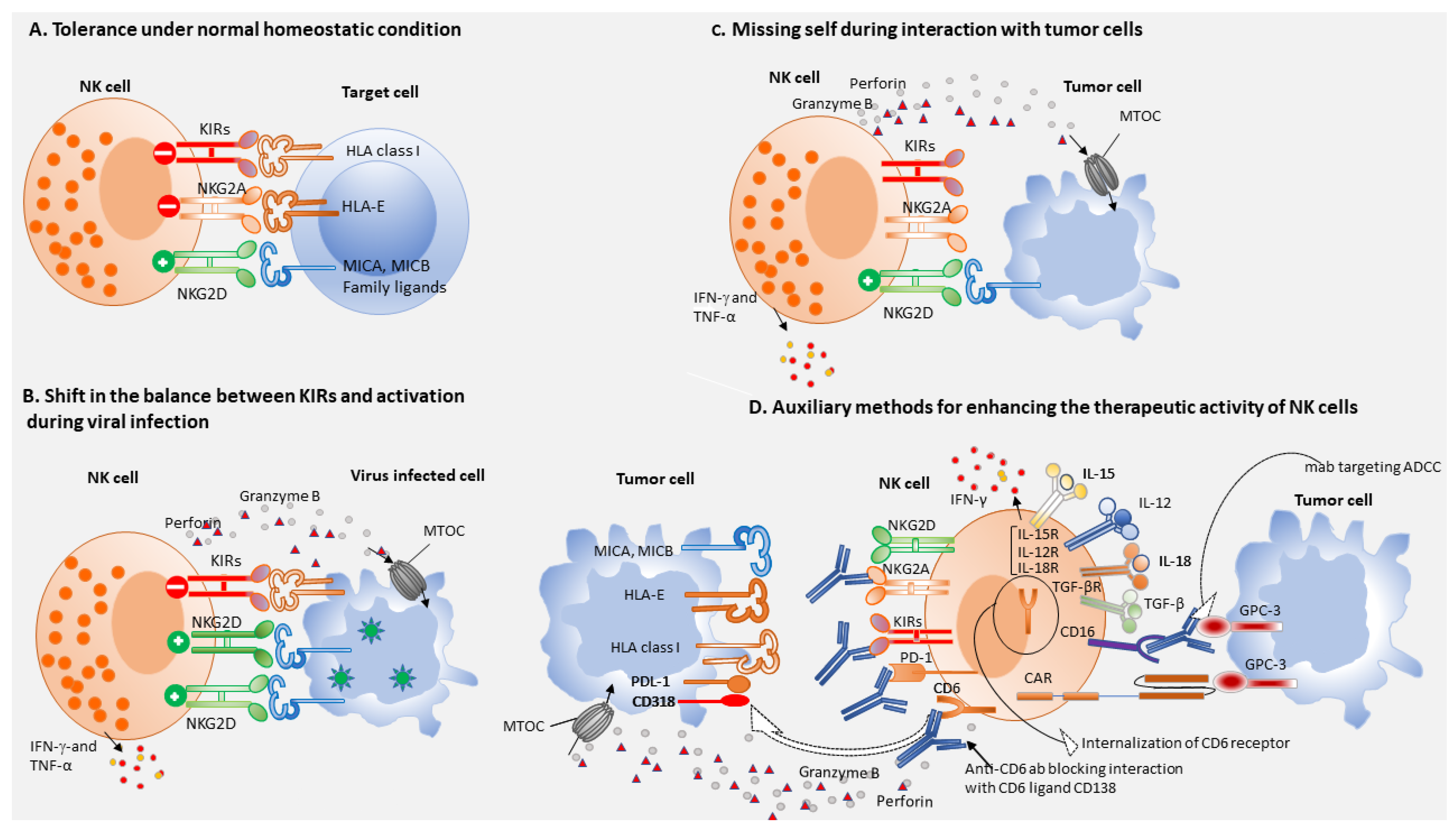
2. NK Cells in Chronic Viral Hepatitis

Chronic HBV and HCV infections are the leading cause for the development of liver cirrhosis and subsequent transformation to HCC [16]. Both HBV and HCV replicate and grow inside the hepatocytes which upregulate ligands of NKG2D, causing NK cell activation. In patients with chronic HBV and HCV infection, low frequencies of circulating NK cells combined with diminished production of proinflammatory cytokines, TNF-α, and IFN-γ have been reported as compared to healthy controls [11,12]. Nevertheless, controversy exists regarding the impact of chronic viral infection on NK cell cytolytic function; while several researchers reported impaired NK cytotoxicity, other reports showed that effector function was not changed [17,18]. The differences in outcomes between these studies were probably due to the lack of standardized protocol and reagents, as well as the heterogeneity of patients. NK cells exert a direct antiviral effect through (1) perforin and tumor necrosis factor-related apoptosis-inducing ligand (TRAIL) dependent cytolysis of infected cells and (2) induction of IFN-γ production [11,12,19]. Phenotypic heterogeneity of NK cells has been reported among HBV and HCV patients with elevated expression levels of activating receptor NKG2D and a decrease in inhibitory receptor expression in chronic HCV patients, while in chronic HBV patients, the percentage of activating NKG2C+ NK cells increased and the inhibitory receptor expression was normal [12]. The reduced percentage and impaired function of NK cells in chronic viral hepatitis patients were believed to contribute to the disease progression and the transformation to HCC. In acute HCV infection, hepatic NK cells show an increased expression of NKp46 and potentiality to degranulate and to produce IFN-γ following strong activation by IFN-α/β and other cytokines (IL-12, IL-15, IL-18) [20]. NK cell activation and proliferation in response to HCV, mediated by monocyte-derived cells and the OX40/OX40L axis, suggests cross talk between monocytes and NK cells [21]. NK cell repertoire in HBV patients co-infected with HCV is biased towards memory like phenotype associated with elevated CD16 mediated ADCC effector function, [15]. Even though, liver NK cells maintain their cytotoxic potential in chronic HBV infection via the upregulation of TRAIL [8], they eliminate autologous HBV specific CD8+ T cells expressing high levels of death receptor for TRAIL. Hence, this NK cell-mediated depletion of antigen-specific CD8+ T cells impairs adaptive antiviral immunity in chronic HBV-infected patients and contributes to viral persistence [22]. In chronic HBV patients, elevated frequency of killer cell lectin-like receptor subfamily G member 1+ (KLRG1) NK cells are detected and are implicated to causes reduction in hepatic fibrosis. Harnessing this antifibrotic function may provide a novel therapeutic approach to treat liver fibrosis in patients with chronic HBV [15]. Effective HBV vaccination and anti-HCV drugs have significantly reduced the number of chronic viral hepatitis patients and this will likely reduce the number of HCC cases in the future [23,24].

3. Role of T Cells in Viral Hepatitis

Earlier studies have demonstrated the central role of antigen specific CD4+ and CD8+ T cell responses in protection against viral persistence [9,25]. Potent CD4+ helper and CD8+ effector T cell responses have been observed in patients with hepatitis [26,27] and experimentally infected chimpanzees that clear HBV and HCV infection [28]. Antigen specific T cell responses can be detectable in the liver and peripheral blood approximately 4 to 8 weeks after infection. HLA- alleles, B27, B57, and A3 show strong association with protective immunity [29,30,31]. Protective T cell responses tend to target epitopes that do not allow virus escape mutations [25]. Even though HBV and HCV adopt different immunological mechanisms for viral persistence, a common feature is the subversion of virus specific T cell immune responses, that is characterized by progressive functional exhaustion and finally deletion of virus specific CD4+ and CD8+ T cells [32]. CD4+ T cells lose their capacity to produce IL-2, a cytokine that is critical for T cell proliferation and this is followed by sequential loss of cytotoxicity and TNFα and IFN-γ production. In addition, intracellular expression of Bcl-2 interacting mediator (Bim), a pro-apoptotic protein increases in virus specific CD8+ T cells of HBV or HCV infected patients [33,34]. A shift in the balance between T cell sustaining cytokine IL-2 and anti-inflammatory cytokines IL-10 and TGF-β dampens the virus specific T cell responses of infected patients [35,36]. Viral mutations in dominant T cell epitopes that have been selected to establish chronic infection render the remaining T cell responses irrelevant. These viral escape mutations have been described in both HBV [37] and HCV-infected patients [38], but they are more frequent in HCV infection as a result of the high replicative fitness of HCV.
The metabolic fitness of virus specific CD8+ T cells plays a central role in mounting an efficacious anti-viral immune response during chronic HBV or HCV infection. One important mechanism responsible for the failure of intrahepatic HCV-specific CD8+ T cells to clear HCV in persistent infection is due to the loss of mitochondrial fitness. In HCV infection, virus-specific CD8+ T cells display a marked upregulation of transcription of genes associated with impaired glycolytic and mitochondrial functions, leading to exhaustion of HCV-specific T cells [39]. HBV-specific CD8+ T cells are markedly impaired at multiple levels and show substantial downregulation of various cellular processes centered on extensive mitochondrial alterations [40]. Multiple strategies to increase the mitochondrial potential of exhausted virus specific CD8+ T cells and thereby rescue them from functional exhaustion has been reported recently. In chronic HBV infection, IL-12 has been shown to revitalize exhausted HBV-specific T cells by reducing their dependence on glycolysis and thereby increasing their mitochondrial potential, which is critical for viral control. Furthermore, IL-12 administration has been reported to significantly decrease the levels of pro-apoptotic protein Bim, which potentially cause attenuation of HBV specific CD8+ T cells [41,42]. IL-15 induced upregulation of autophagy in human liver-resident CD8+ memory T cells has been described as a protective mechanism against mitochondrial depolarization and to optimize effector T cell function [43]. In chronic HBV patients, IL-2 has been described as a potent immunotherapeutic that can rescue CD8+ T cells rendered dysfunctional by intrahepatic priming; this highlights the importance of IL-2-based strategies that should be considered for the treatment of chronic HBV infection [44]. Significant improvement of mitochondrial function with concomitant enhancement in anti-viral CD8+ T cell functions was elicited by mitochondrion-targeted antioxidants, which suggests a central role for reactive oxygen species (ROS) in T cell exhaustion [40]. Thus, mitochondria represent promising therapeutic targets to treat chronic HBV infection. Additionally, combinatorial treatment of polyphenolic compounds (resveratrol, oleuropein) and antioxidants improved anti-HBV T cell responses in chronic HBV patients. Therefore, combination of antioxidants and natural polyphenols represents a promising strategy for chronic HBV therapy [45]. GS-9688 (selgantolimod), a toll-like receptor 8 (TLR8) agonist induces cytokines in human PBMC that are able to activate antiviral effector function by multiple immune cells, including HBV-specific CD8+ T-cells, T follicular helper (TFh), NK cells, and MAITs [46]. In chronic HCV patients treated with direct-acting antiviral (DAA) therapy, exhausted HCV-specific CD8+ T cells remain functionally and metabolically impaired at multiple levels and altered mitochondrial dysfunction exhibited by exhausted HCV-specific CD8+ T cells could not be normalized even after HCV clearance in most patients. This finding implies that HCV cure does not lead to protective immunity and that re-infections have frequently been observed [47]. Nevertheless, functional exhaustion of HCV specific CD8+ T cells associated with impairment in glycolytic and mitochondrial function can be alleviated by histone methyltransferase inhibitors [39].
Individuals who spontaneously resolve acute HCV infection develop long-lived CD45RA CD127+ CD8+ memory T cells and are therefore less likely to become infected upon second exposure [25]. Presence of antigen specific CD8+ T cells in the peripheral blood and liver, detection of IFN-γ, CD3, CD4, and CD8 transcripts are associated with a decline in viral load [35]. The magnitude of the CD8 T cell response is a key determinant of spontaneous resolution and several epitopes are recognized simultaneously in humans with acute resolving HCV, while much fewer epitopes are recognized in individuals developing chronic infection [9,35]. Multiple exhaustion markers, such as PD-1 [48],Tim-3 [49], CTLA-4 [50], CD160, KLRG-1, and 2B4 [51], are differentially expressed on CD8+ T cells during acute and chronic viral infections, suggesting a spectrum of exhaustion markers that correlate with decline in CD8+ T cell function and persistent infection. The decline in CD8+ T cell function were reversible in vitro upon blockade of the PD-1, CTLA-4, and/or Tim-3 pathways. Early induction of IL-21 producing Th17 cells is critical to mediate help and limit exhaustion of virus-specific CD8 T cells [25].
In summary, spontaneous resolution of acute viral hepatitis correlates with early emergence of CD127+ antigen specific CD8+ T cells. Several factors contribute to failure of this response in individuals who become chronically infected. Escape mutations in targeted epitopes facilitate viral evasion of the immune system, loss of CD4+ T cell help, or a switch to an immunoregulatory profile further compromises the antiviral capacity of virus specific CD8+ T cells. Finally, continued viral replication contributes to exhaustion of HCV-specific T cells through persistent antigenic stimulation leading to progressive loss of function and diminished survival of virus-specific CD8 T cells. Additionally, exhaustion is exacerbated by increased expression of the ligands to the inhibitory receptors on infected hepatocytes from a therapeutic angle, the blockade of these inhibitory receptors combined with selective stimulation of co-stimulatory pathways is being pursued both in vitro and in vivo as a means to rescue exhausted T cells and to restore their function.

4. NK Cells and Immune Surveillance

In the normal physiological state, the liver is tolerogenic to immune stimuli from bacterial products, environmental toxins, and food antigens transported via the hepatic portal vein and hepatic artery, thereby avoiding unnecessary tissue damage [52,53]. This immune-tolerant nature of the liver microenvironment contributes to the immune evasion of HCC [54]. NK cells are the predominant innate immune cell subset in the liver and are critical in host defense against invading pathogens, liver injury, and tumor development [55,56]. In humans, almost 50% of intrahepatic lymphocytes are NK cells that are strongly imprinted in a liver-specific manner and play a key role in liver immune responses both under normal homeostasis and pathological conditions [57]. NK cells are considered as the first line of defense against viral infections and tumor invasion [16]. NK cells recognize and kill virus infected cells and tumor cells without major histocompatibility complex restriction or prior sensitization, via cytotoxicity or cytokine secretion [58,59]. NK cells are known for their ability to rapidly recognize and clear viral-infected, tumor-transformed, and stressed cell targets in the absence of antigen specificity [60,61]. NK cells in human are phenotypically defined as CD3CD56+ large granular lymphocytes. NK cells express activating receptors such as CD16, NKp30, NKp44, NKp46, NKp80, NKG2D, CD244, CD226; cytokine receptors such as IL-2R, IL-28R, IL-12R, IFNR, IL-15R, IL-18R, IL-1R8, IL-10R, and TGFβR [58,62]. The inhibitory receptors expressed by NK cells include NKG2A, KLRG1, KIRs, TIGIT, TIM3, Siglecs, PD-1, LAG3, A2AR, LAIRs, and ILTs [58,63]. Under homeostatic conditions, human circulating NK cells represent about 5–15% of circulating lymphocytes and are subdivided into two main subsets defined based on their differential expression of CD56 and CD16, namely CD56bright CD16 and CD56dim CD16+ [64,65]. Relatively low amounts of intracellular cytotoxic granules, perforin, and granzyme B are the features of CD56bright CD16neg NK cells and these cells have low cytotoxic potential and are not able to perform antibody dependent cellular cytotoxicity (ADCC) mediated killing since they do not express CD16. On the other hand CD56dim NK cells exerts both potent cytotoxicity and ADCC given their high constitutive expression of CD16 in response to activation by IL-12, IL-15, and IL-18 [66]. A growing body of evidence suggests that impaired NK cell function leads to the body’s failure to eliminate tumor cells, which indicates that tumor cells could be killed more effectively through enhancing the activity of dysfunctional NK cells [16,67]. Strong evidence for the role of NK cells in tumor surveillance is derived from correlative studies which demonstrated that a low NK cytotoxicity profile of peripheral blood lymphocytes correlated with an increased risk for cancer [68]. Impairment in NK cell function was observed in many types of liver diseases including chronic viral hepatitis and HCC, causing impediments to elimination of virus infected cells or tumor cells efficiently. HCC tumor tissues were markedly infiltrated by activated NK cells and Tregs, which should be considered as candidate therapeutic targets in HCC multidisciplinary treatments [69].
The functions of NK cells are strictly regulated by the balance of activating receptors and inhibitory receptors interacting with target cells [63]. These receptors can bind to specific ligands, mainly the major histocompatibility complex class (MHC-1) that is expressed on healthy hepatocytes, which interacts with inhibitory receptors on NK cells and prevents the activation of NK cells. The inhibitory Killer Ig-like receptors (KIRs) that recognize classical MHC-I alleles and the C-type lectin like receptor NKG2A that forms a heterodimer with the CD94 molecule (CD94/NKG2A) and bind HLA-E, a non-classical MHC-I molecule prevent NK cell activation (Figure 1A) [70]. During viral infection, hepatocytes upregulate stimulatory ligands for NK cell activating receptor NKG2D and as a result activation signal surpasses signaling through the inhibitory receptors KIRS and NKG2A, resulting in cytokine release and cytolysis of target cells (Figure 1B). Either the decreased expression of, or the absence of MHC-I on target cells triggers NK cell killing, a phenomenon known as “missing-self hypothesis”, via the engagement of activating NK receptors (Figure 1C) [71] which bind their putative ligands expressed on virus-infected, malignant, or stressed cells. NK cells can thus directly eradicate infected cells or tumor cells lacking MHC-1 molecule expression [55]. Therefore, approaches to regulate the balance between activating and inhibitory receptors in NK cells, might also lead to the successful treatment for various liver diseases including HCC [70,72]. Hepatocarcinoma cells express ligands of several activating NK receptors (NKR), including NKp30, natural killer receptor group 2, member D (NKG2D), and DNAM-1, such as the B7 protein homolog 6, the major histocompatibility complex class I chain-related protein A and B (MICA/B), and CD155, respectively, whose expression can correlate with HCC outcome [73,74]. Among these receptors, NKG2D/NKG2DL axis plays a pivotal role in the detection and elimination of virus infected cells and tumor cells. Nonetheless, viruses and tumor cells devise numerous strategies to evade detection by NKG2D immunosurveillance system [75]. NKG2D expression is down-regulated by transforming growth factor β (TGF-β), produced in the context of tumor development, which induces immune tolerance and controls inflammation via a suppressive action on innate and adaptive immunity [76,77]. Therefore, targeting TGF-β appears to represent an intervention strategy to boost NK cell mediated tumor immunity and should be considered in future cancer treatment modalities. The importance of NK cells and their activating receptor/ligand axis in HCC immune surveillance has been extensively studied and patients with decreased expression of major histocompatibility complex class I chain-related protein A (MICA) in HCC tissue showed reduced disease-free and overall survival compared with patients with preserved MICA expression [78]. Innate lymphoid cell (ILC-1) or liver-resident NK cells play versatile roles in liver diseases. Liver-resident NK cells are CD56brightEomeshighTbetlowHobit+TIGIT+CD69+CXCR6+CD49e, express higher levels of NKG2D, NKp46, TRAIL, and Fas ligand and possess cytotoxicity against HCC cells [79]. However, during tumor progression liver resident NK cells in tumor tissue down regulate NKG2D [80] and express inhibitory receptors PD-1, CD96, and TIGIT and are rapidly exhausted [81]. Thus, NK cells in the tumor tissues of patients with advanced HCC are dysfunctional or in an exhausted state [5], suggesting that NK-cell exhaustion may be contributing to HCC progression [72].

5. NK Cells-Based Immunotherapy of HCC

The exhaustion of NK cells as reflected by diminished cytotoxicity and impaired cytokine production may serve as a predictive and prognostic marker of HCC [5]. The absolute number of circulating NK cells and intrahepatic NK cells are positively correlated with HCC patients’ survival and prognosis [5,16]. In humans, both intra and extra-hepatic NK cells represent critical cells of our innate immune system, and play a key role in the body’s immune responses against cells infected with HBV or HCV and development of HCC [82]. Therapeutic interventions based on the activating receptor/ligand axis and/or IL-15 stimulation of effector NK cells represent a novel approach in cell based immunotherapy of HCC [73]. NK cells target tumor cells sensitized by monoclonal antibodies specific for tumor associated antigens GPC-3 or AFFP on tumor cells by mediating ADCC (Figure 1D) and this would also be a promising therapeutic strategy after activation of NK cells with cytokines such as IL-15 [73,83]. Activated NK cells generated from the peripheral blood of healthy donors using the K562-mb15-41BBL cell line as a stimulus, exerted remarkably high cytotoxicity against HCC cell lines, that was significantly higher than that of unstimulated or IL-2 activated NK cells. These expanded allogeneic or autologous NK cells, after genetic modification with NKG2D-CD3ζ-DAP10 chimeric receptor were shown to have enhanced anti-HCC cytotoxicity. These results warrant clinical trial of expanded allogeneic or autologous NK-cell infusions in patients with HCC, possibly after genetic modification with NKG2D-CD3ζ-DAP10 [84]. Cryoablation combined with allogeneic NK cell therapy has been shown to markedly improve the progression free survival of patients with advanced HCC [85]. A recent report on Tim-3-mediated dysfunction of both tumor-infiltrating, liver resident NK cells and conventional NK cells, provides a new insight into immune checkpoint based targeting for NK cell for immunotherapeutic intervention in HCC patients [86]. Accumulation of CD49a+ NK cells in the human liver indicates deteriorating disease condition and poor prognosis and thus elimination of CD49a+ NK cells could represent a potential therapeutic strategy in the immunotherapy of HCC [81]. Human intratumoral CD96+ NK cells represent functionally exhausted NK cells and patients with elevated levels of intratumoral CD96 expression exhibit poorer clinical outcomes. Therefore, reversing NK cell exhaustion by blocking the interaction between CD96 and its ligand CD155 might have therapeutic potential in the treatment of HCC patients [72]. NK cell activity is significantly improved when KIRs of donor and HLA class I of the recipient are incompatible, and this results in alleviation of inhibitory signals. Thus, KIR-ligand incompatibility seems to be critical for the enhanced efficacy of allogeneic NK cell [16]. Irreversible electroporation of tumor cells combined with allogeneic NK cell immunotherapy significantly increases the overall survival of patients with stage IV HCC and this combinatorial approach had synergistic effect by reduction of AFP levels with clinical benefit [87]. Dose dependent administration of sorafenib synergizes with NK cell function and therefore combinatorial therapy with sorafenib and NK cells may also improve the outcome of applied therapeutic approaches for HCC patients [88]. Trifunctional NK cell engagers (NKCEs), targeting two activating receptors, NKp46 and CD16, on NK cells and a tumor antigen on cancer cells trigger tumor cell destruction and this approach was superior to therapeutic antibodies both in vivo and in vitro [89]. An anti-NKG2A monoclonal antibody that binds NKG2A, the inhibitory receptor present on both NK cells and CD8+ T cells, promoted anti-tumor immunity of these immune cell subsets and thus, anti-NKG2A might have better therapeutic efficacy since it targets both NK cells and CD8+ T cells [90,91]. Importance of CD6 in NK cell activation has hitherto not been appreciated and recently an anti-CD6 monoclonal antibody, UMCD6 has been shown to upregulate the expression of the activating receptor NKG2D with concomitant downregulation of the inhibitory receptor NKG2A on both NK cells and CD8+ T cells, with concurrent increases in perforin and granzyme B production (Figure 1D) [92]. The dual capability of anti-CD6 monoclonal antibody to enhance killing of cancer cells through its effects on both CD8+ T cells and NK cells opens a new avenue for cancer immunotherapy [92]. Tumor infiltrating NK cells co-expressing CD49a and Eomes with reduced cytotoxic potential, have been reported in HCC and this NK cell subset was implicated to have proangiogenic function [93].
Metabolic fitness of NK cells has a crucial role in tumor surveillance. Fragmentation of mitochondria in liver NK cells of HCC patients was correlated with reduced cytotoxicity and NK cell loss, resulting in tumor evasion from NK cell surveillance, with predicted poor survival in HCC patients. Inhibition of mitochondrial fragmentation improved mitochondrial metabolism, survival, and the antitumor capacity of NK cells [94]. In colorectal liver metastasis (CRLM), liver-resident NK cells infiltrating into the tumors were unable to regulate intracellular pH resulting in mitochondrial stress and apoptosis. Hence, targeting CRLM metabolism represents a novel therapeutic strategy to restoring local NK-cell activity and preventing tumor growth [95] Functional rescue of NK-cells in solid tumors represents a central aim for new immunotherapeutic strategies. Metabolic defect and functional impairment of circulating NK-cells in patients with HCC could be attributed to TGF-β. Incubation of NK-cells from healthy donors with TGF-β-rich plasma from HCC patients can recapitulate the metabolic and functional impairment predicted by phenotypic and gene expression profiles of HCC patients. TGF-β antagonism could partially restore functional defects of NK-cells, suggesting that TGF-β among other molecules may represent a suitable target for immunotherapeutic intervention aimed at functional restoration of NK-cell. [96]. These studies highlight the potential mechanism of immune escape through mitochondrial dysfunction that might be targetable and could reinvigorate NK cell-based cancer treatments.
Potential advantages that NK cell therapeutics have over T cell based therapies include more manageable safety profiles and no requirement for autologous cells for adoptive transfer [97]. Accumulating evidence indicates that harnessing NK anti-tumor immunity represents a potentially powerful therapeutic approach for HCC, given the technical advancement in the activation and expansion of NK cells.

6. CAR-NK Cells

Several strategies using genetic modification techniques have been developed to improve the targetability and efficacy of NK cell cytotoxicity to tumor cells. The strategy used for generating CAR T cells has also been applied to NK cells, improving the specificity and efficacy of NK cell therapy [98,99]. CAR-NK cells are primary NK cells genetically engineered to express chimeric antigen receptor (CAR), such as AFP or GPC-3, which facilitate binding of relevant antigen in a specific manner. Additionally, NK cells express receptors for Fc portion of immunoglobulin (FcR) which can bind sensitized target cells and mediate cytolysis of NK cell susceptible target cells (ADCC). CAR-NK cells have been reported to reduce the risks of autoimmune responses and neoplastic transformation because they have a shorter lifespan than CAR T cells [98,100]. In addition, cytokines released from NK cells, such as IFN-γ and granulocyte-macrophage colony-stimulating factor (GM-CSF), are considered safer than the cytokine storm that results from CAR T cell therapy [98,100]. Moreover, ADCC is an additional NK cell-mediated tumor-killing strategy that could augment CAR-NK antitumor activity. Besides autologous and allogeneic NK cells, NK-92 cells, an NK cell line, is also used in clinical trials of cancer therapy, with promising results observed in sarcoma and leukemia patients [101]. Among genetically modified NK cells, glypican-3 (GPC3) specific CAR-NK-92 cells were reported to have high antitumor activity against HCC xenografts expressing both low and high levels of GPC3. The specificity of GPC3 CAR-NK-92 cells was confirmed by potent cytolytic activity displayed in vitro cytotoxicity assay against GPC3+ HCC cells, while they were not cytotoxic to GPC3 HCC cells [102]. Thus, GPC3-specific CAR-NK cells represent a novel treatment option for GPC-3+ HCC patients (Figure 1D). Granule polarization is a critical step leading to NK cell mediated cytolysis of resistant NK cell target, MDA-MB-453 breast cancer cells unmodified NK cells create conjugates with this resistant cancer cells and respond by granule clustering but fail to induce granule polarization and consequent release of lytic enzymes. Nevertheless, retargeting by ErbB2-targeted CAR-expressing NK cell or high affinity FcR transgenic NK cells plus monoclonal antibody Herceptin (ADCC) provides signals necessary to achieve granule polarization and tumor cell lysis, thereby circumventing tumor cell resistance [103].

7. T Cells-Based Immunotherapy of HCC

Even though, HCC is considered as a poorly immunogenic tumor, studies have shown that HCC patients who have a high level of lymphocyte infiltration in their tumors or high frequencies of circulating antigen specific CTL have a lower risk of recurrence and a better prognosis [104,105]. These findings confirm that HCC patients develop T cell mediated anti-tumor immunity that can suppress tumor progression. Several studies in the past decade identified different tumor associated antigens (TAAs) and their respective T cell epitopes [106,107,108,109,110,111]. Among the HCC TAAs, the immune response to α-fetoprotein (AFP) has been studied extensively since CTL epitopes for AFP were identified at an early stage of tumorigenesis [112,113]. Earlier studies have demonstrated that AFP-epitope specific T cell are more frequently seen in HCC patients than in healthy individuals and the ratio of these T cells in peripheral blood increases after radiofrequency ablation and TACE [105,107]. Human T cells were transduced with mouse TCR specific for HLA-A2/AFP complex and these engineered human T cells have been reported to specifically recognize HLA-A2+ AFP+ HepG2 tumor cells and produce effector cytokines. Importantly, these TCR gene engineered T cells could specifically kill HLA-A2+ AFP+ HepG2 tumor cells, sparing normal hepatocytes in vitro [114]. AFP2-11 -HLA-A*24:02 specific TCR transfected T cells could specifically activate and kill AFP2-11 pulsed T2-A24 cells and AFP+ HLA-A*24:02+ tumor cell lines, demonstrating that AFP2-11 epitope can be naturally presented on the surface of AFP+ tumor cell lines [115]. Antigens that are specific for HCC such as GPC3 and AFP are being tested as part of CAR constructs [116,117].
In addition to AFP, several TAAs have been identified for HCC that include human telomerase reverse transcriptase (hTERT), melanoma antigen gene-A (MAGE-A), glypican-3 (GPC3), and NY-ESO-1 [108,118,119]. The ratio of TAA-specific CTLs in PBMCs of HCC patients varies from 10 to 60.5 cells/300,000 PMBCs, and only 3–19% of patients had CTLs specific to the epitopes derived from different antigens [120]. Nonetheless, RFA enhances TAA-specific T cell responses and the number of TAA-specific T cells induced is associated with recurrence-free survival of HCC patients. To maintain the TAA-specific T cell responses induced by RFA and to improve the immunological outcome for HCC, additional combinatorial strategies such as vaccination or immunomodulatory drugs might be useful [120]. Peptides derived from squamous cell carcinoma antigen when recognized by T cells (SART-3) elicited peptide specific CTL IFN-γ responses upon stimulation of PBMC from HCC patients and were shown to mediate cytolysis of HCC cells expressing the antigen. Vaccination with SART-3 derived peptides resulted in the infiltration of antigen-specific CTL producing IFN-γ into the tumor site [111]. Even though normal mucosal associated invariant T (MAIT) cells could induce apoptosis of HCC cells, they are exhausted and functionally impaired in TME. In the context of liver TME, tumor-derived MAIT cells secreted less IFN-γ, IL-17, and granzyme B, perforin, but they secreted more IL-8, which promotes tumor angiogenesis and progression. Therefore, high density of tumor infiltrating MAIT cells have been reported to indicate disease progression and poorer outcome of HCC and these cells potentially could serve as a novel therapeutic target in T cells-based immunotherapy of liver cancer [121,122].
Adoptive cell transfer techniques, lymphokine-activated killer (LAK) and tumor-specific CTL therapy have been evaluated in HCC patients, and tumor-specific CTL therapy was reported to be more effective in terms of disease outcome [123]. LAK cells administered as an adjuvant to surgery resulted in a 5-year recurrence-free survival rate of 38% HCC patients as compared to 22% patients who did not receive LAK cells [124]. Cytokine induced killer (CIK) cells isolated from PMBCs of patients and cultured with a cytokine cocktail were shown to have highly potent antitumor activity [125]. Furthermore, CIK cell therapy improved the overall survival (OS) of patients when used in combination with either RFA or TACE [125,126]. Ablative therapies induce a peripheral immune response which may enhance the effect of anti-CTLA-4 treatment in patients with advanced hepatocellular carcinoma. Tremelimumab in combination with tumor ablation represents a potentially novel treatment modality for patients with advanced HCC, that leads to the accumulation of intratumoral CD8+ T cells in conjunction with reduction in HCV viral load [127]. Microwave ablation in HCC patients leads to abscopal effects in distant lesions reflected by enhanced tumor antigen specific T cell IFN-γ production that correlated with long term survival. These studies provide additional evidence for the potential synergistic effect that could be achieved by combination of local ablation and immunotherapeutic strategies against this challenging disease [128]. The above studies indicate that immune cell therapy is effective in reducing the recurrence rate, which is typically high for HCC patients following curative treatment.
Effector CD8+ T cells are in an exhausted state in advanced HCC patients, caused by persistent antigen stimulation, local release of IFN-γ and immunosuppressive factors [129,130]. Elevated expression levels of inhibitory receptors such as programmed cell death protein 1 (PD-1), lymphocyte activation gene-3 (LAG-3), 2B4 (CD244) T cell immunoglobulin, and mucin domain 3 (TIM-3), cytotoxic T lymphocyte-associated antigen 4 (CTLA-4) have been reported in exhausted T cells [131,132]. These inhibitory receptors are druggable targets for tumor immunotherapy [131,132]. Recently, checkpoint inhibitors have been used successfully in cancer treatment; however, they are only effective in 10–40% of cases, and some cancers are resistant to checkpoint inhibitors. PD-L1 expression on intratumoral hepatic stellate cells or peritumoral neutrophils also contributes to the impairment of T cell mediated anti-HCC immunity [133,134]. Blockade of PD-1-PD-L1 axis interaction, therefore, offers a promising strategy to reinvigorate exhausted CD8+ T cells and this has been confirmed in clinical trials demonstrating durable objective responses in advanced HCC patients treated with nivolumab or pembrolizumab [135,136,137]. Nevertheless, phase III trials did not reveal any statistically significant improvement in survival benefit [138]. A recent phase-I/II/III study reported that the combination of atezolizumab and bevacizumab resulted in a 62% response rate in advanced HCC patients, which has been approved as a first-line treatment for patients with advanced or metastatic HCC [139,140,141]. Radiotherapy has been shown to enhance immunogenicity of tumors through various mechanisms of immune modulation and therefore combination of ICIs and RT is being evaluated as an approach for HCC to take advantage of the synergistic effect [142]. TIGIT is enriched in PD1highCD8+TILs of HCC patients, and this subset represents the most dysfunctional and exhausted CD8+TIL phenotype and dual blockade of TIGIT and PD-1 improved the cytotoxic potential of CD8+ TIL in HCC patients [143]. The above results indicate that combinatorial strategies are more efficacious than CPI monotherapy and new therapeutic approaches are underway to determine whether the addition of tyrosine kinase inhibition, the anti-vascular endothelial growth factor antibody bevacizumab, or additional immune CPIs can augment the proportion of patients with response to PD-1 or PD-L1 inhibition.
Genetically modified autologous T cells that express an HBsAg specific T cell receptor, mediated a reduction in HBsAg levels in HBV related HCC without exacerbation of liver inflammation or “off-target” toxicities [144]. This is the first human proof of principle study for TCR redirected therapy against HBV associated hepatic malignancies. GPC3-CAR T cells have been shown to eliminate GPC3+HCC cells and tumors in a patient derived xenograft model [145]. However, dual-targeted CAR-T directed at GPC-3 and asialo-glycoprotein receptor (ASRG1) exerted superior anticancer activity and persistence than single-targeted CAR-T cells in two GPC3+ASGR1+HCC xenograft models [146]. Thus, T cells carrying two complementary CARs against GPC3 and ASGR1 may reduce the risk of off-tumor toxicity while maintaining relatively potent antitumor activity on GPC3+ASGR1+ HCC. In order to circumvent T cell exhaustion induced by immune checkpoint expression, an enhanced version of CAR T cells is being currently designed. In this second generation CAR T cells, PD-1 is disrupted via CRISPR/Cas9 to enhance the anti-tumor activity of GPC3-CAR T cells against HCC [147]. GPC3-CAR co-expressing co-stimulatory molecule ICOSL-41BB promotes CAR T cell proliferation and tumor rejection [148]. NKG2D-based CAR T cells efficiently lysed NKG2DL+ HCC cells invitro and potently eradicated NKG2DL+HCC xenografts [149]. The IL-12- inducible GPC3-CAR-T cells could broaden the application of CAR-T-based immunotherapy to patients intolerant of cytoreductive chemotherapy and might provide an alternative therapeutic strategy for patients with GPC3+cancers [150]. Intratumoral injection of AFP-CAR T cells significantly regressed both HepG2 and AFP158-expressing tumors in mouse xenograft models [151]. MUC-1, EpCAM, AFP, and CEA are the targets of CAR T cells with potential utility for treatment of HCC patients [152]. Recently, CAR-T cells targeting PD-L1 were shown to be effective in suppressing tumor growth of human xenograft models of HCC, nonetheless CAR PD-L1 T cells might recognize and kill normal cells expressing PD-L1 and therefore its safety needs to evaluated before clinical application [153].

8. Perspectives

Although very few clinical trials are currently exploring NK cells as a therapeutic option for HCC patients, they represent one of the promising candidates in the development of immunotherapies against advanced HCC. The potential advantages that NK cell therapies have over T cell therapies include more manageable safety profiles and no requirement for autologous cells for adoptive transfer. Furthermore, combinatorial strategies are underway to determine whether the addition of tyrosine kinase inhibitors, thermal ablation, anti-VEGF antibody bevacizumab, or additional immune CPIs could augment the proportion of HCC patients responding. The flexible combination of immunotherapy and other complementary therapies might offer the required breakthrough in clinical efficacy of HCC treatment. The application of recently developed high-throughput singe-cell multi-omics techniques to understand the phenotypic and functional composition of the NK cell populations will contribute to unraveling hepatic NK cell diversity and defining effective markers. Innovations in ICIs and NK cell therapeutic can potentially synergize to re-ignite anti-tumor immunity in HCC patients, improve the magnitude and durability of anti-tumor responses and may be a paradigm shift in the treatment for this disease.

Author Contributions

The manuscript was written by S.G.K. and reviewed by Y.T. All authors have read and agreed to the published version of the manuscript.

Funding

This research cited from the Thanavala Lab was supported in part through an American College of Gastroenterology grant to YT. The clinical trial and biomarker study were approved and funded by National Comprehensive Cancer Network (Clinicaltrials.gov (NCT01835223)), NCCNSORA0002 to Thanavala and Iyer by Bayer U.S. LLC. NCI grant (P30CA016056) supported the use of Roswell Park Comprehensive Cancer Center’s Shared Resources.

Institutional Review Board Statement

Not applicable.

Informed Consent Statement

Not applicable.

Data Availability Statement

Not applicable.

Conflicts of Interest

The authors declare no conflict of interest.

References

  1. Siegel, R.L.; Miller, K.D.; Jemal, A. Cancer Statistics, 2017. CA A Cancer J. Clin. 2017, 67, 7–30. [Google Scholar] [CrossRef] [PubMed] [Green Version]
  2. Wallace, M.C.; Preen, D.; Jeffrey, G.P.; Adams, L.A. The evolving epidemiology of hepatocellular carcinoma: A global perspective. Expert Rev. Gastroenterol. Hepatol. 2015, 9, 765–779. [Google Scholar] [CrossRef] [PubMed]
  3. Lurje, I.; Czigany, Z.; Bednarsch, J.; Roderburg, C.; Isfort, P.; Neumann, U.P.; Lurje, G. Treatment Strategies for Hepatocellular Carcinoma—A Multidisciplinary Approach. Int. J. Mol. Sci. 2019, 20, 1465. [Google Scholar] [CrossRef] [PubMed] [Green Version]
  4. Juengpanich, S.; Shi, L.; Iranmanesh, Y.; Chen, J.; Cheng, Z.; Khoo, A.K.; Pan, L.; Wang, Y.; Cai, X. The role of natural killer cells in hepatocellular carcinoma development and treatment: A narrative review. Transl. Oncol. 2019, 12, 1092–1107. [Google Scholar] [CrossRef] [PubMed]
  5. Sun, C.; Sun, H.Y.; Xiao, W.H.; Zhang, C.; Tian, Z.G. Natural killer cell dysfunction in hepatocellular carcinoma and NK cell-based immunotherapy. Acta Pharm. Sin. 2015, 36, 1191–1199. [Google Scholar] [CrossRef] [PubMed] [Green Version]
  6. Kim, H.D.; Song, G.W.; Park, S.; Jung, M.K.; Kim, M.H.; Kang, H.J.; Yoo, C.; Yi, K.; Kim, K.H.; Eo, S.; et al. Association Between Expression Level of PD1 by Tumor-Infiltrating CD8(+) T Cells and Features of Hepatocellular Carcinoma. Gastroenterology 2018, 155, 1936–1950. [Google Scholar] [CrossRef]
  7. Lee, H.W.; Cho, K.J.; Park, J.Y. Current Status and Future Direction of Immunotherapy in Hepatocellular Carcinoma: What Do the Data Suggest? Immune Netw. 2020, 20, e11. [Google Scholar] [CrossRef]
  8. Dunn, C.; Brunetto, M.; Reynolds, G.; Christophides, T.; Kennedy, P.T.; Lampertico, P.; Das, A.; Lopes, A.R.; Borrow, P.; Williams, K.; et al. Cytokines induced during chronic hepatitis B virus infection promote a pathway for NK cell–mediated liver damage. J. Exp. Med. 2007, 204, 667–680. [Google Scholar] [CrossRef] [Green Version]
  9. Rehermann, B. Pathogenesis of chronic viral hepatitis: Differential roles of T cells and NK cells. Nat. Med. 2013, 19, 859–868. [Google Scholar] [CrossRef] [Green Version]
  10. Heydtmann, M. Macrophages in Hepatitis B and Hepatitis C Virus Infections. J. Virol. 2009, 83, 2796–2802. [Google Scholar] [CrossRef] [Green Version]
  11. Dessouki, O.; Kamiya, Y.; Nagahama, H.; Tanaka, M.; Suzu, S.; Sasaki, Y.; Okada, S. Chronic hepatitis C viral infection reduces NK cell frequency and suppresses cytokine secretion: Reversion by anti-viral treatment. Biochem. Biophys. Res. Commun. 2010, 393, 331–337. [Google Scholar] [CrossRef] [PubMed]
  12. Oliviero, B.; Varchetta, S.; Paudice, E.; Michelone, G.; Zaramella, M.; Mavilio, D.; De Filippi, F.; Bruno, S.; Mondelli, M.U. Natural killer cell functional dichotomy in chronic hepatitis B and chronic hepatitis C virus infections. Gastroenterology 2009, 137, 1151–1160. [Google Scholar] [CrossRef]
  13. Su, C.-W.; Lin, H.-C. Toward a new landscape for the mechanism of immunosuppression in hepatocellular carcinoma. Hepatol. Int. 2021, 15, 287–289. [Google Scholar] [CrossRef]
  14. Fisicaro, P.; Rossi, M.; Vecchi, A.; Acerbi, G.; Barili, V.; Laccabue, D.; Montali, I.; Zecca, A.; Penna, A.; Missale, G.; et al. The Good and the Bad of Natural Killer Cells in Virus Control: Perspective for Anti-HBV Therapy. Int. J. Mol. Sci. 2019, 20, 5080. [Google Scholar] [CrossRef] [PubMed] [Green Version]
  15. Schuch, A.; Zecher, B.F.; Müller, P.A.; Correia, M.P.; Daul, F.; Rennert, C.; Tauber, C.; Schlitt, K.; Boettler, T.; Neumann-Haefelin, C.; et al. NK-cell responses are biased towards CD16-mediated effector functions in chronic hepatitis B virus infection. J. Hepatol. 2019, 70, 351–360. [Google Scholar] [CrossRef] [PubMed] [Green Version]
  16. Liu, P.; Chen, L.; Zhang, H. Natural Killer Cells in Liver Disease and Hepatocellular Carcinoma and the NK Cell-Based Immunotherapy. J. Immunol Res. 2018, 2018, 1206737. [Google Scholar] [CrossRef] [PubMed] [Green Version]
  17. Nattermann, J.; Feldmann, G.; Ahlenstiel, G.; Langhans, B.; Sauerbruch, T.; Spengler, U. Surface expression and cytolytic function of natural killer cell receptors is altered in chronic hepatitis C. Gut 2006, 55, 869–877. [Google Scholar] [CrossRef] [Green Version]
  18. Marotel, M.; Villard, M.; Drouillard, A.; Tout, I.; Besson, L.; Allatif, O.; Pujol, M.; Rocca, Y.; Ainouze, M.; Roblot, G.; et al. Peripheral natural killer cells in chronic hepatitis B patients display multiple molecular features of T cell exhaustion. Elife 2021, 10, e60095. [Google Scholar] [CrossRef]
  19. Rehermann, B. Natural Killer Cells in Viral Hepatitis. Cell. Mol. Gastroenterol. Hepatol. 2015, 1, 578–588. [Google Scholar] [CrossRef] [Green Version]
  20. Amadei, B.; Urbani, S.; Cazaly, A.; Fisicaro, P.; Zerbini, A.; Ahmed, P.; Missale, G.; Ferrari, C.; Khakoo, S.I. Activation of Natural Killer Cells During Acute Infection With Hepatitis C Virus. Gastroenterology 2010, 138, 1536–1545. [Google Scholar] [CrossRef] [Green Version]
  21. Pollmann, J.; Götz, J.-J.; Rupp, D.; Strauss, O.; Granzin, M.; Grünvogel, O.; Mutz, P.; Kramer, C.; Lasitschka, F.; Lohmann, V.; et al. Hepatitis C virus-induced natural killer cell proliferation involves monocyte-derived cells and the OX40/OX40L axis. J. Hepatol. 2018, 68, 421–430. [Google Scholar] [CrossRef] [PubMed]
  22. Peppa, D.; Gill, U.S.; Reynolds, G.; Easom, N.J.W.; Pallett, L.J.; Schurich, A.; Micco, L.; Nebbia, G.; Singh, H.D.; Adams, D.H.; et al. Up-regulation of a death receptor renders antiviral T cells susceptible to NK cell–mediated deletion. J. Exp. Med. 2012, 210, 99–114. [Google Scholar] [CrossRef] [PubMed]
  23. Locarnini, S.; Hatzakis, A.; Chen, D.S.; Lok, A. Strategies to control hepatitis B: Public policy, epidemiology, vaccine and drugs. J. Hepatol. 2015, 62, S76–S86. [Google Scholar] [CrossRef] [PubMed]
  24. Alkhouri, N.; Lawitz, E.; Poordad, F. Novel treatments for chronic hepatitis C: Closing the remaining gaps. Curr. Opin. Pharm. 2017, 37, 107–111. [Google Scholar] [CrossRef] [PubMed]
  25. Abdel-Hakeem, M.S.; Shoukry, N.H. Protective immunity against hepatitis C: Many shades of gray. Front. Immunol. 2014, 5, 274. [Google Scholar] [CrossRef]
  26. Rehermann, B.; Ferrari, C.; Pasquinelli, C.; Chisari, F.V. The hepatitis B virus persists for decades after patients’ recovery from acute viral hepatitis despite active maintenance of a cytotoxic T–lymphocyte response. Nat. Med. 1996, 2, 1104–1108. [Google Scholar] [CrossRef] [PubMed]
  27. Lechner, F.; Wong, D.K.; Dunbar, P.R.; Chapman, R.; Chung, R.T.; Dohrenwend, P.; Robbins, G.; Phillips, R.; Klenerman, P.; Walker, B.D. Analysis of successful immune responses in persons infected with hepatitis C virus. J. Exp. Med. 2000, 191, 1499–1512. [Google Scholar] [CrossRef] [Green Version]
  28. Schmidt, J.; Blum, H.E.; Thimme, R. T-cell responses in hepatitis B and C virus infection: Similarities and differences. Emerg Microbes Infect. 2013, 2, e15. [Google Scholar] [CrossRef]
  29. Dazert, E.; Neumann-Haefelin, C.; Bressanelli, S.; Fitzmaurice, K.; Kort, J.; Timm, J.; McKiernan, S.; Kelleher, D.; Gruener, N.; Tavis, J.E.; et al. Loss of viral fitness and cross-recognition by CD8+ T cells limit HCV escape from a protective HLA-B27-restricted human immune response. J. Clin. Investig. 2009, 119, 376–386. [Google Scholar] [CrossRef] [PubMed] [Green Version]
  30. Kim, A.Y.; Kuntzen, T.; Timm, J.; Nolan, B.E.; Baca, M.A.; Reyor, L.L.; Berical, A.C.; Feller, A.J.; Johnson, K.L.; Wiesch, J.S.Z.; et al. Spontaneous Control of HCV Is Associated With Expression of HLA-B*57 and Preservation of Targeted Epitopes. Gastroenterology 2011, 140, 686–696. [Google Scholar] [CrossRef] [Green Version]
  31. Fitzmaurice, K.; Petrovic, D.; Ramamurthy, N.; Simmons, R.; Merani, S.; Gaudieri, S.; Sims, S.; Dempsey, E.; Freitas, E.; Lea, S.; et al. Molecular footprints reveal the impact of the protective HLA-A*03 allele in hepatitis C virus infection. Gut 2011, 60, 1563–1571. [Google Scholar] [CrossRef] [Green Version]
  32. Penna, A.; Pilli, M.; Zerbini, A.; Orlandini, A.; Mezzadri, S.; Sacchelli, L.; Missale, G.; Ferrari, C. Dysfunction and functional restoration of HCV-specific CD8 responses in chronic hepatitis C virus infection. Hepatology 2007, 45, 588–601. [Google Scholar] [CrossRef] [PubMed]
  33. Larrubia, J.R.; Lokhande, M.U.; García-Garzón, S.; Miquel, J.; González-Praetorius, A.; Parra-Cid, T.; Sanz-de-Villalobos, E. Persistent hepatitis C virus (HCV) infection impairs HCV-specific cytotoxic T cell reactivity through Mcl-1/Bim imbalance due to CD127 down-regulation. J. Viral Hepat. 2013, 20, 85–94. [Google Scholar] [CrossRef] [PubMed]
  34. Lopes, A.R.; Kellam, P.; Das, A.; Dunn, C.; Kwan, A.; Turner, J.; Peppa, D.; Gilson, R.J.; Gehring, A.; Bertoletti, A.; et al. Bim-mediated deletion of antigen-specific CD8+ T cells in patients unable to control HBV infection. J. Clin. Investig. 2008, 118, 1835–1845. [Google Scholar] [CrossRef] [PubMed]
  35. Accapezzato, D.; Francavilla, V.; Paroli, M.; Casciaro, M.; Chircu, L.V.; Cividini, A.; Abrignani, S.; Mondelli, M.U.; Barnaba, V. Hepatic expansion of a virus-specific regulatory CD8(+) T cell population in chronic hepatitis C virus infection. J. Clin. Investig. 2004, 113, 963–972. [Google Scholar] [CrossRef]
  36. Alatrakchi, N.; Graham, C.S.; van der Vliet, H.J.J.; Sherman, K.E.; Exley, M.A.; Koziel, M.J. Hepatitis C virus (HCV)-specific CD8+ cells produce transforming growth factor beta that can suppress HCV-specific T-cell responses. J. Virol. 2007, 81, 5882–5892. [Google Scholar] [CrossRef] [PubMed] [Green Version]
  37. Bertoletti, A.; Sette, A.; Chisari, F.V.; Penna, A.; Levrero, M.; Carli, M.D.; Fiaccadori, F.; Ferrari, C. Natural variants of cytotoxic epitopes are T-cell receptor antagonists for antiviral cytotoxic T cells. Nature 1994, 369, 407–410. [Google Scholar] [CrossRef]
  38. Wölfl, M.; Rutebemberwa, A.; Mosbruger, T.; Mao, Q.; Li, H.-m.; Netski, D.; Ray, S.C.; Pardoll, D.; Sidney, J.; Sette, A.; et al. Hepatitis C virus immune escape via exploitation of a hole in the T cell repertoire. J. Immunol. 2008, 181, 6435–6446. [Google Scholar] [CrossRef] [Green Version]
  39. Barili, V.; Fisicaro, P.; Montanini, B.; Acerbi, G.; Filippi, A.; Forleo, G.; Romualdi, C.; Ferracin, M.; Guerrieri, F.; Pedrazzi, G.; et al. Targeting p53 and histone methyltransferases restores exhausted CD8+ T cells in HCV infection. Nat. Commun. 2020, 11, 604. [Google Scholar] [CrossRef] [PubMed] [Green Version]
  40. Fisicaro, P.; Barili, V.; Montanini, B.; Acerbi, G.; Ferracin, M.; Guerrieri, F.; Salerno, D.; Boni, C.; Massari, M.; Cavallo, M.C.; et al. Targeting mitochondrial dysfunction can restore antiviral activity of exhausted HBV-specific CD8 T cells in chronic hepatitis B. Nat. Med. 2017, 23, 327–336. [Google Scholar] [CrossRef]
  41. Schurich, A.; Pallett, L.J.; Jajbhay, D.; Wijngaarden, J.; Otano, I.; Gill, U.S.; Hansi, N.; Kennedy, P.T.; Nastouli, E.; Gilson, R.; et al. Distinct Metabolic Requirements of Exhausted and Functional Virus-Specific CD8 T Cells in the Same Host. Cell Rep. 2016, 16, 1243–1252. [Google Scholar] [CrossRef] [Green Version]
  42. Schurich, A.; Pallett, L.J.; Lubowiecki, M.; Singh, H.D.; Gill, U.S.; Kennedy, P.T.; Nastouli, E.; Tanwar, S.; Rosenberg, W.; Maini, M.K. The third signal cytokine IL-12 rescues the anti-viral function of exhausted HBV-specific CD8 T cells. PLoS Pathog. 2013, 9, e1003208. [Google Scholar] [CrossRef] [PubMed]
  43. Swadling, L.; Pallett, L.J.; Diniz, M.O.; Baker, J.M.; Amin, O.E.; Stegmann, K.A.; Burton, A.R.; Schmidt, N.M.; Jeffery-Smith, A.; Zakeri, N.; et al. Human Liver Memory CD8(+) T Cells Use Autophagy for Tissue Residence. Cell Rep. 2020, 30, 687–698. [Google Scholar] [CrossRef] [PubMed] [Green Version]
  44. Bénéchet, A.P.; De Simone, G.; Di Lucia, P.; Cilenti, F.; Barbiera, G.; Le Bert, N.; Fumagalli, V.; Lusito, E.; Moalli, F.; Bianchessi, V.; et al. Dynamics and genomic landscape of CD8(+) T cells undergoing hepatic priming. Nature 2019, 574, 200–205. [Google Scholar] [CrossRef] [PubMed]
  45. Acerbi, G.; Montali, I.; Ferrigno, G.D.; Barili, V.; Schivazappa, S.; Alfieri, A.; Laccabue, D.; Loglio, A.; Borghi, M.; Massari, M.; et al. Functional reconstitution of HBV-specific CD8 T cells by in vitro polyphenol treatment in chronic hepatitis B. J. Hepatol. 2021, 74, 783–793. [Google Scholar] [CrossRef] [PubMed]
  46. Amin, O.E.; Colbeck, E.J.; Daffis, S.; Khan, S.; Ramakrishnan, D.; Pattabiraman, D.; Chu, R.; Micolochick Steuer, H.; Lehar, S.; Peiser, L.; et al. Therapeutic potential of TLR8 agonist GS-9688 (selgantolimod) in chronic hepatitis B: Re-modelling of antiviral and regulatory mediators. Hepatology 2020. [Google Scholar] [CrossRef]
  47. Aregay, A.; Owusu Sekyere, S.; Deterding, K.; Port, K.; Dietz, J.; Berkowski, C.; Sarrazin, C.; Manns, M.P.; Cornberg, M.; Wedemeyer, H. Elimination of hepatitis C virus has limited impact on the functional and mitochondrial impairment of HCV-specific CD8+ T cell responses. J. Hepatol. 2019, 71, 889–899. [Google Scholar] [CrossRef]
  48. Barber, D.L.; Wherry, E.J.; Masopust, D.; Zhu, B.; Allison, J.P.; Sharpe, A.H.; Freeman, G.J.; Ahmed, R. Restoring function in exhausted CD8 T cells during chronic viral infection. Nature 2006, 439, 682–687. [Google Scholar] [CrossRef]
  49. McMahan, R.H.; Golden-Mason, L.; Nishimura, M.I.; McMahon, B.J.; Kemper, M.; Allen, T.M.; Gretch, D.R.; Rosen, H.R. Tim-3 expression on PD-1+ HCV-specific human CTLs is associated with viral persistence, and its blockade restores hepatocyte-directed in vitro cytotoxicity. J. Clin. Investig. 2010, 120, 4546–4557. [Google Scholar] [CrossRef] [Green Version]
  50. Radziewicz, H.; Ibegbu, C.C.; Fernandez, M.L.; Workowski, K.A.; Obideen, K.; Wehbi, M.; Hanson, H.L.; Steinberg, J.P.; Masopust, D.; Wherry, E.J.; et al. Liver-Infiltrating Lymphocytes in Chronic Human Hepatitis C Virus Infection Display an Exhausted Phenotype with High Levels of PD-1 and Low Levels of CD127 Expression. J. Virol. 2007, 81, 2545–2553. [Google Scholar] [CrossRef] [PubMed] [Green Version]
  51. Blackburn, S.D.; Shin, H.; Haining, W.N.; Zou, T.; Workman, C.J.; Polley, A.; Betts, M.R.; Freeman, G.J.; Vignali, D.A.A.; Wherry, E.J. Coregulation of CD8+ T cell exhaustion by multiple inhibitory receptors during chronic viral infection. Nat. Immunol. 2009, 10, 29–37. [Google Scholar] [CrossRef] [Green Version]
  52. Kubes, P.; Jenne, C. Immune Responses in the Liver. Annu. Rev. Immunol. 2018, 36, 247–277. [Google Scholar] [CrossRef] [PubMed]
  53. Heymann, F.; Tacke, F. Immunology in the liver--from homeostasis to disease. Nat. Rev. Gastroenterol. Hepatol. 2016, 13, 88–110. [Google Scholar] [CrossRef] [PubMed]
  54. Makarova-Rusher, O.V.; Medina-Echeverz, J.; Duffy, A.G.; Greten, T.F. The yin and yang of evasion and immune activation in HCC. J. Hepatol. 2015, 62, 1420–1429. [Google Scholar] [CrossRef] [Green Version]
  55. Morvan, M.G.; Lanier, L.L. NK cells and cancer: You can teach innate cells new tricks. Nat. Rev. Cancer 2016, 16, 7–19. [Google Scholar] [CrossRef]
  56. Jenne, C.N.; Kubes, P. Immune surveillance by the liver. Nat. Immunol. 2013, 14, 996–1006. [Google Scholar] [CrossRef]
  57. Mikulak, J.; Bruni, E.; Oriolo, F.; Di Vito, C.; Mavilio, D. Hepatic Natural Killer Cells: Organ-Specific Sentinels of Liver Immune Homeostasis and Physiopathology. Front. Immunol. 2019, 10, 946. [Google Scholar] [CrossRef] [Green Version]
  58. Li, J.; Tao, L.; Wang, X. Cytotoxic immune cell-based immunotherapy for hepatocellular carcinoma. Hepatoma Res. 2020, 6, 15. [Google Scholar] [CrossRef]
  59. Chiossone, L.; Dumas, P.Y.; Vienne, M.; Vivier, E. Natural killer cells and other innate lymphoid cells in cancer. Nat. Rev. Immunol. 2018, 18, 671–688. [Google Scholar] [CrossRef] [PubMed]
  60. Vivier, E.; Raulet, D.H.; Moretta, A.; Caligiuri, M.A.; Zitvogel, L.; Lanier, L.L.; Yokoyama, W.M.; Ugolini, S. Innate or Adaptive Immunity? The Example of Natural Killer Cells. Science 2011, 331, 44–49. [Google Scholar] [CrossRef] [PubMed] [Green Version]
  61. Waldhauer, I.; Steinle, A. NK cells and cancer immunosurveillance. Oncogene 2008, 27, 5932–5943. [Google Scholar] [CrossRef] [Green Version]
  62. Martinet, L.; Smyth, M.J. Balancing natural killer cell activation through paired receptors. Nat. Rev. Immunol. 2015, 15, 243–254. [Google Scholar] [CrossRef]
  63. Pegram, H.J.; Andrews, D.M.; Smyth, M.J.; Darcy, P.K.; Kershaw, M.H. Activating and inhibitory receptors of natural killer cells. Immunol. Cell Biol. 2011, 89, 216–224. [Google Scholar] [CrossRef]
  64. Cooper, M.A.; Fehniger, T.A.; Caligiuri, M.A. The biology of human natural killer-cell subsets. Trends Immunol. 2001, 22, 633–640. [Google Scholar] [CrossRef]
  65. Cooper, M.A. Natural killer cells might adapt their inhibitory receptors for memory. Proc. Natl. Acad. Sci. USA 2018, 115, 11357–11359. [Google Scholar] [CrossRef] [PubMed] [Green Version]
  66. De Maria, A.; Bozzano, F.; Cantoni, C.; Moretta, L. Revisiting human natural killer cell subset function revealed cytolytic CD56dimCD16+ NK cells as rapid producers of abundant IFN-γ on activation. Proc. Natl. Acad. Sci. USA 2011, 108, 728–732. [Google Scholar] [CrossRef] [Green Version]
  67. Sung, P.S.; Jang, J.W. Natural Killer Cell Dysfunction in Hepatocellular Carcinoma: Pathogenesis and Clinical Implications. Int. J. Mol. Sci. 2018, 19, 3648. [Google Scholar] [CrossRef] [Green Version]
  68. Imai, K.; Matsuyama, S.; Miyake, S.; Suga, K.; Nakachi, K. Natural cytotoxic activity of peripheral-blood lymphocytes and cancer incidence: An 11-year follow-up study of a general population. Lancet 2000, 356, 1795–1799. [Google Scholar] [CrossRef]
  69. Wang, L.; Yang, Z.; Cao, Y. Regulatory T cell and activated natural killer cell infiltration in hepatocellular carcinoma: Immune cell profiling using the CIBERSORT. Ann. Transl. Med. 2020, 8, 1483. [Google Scholar] [CrossRef]
  70. Ravindranath, M.H.; Filippone, E.J.; Devarajan, A.; Asgharzadeh, S. Enhancing Natural Killer and CD8(+) T Cell-Mediated Anticancer Cytotoxicity and Proliferation of CD8(+) T Cells with HLA-E Monospecific Monoclonal Antibodies. Monoclon. Antibodies Immunodiagn. Immunother. 2019, 38, 38–59. [Google Scholar] [CrossRef] [PubMed] [Green Version]
  71. Kärre, K. Natural killer cell recognition of missing self. Nat. Immunol. 2008, 9, 477–480. [Google Scholar] [CrossRef]
  72. Sun, H.; Huang, Q.; Huang, M.; Wen, H.; Lin, R.; Zheng, M.; Qu, K.; Li, K.; Wei, H.; Xiao, W.; et al. Human CD96 Correlates to Natural Killer Cell Exhaustion and Predicts the Prognosis of Human Hepatocellular Carcinoma. Hepatology 2019, 70, 168–183. [Google Scholar] [CrossRef]
  73. Mantovani, S.; Oliviero, B.; Lombardi, A.; Varchetta, S.; Mele, D.; Sangiovanni, A.; Rossi, G.; Donadon, M.; Torzilli, G.; Soldani, C.; et al. Deficient Natural Killer Cell NKp30-Mediated Function and Altered NCR3 Splice Variants in Hepatocellular Carcinoma. Hepatology 2019, 69, 1165–1179. [Google Scholar] [CrossRef] [PubMed]
  74. Gong, J.; Fang, L.; Liu, R.; Wang, Y.; Xing, J.; Chen, Y.; Zhuang, R.; Zhang, Y.; Zhang, C.; Yang, A.; et al. UPR decreases CD226 ligand CD155 expression and sensitivity to NK cell-mediated cytotoxicity in hepatoma cells. Eur. J. Immunol. 2014, 44, 3758–3767. [Google Scholar] [CrossRef] [Green Version]
  75. Lanier, L.L. NKG2D Receptor and Its Ligands in Host Defense. Cancer Immunol. Res. 2015, 3, 575–582. [Google Scholar] [CrossRef] [Green Version]
  76. Cacalano, N.A. Regulation of Natural Killer Cell Function by STAT3. Front. Immunol. 2016, 7, 128. [Google Scholar] [CrossRef] [PubMed]
  77. Lazarova, M.; Steinle, A. Impairment of NKG2D-Mediated Tumor Immunity by TGF-β. Front. Immunol. 2019, 10, 2689. [Google Scholar] [CrossRef] [Green Version]
  78. Zhang, J.; Xu, Z.; Zhou, X.; Zhang, H.; Yang, N.; Wu, Y.; Chen, Y.; Yang, G.; Ren, T. Loss of expression of MHC class I-related chain A (MICA) is a frequent event and predicts poor survival in patients with hepatocellular carcinoma. Int. J. Clin. Exp. Pathol. 2014, 7, 3123–3131. [Google Scholar]
  79. Hwang, S.; Han, J.; Baek, J.S.; Tak, E.; Song, G.W.; Lee, S.G.; Jung, D.H.; Park, G.C.; Ahn, C.S.; Kim, N. Cytotoxicity of Human Hepatic Intrasinusoidal CD56(bright) Natural Killer Cells against Hepatocellular Carcinoma Cells. Int. J. Mol. Sci. 2019, 20, 1564. [Google Scholar] [CrossRef] [PubMed] [Green Version]
  80. Easom, N.J.W.; Stegmann, K.A.; Swadling, L.; Pallett, L.J.; Burton, A.R.; Odera, D.; Schmidt, N.; Huang, W.C.; Fusai, G.; Davidson, B.; et al. IL-15 Overcomes Hepatocellular Carcinoma-Induced NK Cell Dysfunction. Front. Immunol 2018, 9, 1009. [Google Scholar] [CrossRef] [PubMed] [Green Version]
  81. Sun, H.; Liu, L.; Huang, Q.; Liu, H.; Huang, M.; Wang, J.; Wen, H.; Lin, R.; Qu, K.; Li, K.; et al. Accumulation of Tumor-Infiltrating CD49a+ NK Cells Correlates with Poor Prognosis for Human Hepatocellular Carcinoma. Cancer Immunol. Res. 2019, 7, 1535–1546. [Google Scholar] [CrossRef] [PubMed]
  82. Nomura, M.; Tsuge, M.; Uchida, T.; Hiraga, N.; Kurihara, M.; Tsushima, K.; Fujino, H.; Nakahara, T.; Murakami, E.; Abe-Chayama, H.; et al. CTL-associated and NK cell-associated immune responses induce different HBV DNA reduction patterns in chronic hepatitis B patients. J. Viral Hepat. 2018, 25, 1555–1564. [Google Scholar] [CrossRef] [PubMed]
  83. Mantovani, S.; Oliviero, B.; Varchetta, S.; Mele, D.; Mondelli, M.U. Natural Killer Cell Responses in Hepatocellular Carcinoma: Implications for Novel Immunotherapeutic Approaches. Cancers 2020, 12, 926. [Google Scholar] [CrossRef] [PubMed] [Green Version]
  84. Kamiya, T.; Chang, Y.H.; Campana, D. Expanded and Activated Natural Killer Cells for Immunotherapy of Hepatocellular Carcinoma. Cancer Immunol. Res. 2016, 4, 574–581. [Google Scholar] [CrossRef] [PubMed] [Green Version]
  85. Lin, M.; Liang, S.; Wang, X.; Liang, Y.; Zhang, M.; Chen, J.; Niu, L.; Xu, K. Cryoablation combined with allogenic natural killer cell immunotherapy improves the curative effect in patients with advanced hepatocellular cancer. Oncotarget 2017, 8, 81967–81977. [Google Scholar] [CrossRef] [Green Version]
  86. Tan, S.; Xu, Y.; Wang, Z.; Wang, T.; Du, X.; Song, X.; Guo, X.; Peng, J.; Zhang, J.; Liang, Y.; et al. Tim-3 Hampers Tumor Surveillance of Liver-Resident and Conventional NK Cells by Disrupting PI3K Signaling. Cancer Res. 2020, 80, 1130–1142. [Google Scholar] [CrossRef] [Green Version]
  87. Alnaggar, M.; Lin, M.; Mesmar, A.; Liang, S.; Qaid, A.; Xu, K.; Chen, J.; Niu, L.; Yin, Z. Allogenic Natural Killer Cell Immunotherapy Combined with Irreversible Electroporation for Stage IV Hepatocellular Carcinoma: Survival Outcome. Cell. Physiol. Biochem. 2018, 48, 1882–1893. [Google Scholar] [CrossRef] [PubMed]
  88. Hosseinzadeh, F.; Verdi, J.; Ai, J.; Hajighasemlou, S.; Seyhoun, I.; Parvizpour, F.; Hosseinzadeh, F.; Iranikhah, A.; Shirian, S. Combinational immune-cell therapy of natural killer cells and sorafenib for advanced hepatocellular carcinoma: A review. Cancer Cell Int. 2018, 18, 133. [Google Scholar] [CrossRef]
  89. Gauthier, L.; Morel, A.; Anceriz, N.; Rossi, B.; Blanchard-Alvarez, A.; Grondin, G.; Trichard, S.; Cesari, C.; Sapet, M.; Bosco, F.; et al. Multifunctional Natural Killer Cell Engagers Targeting NKp46 Trigger Protective Tumor Immunity. Cell 2019, 177, 1701–1713. [Google Scholar] [CrossRef]
  90. André, P.; Denis, C.; Soulas, C.; Bourbon-Caillet, C.; Lopez, J.; Arnoux, T.; Bléry, M.; Bonnafous, C.; Gauthier, L.; Morel, A.; et al. Anti-NKG2A mAb Is a Checkpoint Inhibitor that Promotes Anti-tumor Immunity by Unleashing Both T and NK Cells. Cell 2018, 175, 1731–1743. [Google Scholar] [CrossRef] [Green Version]
  91. Kamiya, T.; Seow, S.V.; Wong, D.; Robinson, M.; Campana, D. Blocking expression of inhibitory receptor NKG2A overcomes tumor resistance to NK cells. J. Clin. Investig. 2019, 129, 2094–2106. [Google Scholar] [CrossRef] [PubMed] [Green Version]
  92. Ruth, J.H.; Gurrea-Rubio, M.; Athukorala, K.S.; Rasmussen, S.M.; Weber, D.P.; Randon, P.M.; Gedert, R.J.; Lind, M.E.; Amin, M.A.; Campbell, P.L.; et al. CD6 is a target for cancer immunotherapy. JCI Insight 2021, 6, e145662. [Google Scholar] [CrossRef] [PubMed]
  93. Zecca, A.; Barili, V.; Rizzo, D.; Olivani, A.; Biasini, E.; Laccabue, D.; Dalla Valle, R.; Ferrari, C.; Cariani, E.; Missale, G. Intratumor Regulatory Noncytotoxic NK Cells in Patients with Hepatocellular Carcinoma. Cells 2021, 10, 614. [Google Scholar] [CrossRef] [PubMed]
  94. Zheng, X.; Qian, Y.; Fu, B.; Jiao, D.; Jiang, Y.; Chen, P.; Shen, Y.; Zhang, H.; Sun, R.; Tian, Z.; et al. Mitochondrial fragmentation limits NK cell-based tumor immunosurveillance. Nat. Immunol. 2019, 20, 1656–1667. [Google Scholar] [CrossRef]
  95. Harmon, C.; Robinson, M.W.; Hand, F.; Almuaili, D.; Mentor, K.; Houlihan, D.D.; Hoti, E.; Lynch, L.; Geoghegan, J.; O’Farrelly, C. Lactate-Mediated Acidification of Tumor Microenvironment Induces Apoptosis of Liver-Resident NK Cells in Colorectal Liver Metastasis. Cancer Immunol. Res. 2019, 7, 335–346. [Google Scholar] [CrossRef] [Green Version]
  96. Zecca, A.; Barili, V.; Canetti, D.; Regina, V.; Olivani, A.; Carone, C.; Capizzuto, V.; Zerbato, B.; Trenti, T.; Dalla Valle, R.; et al. Energy metabolism and cell motility defect in NK-cells from patients with hepatocellular carcinoma. Cancer Immunol. Immunother. CII 2020, 69, 1589–1603. [Google Scholar] [CrossRef]
  97. Myers, J.A.; Miller, J.S. Exploring the NK cell platform for cancer immunotherapy. Nat. Rev. Clin. Oncol. 2021, 18, 85–100. [Google Scholar] [CrossRef]
  98. Mizukoshi, E.; Kaneko, S. Immune cell therapy for hepatocellular carcinoma. J. Hematol. Oncol. 2019, 12, 52. [Google Scholar] [CrossRef]
  99. Nowakowska, P.; Romanski, A.; Miller, N.; Odendahl, M.; Bonig, H.; Zhang, C.; Seifried, E.; Wels, W.S.; Tonn, T. Clinical grade manufacturing of genetically modified, CAR-expressing NK-92 cells for the treatment of ErbB2-positive malignancies. Cancer Immunol. Immunother. Cii 2018, 67, 25–38. [Google Scholar] [CrossRef]
  100. Klingemann, H. Are natural killer cells superior CAR drivers? Oncoimmunology 2014, 3, e28147. [Google Scholar] [CrossRef] [Green Version]
  101. Tonn, T.; Schwabe, D.; Klingemann, H.G.; Becker, S.; Esser, R.; Koehl, U.; Suttorp, M.; Seifried, E.; Ottmann, O.G.; Bug, G. Treatment of patients with advanced cancer with the natural killer cell line NK-92. Cytotherapy 2013, 15, 1563–1570. [Google Scholar] [CrossRef] [PubMed]
  102. Yu, M.; Luo, H.; Fan, M.; Wu, X.; Shi, B.; Di, S.; Liu, Y.; Pan, Z.; Jiang, H.; Li, Z. Development of GPC3-Specific Chimeric Antigen Receptor-Engineered Natural Killer Cells for the Treatment of Hepatocellular Carcinoma. Mol. J. Am. Soc. Gene 2018, 26, 366–378. [Google Scholar] [CrossRef] [PubMed] [Green Version]
  103. Eitler, J.; Wotschel, N.; Miller, N.; Boissel, L.; Klingemann, H.G.; Wels, W.; Tonn, T. Inability of granule polarization by NK cells defines tumor resistance and can be overcome by CAR or ADCC mediated targeting. J. Immunother. Cancer 2021, 9, e001334. [Google Scholar] [CrossRef] [PubMed]
  104. Wada, Y.; Nakashima, O.; Kutami, R.; Yamamoto, O.; Kojiro, M. Clinicopathological study on hepatocellular carcinoma with lymphocytic infiltration. Hepatology 1998, 27, 407–414. [Google Scholar] [CrossRef]
  105. Mizukoshi, E.; Yamashita, T.; Arai, K.; Sunagozaka, H.; Ueda, T.; Arihara, F.; Kagaya, T.; Yamashita, T.; Fushimi, K.; Kaneko, S. Enhancement of tumor-associated antigen-specific T cell responses by radiofrequency ablation of hepatocellular carcinoma. Hepatology 2013, 57, 1448–1457. [Google Scholar] [CrossRef] [Green Version]
  106. Bricard, G.; Bouzourene, H.; Martinet, O.; Rimoldi, D.; Halkic, N.; Gillet, M.; Chaubert, P.; Macdonald, H.R.; Romero, P.; Cerottini, J.C.; et al. Naturally acquired MAGE-A10- and SSX-2-specific CD8+ T cell responses in patients with hepatocellular carcinoma. J. Immunol. 2005, 174, 1709–1716. [Google Scholar] [CrossRef] [Green Version]
  107. Mizukoshi, E.; Nakamoto, Y.; Tsuji, H.; Yamashita, T.; Kaneko, S. Identification of alpha-fetoprotein-derived peptides recognized by cytotoxic T lymphocytes in HLA-A24+ patients with hepatocellular carcinoma. Int. J. Cancer 2006, 118, 1194–1204. [Google Scholar] [CrossRef]
  108. Mizukoshi, E.; Nakamoto, Y.; Marukawa, Y.; Arai, K.; Yamashita, T.; Tsuji, H.; Kuzushima, K.; Takiguchi, M.; Kaneko, S. Cytotoxic T cell responses to human telomerase reverse transcriptase in patients with hepatocellular carcinoma. Hepatology 2006, 43, 1284–1294. [Google Scholar] [CrossRef] [PubMed]
  109. Komori, H.; Nakatsura, T.; Senju, S.; Yoshitake, Y.; Motomura, Y.; Ikuta, Y.; Fukuma, D.; Yokomine, K.; Harao, M.; Beppu, T.; et al. Identification of HLA-A2- or HLA-A24-restricted CTL epitopes possibly useful for glypican-3-specific immunotherapy of hepatocellular carcinoma. Clin. Cancer Res. Off. J. Am. Assoc. Cancer Res. 2006, 12, 2689–2697. [Google Scholar] [CrossRef] [Green Version]
  110. Xing, Q.; Pang, X.W.; Peng, J.R.; Yin, Y.H.; Li, Y.; Yu, X.; Zhou, S.P.; Zhang, Y.; Chen, W.F. Identification of new cytotoxic T-lymphocyte epitopes from cancer testis antigen HCA587. Biochem. Biophys. Res. Commun. 2008, 372, 331–335. [Google Scholar] [CrossRef]
  111. Kaji, K.; Mizukoshi, E.; Yamashita, T.; Arai, K.; Sunagozaka, H.; Fushimi, K.; Nakagawa, H.; Yamada, K.; Terashima, T.; Kitahara, M.; et al. Cellular Immune Responses for Squamous Cell Carcinoma Antigen Recognized by T Cells 3 in Patients with Hepatocellular Carcinoma. PLoS ONE 2017, 12, e0170291. [Google Scholar]
  112. Thimme, R.; Neagu, M.; Boettler, T.; Neumann-Haefelin, C.; Kersting, N.; Geissler, M.; Makowiec, F.; Obermaier, R.; Hopt, U.T.; Blum, H.E.; et al. Comprehensive analysis of the alpha-fetoprotein-specific CD8+ T cell responses in patients with hepatocellular carcinoma. Hepatology 2008, 48, 1821–1833. [Google Scholar] [CrossRef] [PubMed]
  113. Butterfield, L.H.; Koh, A.; Meng, W.; Vollmer, C.M.; Ribas, A.; Dissette, V.; Lee, E.; Glaspy, J.A.; McBride, W.H.; Economou, J.S. Generation of human T-cell responses to an HLA-A2.1-restricted peptide epitope derived from alpha-fetoprotein. Cancer Res. 1999, 59, 3134–3142. [Google Scholar] [PubMed]
  114. Zhu, W.; Peng, Y.; Wang, L.; Hong, Y.; Jiang, X.; Li, Q.; Liu, H.; Huang, L.; Wu, J.; Celis, E.; et al. Identification of α-fetoprotein-specific T-cell receptors for hepatocellular carcinoma immunotherapy. Hepatology 2018, 68, 574–589. [Google Scholar] [CrossRef] [Green Version]
  115. Li, Z.; Gong, H.; Liu, Q.; Wu, W.; Cheng, J.; Mei, Y.; Chen, Y.; Zheng, H.; Yu, X.; Zhong, S.; et al. Identification of an HLA-A*24:02-restricted α-fetoprotein signal peptide-derived antigen and its specific T-cell receptor for T-cell immunotherapy. Immunology 2020, 159, 384–392. [Google Scholar] [CrossRef] [PubMed]
  116. Zhai, B.; Shi, D.; Gao, H.; Qi, X.; Jiang, H.; Zhang, Y.; Chi, J.; Ruan, H.; Wang, H.; Ru, Q.C.; et al. A phase I study of anti-GPC3 chimeric antigen receptor modified T cells (GPC3 CAR-T) in Chinese patients with refractory or relapsed GPC3+ hepatocellular carcinoma (r/r GPC3+ HCC). J. Clin. Oncol. 2017, 35, 3049. [Google Scholar] [CrossRef]
  117. Sun, L.; Guo, H.; Jiang, R.; Lu, L.; Liu, T.; He, X. Engineered cytotoxic T lymphocytes with AFP-specific TCR gene for adoptive immunotherapy in hepatocellular carcinoma. Tumour Biol. J. Int. Soc. Oncodevelopmental Biol. Med. 2016, 37, 799–806. [Google Scholar] [CrossRef]
  118. Flecken, T.; Schmidt, N.; Hild, S.; Gostick, E.; Drognitz, O.; Zeiser, R.; Schemmer, P.; Bruns, H.; Eiermann, T.; Price, D.A.; et al. Immunodominance and functional alterations of tumor-associated antigen-specific CD8+ T-cell responses in hepatocellular carcinoma. Hepatology 2014, 59, 1415–1426. [Google Scholar] [CrossRef] [PubMed] [Green Version]
  119. Korangy, F.; Ormandy, L.A.; Bleck, J.S.; Klempnauer, J.; Wilkens, L.; Manns, M.P.; Greten, T.F. Spontaneous tumor-specific humoral and cellular immune responses to NY-ESO-1 in hepatocellular carcinoma. Clin. Cancer Res. Off. J. Am. Assoc. Cancer Res. 2004, 10, 4332–4341. [Google Scholar] [CrossRef] [Green Version]
  120. Mizukoshi, E.; Nakamoto, Y.; Arai, K.; Yamashita, T.; Sakai, A.; Sakai, Y.; Kagaya, T.; Yamashita, T.; Honda, M.; Kaneko, S. Comparative analysis of various tumor-associated antigen-specific t-cell responses in patients with hepatocellular carcinoma. Hepatology 2011, 53, 1206–1216. [Google Scholar] [CrossRef]
  121. Zhang, Y.; Kong, D.; Wang, H. Mucosal-Associated Invariant T cell in liver diseases. Int. J. Biol. Sci. 2020, 16, 460–470. [Google Scholar] [CrossRef]
  122. Zheng, C.; Zheng, L.; Yoo, J.-K.; Guo, H.; Zhang, Y.; Guo, X.; Kang, B.; Hu, R.; Huang, J.Y.; Zhang, Q.; et al. Landscape of Infiltrating T Cells in Liver Cancer Revealed by Single-Cell Sequencing. Cell 2017, 169, 1342–1356. [Google Scholar] [CrossRef] [PubMed] [Green Version]
  123. Haruta, I.; Yamauchi, K.; Aruga, A.; Komatsu, T.; Takasaki, K.; Hayashi, N.; Hanyu, F. Analytical study of the clinical response to two distinct adoptive immunotherapies for advanced hepatocellular carcinoma: Comparison between LAK cell and CTL therapy. J. Immunother. Emphas. Tumor Immunol. Off. J. Soc. Biol. 1996, 19, 218–223. [Google Scholar] [CrossRef] [PubMed]
  124. Takayama, T.; Sekine, T.; Makuuchi, M.; Yamasaki, S.; Kosuge, T.; Yamamoto, J.; Shimada, K.; Sakamoto, M.; Hirohashi, S.; Ohashi, Y.; et al. Adoptive immunotherapy to lower postsurgical recurrence rates of hepatocellular carcinoma: A randomised trial. Lancet 2000, 356, 802–807. [Google Scholar] [CrossRef]
  125. Lee, J.H.; Lee, J.H.; Lim, Y.S.; Yeon, J.E.; Song, T.J.; Yu, S.J.; Gwak, G.Y.; Kim, K.M.; Kim, Y.J.; Lee, J.W.; et al. Adjuvant immunotherapy with autologous cytokine-induced killer cells for hepatocellular carcinoma. Gastroenterology 2015, 148, 1383–1391. [Google Scholar] [CrossRef] [Green Version]
  126. Lee, J.H.; Lee, J.H.; Lim, Y.S.; Yeon, J.E.; Song, T.J.; Yu, S.J.; Gwak, G.Y.; Kim, K.M.; Kim, Y.J.; Lee, J.W.; et al. Sustained efficacy of adjuvant immunotherapy with cytokine-induced killer cells for hepatocellular carcinoma: An extended 5-year follow-up. Cancer Immunol. Immunother. CII 2019, 68, 23–32. [Google Scholar] [CrossRef] [Green Version]
  127. Duffy, A.G.; Ulahannan, S.V.; Makorova-Rusher, O.; Rahma, O.; Wedemeyer, H.; Pratt, D.; Davis, J.L.; Hughes, M.S.; Heller, T.; ElGindi, M.; et al. Tremelimumab in combination with ablation in patients with advanced hepatocellular carcinoma. J. Hepatol. 2017, 66, 545–551. [Google Scholar] [CrossRef] [PubMed] [Green Version]
  128. Leuchte, K.; Staib, E.; Thelen, M.; Gödel, P.; Lechner, A.; Zentis, P.; Garcia-Marquez, M.; Waldschmidt, D.; Datta, R.R.; Wahba, R.; et al. Microwave ablation enhances tumor-specific immune response in patients with hepatocellular carcinoma. Cancer Immunol. Immunother. 2021, 70, 893–907. [Google Scholar] [CrossRef]
  129. Kalathil, S.; Lugade, A.A.; Miller, A.; Iyer, R.; Thanavala, Y. Higher frequencies of GARP(+)CTLA-4(+)Foxp3(+) T regulatory cells and myeloid-derived suppressor cells in hepatocellular carcinoma patients are associated with impaired T-cell functionality. Cancer Res. 2013, 73, 2435–2444. [Google Scholar] [CrossRef] [PubMed] [Green Version]
  130. Kalathil, S.G.; Thanavala, Y. High immunosuppressive burden in cancer patients: A major hurdle for cancer immunotherapy. Cancer Immunol. Immunother. CII 2016, 65, 813–819. [Google Scholar] [CrossRef] [PubMed] [Green Version]
  131. Kalathil, S.G.; Lugade, A.A.; Miller, A.; Iyer, R.; Thanavala, Y. PD-1(+) and Foxp3(+) T cell reduction correlates with survival of HCC patients after sorafenib therapy. JCI Insight 2016, 1, e86182. [Google Scholar] [CrossRef] [Green Version]
  132. Bian, J.; Lin, J.; Long, J.; Yang, X.; Yang, X.; Lu, X.; Sang, X.; Zhao, H. T lymphocytes in hepatocellular carcinoma immune microenvironment: Insights into human immunology and immunotherapy. Am. J. Cancer Res. 2020, 10, 4585–4606. [Google Scholar] [PubMed]
  133. Xia, Y.; Chen, R.; Ye, S.L.; Sun, R.; Chen, J.; Zhao, Y. Inhibition of T-cell responses by intratumoral hepatic stellate cells contribute to migration and invasion of hepatocellular carcinoma. Clin. Exp. Metastasis 2011, 28, 661–674. [Google Scholar] [CrossRef] [PubMed]
  134. He, G.; Zhang, H.; Zhou, J.; Wang, B.; Chen, Y.; Kong, Y.; Xie, X.; Wang, X.; Fei, R.; Wei, L.; et al. Peritumoural neutrophils negatively regulate adaptive immunity via the PD-L1/PD-1 signalling pathway in hepatocellular carcinoma. J. Exp. Clin. Cancer Res. Cr 2015, 34, 141. [Google Scholar] [CrossRef] [PubMed] [Green Version]
  135. El-Khoueiry, A.B.; Sangro, B.; Yau, T.; Crocenzi, T.S.; Kudo, M.; Hsu, C.; Kim, T.-Y.; Choo, S.-P.; Trojan, J.; Welling, T.H.R.; et al. Nivolumab in patients with advanced hepatocellular carcinoma (CheckMate 040): An open-label, non-comparative, phase 1/2 dose escalation and expansion trial. Lancet 2017, 389, 2492–2502. [Google Scholar] [CrossRef]
  136. Zhu, A.X.; Finn, R.S.; Edeline, J.; Cattan, S.; Ogasawara, S.; Palmer, D.; Verslype, C.; Zagonel, V.; Fartoux, L.; Vogel, A.; et al. Pembrolizumab in patients with advanced hepatocellular carcinoma previously treated with sorafenib (KEYNOTE-224): A non-randomised, open-label phase 2 trial. Lancet Oncol. 2018, 19, 940–952. [Google Scholar] [CrossRef]
  137. Huppert, L.A.; Gordan, J.D.; Kelley, R.K. Checkpoint Inhibitors for the Treatment of Advanced Hepatocellular Carcinoma. Clin. Liver Dis. 2020, 15, 53–58. [Google Scholar] [CrossRef]
  138. Finn, R.S.; Ryoo, B.Y.; Merle, P.; Kudo, M.; Bouattour, M.; Lim, H.Y.; Breder, V.; Edeline, J.; Chao, Y.; Ogasawara, S.; et al. Pembrolizumab As Second-Line Therapy in Patients With Advanced Hepatocellular Carcinoma in KEYNOTE-240: A Randomized, Double-Blind, Phase III Trial. J. Clin. Oncol. Off. J. Am. Soc. Clin. Oncol. 2020, 38, 193–202. [Google Scholar] [CrossRef]
  139. Floudas, C.S.; Brar, G.; Greten, T.F. Immunotherapy: Current Status and Future Perspectives. Dig. Dis. Sci. 2019, 64, 1030–1040. [Google Scholar] [CrossRef]
  140. Armstrong, S.; Prins, P.; He, A.R. Immunotherapy and immunotherapy biomarkers for hepatocellular carcinoma. Hepatoma Res. 2021, 7, 18. [Google Scholar]
  141. Finn, R.S.; Qin, S.; Ikeda, M. Atezolizumab puls bevacizumab in unresectable hepatocellular carcinoma. N. Engl. J. Med. 2020, 382, 1894. [Google Scholar] [CrossRef]
  142. Lee, B.M.; Seong, J. Radiotherapy as an immune checkpoint blockade combination strategy for hepatocellular carcinoma. World J. Gastroenterol. 2021, 27, 919–927. [Google Scholar] [CrossRef] [PubMed]
  143. Ge, Z.; Zhou, G.; Carrascosa, L.C.; Gausvik, E.; Boor, P.P.C.; Noordam, L.; Douka, M.; Polak, W.G.; Terkivatan, T.; Pan, Q.; et al. TIGIT and PD1 Co-blockade Restores ex vivo Functions of Human Tumor-Infiltrating CD8+ T Cells in Hepatocellular Carcinoma. Cell. Mol. Gastroenterol. Hepatol. 2021. [Google Scholar] [CrossRef]
  144. Qasim, W.; Brunetto, M.; Gehring, A.J.; Xue, S.A.; Schurich, A.; Khakpoor, A.; Zhan, H.; Ciccorossi, P.; Gilmour, K.; Cavallone, D.; et al. Immunotherapy of HCC metastases with autologous T cell receptor redirected T cells, targeting HBsAg in a liver transplant patient. J. Hepatol. 2015, 62, 486–491. [Google Scholar] [CrossRef] [PubMed]
  145. Gao, H.; Li, K.; Tu, H.; Pan, X.; Jiang, H.; Shi, B.; Kong, J.; Wang, H.; Yang, S.; Gu, J.; et al. Development of T cells redirected to glypican-3 for the treatment of hepatocellular carcinoma. Clin. Cancer Res. Off. J. Am. Assoc. Cancer Res. 2014, 20, 6418–6428. [Google Scholar] [CrossRef] [Green Version]
  146. Chen, C.; Li, K.; Jiang, H.; Song, F.; Gao, H.; Pan, X.; Shi, B.; Bi, Y.; Wang, H.; Wang, H.; et al. Development of T cells carrying two complementary chimeric antigen receptors against glypican-3 and asialoglycoprotein receptor 1 for the treatment of hepatocellular carcinoma. Cancer Immunol. Immunother. CII 2017, 66, 475–489. [Google Scholar] [CrossRef] [PubMed]
  147. Guo, X.; Jiang, H.; Shi, B.; Zhou, M.; Zhang, H.; Shi, Z.; Du, G.; Luo, H.; Wu, X.; Wang, Y.; et al. Disruption of PD-1 Enhanced the Anti-tumor Activity of Chimeric Antigen Receptor T Cells Against Hepatocellular Carcinoma. Front. Pharm. 2018, 9, 1118. [Google Scholar] [CrossRef]
  148. Hu, W.; Huang, X.; Huang, X.; Chen, W.; Hao, L.; Chen, Z. Chimeric antigen receptor modified T cell (CAR-T) co-expressed with ICOSL-41BB promote CAR-T proliferation and tumor rejection. Biomed. Pharm. = Biomed. Pharm. 2019, 118, 109333. [Google Scholar] [CrossRef]
  149. Sun, B.; Yang, D.; Dai, H.; Liu, X.; Jia, R.; Cui, X.; Li, W.; Cai, C.; Xu, J.; Zhao, X. Eradication of Hepatocellular Carcinoma by NKG2D-Based CAR-T Cells. Cancer Immunol. Res. 2019, 7, 1813–1823. [Google Scholar] [CrossRef]
  150. Liu, Y.; Di, S.; Shi, B.; Zhang, H.; Wang, Y.; Wu, X.; Luo, H.; Wang, H.; Li, Z.; Jiang, H. Armored Inducible Expression of IL-12 Enhances Antitumor Activity of Glypican-3-Targeted Chimeric Antigen Receptor-Engineered T Cells in Hepatocellular Carcinoma. J. Immunol. 2019, 203, 198–207. [Google Scholar] [CrossRef]
  151. Liu, H.; Xu, Y.; Xiang, J.; Long, L.; Green, S.; Yang, Z.; Zimdahl, B.; Lu, J.; Cheng, N.; Horan, L.H.; et al. Targeting Alpha-Fetoprotein (AFP)-MHC Complex with CAR T-Cell Therapy for Liver Cancer. Clin. Cancer Res. Off. J. Am. Assoc. Cancer Res. 2017, 23, 478–488. [Google Scholar] [CrossRef] [PubMed] [Green Version]
  152. Chen, Y.; E, C.Y.; Gong, Z.W.; Liu, S.; Wang, Z.X.; Yang, Y.S.; Zhang, X.W. Chimeric antigen receptor-engineered T-cell therapy for liver cancer. Hepatobiliary Pancreat. Dis. Int. 2018, 17, 301–309. [Google Scholar] [CrossRef] [PubMed]
  153. Qin, L.; Zhao, R.; Chen, D.; Wei, X.; Wu, Q.; Long, Y.; Jiang, Z.; Li, Y.; Wu, H.; Zhang, X.; et al. Chimeric antigen receptor T cells targeting PD-L1 suppress tumor growth. Biomark. Res. 2020, 8, 19. [Google Scholar] [CrossRef] [PubMed]
Figure 1. Shift in the balance between inhibitory and activation signaling; auxiliary methods for enhancing the therapeutic efficacy of NK cell-based immunotherapy. NK cells express a broad spectrum of receptors with activating or inhibitory functions and the balance between signaling inputs through these receptors shifts the equilibrium towards tolerance or activation of cytotoxicity against the target cells. (A). Under normal homeostatic condition, when the overall inhibitory signaling strength outweighs activating receptor signaling, NK cell activation is aborted resulting in tolerance. (B). During viral infection, cells upregulate stimulatory ligands for NK cell activating receptor NKG2D and as a result activation signal surpasses signaling through the inhibitory receptors KIRS and NKG2A, resulting in cytokine release and cytolysis of target cells. (C). Class I MHC ligands of NK cell inhibitory receptors are downregulated in tumor cells during malignant transformation and the loss of inhibitory signals causes positive signaling which leads to NK cell activation and tumor cell killing, referred to the “missing-self” phenomena. (D). Immune-checkpoint inhibitors could potentially relieve suppression of NK cell-mediated cytotoxicity by preventing inhibitory signaling through PD-1, CD6, NKG2A, and killer immunoglobulin-like receptors (KIRs). In addition, activation of members of the natural cytotoxicity receptor family, such as NKG2D, results in the release of preformed cytolytic granules containing granzyme B and perforin. Pro-inflammatory cytokines such as IL-12 and IL-18 enhance NK cell effector function and cytokine secretion, whereas anti-inflammatory cytokine TGFβ inhibits NK cell function. IL-15 is a crucial homeostatic cytokine for NK cells and exogenous IL-15 can cause NK cell activation. NK cells expressing Fc receptors for antibodies, can recognize and kill sensitized tumor cells through ADCC. Genetically modified CAR NK cells accelerate the antitumor activity of adoptive NK cell therapies.
Cells 10 01332 g001
Publisher’s Note: MDPI stays neutral with regard to jurisdictional claims in published maps and institutional affiliations.

