You are currently viewing a new version of our website. To view the old version click .
Polymers
  • Review
  • Open Access

17 September 2013

Nanomembranes and Nanofibers from Biodegradable Conducting Polymers

,
,
,
,
and
Chemical Engineering Department, Polytechnic University of Catalonia, Av. Diagonal 647, Barcelona E-08028, Spain
*
Author to whom correspondence should be addressed.
This article belongs to the Special Issue Semiconducting Polymers for Organic Electronic Devices

Abstract

This review provides a current status report of the field concerning preparation of fibrous mats based on biodegradable (e.g., aliphatic polyesters such as polylactide or polycaprolactone) and conducting polymers (e.g., polyaniline, polypirrole or polythiophenes). These materials have potential biomedical applications (e.g., tissue engineering or drug delivery systems) and can be combined to get free-standing nanomembranes and nanofibers that retain the better properties of their corresponding individual components. Systems based on biodegradable and conducting polymers constitute nowadays one of the most promising solutions to develop advanced materials enable to cover aspects like local stimulation of desired tissue, time controlled drug release and stimulation of either the proliferation or differentiation of various cell types. The first sections of the review are focused on a general overview of conducting and biodegradable polymers most usually employed and the explanation of the most suitable techniques for preparing nanofibers and nanomembranes (i.e., electrospinning and spin coating). Following sections are organized according to the base conducting polymer (e.g., Section 4, Section 5 and Section 6 describe hybrid systems having aniline, pyrrole and thiophene units, respectively). Each one of these sections includes specific subsections dealing with applications in a nanofiber or nanomembrane form. Finally, miscellaneous systems and concluding remarks are given in the two last sections.

1. Introduction

There is a wide interest in the biomedical field to develop polymer systems with both biodegradable and electrically conducting properties. These systems have clear advantages like beneficial effects on wound healing (e.g., repair of damaged cranial, spinal and peripheral nerves, connective tissue and skin) and the absence of long-term health risk. In addition, the electrical response of conducting polymers makes feasible the local stimulation of desired tissue, time controlled drug release and the stimulation of either the proliferation or differentiation of various cell types. Nevertheless, it remains a considerable challenge to synthesize an ideal electroactive polymer that fulfills requisites of biocompatibility and biodegradability in order to minimize the inflammatory reaction in the host tissue that could be raised by the use of non-degradable particles.
One alternative strategy corresponds to the use of conducting polymer/biopolymer blends since unique properties that justify their potential technological applications in electrical, magnetic and biomedical devices can be achieved. Nanotechnology assists in the development of biocomposite nanofibrous scaffolds that can react positively to changes in the immediate cellular environment and stimulate specific regenerative events at molecular level to generate healthy tissues [1,2,3,4,5,6,7]. Recently, electrospinning has gained huge attention probably due to the great accessibility of fabrication of composites and incorporation of drugs, capability to prepare controlled and oriented nanofibers and feasibility to render scaffolds with the porosity required for effective tissue regeneration applications. Specifically, biopolymer-based conducting fibrous mats are of special interest for tissue engineering because they are able to stimulate specific cell functions or trigger cell responses in addition to the expected ability to physically support tissue growth [8]. Several properties are desired for tissue engineering applications and include conductivity, reversible oxidation, redox stability, biocompatibility, hydrophobicity, three-dimensional geometry and surface topography. Control over the surface properties of a biomaterial substrate is highly important because these properties determine the initial response of cultured cells. The modification of the surface of a biomaterial by distinct patterning can thus be used to mimic the native cellular environment. Micro- and nanofabrication technologies offer the capability to design a well-defined chemical composition and topology of the material substrate, suitable to control cell–substrate interactions [9].
Polymeric ultra-thin films are useful for several applications in biomedicine and specifically nanofilms can be used as plasters to be delivered, targeted and finely positioned in situ on surgical incisions, or to perform therapeutic or treatment tasks.

2. Base Materials

The requirements for a material to be used for tissue engineering purposes are biocompatibility and biodegradability since it should degrade with time and should be replaced with newly regenerated tissues. Polylactide (PLA), polycaprolactone (PCL), polyglycolide (PGL) and their copolymers [e.g., poly(lactic-co-glycolic acid) (PLGA)] (Figure 1) are the synthetic polymers most usually employed, although continuous efforts are focused to design novel biomaterials with enhanced performance. The architecture of the biomaterial is also very important and specifically scaffolds constituted by electrospun nanofibers have promising features like big surface area for absorbing proteins and abundance of binding sites to cell membrane receptors. Different reviews are concerned with the formation of biodegradable nanomats by electrospinning and their potential use for tissue engineering applications [10,11], the generation of smart scaffolds [12] or the use of functional electrospun nanofibrous scaffolds for biomedical applications [13].
Figure 1. Scheme showing the chemical structure of main biodegradable polymer used in biomedicine.
Nanomembranes (NMs) constitute nowadays an interesting topic in the wide field of nanotechnologies. They can be defined as a freestanding or free-floating self-supported structure whose width-to-thickness and length-to-thickness aspect ratios both exceed 100 [14,15,16]. These quasi-2D structures have a thickness very close to the fundamental limits of the solid matter (they may be a few tens of atomic/molecular layers thick) and exhibit a host of unusual properties that make them useful for various applications in energy harvesting, sensing, optics, actuators, plasmonics and biomedicine [17,18,19]. The 2D geometry of such materials facilitates also integration into devices which can exploit quantum and other size-dependent effects.
Fabrication of nanofibers and membranes made of conductive electronic polymers has recently been demonstrated to be useful in the design and construction of nanoelectronic devices [20,21].
Common classes of organic conductive polymers (CPs) include polyacetylene (PA), polypyrrole (PPy), polythiophenes (PThs), polyaniline (PANi), and poly(p-phenylene vinylenes) (PPVs) (Figure 2), although not all have been considered for biomedical uses. Interestingly, different approaches have been formulated to synthesize nanofibers of conducting polymers (e.g., polyaniline nanofibers have recently been prepared by electrochemical polymerization using chronopotentiometery technique [22] and their surface subsequently modified by silver nanoparticles using cyclic voltametry [23]).
Figure 2. Scheme showing the chemical structure of main conducting polymers.
Researchers have explored the utility of incorporating conducting polymers into biomaterials to take advantage of the beneficial effect of electrical stimulation on tissue regeneration such as nerve skeletal or other living tissues. For example, polyaniline and its derivatives were found to be able to function as biocompatible substrates, upon which both H9c2 cardiac myoblasts and PC12 pheochromocytoma cells can adhere, grow, and differentiate well [24,25]. Polypyrrole was shown to enhance the effect of nerve growth factor (NGF) in inducing neuronal differentiation of PC12 cells with electric stimulation [26]. Polyvinylidine difluoride (PVDF) and poled polytetrafluoro ethylene (PTFE) were found to promote enhanced neurite outgrowth in vitro and enhanced nerve regeneration in vivo as a consequence of either transient or static surface charges in the material [27].

3. Preparation of Nanofibers and Nanomembranes: Electrospinning and Spin Coating

Ultrathin fibers from a wide range of polymer materials can be easily prepared by electrospinning [28,29,30,31,32,33,34,35,36,37,38] (Figure 3). This electrostatic technique involves the use of a high voltage field to charge the surface of a polymer solution droplet, held at the end of a capillary tube, and induce the ejection of a liquid jet towards a grounded target (collector).
Figure 3. Schematic diagram showing the electrospinning process.
The single jet initially formed is divided into multiple filaments by radial charge repulsion, which results in the formation of solidified ultrathin fibers as the solvent is evaporating. Morphology of fibers obtained onto the collector depends on the solution properties (e.g., viscosity, dielectric constant, volatility, concentration) and operational parameters (e.g., strength of the applied electrical field, deposition distance, flux) [30,32,33,34,35]. Electrospinning of some polymers may be problematic but in some cases experimental conditions can be properly adjusted. For example, it has recently been demonstrates that the application of a voltage of opposite polarity to the charges existing on a polyelectrolyte is an efficient solution that may significantly contribute to the development of new functional nanofiber materials [39].
Selection of the appropriate experimental conditions can lead to fibers with diameters that can range from several micrometers to few nanometers in an extremely rapid process (millisecond scale) [29]. The technique is also characterized by a huge material elongation rate (1000 s−1), high cross-sectional area reduction (105–106) that favors molecular orientation within the fiber, and becomes nowadays a simple one step approach for producing active matrices with high surface area [29]. The electrospun fibers can provide interconnected porous networks, which are interesting for drug gene/cell delivery, artificial blood vessels, wound dressings and substrates for tissue regeneration, immobilization of enzymes and catalyst systems.
Non-woven mats of electrospun nanofibers can mimic the extracellular matrices (ECM) since their architecture becomes similar to the collagen structure of the ECM (a 3D network of collagen nanofibers 50–500 nm in diameter). In addition, electrospun synthetic materials can offer several advantages for tissue regeneration: correct and controllable topography (e.g., 3D porosity, nanoscale size, and alignment), encapsulation and local sustained release of drugs (e.g., growth factors, antioxidants, anti-inflammatory agents), and surface functionalization. Electrospun nanofiber-mats can also be used for development of complex nanosensory systems to detect biomolecules (e.g., glucose-recognition) in a less than nanomolar concentrations [40].
Obtention of conductive nanofibers by electrospinning is not trivial and different strategies have been undertaken: (a) Incorporation of conductive particles [e.g., carbon nanotubes (CNT)] into the fibers, being usually necessary a surface treatment of particles in order to increase their affinity for the polymer matrix; (b) Direct electrospinning of conducting polymers with problems related to their stiffness and low solubility; (c) Blending the conducting polymer with another electrospinnable polymer (used as a carrier), being the detriment of the electronic properties the major inconvenience; and (d) Coating electrospun nanofibers with conductive materials.
Spin coating is an useful technique for preparing uniform thin films with thicknesses below 10 nm. Basically a polymer solution is placed on a flat substrate, which is then rotated at high speed in order to spread the fluid by centrifugal force (Figure 4). The thickness of the film depends on the angular speed of spinning and the amount and concentration of the solution. Usually a sacrificial layer is deposited over the permanent support. The formed film is separated by destroying this layer after the fabrication procedure.
Figure 4. Spin coating process involves the following steps: (a) Solution deposition; (b) Substrate acceleration; (c) Constant spinning rate; and (d) Drying and separation (not shown).

4. Conducting and Biodegradable Systems Based on Aniline Units

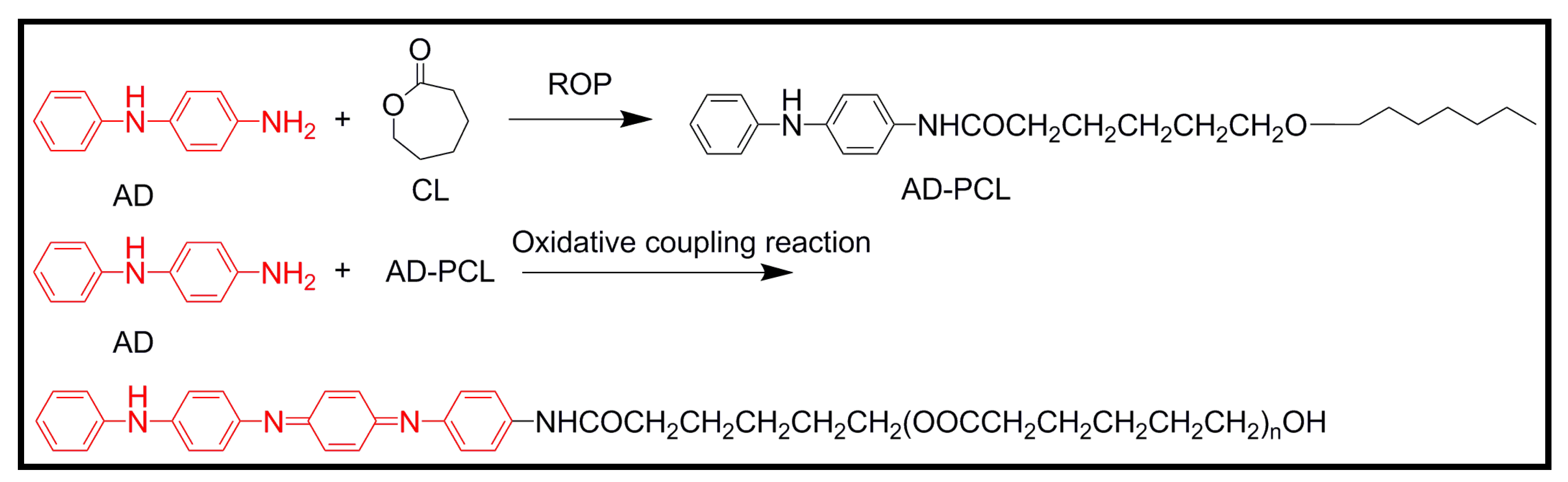
4.1. Development of Novel Biodegradable Samples Having Aniline Units

The primary chain of polyaniline consists of a combination of equal numbers of benzenoid-amine sites which react with oxydizing analytes and quinoid-imine sites which react with reducing and protonating analytes. This base form, known as emeraldine, is insulating but its conductivity can be tuned by doping from 10−1 up to 100 S/cm and more. Upon to exposure to aqueous protonic or functionalized acids, –N= sites become protonated, while maintaining the number of electrons in the polymer chain constant, and the conducting emeraldine salt form (PANiES) is achieved (Figure 5).
Figure 5. Chemical structure of polyaniline (emeraldine base) and transformation to a conductive salt by prototonation in an acid medium.
One strategy to get conductive and biodegradable polymers related to polyaniline is based on joining a biodegradable polymer (e.g., polyactide or chitosan) with heterocyclic oligomers of aniline. In fact, oligoanilines with well-defined chain lengths have been the model compounds for the electrical, magnetic, optical, and structural properties of PANi. Thus, many polymers containing oligoanilines as the side chains or even in the main chain have been designed and synthesized to obtain new electroactive materials. Different derivatives involving aniline trimers, tetramers and pentamers merit to be explained in a more detailed way.
Wei et al. [41] demonstrated that an electroactive silsequioxane precursor containing an aniline trimer [i.e., N-(4-aminophenyl)-N′-(4′-(3-triethoxysilyl-propyl-ureido) phenyl-1,4-quinonenediimine) (ATQD)] could be a promising biomaterial for tissue engineering. To this end, self-assembled monolayers of ATQD on glass substrates were covalently modified with an adhesive oligopeptide, cyclic Arg-Gly-Asp (RGD) (Figure 6). The mean height of the monolayer coating on the surfaces was ~3 nm, as measured by atomic force microscopy. The bioactive, derivatized electroactive scaffold material, ATQD-RGD, supported adhesion and proliferation of PC12 neuronal-like cells. Importantly, electroactive surfaces stimulated spontaneous neuritogenesis in PC12 cells, in the absence of neurotrophic growth factors, such as nerve growth factor (NGF). Hence covalent grafting of bioactive molecules, such as adhesion peptides, appears as an effective strategy to improve the biocompatibility of conventionally non-biocompatible materials and consequently may allow to overcome the apparently poor cell biocompatibility of PANi.
Figure 6. Scheme showing the preparation of self-assembled monolayers of ATQD-RGD, inset reproduced with permission from [41]. Copyright 2007 American Chemical Society.
A novel diblock copolymer (mPEG-b-TEA, PGAT) was synthesized by conjugating the electroactive aniline tetramer (AT) and poly(ethylene glycol) methyl ether (mPEG) (Figure 7). The advantage of this block copolymer was its relatively good solubility in water and in most organic solvents. The copolymer was also mixed with different ratios of poly(l-lactic acid) (PLLA) in order to prepare biodegradable and electroactive PLLA/PGAT polymer blends [42]. Thin films (35–45 nm) of these materials were prepared by spin coating polymer blend chloroform solutions onto the surface of a silicon substrate. The surface topography of films changed with the composition of the blend (Figure 8), being observed large and regular micro-domains that expanded from 200 to 450 nm as the PGAT ratio increased. As the solvent evaporated during the process, micro-phase separation propagated throughout the film, and finally, a cylindrical micro-domain array was achieved. A large nanochannel structure was observed as a consequence of the flexibility and stronger affinity of mPEG chains to chloroform than that of PLLA. Therefore, chloroform served as a selective solvent and preferentially expanded the volume fraction of the mPEG microdomain. Blends exhibited reduced cytotoxicity as compared to AT due to the introduction of the biocompatible PLLA moiety. Blends showed an electroactivity that could accelerate the differentiation of rat C6 glioma cells.
Figure 7. Synthesis scheme for the preparation of PGAT diblock copolymers.
Figure 8. Atomic force microscopy (AFM) images of PLLA/PGAT samples containing (a) 33 wt % and (b) 10 wt % of PGAT. Reprinted with permission from [42]. Copyright 2011 WILEY-VCH Verlag GmbH & Co.
A polysaccharide crosslinker of tetraaniline grafting oxidized sodium alginate with a large amount of aldehyde and carboxylic groups (Figure 9a) has been synthesized by the condensation of terminal amino groups in phenyl/amino-capped tetraaniline (AT) with the aldehyde groups in multialdehyde sodium alginate (MASA) [43]. Copolymers can be obtained with different content of graft AT which is linked to the main chain through highly stable conjugated imine groups. The novel copolymer has interesting properties that justify its potential applications in biomedical fields such as tissue engineering, drug delivery, and nerve probes where electroactivity is required. Thus, the copolymer is water soluble under any pH, biodegradable, electroactive, and noncytotoxic. Furthermore, it can self-assemble into nanoparticles with large active functional groups on the outer surface and alternatively it can be used to crosslink materials with amino and aminoderivative groups like gelatin (via formation of Schiff base or amide through carbodiimide chemistry or electrostatic interaction) to form hydrogels (Figure 9b). MASA-AT only had one pair of reversible redox peaks (the mean redox potential was 0.48 V), which were ascribed to the conversion between leucoemeraldine state and emeraldine state (Figure 9c).
Figure 9. (a) Scheme based on [43] showing the synthesis of the multialdehide sodium alginate (MASA) and the tetraaniline-graft-multialdehide sodium alginate (MASA-AT); (b) Self-assembling and crosslinking capabilities of MASA-AT molecules; and (c) Conversions of MASA-AT between different oxidation states. Copyright 2011 WILEY-VCH Verlag GmbH & Co.
A universal strategy for the facile synthesis of degradable and electroactive block copolymers based on aniline oligomers and polyesters in a two-step approach has recently been reported [44]. Polyesters with an aniline dimer (AD) segment were first obtained by controlled ring opening polymerization (ROP) of a lactone (e.g., caprolactone) initiated by the amine group of AD. The postpolymerization modification via an oxidative coupling reaction between AD and a polyester was then used to form the electroactive segment AT in the copolymers (Figure 10). Thus, diblock copolymers with a controlled structure and molecular weight (i.e., 1300–2800 g/mol) were formed with a rigid AT segment at the chain end as one block and the long degradable flexible PCL as the other block. Furthermore, electrical conductivity of the block copolymers ranged from 6.3 × 10⁻⁷ to 1.03 × 10⁻⁵ S/cm depending on their AT content.
Figure 10. Two-step approach to prepare degradable and conductive block copolymers having aniline tetramer end groups.
A novel electrically conductive biodegradable polyphosphazene polymer containing an aniline pentamer (AP) and glycine ethyl ester (GEE) as side chains was obtained by a nucleophilic substitution reaction (Figure 11) [45]. The electrical conductivity of the polymer was ~2 × 10⁻⁵ S/cm (i.e., in the semiconducting region) upon protonic-doped experiments. Furthermore, the polymer was proved to promote cell adhesion and proliferation according to in vitro assays with RSC96 Schwann cells. The as-synthesized polymer also showed good solubility in common organic solvents and good film-forming properties, and consequently potential applications as scaffolds for neuronal and cardiovascular tissue engineering were claimed [46].
Figure 11. Structure and synthesis scheme for poly[(glycine ethyl ester)x(aniline pentamer)y phosphacene] (PGAP).
A multiblock copolymer (PLA-co-AP) was synthesized by the condensation of hydroxyl-capped poly(l-lactide) and carboxyl-capped aniline pentamer (Figure 12). The copolymer exhibited excellent electroactivity, solubility, and biodegradability. Mechanical properties appeared promising for its application as scaffold material with the tensile strength of 3 MPa, tensile Young’s modulus of 32 MPa, and breaking elongation rate of 95%. The compatibility of PLA-co-AP copolymer was assayed in vitro, being found that it was innocuous, biocompatible, and helpful for the adhesion and proliferation of rat C6 cells. Moreover, PLA-co-AP was able under stimulation by electrical signals to accelerate the differentiation of rat neuronal pheochromocytoma PC12 cells [47].
Figure 12. Schematic synthesis route and structure of PLA-co-AP copolymer.
A PLA-b-AP-b-PLA triblock copolymer was also synthesized [48], being demonstrated that it posses electroactivity and good biodegradability. This type of block copolymers could undergo self-assembly and form micro-phase separation. Thus, the soft PLA segments tended to aggregate together to form a continuous matrix, while the AP hard segments formed discontinuous domains (Figure 13). Electric conduction was easy within the AP domains while between two adjacent domains conduction should occur by the tunnel effect through the PLA matrix and therefore, the apparent conductivity significantly decreased.
Figure 13. Schematic representation of the self-assembling of PLA-b-AP-b-PLA triblock copolymers.
However, it was difficult to control the final oligoaniline content and hydrophilicity of the materials with the proposed procedure. It should be pointed out that most of these polymers were only soluble in organic solvents resulting in environmental concerns. Therefore, the development of electroactive polymers able to dissolve in nontoxic aqueous media is required. The incorporation of oligoaniline into water-soluble polymers [e.g., chitosan (CS)] is a challenging task (Figure 14) [49]. Specifically, an aniline pentamer with a terminal carboxylic group at each end was synthesized and subsequently activated with N-hydroxysuccinimide to allow the further condensation with amine groups of chitosan. The obtained amphiphilic polymers were able to self-assemble into 200−300 nm micelles by dialysis against deionized water from an acetic acid buffer solution. These micelles consisted of the hydrophobic AP block cores with the surface covered by soft and freely stretched CS chains. Self-assembling is highly interesting since enables the use of polymers in delivery applications. Moreover, AP, which has no bioproperties, becomes by self-assembly covered with a natural biomaterial. In this way, it was claimed that the biocompatibility of the polymer can be improved and may give also a change to remove the undegradable oligoaniline from the body. PC12 cells on these samples containing AP showed neurite extension and even were able to form intricate networks, being the best effect observed with samples containing 4.9 wt % of AP.
Figure 14. Chemical structure of aniline pentamer cross-linking chitosan (AP-cs-CS) (left) and scanning micrographs (right) showing nanomicelles of AP-cs-CS and the typical morphology of CS. Reprinted with permission from [49]. Copyright 2008 American Chemical Society.
Linear and hyperbranched copolymers with electrical conductivity and biodegradability were also synthesized using a carboxyl-capped aniline pentamer and branched polycaprolactones by coupling reactions [50]. Copolymers were electroactive and showed three pairs of redox peaks. The hyperbranched copolymers had a higher conductivity than the linear ones, probably as a consequence of the ordered distribution of peripheral AP segments that more easily form a conductive network (Figure 15). In this way, it is clear that the conductivity of polymers could be improved and controlled by the macromolecular architecture.
Figure 15. Model explaining the higher conductivity of hyperbranched copolymers than the linear ones with the same content of conductive units.
So far these hyperbranched degradable conducting copolymers have been blended with polycaprolactone to construct electroactive tubular porous nerve conduits by a solution-casting/particle-leaching method [51]. Thermal and mechanical properties, hydrophilicity, morphology, toxicity and conductivity (values between 3.4 × 10⁻⁶ and 3.1 × 10⁻⁷ S/cm were found depending on the composition) were determined for blends doped with (±)-10-camphorsulfonic acid. The results obtained supported their potential in neural tissue engineering applications.

4.2. Biodegradable Scaffolds Constituted by Nanofibers of Polyaniline

Composite materials are currently utilized as a temporary substrate to stimulate tissue formation by controlled electrochemical signals as well as continuous mechanical stimulation until the regeneration processes are completed.
First works providing novel conductive material well suited as biocompatible scaffolds for tissue engineering concern the development of PANi-gelatin blend nanofibers [52]. Both compounds were dissolved in 1,1,1,3,3,3-hexafluoroisopropanol and co-electrospun into nanofibers. SEM analysis of the blend fibers containing less than 3 wt % of PANi revealed uniform fibers with no evidence for phase segregation and with a substantial change on the physicochemical properties of gelatin. The average diameter size of fibers decreased from ~800 to ~60 nm by increasing the amount of PANi (from 0 to ~5 wt %) while the tensile modulus increased from ~500 to ~1400 MPa. PANi-gelatin blend fibers supported H9c2 rat cardiac myoblast cell attachment and proliferation to a similar degree as positive controls (i.e., tissue culture-treated plastic).
Picciani et al. [53] considered the use of poly(l-lactide) as the support polymeric matrix for the preparation of PANi-based conducting nanofibers and evaluated the influence of some operational parameters, such as the polymer concentration, applied voltage, and flow rate, on the morphology of electrospun fibers. Thus, ultrafine fibers (i.e., diameters ranging between 100 and 200 nm) consisting of blends of polyaniline doped with p-toluene sulfonic acid and PLA were prepared by electrospinning. The high interaction between both components and the rapid evaporation of the solvent during electrospinning resulted in nanofibers with a lower degree of crystallinity in comparison with cast films. The electrical conductivity of the electrospun fiber mats was also reported to be lower probably as a consequence of their lower crystallinity and the high porosity of the nonwoven mats.
Several polyaniline and poly(d,l-lactide) (PANi/PDLA) mixtures at different weight percentages were successfully electrospunned from 1,1,1,3,3,3-hexafluoroisopropanol solutions and their conductivity and biocompatibility evaluated [54]. Promising results were only attained when the PANi content reached 25%. Specifically, this scaffold was able to conduct a current of 5 mA and had an electrical conductivity of 0.0437 S/cm. Calorimetric analysis indicated that fibers were a mixture of the two involved polymers rather than a blend as the Tg value was close to the Tg of PDLA alone. Primary rat muscle cells were able to attach and proliferate over all the new scaffolds although they degraded during the process. The polymer degradation and shrinkage may prevent the blend from being used as the primary component of a biomedical device, but it was claimed its usefulness as a biocompatible coating on devices such as sensors [54].
Composites from the blending of conductive (CPs) and biocompatible polymers are powerfully emerging as a successful strategy for the regeneration of myocardium due to their unique conductive and biological recognition properties able to assure a more efficient electroactive stimulation of cells.
Composite substrates made of synthesized polyaniline (sPANi) doped with camphorsulfonic acid and polycaprolactone (PCL) electrospun fibers were investigated as platforms for cardiac tissue regeneration [55]. In particular, conductibility tests indicated that sPANi short fibres provided a highly efficient transfer of electric signal due to the spatial organization of the electroactive needle-like phases up to form a percolative network. On the basis of this characterization, sPANi/PCL electrospun membranes have been optimized to mimic either the morphological and functional features of the cardiac muscle extracellular matrix. Biological assays (i.e., evaluation of cell survival rate and immunostaining of sarcomeric α-actinin of cardiomyocites-like cells) indicated that conductive signals offered by PANi needles, promoted the cardiogenic differentiation of human mesenchymal stem cells into cardiomyocite-like cells. These preliminary results demonstrated that the development of electroactive biodegradable substrates opens the way towards a new generation of synthetic patches for the support of the regeneration of damaged myocardium.
The insolubility of PANI, in most common solvents can be circumvented by copolymerizing aniline with substituted anilines that impart solubility to the resulting functionalized PANI copolymers (fPANIs). Copolymerization of aniline with aminobenzoic acids (ABAs) gives copolymers (Figure 16) that are soluble in basic aqueous media, and in polar solvents [e.g., N-methyl-2-pyrrolidone (NMP) and dimethyl sulfoxide (DMSO)] and consequently conducting samples that can be easily electrospun.
Figure 16. Chemical structure of copolymers constituted by anyline and aminobenzoic units.
Nanofibrous blends of HCl-doped poly(aniline-co-3-aminobenzoic acid) (3ABAPANI) copolymer and poly(lactic acid) (PLA) were fabricated by electrospinning solutions of the polymers, in varying relative proportions, in a dimethyl sulfoxide/tetrahydrofuran mixture [56]. The conducting copolymer was synthesized from a comonomer mixture with equimolar proportions of aniline and 3ABA, using potassium iodate as oxidizing agent and hydrochloric acid. The nanofibrous electrospun 3ABAPANI-PLA blends gave enhanced cell growth (assayed with COS-1 fibroblast cells), potent antimicrobial capability against Staphylococcus aureus and electrical conductivity (e.g., 8.1 mS/cm was determined for the sample with 55 wt % of PLA). This new class of nanofibrous blends can potentially be employed as tissue engineering scaffolds, and in particular are promising as the basis of a new generation of functional wound dressings that may eliminate deficiencies of currently available antimicrobial dressings.
Combination of temperature responsive-conducting polymers together with carbon nanotubes (CNTs) has been revealed as an excellent smart matrix with outstanding cell viability and proliferation. Specifically, electrospun microfabric scaffolds of poly(N-isopropylacrylamide)–CNT–polyaniline were studied [57]. The polymer was synthesized by coupling chemistry using polyaniline, HOOC-MWNT, and amine-terminated poly(N-isopropylacrylamide) and electrospun from 1,1,1,3,3,3-hexafluoroisopropanol and N,N-dimethylformamide (8:2, v/v) solvent mixture. New scaffolds supported an excellent cell proliferation and viability that was attributed to the balanced hydrophilic functions, conductance, and mechanical strength provided by the poly(N-isopropylacrylamide), polyaniline, and MWNTs, respectively. Furthermore, a temperature dependent cells detachment behavior was observed by varying incubation at below lower critical solution temperature of poly(N-isopropylacrylamide). Suitable three-dimensional conducting smart tissue scaffolds were also reported when poly(N-isopropylacrylamide-co-methacrylic acid) was employed as temperature responsive polymer component [58].

7. Other Conducting and Biodegradable Systems

Melanin films as a biodegradable semiconducting biomaterial for tissue engineering applications have recently been studied since melanins are naturally occurring pigments that exhibit unique electrical properties [134]. Melanin thin films with little concern for potential cytotoxicity were produced by the spin coating technique and using dimethylsulfoxide as a solvent. The thickness of films could be controlled from 160 nm smooth with a roughness of 0.341 nm and a conductivity of 7.0 × 10⁻⁵ S∙cm−1 in the hydrated state. Melanin thin films enhanced Schwann cell growth and neurite extension compared to collagen films in vitro. Furthermore, melanin implants were significantly resorbed after 8 weeks.
New biodegradable proton-conducting carbohydrate polymer films based on alginic acid (constituted by nearly 61% of mannuronic and 39% of guluronic acid) and benzimidazole (Figure 32) have recently been developed by Rachocki et al. [135]. A significant conductivity above water boiling temperature (up to approximately 10−3 S/cm at 473 K) was determined and consequently the obtained films can be considered for application in high-temperature electrochemical devices. 1H-NMR studies revealed that the main mechanism of the proton transport corresponded to a structural diffusion mechanism (in which the water molecules and/or benzimidazole rings take part. The thermal stability of the heterocyclic dopant in the alginicacid-benzimidazole films was significantly higher than that of the water molecules in the film without heterocyclic molecules or in the pure alginic powder and thus the effective dynamics of heterocyclic aromatic rings and high conductivity was claimed to take place above the water boiling temperature.
Figure 32. Chemical structure of the alginic acid-benzimidazole complex.

8. Conclusions

Development of advanced materials able to combine biodegradable and conducting properties is currently considered a hot topic in Nanotechnology and Biomedicine because of their highly promising applications. These mainly concern tissue engineering and drug delivery systems since such new class of materials enable to cover aspects like local stimulation of desired tissue, time controlled drug release and stimulation of either the proliferation or differentiation of various cell types. Moreover, the development of many other applications, as for example the fabrication of biodegradable nanocondensers for the local and temporal implantation in the human body, is just starting at the many research laboratories working in these materials.
In recent years important efforts have been focused in the synthesis of conductive polymers to fulfill requisites of biocompatibility and biodegradability by combining conducting and degradable units. The ease synthesis and good processability of aniline oligomers are the basis of recent developments concerning the preparation of new polymers having the desired combination of properties. However, nowadays it is also highly interesting to explore new strategies concerning pyrrole and thiophene oligomers. In addition, efforts focused on reducing the content of conducting units while retaining sufficient conductivity are highly encouraging. To this end, control of macromolecular architecture (i.e., design of star and branched polymers) has been revealed as a basic tool since it can allow enhancing conductivity.
Nanostructured blends constituted by conducting (e.g., polyaniline, polypirrol and polythiophenes) and biodegradable (e.g., polylactide, polycaprolactone and poly(lactic-co-glycolic acid)) polymers as well as the use of degradable polymers as templates for a subsequent deposition of conducting polymers are different and interesting alternatives to get materials with tuned properties.
The use of conducting and biodegradable hybrid systems in the areas of bioanalytical sciences is also of immense interest since their biocompatibility opens up the possibility of using them in in vivo biosensor applications for continuous monitoring of drugs or metabolites in biological fluids, or as a means of opening up the field to a variety of new analytes.
Conducting fibrous mats based on biodegradable polymers are of special interest for tissue engineering because they are able to stimulate specific cell functions or trigger cell responses in addition to the expected ability to physically support tissue growth. Self-standing ultra-thin films based on electroactive and degradable polymers have been found to useful for different biomedical applications. For example, they can be used as plasters to be delivered, targeted and finely positioned in situ on surgical incisions, or to perform therapeutic or treatment tasks. In summary, the reported systems provide an excellent opportunity for fabricating highly specific, biocompatible, selective, stable, economic and handy biomedical devices.
Many of these features are clearly reflected in this review, which depicts a vivid panorama of the scientific activity in the field and represent distinguished examples of the potentialities and richness that come from the interplay of polymers with so different properties. Yet, biodegradable and conducting polymers provide an enormous range of engaging and stimulating applications by their own. It is clear that these systems will make further meaningful contributions to biomedical field in next times. Design of advanced nanostructured materials able to combine their properties for the development of more sophisticated applications represents a tremendous challenge.

Acknowledgments

Authors are indebted to supports from MINECO and FEDER (MAT2012-36205 and MAT2012-34498) and the Generalitat de Catalunya (2009SGR925 and 2009SGR1208). M.M.P.-M. thanks financial support through a FPI-UPC grant. Support for the research of C.A. was received through the prize “ICREA Academia” for excellence in research funded by the Generalitat de Catalunya.

Conflicts of Interest

The authors declare no conflict of interest.

References

  1. Tiwari, A.; Tiwari, A. Bioengineered Nanomaterials; CRC Press: Boca Raton, FL, USA, 2013. [Google Scholar] [Green Version]
  2. Tiwari, A.; Ramalingam, M.; Kobayashiu, H.; Turner, A.P.F. Biomedical Materials and Diagnostic Devices; Wiley-Scrivener Publishing LLC: Beverly, MA, USA, 2012. [Google Scholar] [Green Version]
  3. Tiwari, A.; Mishra, A.K.; Kobayashi, H.; Turner, A.P.F. Intelligent Nanomaterials; Wiley-Scrivener Publishing LLC: Salem, MA, USA, 2012. [Google Scholar] [Green Version]
  4. Tiwari, A.; Tiwari, A. Nanomaterials in Drug Delivery, Imaging and Tissue Engineering; Wiley-Scrivener Publishing LLC: Salem, MA, USA, 2013. [Google Scholar] [Green Version]
  5. Ramalinga, M.; Tiwari, A.; Ramakrishna, S.; Kobayashi, H. Integrated Biomaterials for Biomedical Technology; Wiley-Scrivener Publishing LLC: Beverly, MA, USA, 2012. [Google Scholar] [Green Version]
  6. Sharma, P.K.; Dutta, R.K.; Pandey, A.C. Advances in multifunctional magnetic nanoparticles. Adv. Mater. Lett. 2011, 2, 246–263. [Google Scholar] [CrossRef]
  7. Tiwari, A. Recent Developments in Bio-Nanocomposites for Biomedical Applications; Nova Science Publishers Inc.: New York, NY, USA, 2011. [Google Scholar] [Green Version]
  8. Sharma, C.; Gautam, S.; Dinda, A.K.; Mishra, N.C. Cartilage tissue engineering: Current Scenario and Challenges. Adv. Mater. Lett. 2011, 2, 90–99. [Google Scholar]
  9. Ramalingama, M.; Tiwari, A. Spatially controlled cell growth using patterned biomaterials. Adv. Mater. Lett. 2010, 1, 179–187. [Google Scholar] [CrossRef]
  10. Ashammakhi, N.; Ndreu, A.; Piras, A.M.; Nikkola, L.; Sindelar, T.; Ylikauppila, H.; Harlin, A.; Chiellini, E.; Hasirci, V.; Redl, H. Biodegradable nanomats produced by electrospinning: Expanding multifunctionality and potential for tissue engineering. J. Nanosci. Nanotechnol. 2007, 7, 862–882. [Google Scholar] [CrossRef]
  11. Teo, W.E.; He, W.; Ramakrishna, S. Electrospun scaffold tailored for tissue-specific extracellular matrix. Biotechnol. J. 2006, 1, 918–929. [Google Scholar] [CrossRef]
  12. Moroni, L.; de Wijn, J.R.; van Blitterswijk, C.A. Integrating novel technologies to fabricate smart scaffolds. J. Biomater. Sci. Polym. E 2008, 19, 543–572. [Google Scholar] [CrossRef]
  13. Liang, D.; Hsiao, B.S.; Chu, B. Functional electrospun nanofibrous scaffolds for biomedical applications. Adv. Drug Deliver. Rev. 2007, 59, 1392–1412. [Google Scholar] [CrossRef]
  14. Jiang, C.; Markutsya, S.; Pikus, Y.; Tsukruk, V.V. Freely suspended nanocomposite membranes as highly sensitive sensors. Nat. Mater. 2004, 3, 721–728. [Google Scholar] [CrossRef]
  15. Vendamme, R.; Onoue, S.Y.; Nakao, A.; Kunitake, T. Robust free-standing nanomembranes of organic/inorganic interpenetrating networks. Nat. Mater. 2006, 5, 494–501. [Google Scholar] [CrossRef]
  16. Kim, D.-H.; Ahn, J.-H.; Choi, W.M.; Kim, H.-S.; Kim, T.-H.; Song, J.; Huang, Y.Y.; Liu, Z.; Lu, C.; Rogers, J.A. Stretchable and foldable silicon integrated circuits. Science 2008, 320, 507–511. [Google Scholar] [CrossRef]
  17. Eda, G.; Fanchini, G.; Chhowalla, M. Large-area ultrathin films of reduced graphene oxide as a transparent and flexible electronic material. Nat. Nanotechnol. 2008, 3, 270–274. [Google Scholar] [CrossRef]
  18. Park, S.I.; Xiong, Y.J.; Kim, R.H.; Elvikis, P.; Meitl, M.; Kim, D.H.; Wu, J.; Yoon, J.; Yu, C.J.; Liu, Z.J.; et al. Printed assemblies of inorganic light-emitting diodes for deformable and semitransparent displays. Science 2009, 325, 977–981. [Google Scholar] [CrossRef]
  19. Tiwaki, A.; Kumar, R.; Prabaharan, M.; Pandey, R.R.; Kumasi, P.; Chaturvedi, A.; Mishra, A.K. Nanofibrous polyaniline thin film prepared by plasma-induced polymerization technique for detection of NO2 gas. Polym. Adv. Technol. 2010, 21, 615–620. [Google Scholar] [CrossRef]
  20. Norris, I.D.; Shaker, M.M.; Ko, F.K.; MacDiarmid, A.G. Electrostatic fabrication of ultrafine conducting fibers: Polyaniline/polyethylene oxide blends. Synth. Met. 2000, 114, 109–114. [Google Scholar] [CrossRef]
  21. MacDiarmid, A.G.; Jones, W.E.; Norris, I.D.; Gao, J.; Johnson, A.T.; Pinto, N.J.; Hone, J.; Han, B.; Ko, F.K.; Okuzaki, H.; et al. Electrostatically-generated nanofibers of electronic polymers. Synth. Met. 2001, 119, 27–30. [Google Scholar] [CrossRef]
  22. Ali, Y.; Kumar, V.; Sonkawade, R.G.; Dhaliwal, A.S. Fabrication of polyaniline nanofibers by chronopotentiometry. Adv. Mater. Lett. 2012, 3, 388–392. [Google Scholar]
  23. Ali, Y.; Kumar, V.; Dhaliwal, R.G.; Sonkawade, R.G. Surface modification of polyaniline nanofiber using silver nanoparticles to enhance sensing properties. Adv. Mater. Lett. 2013, 4, 368–372. [Google Scholar]
  24. Wei, Y.; Lelkes, P.I.; MacDiarmid, A.G.; Guterman, E.; Cheng, S.; Palouian, K. Contemporary Topics in Advanced Polymer Science and Technology; Peking University Press: Beijing, China, 2004; pp. 430–436. [Google Scholar] [Green Version]
  25. Bidez, P.; Li, S.; MacDiarmid, A.G.; Venancio, E.C.; Wei, Y.; Lelkes, P.I. Polyaniline, an electroactive polymer, supports adhesion and proliferation of cardiac myoblasts. J. Biomater. Sci. Polym. E 2006, 17, 199–212. [Google Scholar] [CrossRef]
  26. Kotwal, A.; Schmidt, C.E. Electrical stimulation alters protein adsorption and nerve cell interactions with electrically conducting biomaterials. Biomaterials 2001, 22, 1055–1064. [Google Scholar] [CrossRef]
  27. Valentini, R.F. Nerve Guidance Channels in the Biomedical Engineering HandBook, 2nd ed.; Bronzino, J.D., Ed.; CRC Press LLC: Boca Raton, FL, USA, 2000. [Google Scholar] [Green Version]
  28. Reneker, D.H.; Chun, I. Nanometre diameter fibres of polymer, produced by electrospinning. Nanotechnology 1996, 7, 216–223. [Google Scholar] [CrossRef]
  29. Reneker, D.H.; Yarin, A.L.; Fong, H.; Koombhongse, S. Bending instability of electrically charged liquid jets of polymer solutions in electrospinning. J. Appl. Phys. 2000, 87, 4531–4547. [Google Scholar]
  30. Frenot, A.; Chronakis, I.S. Polymer nanofibers assembled by electrospinning. Curr. Opin. Colloid Interface Sci. 2003, 8, 64–75. [Google Scholar] [CrossRef]
  31. Dzenis, Y. Spinning continuous fibers for nanotechnology. Science 2004, 304, 1917–1919. [Google Scholar] [CrossRef]
  32. Li, D.; Xia, Y. Electrospinning of nanofibers: Reinventing the wheel? Adv. Mater. 2004, 16, 1151–1170. [Google Scholar] [CrossRef]
  33. Jayaraman, K.; Kotaki, M.; Zhang, Y.; Mo, X.; Ramakrishna, S. Recent advances in polymer nanofibers. J. Nanosci. Nanotechnol. 2004, 4, 52–65. [Google Scholar]
  34. Dhakate, S.R.; Singla, B.; Uppal, M.; Mathur, R.B. Effect of processing parameters on morphology and thermal properties of polycarbonate nanofibers. Adv. Mater. Lett. 2010, 1, 200–204. [Google Scholar]
  35. Sharma, S. Ferroelectric nanofibers: Principle, processing and applications. Adv. Mater. Lett. 2013, 4, 522–533. [Google Scholar]
  36. Dersch, R.; Steinhart, M.; Boudriot, U.; Greiner, A.; Wendorff, J.H. Nanoprocessing of polymers: Applications in medicine, sensors, catalysis, photonics. Polym. Adv. Technol. 2005, 16, 276–282. [Google Scholar] [CrossRef]
  37. Chronakis, I.S. Novel nanocomposites and nanoceramics based on polymer nanofibers using electrospinning process—A review. J. Mater. Process. Tech. 2005, 167, 283–293. [Google Scholar] [CrossRef]
  38. Deitzel, J.M.; Kleinmeyer, J.; Harris, D.; Tan, N.C.B. The effect of processing variables on the morphology of electrospun nanofibers and textiles. Polymer 2001, 42, 261–272. [Google Scholar]
  39. Terada, D.; Kobayashi, H.; Zhang, K.; Tiwari, A.; Yoshikawa, C.; Hanagata, N. Transient charge-masking effect of applied voltage on electrospinning of pure chitosan nanofibers from aqueous solutions. Sci. Technol. Adv. Mater. 2012, 13, 1–9. [Google Scholar]
  40. Tiwari, A.; Terada, D.; Yoshikawa, C.; Kobayashi, H. An enzyme-free highly glucose-specific assay using self-assembled aminobenzene boronic acid upon polyelectrolytes electrospun nanofibers-mat. Talanta 2010, 82, 1725–1732. [Google Scholar] [CrossRef]
  41. Guo, Y.; Li, M.Y.; Mylonakis, A.; Han, J.J.; MacDiarmid, A.G.; Chen, X.S.; Lelkes, P.I.; Wei, Y. Electroactive oligoaniline-containing self-assembled monolayers for tissue engineering applications. Biomacromolecules 2007, 8, 3025–3034. [Google Scholar] [CrossRef]
  42. Liu, Y.; Hu, J.; Zhuang, X.; Zhang, P.; Chen, X.; Wei, Y.; Wang, X. Preparation and characterization of biodegradable and electroactive polymer blend materials based on mPEG/tetraaniline and PLLA. Macromol. Biosci. 2011, 11, 806–813. [Google Scholar] [CrossRef]
  43. Wang, Q.; He, W.; Huang, J.; Liu, S.; Wu, G.; Teng, W.; Wang, Q.; Don, Y. Synthesis of water soluble, biodegradable, and electroactive polysaccharide crosslinker with aldehyde and carboxylic groups for biomedical applications. Macromol. Biosci. 2011, 11, 362–372. [Google Scholar] [CrossRef]
  44. Guo, B.; Finne-Wistrand, A.; Albertsson, A.-C. Universal two-step approach to degradable and electroactive block copolymers and networks from combined ring-opening polymerization and post-functionalization via oxidative coupling reactions. Macromolecules 2011, 44, 5227–5236. [Google Scholar] [CrossRef]
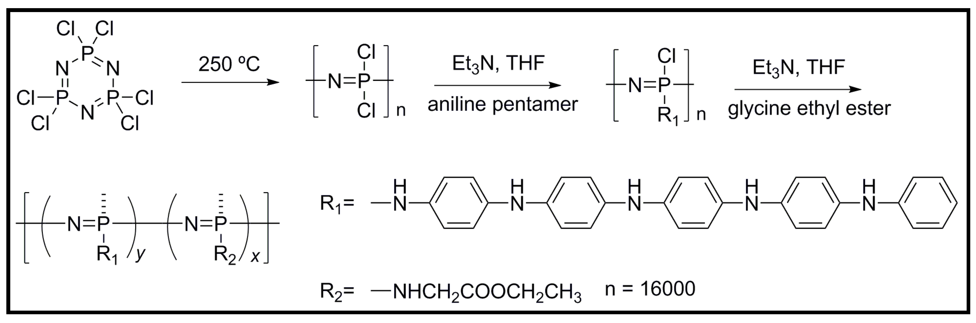
  45. Zhang, Q.-S.; Yan, Y.-H.; Li, S.-P.; Feng, T. Synthesis of a novel biodegradable and electroactive polyphosphazene for biomedical application. Biomed. Mater. 2009, 4. [Google Scholar] [CrossRef]
  46. Zhang, Q.; Yan, Y.; Li, S.; Feng, T. The synthesis and characterization of a novel biodegradable and electroactive polyphosphazene for nerve regeneration. Mater. Sci. Eng. C 2010, 30, 160–166. [Google Scholar] [CrossRef]
  47. Huang, L.; Zhuang, X.; Hu, J.; Lang, L.; Zhang, P.; Wang, Y.; Chen, X.; Wei, Y.; Jing, X. Synthesis of biodegradable and electroactive multiblock polylactide and aniline pentamer copolymer for tissue engineering applications. Biomacromolecules 2008, 9, 850–858. [Google Scholar] [CrossRef]
  48. Huang, L.; Hu, J.; Lang, L.; Wang, X.; Zhang, P.; Jing, X.; Wang, X.; Chen, X.; Lelkes, P.I.; MacDiarmid, A.G.; et al. Synthesis and characterization of electroactive and biodegradable ABA block copolymer of polylactide and aniline pentamer. Biomaterials 2007, 28, 1741–1751. [Google Scholar] [CrossRef]
  49. Hu, J.; Huang, L.H.; Zhuang, X.L.; Zhang, P.B.; Lang, L.; Chen, X.S.; Wei, Y.; Jing, X.B. Electroactive aniline pentamer cross-linking chitosan for stimulation growth of electrically sensitive cells. Biomacromolecules 2008, 9, 2637–2644. [Google Scholar] [CrossRef]
  50. Guo, B.; Finne-Wistrand, A.; Albertsson, A.-C. Enhanced electrical conductivity by macromolecular architecture: Hyperbranched electroactive and degradable block copolymers based on poly(ε-caprolactone) and aniline pentamer. Macromolecules 2010, 43, 4472–4480. [Google Scholar] [CrossRef]
  51. Guo, B.; Sun, Y.; Finne-Wistrand, A.; Mustafa, K.; Albertsson, A.-C. Electroactive porous tubular scaffolds with degradability and non-cytotoxicity for neural tissue regeneration. Acta Biomater. 2012, 8, 144–153. [Google Scholar] [CrossRef]
  52. Li, M.; Guo, Y.; Wei, Y.; MacDiarmid, A.G.; Lelkes, P.I. Electrospinning polyaniline-contained gelatin nanofibers for tissue engineering applications. Biomaterials 2006, 27, 2705–2715. [Google Scholar] [CrossRef]
  53. Picciani, P.H.S.; Medeiros, E.S.; Pan, Z.; Orts, W.J.; Mattoso, L.H.C.; Soares, B.G. Development of conducting polyaniline/poly(lactic acid) nanofibers by electrospinning. J. Appl. Polym. Sci. 2009, 112, 744–753. [Google Scholar] [CrossRef]
  54. McKeon, K.D.; Lewis, A.; Freeman, J.W. Electrospun poly(d,l-Lactide) and polyaniline scaffold characterization. J. Appl. Polym. Sci. 2010, 115, 1566–1572. [Google Scholar] [CrossRef]
  55. Borriello, A.; Guarino, V.; Schiavo, L.; Alvarez-Perez, M.A.; Ambrosio, L. Optimizing PANi doped electroactive substrates as patches for the regeneration of cardiac muscle. Mater. Sci. Eng. C 2011, 22, 1053–1062. [Google Scholar]
  56. Gizdavic-Nikolaidis, M.; Ray, S.; Bennett, J.R.; Easteal, A.J.; Cooney, R.P. Electrospun functionalized polyaniline copolymer-based nanofibers with potential application in tissue engineering. Macromol. Biosci. 2010, 10, 1424–1431. [Google Scholar] [CrossRef]
  57. Tiwari, A.; Sharma, Y.; Hattori, S.; Terada, D.; Sharma, A.K.; Turner, A.P.F.; Kobayashi, H. Influence of poly(N-isopropylacrylamide)-CNT-polyaniline three dimensional electrospun microfabric scaffolds on cell growth and viability. Biopolymers 2013, 99, 334–341. [Google Scholar] [CrossRef]
  58. Sharma, Y.; Tiwari, A.; Hattori, S.; Terada, D.; Sharma, A.K.; Ramalingan, M.; Kobayashi, H. Fabrication of conducting electrospun nanofibers scaffold for three-dimensional cells culture. Int. J. Biol. Macromol. 2012, 51, 627–631. [Google Scholar] [CrossRef]
  59. Chronakis, I.S.; Grapenson, S.; Jakob, A. Conductive polypyrrole nanofibers via electrospinning: Electrical and morphological properties. Polymer 2006, 47, 1597–1603. [Google Scholar] [CrossRef]
  60. Schmidt, C.E.; Shastri, V.R.; Vacanti, J.P.; Langer, R. Stimulation of neurite utgrowth using an electrically conducting polymer. Proc. Natl. Acad. Sci. USA 1997, 94, 8948–8953. [Google Scholar]
  61. Rivers, T.J.; Hudson, T.W.; Schmidt, C.E. Synthesis of a novel, biodegradable electrically conducting polymer for biomedical applications. Adv. Funct. Mater. 2002, 12, 33–37. [Google Scholar] [CrossRef]
  62. Schmidt, C.E.; Rivers, T.J. Biodegradable, Electrically Conducting Polymer for Tissue Engineering Applications. U.S. Patent 6,696,575, 23 February 2004. [Google Scholar]
  63. Wang, S.; Lu, L.; Gruetzmacher, J.A.; Currier, B.L.; Yaszemski, M.J. Synthesis and characterizations of biodegradable and crosslinkable poly(epsilon-caprolactone fumarate), poly(ethylene glycol fumarate), and their amphiphilic copolymer. Biomaterials 2006, 27, 832–841. [Google Scholar] [CrossRef]
  64. Runge, M.B.; Dadsetan, M.; Baltrusaitis, J.; Knight, A.M.; Ruesink, T.; Lazcano, E.; Lu, L.; Windebank, A.J.; Yaszemsk, M.J. The development of electrically conductive polycaprolactone fumarate-polypyrrole composite materials for nerve regeneration. Biomaterials 2010, 31, 5916–5926. [Google Scholar] [CrossRef]
  65. Jabbari, E.; Wang, S.; Lu, L.; Gruetzmacher, J.A.; Ameenuddin, S.; Hefferan, T.E.; Currier, B.L.; Windebank, A.J.; Yaszemski, M.J. Synthesis, material properties, and biocompatibility of a novel self-cross-linkable poly(caprolactone fumarate) as an injectable tissue engineering scaffold. Biomacromolecules 2005, 6, 2503–2511. [Google Scholar] [CrossRef]
  66. Durgam, H.; Sapp, S.; Deister, C.; Khaing, Z.; Chang, E.; Luebben, S.; Schmidt, C.E. Novel degradable copolymers of polypyrrole support cell proliferation and enhance neurite out-growth with electrical stimulation. J. Biomater. Sci. Polym. E 2010, 21, 1265–1282. [Google Scholar] [CrossRef]
  67. Shi, G.; Rouabhia, M.; Wang, Z.; Dao, L.H.; Zhang, Z. A novel electrically conductive and biodegradable composite made of polypyrrole nanoparticles and polylactide. Biomaterials 2004, 25, 2477–2488. [Google Scholar] [CrossRef]
  68. Cen, L.; Neoh, K.G.; Kang, E.T. Surface functionalization of polypurrole film with glucose oxidase and viologen. Biosens. Bioelectron. 2003, 18, 363–374. [Google Scholar] [CrossRef]
  69. Wang, Z.; Roberge, C.; Wan, Y.; Dao, L.H.; Guidoin, R.; Zhang, Z. A biodegradable electrical bioconductor made of polypyrrole nanoparticle/poly(d,l-Lactide) composite a preliminary in vitro biostability study. J. Biomed. Mater. Res. A 2003, 66, 738–746. [Google Scholar]
  70. Zhang, Z.; Rouabhia, M.; Wang, Z.; Roberge, C.; Shi, G.; Roche, P.; Li, J.; Dao, L.H. Electrically conductive biodegradable polymer composite for nerve regeneration: Electricity-stimulated neurite outgrowth and axon regeneration. Artif. Organs 2007, 31, 13–22. [Google Scholar] [CrossRef]
  71. Boutry, C.M.; Müller, M.; Hierold, C. Junctions between metals and blends of conducting and biodegradable polymers (PLLA-PPy and PCL-PPy). Mater. Sci. Eng. C 2012, 32, 1610–1620. [Google Scholar] [CrossRef]
  72. Boutry, C.M.; Gerber-Hörler, I.; Hierold, C. Electrically conducting biodegradable polymer composites (polylactide-polypyrrole and polycaprolactone-polypyrrole) for passive resonant circuits. Polym. Eng. Sci. 2013, 53, 1196–1208. [Google Scholar] [CrossRef]
  73. Pham, Q.P.; Sharma, U.; Mikos, A.G. Electrospinning of polymeric nanofibers for tissue engineering applications. Tissue Eng. 2006, 12, 1197–1211. [Google Scholar] [CrossRef]
  74. Bashur, C.A.; Dahlgren, L.A.; Goldstein, A.S. Effect of fiber diameter and orientation on fibroblast morphology and proliferation on electrospun poly(d,l-Lactic-co-Glycolic acid) meshes. Biomaterials 2006, 27, 5681–5688. [Google Scholar] [CrossRef]
  75. Yang, F.; Murugan, R.; Wang, S.; Ramakrishna, S. Electrospinning of nano/micro scale poly(l-Lactic acid) aligned fibers and their potential in neural tissue engineering. Biomaterials 2005, 26, 2603–2610. [Google Scholar] [CrossRef]
  76. Tian, T.; Deng, J.; Xie, Z.; Zhao, Y.; Feng, Z.; Kang, X.; Gu, Z. Polypyrrole hollow fiber for solid phase extraction. Analyst 2012, 137, 1846–1852. [Google Scholar]
  77. Kang, T.S.; Lee, S.W.; Joo, J.; Lee, J.Y. Electrically conducting polypyrrole fibers spun by electrospinning. Synth. Met. 2005, 153, 61–64. [Google Scholar] [CrossRef]
  78. Kai, D.; Prabhakaran, M.P.; Jin, G.; Ramakrishna, S. Polypyrrole-contained electrospun conductive nanofibrous membranes for cardiac tissue engineering. J. Biomed. Mater. Res. A 2011, 99, 376–385. [Google Scholar]
  79. Kuhn, H.H.; Child, A.D. Electrically Conducting Textiles. Handbook of Conducting Polymers, 2nd ed.; Skotheim, T.A., Elsenaumer, R.L., Reynolds, J.R., Eds.; Marcel Dekker: New York, NY, USA, 1998; pp. 993–1104. [Google Scholar]
  80. Nair, S.; Natarajan, S.; Kim, S.H. Fabrication of electrically conducting polypyrrole-poly(ethylene oxide) composite nanofibers. Macromol. Rapid Commun. 2005, 26, 1599–1603. [Google Scholar] [CrossRef]
  81. Xie, J.; MacEwan, M.R.; Willerth, S.M.; Li, X.; Moran, D.W.; Sakiyama-Elbert, S.E.; Xia, Y. Conductive core-sheath nanofibers and their potential application in neural tissue engineering. Adv. Funct. Mater. 2009, 19, 2312–2318. [Google Scholar]
  82. Lee, J.Y.; Bashur, C.A.; Goldstein, A.S.; Schmidt, C.E. Polypyrrole-coated electrospun PLGA nanofibers for neural tissue applications. Biomaterials 2009, 30, 4325–4335. [Google Scholar] [CrossRef]
  83. Zhou, J.; Zhao, S.; Zhang, X.; Feng, W.; Yuan, X. Preparation and properties of electrospun poly(ε-caprolactone)/polypyrrole membranes. Acta Polym. Sin. 2010, 1, 1094–1099. [Google Scholar]
  84. Wang, Y.Z.; Kim, H.J.; Vunjak-Novakovic, G.; Kaplan, D.L. Stem cell-based tissue engineering with silk biomaterials. Biomaterials 2006, 27, 6064–6082. [Google Scholar] [CrossRef]
  85. Yang, Y.; Chen, X.; Ding, F.; Zhang, P.; Liu, J.; Gu, X. Biocompatibility evaluation of silk fibroin with peripheral nerve tissues and cells in vitro. Biomaterials 2007, 28, 1643–1652. [Google Scholar] [CrossRef]
  86. Aznar-Cervantes, S.; Roca, M.I.; Martinez, J.G.; Meseguer-Olmo, L.; Cenis, J.L.; Moraleda, J.M.; Otero, T.F. Fabrication of conductive electrospun silk fibroin scaffolds by coating with poypyrrole for biomedical applications. Biochemistry 2012, 85, 36–43. [Google Scholar]
  87. Jin, L.; Wang, T.; Feng, Z.-Q.; Zhu, M.; Leach, M.K.; Naim, Y.I.; Jiang, Q. Fabrication and characterization of a novel polypyrrole fibrous scaffold designed for 3D cell culture. J. Mater. Chem. 2012, 22, 18321–18326. [Google Scholar]
  88. Mohammadi, A.; Ameli, A.; Alizadeh, N. Headspace solid-phase microextraction using a dodecylsulfate-doped polypyrrole film coupled to ion mobility spectrometry for the simultaneous determination of atrazine and ametryn in soil and water samples. Talanta 2009, 78, 1107–1114. [Google Scholar] [CrossRef]
  89. Li, X.; Zhong, M.; Chen, J. Electrodeposited polyaniline as a fiber coating for solid-phase microextraction of organochlorine pesticides from water. J. Sep. Sci. 2008, 31, 2839–2845. [Google Scholar]
  90. Li, M.; Wei, Z.; Jiang, L. Polypyrrole nanofiber arrays synthesized by a biphasic electrochemical strategy. J. Mater. Sci. 2008, 18, 2276–2280. [Google Scholar]
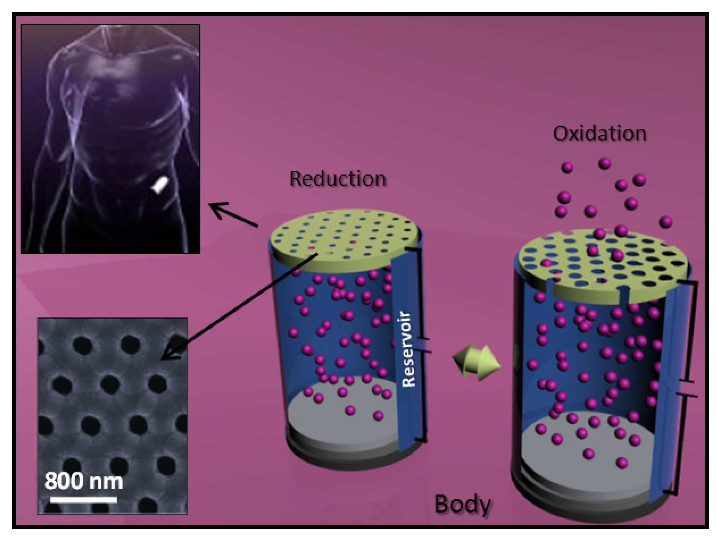
  91. Yang, S.Y.; Yang, J.-A.; Kim, E.-S.; Jeon, G.; Oh, E.J.; Choi, K.Y.; Hahn, S.K.; Kim, J.K. Single-file diffusion of protein drugs through cylindrical nanochannels. ACS Nano 2010, 4, 3817–3822. [Google Scholar] [CrossRef]
  92. Jeon, G.; Yang, S.Y.; Byun, J.; Kim, J.K. Electrically actuable smart nanoporous membrane for pulsatile drug release. Nano Lett. 2011, 11, 1284–1288. [Google Scholar] [CrossRef]
  93. Smela, E.; Gadegaard, N. Surprising volume change in PPy(DBS): An atomic force microscopy study. Adv. Mater. 1999, 11, 953–957. [Google Scholar] [CrossRef]
  94. Smela, E.; Gadegaard, N. Volume change in polypyrrole studied by atomic force microscopy. J. Phys. Chem. B 2001, 105, 9395–9405. [Google Scholar] [CrossRef]
  95. Smela, E. Conjugated polymer actuators for biomedical applications. Adv. Mater. 2003, 15, 481–494. [Google Scholar] [CrossRef]
  96. Ge, D.; Ru, X.; Hong, S.; Jiang, S.; Tu, J.; Wang, J.; Zhang, A.; Ji, S.; Linkov, V.; Ren, B.; et al. Coating metals on cellulose–polypyrrole composites: A new route to self-powered drug delivery system. Electrochem. Commun. 2010, 12, 1367–1370. [Google Scholar] [CrossRef]
  97. Collier, J.H.; Camp, J.P.; Hudson, T.W.; Schmidt, C.E. Synthesis and characterization of polypyrrole–hyaluronic acid composite biomaterials for tissue. J. Biomed. Mater. Res. A 2000, 50, 574–584. [Google Scholar] [CrossRef]
  98. Laurent, T.C.; Fraser, J.R.E. Hyaluronan. FASEB J. 1992, 6, 2397–2404. [Google Scholar]
  99. Akkouch, A.; Shi, G.; Zhang, Z.; Rouabhia, M. Bioactivating electrically conducting polypyrrole with fibronectin and bovine serum albumin. J. Biomed. Mater. Res. A 2010, 92, 221–231. [Google Scholar]
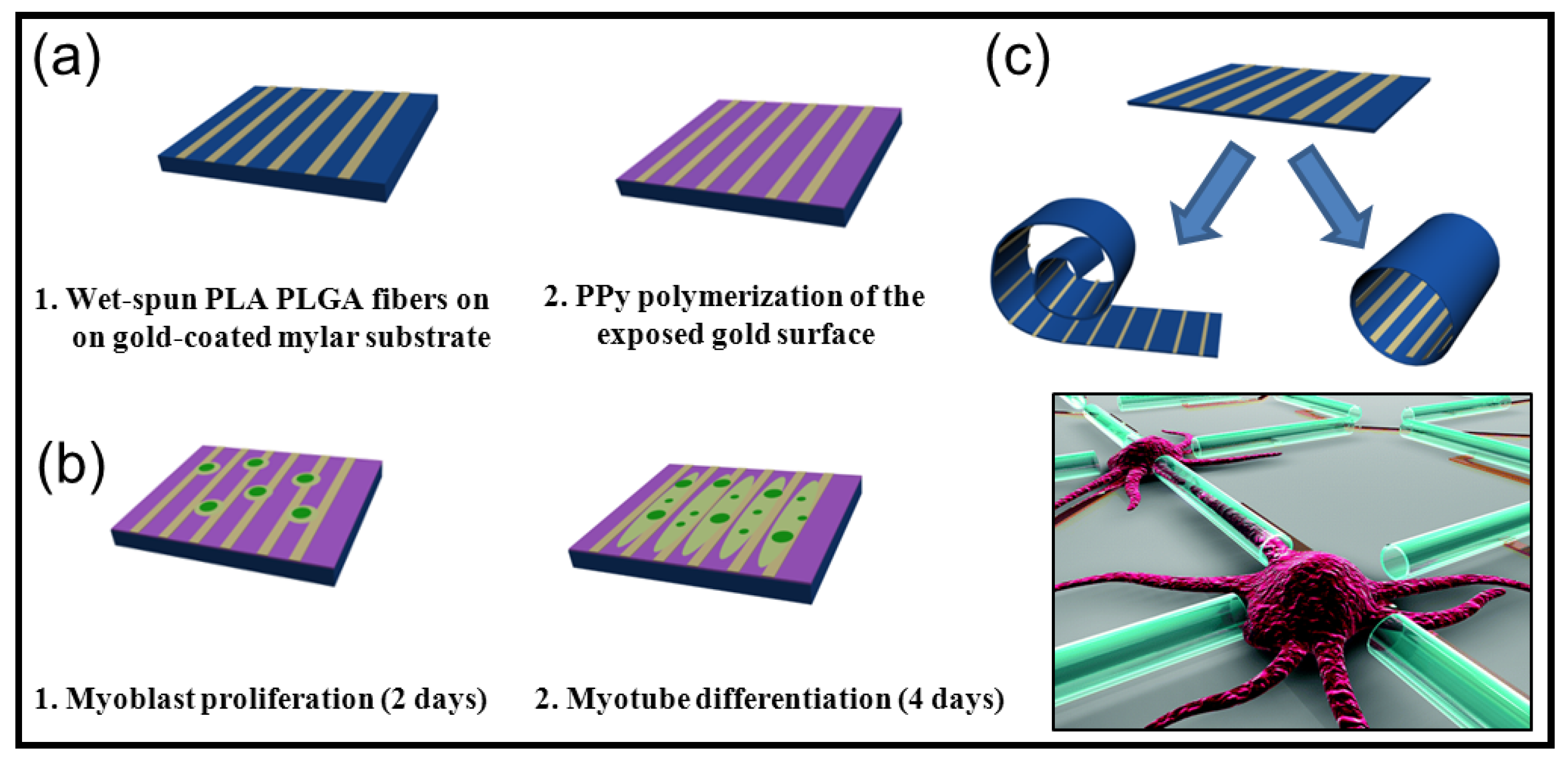
  100. Razal, J.; Kita, M.; Quigley, A.F.; Kennedy, E.; Moulton, S.E.; Kapsa, R.M.I.; Clark, G.M.; Wallace, G.G. Wet-spun biodegradable fibers on conducting platforms: Novel architectures for muscle regeneration. Adv. Funct. Mater. 2009, 19, 3381–3388. [Google Scholar] [CrossRef]
  101. Quigley, A.F.; Razal, J.M.; Thompson, B.C.; Moulton, S.E.; Kita, M.; Kennedy, E.L.; Clark, G.M.; Wallace, G.G.; Lapsa, R.M.I. A conducting-polymer platform with biodegradable fibers for stimulation and guidance of axonal growth. Adv. Mater. 2009, 21, 4393–4397. [Google Scholar] [CrossRef]
  102. Yu, M.; Huang, Y.; Ballweg, J.; Shin, H.; Huang, M.; Savage, D.E.; Lagally, M.G.; Dent, E.W.; Blick, R.H.; Williams, J.C. Semiconductor nanomembrane tubes: Three-dimensional confinement for controlled neurite outgrowth. ACS Nano 2011, 5, 2447–2457. [Google Scholar] [CrossRef]
  103. Kim, J.; Seo, Y.B. Electro-active paper actuators. Smart Mater. Struct. 2002, 11, 355–360. [Google Scholar] [CrossRef]
  104. Kim, J.; Yun, S.; Ounaies, Z. Discovery of cellulose as a smart material. Macromolecules 2006, 39, 4202–4206. [Google Scholar] [CrossRef]
  105. Kim, J.; Deshpande, S.D.; Yun, S.; Li, Q. A comparative study of conductive polypyrrole and polyaniline coatings on electro-active papers. Polym. J. 2006, 38, 659–668. [Google Scholar] [CrossRef]
  106. Kim, J.; Yun, S.; Deshpande, S.D. Synthesis, characterization and actuation behavior of polyaniline-cellophane based electro-active paper actuator. Polym. Int. 2007, 56, 1530–1536. [Google Scholar] [CrossRef]
  107. Kim, J.; Yun, S.; Mahadeva, S.K.; Yun, K.; Yang, S.Y.; Maniruzzaman, M. Paper actuators made with cellulose and hybrid materials. Sensors 2010, 10, 1473–1485. [Google Scholar] [CrossRef]
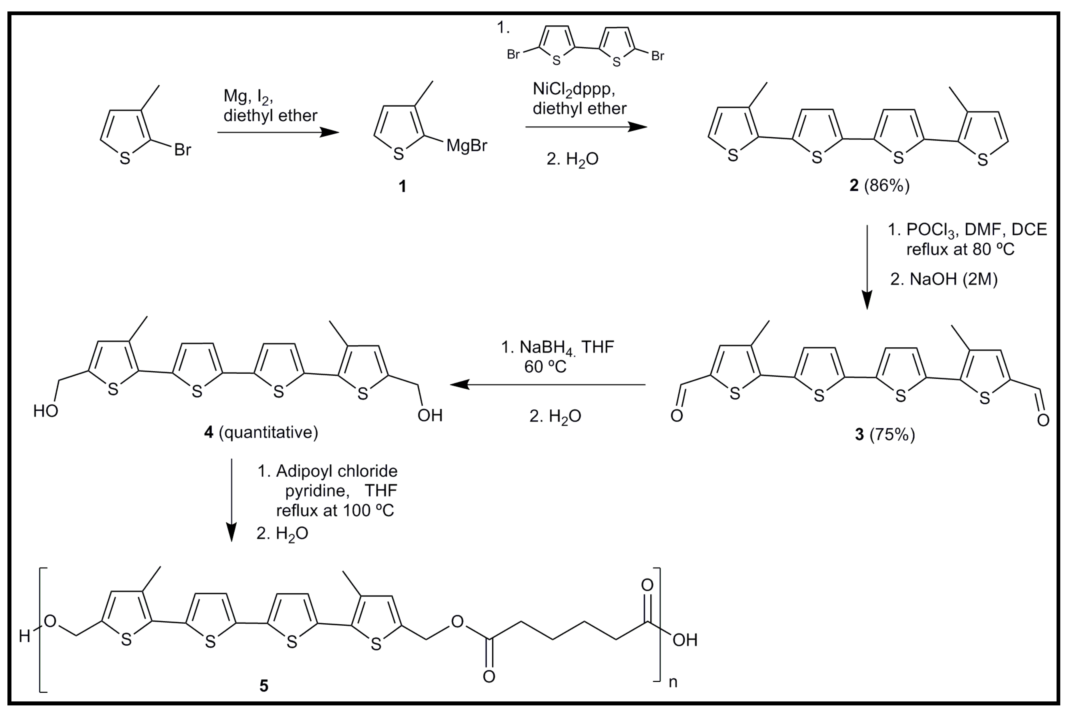
  108. Guimard, N.K.E.; Sessler, J.L.; Schmidt, C.E. Towards a biocompatible and biodegradable copolymer incorporating electroactive oligothiophene units. Macromolecules 2009, 42, 502–511. [Google Scholar] [CrossRef]
  109. Baughman, R.H.; Shacklette, R.L.; Elsenbaumer, R.L.; Plichta, E.J.; Becht, C. Conjugated Polymeric Materials: Opprtunities in Electronics, Optoelectronics, and Molecular Electronics; NATO ASI Series E; Brédas, J.L., Chance, R.R., Eds.; Applied Science: Mons, Belgium, 1990; Volume 182, pp. 559–582. [Google Scholar]
  110. Pei, Q.B.; Inganläs, O. Conjugated polymers and the bending cantilever method: Electrical muscles and smart devices. Adv. Mater. 1992, 4, 277–278. [Google Scholar] [CrossRef]
  111. Otero, T.F.; Cortes, M.T. Artificial muscles with tactile sensitivity. Adv. Mater. 2003, 15, 279–282. [Google Scholar] [CrossRef]
  112. Gandhi, M.R.; Murray, P.; Spinks, G.M.; Wallace, G.G. Mechanism of electromechanical actuation in polypyrrole. Synth. Met. 1995, 73, 247–256. [Google Scholar] [CrossRef]
  113. Adeloju, S.B.; Shaw, S.J.; Wallace, G.G. Pulsed-amperometric detection of urea in blood samples on a conducting polypyrrole-urease biosensor. Anal. Chim. Acta 1997, 341, 155–160. [Google Scholar] [CrossRef]
  114. Pernaut, J.M.; Reynolds, J.R. Use of conducting electroactive polymers for drug delivery and sensing of bioactive molecules. A redox chemistry approach. J. Phys. Chem. B 2000, 104, 4080–4090. [Google Scholar] [CrossRef]
  115. Reinhard, C.S.; Radomsky, M.L.; Saltzman, W.M.; Hilton, J.; Brem, H. Polymeric controlled release of dexamethasone in normal rat brain. J. Control. Release 1991, 1683, 331–339. [Google Scholar]
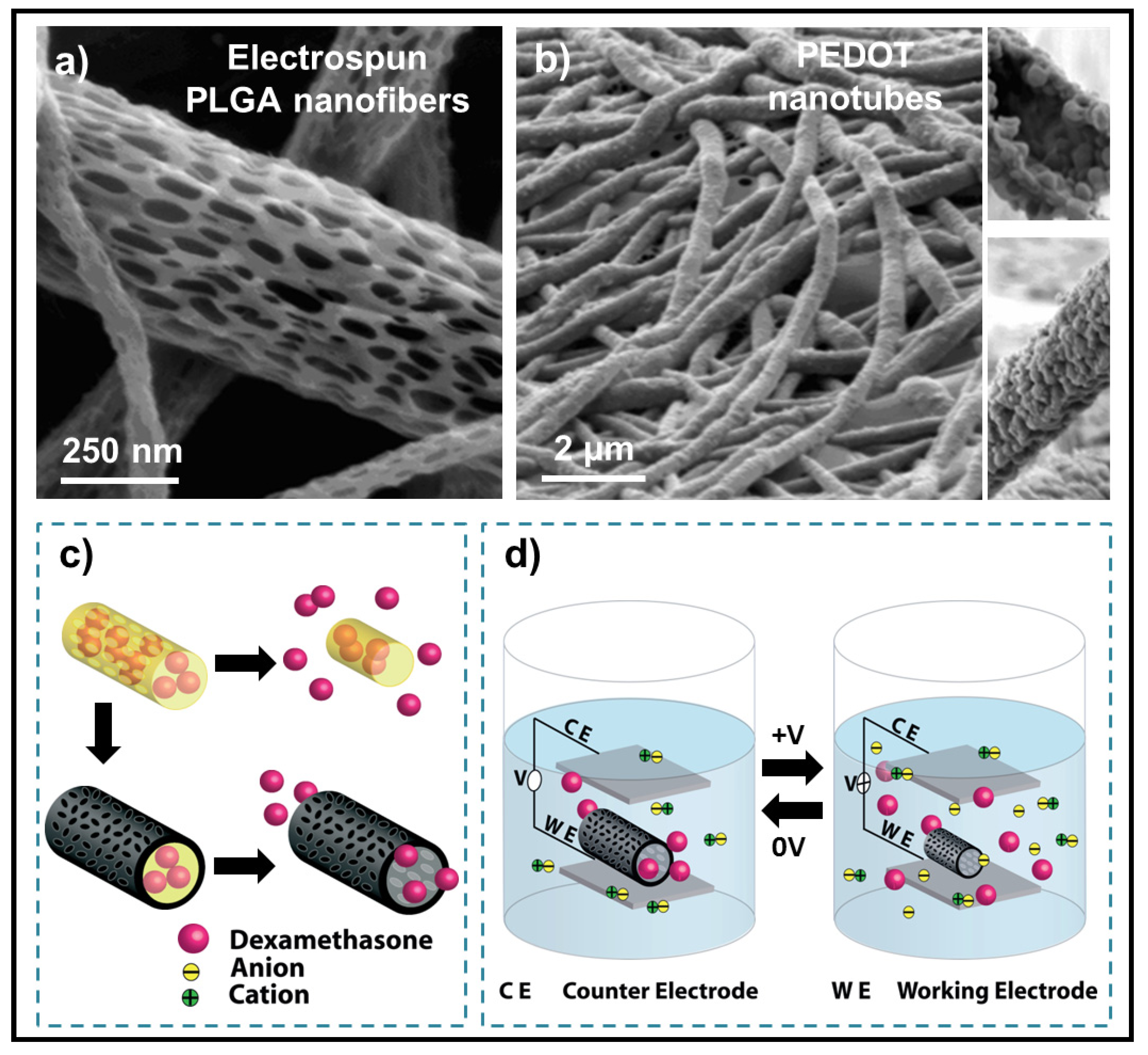
  116. Abidian, M.R.; Kim, D.H.; Martin, D.C. Conducting polymer nanotubes for controlled drug release. Adv. Mater. 2006, 18, 405–409. [Google Scholar] [CrossRef]
  117. Jager, E.W.H.; Inganäs, O.; Lundström, I. Microrobots for micrometer-size objects in aqueous media: Potential tools for single-cell manipulation. Science 2000, 288, 2335–2338. [Google Scholar] [CrossRef]
  118. Kipke, D.R.; Shain, W.; Buzsáki, G.; Fetz, E.; Henderson, J.M.; Hetke, J.F.; Schalk, G. Symposium: Advanced neurotechnologies for chronic neural interfaces: New horizons and clinical opportunities. J. Neurosci. 2008, 28, 11830–11838. [Google Scholar] [CrossRef]
  119. Hochberg, L.R.; Serruya, M.D.; Friehs, G.M.; Mukand, J.A.; Saleh, M.; Caplan, A.H.; Branner, A.; Chen, D.; Penn, R.D.; Donoghue, J.P. Neuronal ensemble control of prosthetic devices by a human with tetraplegia. Nature 2006, 442, 164–171. [Google Scholar] [CrossRef]
  120. Polikov, V.S.; Tresco, P.A.; Reichert, W.M. Response of brain tissue to chronically implanted neural electrodes. J. Neurosci. Meth. 2005, 148, 1–18. [Google Scholar] [CrossRef]
  121. Seymour, J.P.; Kipke, D.R. Neural probe design for reduced tissue encapsulation in CNS. Biomaterials 2007, 28, 3594–3607. [Google Scholar] [CrossRef]
  122. Isaksson, J.; Kjäll, P.; Nilsson, D.; Robinson, N.D.; Berggren, M.; Richter-Dahlfors, A. Electronic control of Ca2+ signalling in neuronal cells using an organic electronic ion pump. Nat. Mater. 2007, 6, 673–679. [Google Scholar] [CrossRef]
  123. George, P.M.; Lyckman, A.W.; LaVan, D.A.; Hegde, A.; Leung, Y.; Avasare, R.; Testa, C.; Alexander, P.M.; Langer, R.; Sur, M. Fabrication and biocompatibility of polypyrrole implants suitable for neural prosthetics. Biomaterials 2005, 26, 3511–3519. [Google Scholar] [CrossRef]
  124. Abidian, M.R.; Ludwig, K.A.; Marzullo, T.C.; Martin, D.C.; Kipke, D.R. Interfacing conducting polymer nanotubes with the central nervous system: Chronic neural recording using poly(3,4-ethylenedioxythiophene) nanotubes. Adv. Mater. 2009, 21, 3764–3770. [Google Scholar] [CrossRef]
  125. Abidian, M.R.; Martin, D.C. Multifunctional nanobiomaterials for neural interfaces. Adv. Funct. Mater. 2009, 19, 573–585. [Google Scholar] [CrossRef]
  126. Sirringhaus, H.; Brown, P.J.; Friend, R.H.; Nielsen, M.M.; Bechgaard, K.; Langeveld-Voss, B.M.W.; Spiering, A.J.H.; Janssen, R.A.J.; Meijer, E.W. Microstructure–mobility correlation in self-organised, conjugated polymer field-effect transistors. Synthetic Met. 2000, 111–112, 129–132. [Google Scholar] [CrossRef]
  127. Liu, H.; Reccius, C.H.; Craighead, H.G. Single electrospun regioregular poly(3-hexylthiophene) nanofiber field-effect transistor. Appl. Phys. Lett. 2005, 87, 253106:1–253106:3. [Google Scholar]
  128. Lee, S.; Moon, G.D.; Jeong, U. Continuous production of uniform poly(3-hexylthiophene) (P3HT) nanofibers by electrospinning and their electrical properties. J. Mater. Chem. 2009, 19, 743–748. [Google Scholar] [CrossRef]
  129. Subramanian, A.; Krishnan, U.M.; Sethuraman, S. Axially aligned electrically conducting biodegradable nanofibers for neural regeneration. J. Mater. Sci. Mater. M 2012, 23, 1797–1809. [Google Scholar] [CrossRef]
  130. Armelin, E.; Gomes, A.L.; Pérez-Madrigal, M.M.; Puiggalí, J.; Franco, L.; del Valle, L.J.; Rodríguez-Galán, A.; de Carvalho Campos, J.S.; Ferrer-Anglada, N.; Alemán, C. Biodegradable free-standing nanomembranes of conducting polymer: Polyester blends as bioactive platforms for tissue engineering. J. Mater. Chem. 2012, 22, 585–594. [Google Scholar] [CrossRef]
  131. Pérez-Madrigal, M.M.; Armelin, E.; del Valle, L.J.; Estrany, F.; Alemán, C. Bioactive and electroactive response of flexible polythiophene: Polyester nanomembranes for tissue engineering. Polym. Chem. 2012, 3, 979–991. [Google Scholar] [CrossRef]
  132. Pérez Madrigal, M.M.; Giannotti, M.I.; Oncins, G.; Franco, L.; Armelin, E.; Puiggalí, J.; Sanz, F.; del Valle, L.J.; Alemán, C. Bioactive nanomembranes of semiconductor polythiophene and thermoplastic polyurethane: Thermal, nanostructural and nanomechanical properties. Polym. Chem. 2013, 4, 568–583. [Google Scholar] [CrossRef]
  133. Teixeira-Dias, B.; del Valle, L.J.; Aradilla, D.; Estrany, F.; Alemán, C. A conducting polymer/protein composite with bactericidal and electroactive properties. Macromol. Mater. Eng. 2012, 297, 427–436. [Google Scholar] [CrossRef]
  134. Bettinger, C.J.; Bruggeman, J.P.; Misra, A.; Borenstein, J.P.; Langer, R. Biocompatibility of biodegradable semiconducting melanin films for nerve tissue engineering. Biomaterials 2009, 30, 3050–3057. [Google Scholar] [CrossRef]
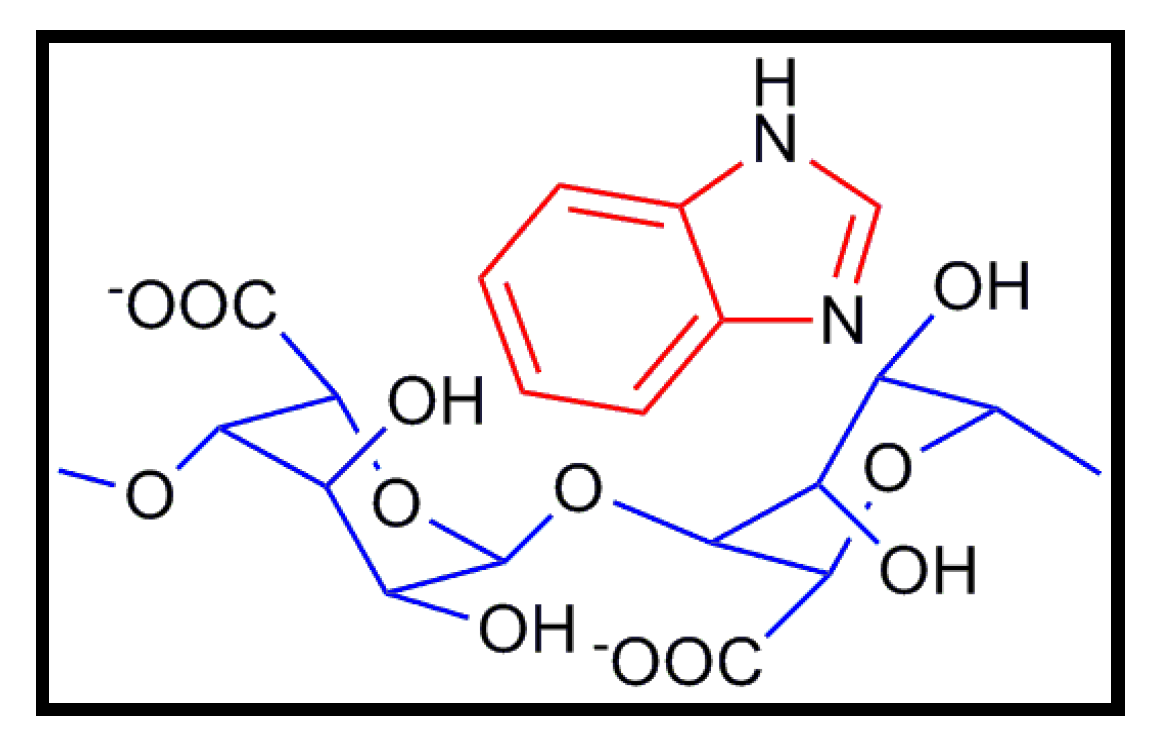
  135. Rachocki, A.; Pogorzelec-Glaser, K.; Pawlaczyk, C.; Tritt-Goc, J. Morphology, molecular dynamics and electric conductivity of carbohydrate polymer film based on algínic acid and benzimidazole. Carbohyd. Res. 2011, 346, 2718–2726. [Google Scholar] [CrossRef]

Article Metrics

Citations

Article Access Statistics

Multiple requests from the same IP address are counted as one view.