Abstract
The aim of this study is to evaluate the suitability of crystalline scintillator LaCl3:Ce for possible use in hybrid medical imaging systems, such as PET/CT and SPECT/CT scanners. For this purpose, a single crystal (10 × 10 × 10 mm3) was irradiated by X-rays within the tube voltage range from 50 to 150 kVp, and the absolute efficiency (AE) was measured experimentally. The energy absorption efficiency (EAE), quantum detection efficiency (QDE), and the spectral compatibility with various optical detectors were also calculated with the use of mathematical formulas. The results were compared with published data for Bi4Ge3O12 (BGO), Lu2SiO5:Ce (LSO), and CdWO4 single crystals of equal dimensions, commonly used in medical imaging applications. The luminescence efficiency values of the examined crystal were found to be higher than those of LSO, BGO, and CdWO4 crystals, within the whole X-ray tube voltage range. In the matter of EAE, LaCl3:Ce demonstrated reduced performance with respect to LSO and CdWO4 crystals. The emission spectrum of LaCl3:Ce was found to be compatible with various types of photocathodes and silicon photomultipliers (SiPMs). Considering these properties, LaCl3:Ce crystal could be considered suitable for use in hybrid medical imaging systems.
1. Introduction
Scintillators are materials particularly important in medical imaging systems, because their use may reduce the ionizing radiation dose to the patient. Scintillation detectors are usually connected to optical sensors, such as film, photocathodes, photodiodes, CCD, a-Si/TFT, and CMOS [1,2,3,4]. The latter setup has been widely employed in several technological fields, from industry up to nuclear physics, but with a prominent application in medical imaging applications such as X-ray imaging, computed tomography (CT), single photon emission computed tomography (SPECT), and positron emission tomography (PET) [4,5,6,7,8]. The sensitivity, which is antagonistic to patient dose, of these systems increases remarkably when more efficient and faster scintillation crystals are utilized [5,8].
Crystal scintillator’s investigation is of major importance in nuclear medicine systems. As an example, crystalline scintillators with halogenated impurities, such as lanthanum chloride (LaCl3) activated with cerium (Ce), has been widely studied in nuclear medicine applications [9,10,11] because of its physical properties, such as a density of 3.86 g/cm3 suitable for radiation absorption, a high light output reported from 40.000 photons/MeV to 49.000 photons/MeV), a decay time of 28 ns, and good energy resolution, which depends upon the crystal physical and chemical properties such as size and cerium concentration [10,12,13,14,15,16,17,18].
The LaCl3:Ce crystal has an hexagonal symmetry in the UCl3 type lattice in the space group P63/m or C26h, with point symmetry C3h at the lanthanide site. The lanthanide coordination polyhedron consists of nine chloride ligands arranged in a tricapped trigonal prism configuration [15,19]. The lattice constant of the crystal is 0.6196 nm [13]. The con-centration of cerium in the crystal has been reported in the literature to be from 0.1% to 30% [11,12,15,20].
Current nuclear medicine instrumentation includes hybrid systems where contemporary imaging systems such as SPECT and PET are combined with X-ray computed tomography scanners (CT) and form hybrid systems such as SPECT/CT and PET/CT. A breakthrough of such a hybrid modality could be the use of the same detector type for CT and SPECT or PET.
Under this consideration, in the current work, the response of a commercially available LaCl3:Ce single crystal scintillator [13] excitation was experimentally examined for X-ray tube voltages in the range from 50 kVp to 150 kVp. The response was examined via (a) the absolute luminescence efficiency (AE), describing the light output power per incident exposure, (b) the spectral matching factor (αs) and the overall efficiency (EE), investigating the suitability of various photodetectors attached to LaCl3:Ce, and (c) the radiation absorption properties of the crystal. After research into the relevant literature, excitation of LaCl3:Ce crystal with X-rays to investigate some of its properties was carried out in specific energy ranges. The crystal’s emission spectrum, after X-ray excitation produced by X-ray tube at 35 kV with 25 mA, has been studied by Guillot-Noel et al. [15]. This study was supplemented by the usage of LaCl3:Ce crystals of different concentrations of cerium, under the same experimental conditions [12]. The emission spectrum of the crystal was also measured at 30 kV with 25 mA [20]. In order to investigate the crystal suitability, mainly for X-ray counting applications, studies of the proportionality of response and the energy resolution of the crystal have also been carried out in the energy ranges 10.5–100 keV [21] and 5–60 keV (X-rays from radioactive sources Fe-55: 5.9 keV, Cd-109: 22 keV, and Am-241: 60 keV) [22]. This work extends the current LaCl3:Ce literature and presents a combined examination of LaCl3:Ce performance in terms of measuring absolute efficiency in clinical utilized X-ray tube voltages, calculating the X-ray energy absorption efficiency and estimating the suitability of LaCl3:Ce in conjunction with commercially available photoreceptors, with the scope of using LaCl3:Ce in hybrid medical imaging systems.
The results of this study were compared with calculated and previously published results for Bi4Ge3O12 (BGO), Lu2SiO5:Ce (LSO), and CdWO4 single crystals that are commonly used in several imaging systems [23,24], and the specific LaCl3:Ce crystal absolute efficiency was found comparable or better.
High light yield (i.e., AE) as well as satisfactory spectral matching to optical sensors (αs and EE) yields scintillator–photodetector combinations which can provide higher signal output and better image quality for a given level of patient exposure. Accordingly, a required quality of output image can be obtained with less radiation burden to the examinee. The latter is important in CT examinations where a lot of X-ray projections are obtained per gantry rotation and scan.
2. Materials and Methods
A single cubic-shaped crystal was purchased from Advatech UK Limited [13]. The cube dimensions were 10 × 10 × 10 mm3. The light yield (LY) of the crystal was 49.000 photons/MeV (provided by the supplier), its density 3.86 g/cm3, and its max emission peak is at 350 nm [13]. The surfaces of the crystal were polished. In addition, the crystal was purchased encapsulated in a thin aluminum protective layer, due to its hygroscopicity, where only one crystal surface, i.e., output, was not encapsulated. Energy absorption efficiency (EAE), quantum detection efficiency (QDE), absolute luminescence efficiency (AE), spectral matching factors (SMF), and effective efficiency (EE) with several optical detectors were determined experimentally or with the use of mathematical formulas. The X-ray flux needed for absolute efficiency calculation was obtained by an X-ray tube coupled with a CPI, series CMP 200DR 50 kW generator. The high voltage ranged from 50 to 150 kVp and the current–time product was kept constant at 63 mA s. The inherent filtration of the X-ray tube was 1.5 mm Al. Furthermore, an additional Al filtration of 20 mm was placed at the tube exit [7,23,24,25].
The quantum detection efficiency (QDE) describes the ability of a scintillator to detect photons and is defined as the fraction of the incident X-rays interacting with the scintillator [26]. The fraction of the incident X-ray energy absorbed in the crystal is described through the energy absorption efficiency (EAE) [26]. EAE and QDE can be calculated as [23,26]:
and
where Φ0(E) is the incident X-ray photon fluence on the scintillator, E is the photon energy, μatt(E)/ρ is the radiation photon total mass attenuation coefficient, and μen(E)/ρ is the corresponding total mass energy absorption coefficient. Τ is the detector thickness and ρ is the density (in g/cm3) [23,26]. The coefficients used in Equations (1) and (2) were obtained from XMudat software [27], and the X-ray fluence from TASMIP Spectra Calculator [28].
The absolute luminescence efficiency is defined as the ratio of the energy flux λ (units μW∙m−2) of the optical photons emitted by a stimulated crystal to the rate of exposure (units mR∙s−1) of the X-rays incident on it [29]. The instrumentation necessary for measuring the optical photon flux comprised an Oriel light integration sphere, an EMI photomultiplier tube, and a Cary electrometer. More details regarding the measurement of AE can be obtained in the literature [7,23,24,25]. According to its definition [24,25],
The units of AE are known as efficiency units (EU), where 1 EU = 1 µW∙m−2/(mR∙s−1).
Crystal scintillators are always combined with optical photon detectors. The performance of such a combination can be estimated by the spectral matching factor αs, which expresses the spectral compatibility of the scintillator’s emitted light to the spectral sensitivity of the photodetector, and it can be defined as [23]
where Sp(λ) is the spectrum of the emitted light by the scintillator, SD(λ) is the spectral sensitivity of the photodetector coupled to the scintillator, and λ is the wavelength of the light emitted [30]. The spectral sensitivity of various photodetectors was obtained from manufacturers’ data and the literature [25,31,32,33,34].
The overall efficiency of a scintillator–photodetector combination has been expressed by the effective efficiency (EE) [35] and is calculated as [8,25]
EE= AE
·αs
3. Results and Discussion
Figure 1 shows values for the energy absorption efficiency of the LaCl3:Ce crystal. These values were compared with calculated data for BGO, LSO, and CdWO4 single crystals of equal dimensions. The EAE values of LaCl3:Ce crystal were lower than BGO, LSO, and CdWO4 in the low-energy range (50 kVp) (0.497 for LaCl3:Ce, 0.840 for BGO, 0.871 for LSO, and 0.714 for CdWO4) as a consequence of the significantly higher density of these materials (7.13, 7.4, and 7.9 g/cm3, respectively) [23,36,37] in regard to the 3.86 g/cm3 of LaCl3:Ce. The deviation between the absorption efficiency values of LaCl3:Ce and those of BGO, LSO, and CdWO4 decreases as the X-ray energy increases. At 70 kVp, LaCl3:Ce shows a tendency to increase, but remains lower than the EAE of LSO and CdWO4 crystals. At 150 kVp, the EAE values of LaCl3:Ce, BGO, LSO, and CdWO4 are approximately 0.584, 0.698, 0.566, and 0.587, respectively. The highest values for LaCl3:Ce were calculated at 140 and 150 kVp, demonstrating that the use of LaCl3:Ce of this thickness favors higher energy radiographic applications such as computed tomography.
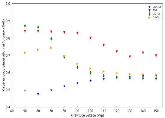
Figure 1.
EAE of the LaCl3:Ce, BGO, LSO:Ce, and CdWO4 single crystals.
The attenuation coefficients, as well as the ratio of the attenuation coefficients μen/μatt used in Equations (1) and (2), are shown in Figure 2 and Figure 3, respectively, for all four materials.
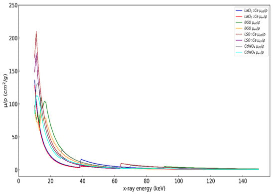
Figure 2.
Attenuation coefficients of the LaCl3:Ce, BGO, LSO, and CdWO4 single crystals.
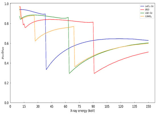
Figure 3.
Attenuation coefficients ratio μen/μatt of the LaCl3:Ce, BGO, LSO, and CdWO4 single crystals.
As shown in Figure 1, the EAE generally decreases when increasing the voltage across the X-ray tube. The ability of a crystal to absorb photons is expressed through the attenuation coefficient μatt and the absorption coefficient μen. μatt expresses the probability of interaction of the radiation with the crystal, which is manifested mainly through photoelectric effect, based on the energies and the effective atomic number (Zeff) of these crystals. μen expresses the probability of absorbing radiation inside the crystal. With increasing voltage in the X-ray tube, the emitted radiation spectrum moves to higher energies. As the energies of the emitted photons increase, the μatt coefficients decrease, as shown in Figure 2, except for some energy values at which some discontinuities occur. These discontinuities are called absorption edges and correspond to binding energies of the K, L, M, and N shells, where the probability of photon absorption through the photoelectric effect is greatly increased due to resonance. In addition, as shown in Figure 3, the ratio μen/μatt at some energy values decreases sharply, and, as a consequence, the EAE of the crystal decreases. This reduction for BGO and LSO:Ce crystals occurs at the K-edges energy values. Specifically, for BGO, this reduction occurs at 90 keV, therefore the EAE of the crystal decreases after 90 kVp with a tendency to stabilize after 120 kVp, due to the observed small recovery of the corresponding curve in Figure 3. For LSO:Ce and CdWO4 crystals, this reduction occurs at 63 KeV and 69 keV, respectively, and, consequently, the EAE of these crystals decreases after 63 kVp and 69 kVp, respectively, while stabilizing after 110 kVp for both crystals at about the same levels as shown in Figure 3. For LaCl3:Ce crystal, the reduction is at 38 keV and the effect in the corresponding curve in Figure 1, although not clearly visible, explains the reduction of EAE from 50 kVp to 60 kVp. The increase of EAE after 60 kVp and its stabilization after 110 kVp, at the same levels as the LSO:Ce and CdWO4 crystals, is explained by the high increase of the μen/μatt ratio of the crystal after 38 keV and its tendency to equate with the values of the μen/μatt ratio of the other two crystals.
Figure 4 illustrates the fluctuation of QDE values with X-ray tube voltage. It is apparent from these values that LaCl3:Ce of thickness 10 mm exhibits almost perfect efficiency to detect the incident photos, as the values range from 0.996 to 1.
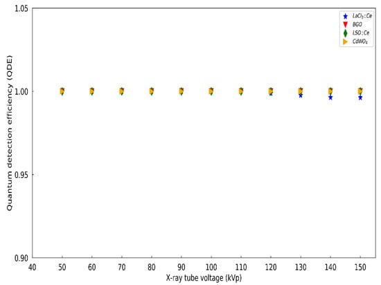
Figure 4.
Quantum detection efficiency of the LaCl3:Ce single crystal in comparison with BGO, LSO, and CdWO4 single crystals.
The variation of the purchased LaCl3:Ce absolute efficiency with X-ray tube voltages 50 to 150 kVp is shown in Figure 5. In Figure 5, the corresponding AE values for LSO, BGO, and CdWO4 crystals of equal dimensions (10 × 10 × 10 mm3) are also demonstrated [23,24]. The AE values of all crystals demonstrate a tendency to increase with increasing of the kVp. However, LaCl3:Ce values were in all cases higher than those of the other crystals. For example, at the tube voltages applied of 70 kVp and 130 kVp, the absolute efficiencies were (a) for 70 kVp: 24.38 EU for LaCl3:Ce, 18.7 EU for CdWO4, 12.4 EU for LSO, and 2.3 EU for BGO; and (b) for 130 kVp: 38.7 EU for LaCl3:Ce, 26.9 EU for CdWO4, 17.7 EU for LSO, and 3.7 EU for BGO [23,24]. The AE results confirm the suitability of LaCl3:Ce crystal for possible use in medical imaging applications, since it exhibits higher values than LSO and BGO single crystals, which are commonly used in such applications. Furthermore, the AE values in higher energies verify the efficiency of LaCl3:Ce crystal in nuclear medicine applications.
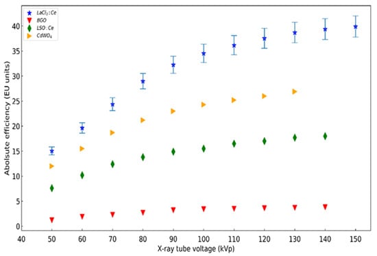
Figure 5.
AE values of the LaCl3:Ce single crystal in comparison with previously published data for BGO, LSO, and CdWO4 single crystals. The error bars correspond to 5.3%.
It is worth commenting that the increased efficiency of LaCl3:Ce, compared to CdWO4, LSO, and BGO, cannot be attributed only to its radiation absorption properties. A reason that can explain its increased absolute efficiency is the additional effect of its higher light yield (LY) 49.000 photons/MeV compared to the 8.900 photons/MeV for BGO crystal, 30.000 photons/MeV for LSO crystal, and 28.000 photons/MeV for CdWO4 scintillator. The total number of the optical photons produced per incident X-ray, L, are due to the combined effect of EAE and LY. In order to theoretically estimate the total number of optical photons produced for several X-ray tube voltages, we calculated the mean energy for the X-ray spectra at 50 kV, 60 kV, 80 kV, 140 kV, and 150 kV as . The corresponding mean energies were calculated as 41.5 keV, 47.2 keV, 56.7 keV, 74.8 keV, and 75.9 keV, respectively. The total number of optical photons produced was calculated as .
In Table 1, the L values of LaCl3:Ce, compared to CdWO4, LSO, and BGO, at 50 kV, 60 kV, 80 kV, 140 kV, and 150 kV are shown.

Table 1.
Theoretical calculation of the number of optical photons generated in the crystal scintillators.
It may be seen that for low X-ray energies, LaCl3:Ce is inferior to LSO in terms of optical photon production, because of the low EAE, due to its low density and despite its higher light yield compared to LSO. For higher energies though, where the EAE differences between LaCl3:Ce and LSO are reduced, as shown in Figure 1, LaCl3:Ce produces more optical photons. In each case, however, the AE values of LaCl3:Ce are superior to the scintillators at the X-ray tube voltages under investigation. This indicates that the optical photon propagation properties, such as transmission through the material and optical escape from the crystal, also play an important role in the total scintillation efficiency. The BGO, LSO, and CdWO4 crystals were wrapped with Teflon layers as part of the irradiation procedure [38]. According to the literature, the reflectivity of the surfaces of polished crystals alone, or in contact with Teflon or specular surfaces, is close to 100% for optical photon angle of incidence over 30 degrees [39].
Figure 6, Figure 7, Figure 8 and Figure 9 illustrate the normalized optical spectrum of LaCl3:Ce crystal along with the spectral sensitivities of various optical sensors [25,31,32,33,34]. The LaCl3:Ce spectrum, obtained from the vendor’s website [13], shows the main luminescence peak at 350 nm [13,14].
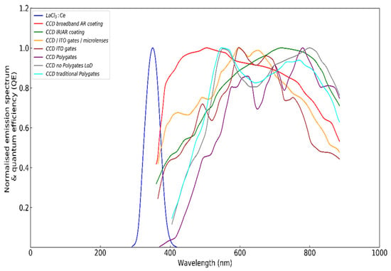
Figure 6.
Normalized emitted light spectrum of the LaCl3:Ce crystal and spectral sensitivity of various charge-coupled devices.
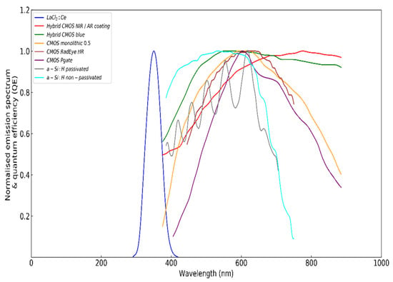
Figure 7.
Normalized emitted light spectrum of the LaCl3:Ce crystal and spectral sensitivity of various complementary metal–oxide semiconductors.
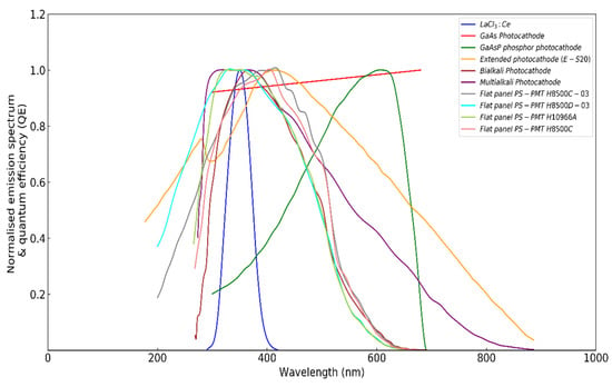
Figure 8.
Normalized emitted light spectrum of the LaCl3:Ce crystal and spectral sensitivity of various photocathodes.
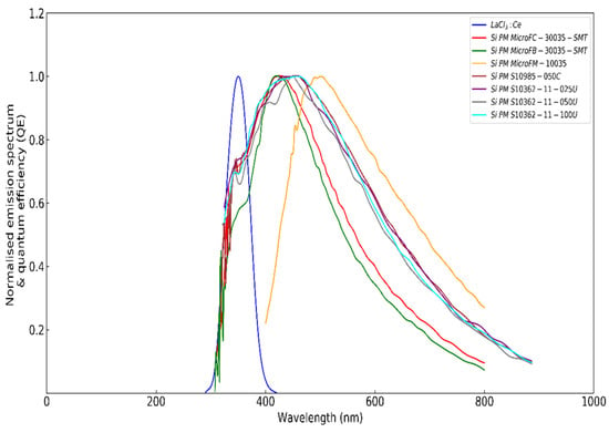
Figure 9.
Normalized emitted light spectrum of the LaCl3:Ce crystal and spectral sensitivity of various silicon photomultipliers.
The spectral matching factor values for LaCl3:Ce, along with several optical detectors, were calculated according to Equation (4). These optical detectors, which are shown in Figure 6, Figure 7, Figure 8 and Figure 9, were silicon photomultipliers (SiPMs) utilized in nuclear medicine techniques, charge-coupled devices (CCD), and complementary metal–oxide semiconductors (CMOS) used in imaging applications, as well as various photocathodes. It was found that LaCl3:Ce exhibits excellent compatibility with multialkali photocathode (0.99), GaAs photocathode (0.93), and bialkali photocathode (0.94). Moreover, it exhibits exceptional compatibility when coupled with various flat panel (FP) photocathodes, with the SMF values fluctuating from 0.91 to 0.99 (0.99 for H8500D-03 and H10966A). On the other hand, poor compatibility was registered with the GaAsP phosphor photocathode, since the SMF value was only 0.27. LaCl3:Ce also showed good compatibility with most of the silicon photomultipliers used in our experiments, as it showed SMF values in the range from 0.62 to 0.67 (0.67 for Si PM S10985-050C and Si PM S10362-11-025U). On the contrary, LaCl3:Ce was found to be incompatible with CCDs and CMOS detectors. Specifically, the lowest SMF values were registered for CCD with polygates (0.002), for CMOS RadEye HR (0.0), for CMOS Pgate (0.0002), and for CCD with traditional polygates and CCD no polygates (both at 0.0003). The combinations with SMF greater than 0.60 are shown in Table 2.

Table 2.
LaCl3:Ce photodetector combinations with SMF above 0.60.
Figure 10, Figure 11, Figure 12 and Figure 13 illustrate the effective luminescence efficiency of the LaCl3:Ce crystal with the optical detectors shown in Figure 6, Figure 7, Figure 8 and Figure 9. The optimum effective efficiency values were attributed to photocathodes and silicon photomultipliers (SiPMs). The lowest values are obtained when coupled with CCDs and CMOS optical sensors. In detail, EE values almost equal to the AE ones are obtained (~15 EU at 50 kVp and ~39 EU at 150 kVp) when LaCl3:Ce is coupled with GaAs photocathode, bialkali photocathode, multialkali photocathode, flat panel PS-PMT H8500D-03, and flat panel PS-PMT H10966A. On the contrary, the decrease (with kVp) in the detected luminescence signal varied from 99.7% to 100% when LaCl3:Ce was combined with CMOS RadEye HR, CMOS Pgate, CCD with polygates, CCD no polygates LoD, and CCD with traditional polygates.
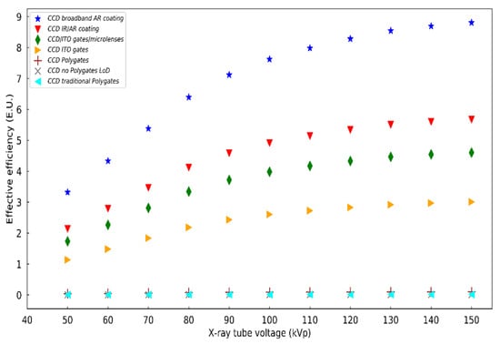
Figure 10.
Effective efficiency of the LaCl3:Ce crystal combined with various charge-coupled devices.
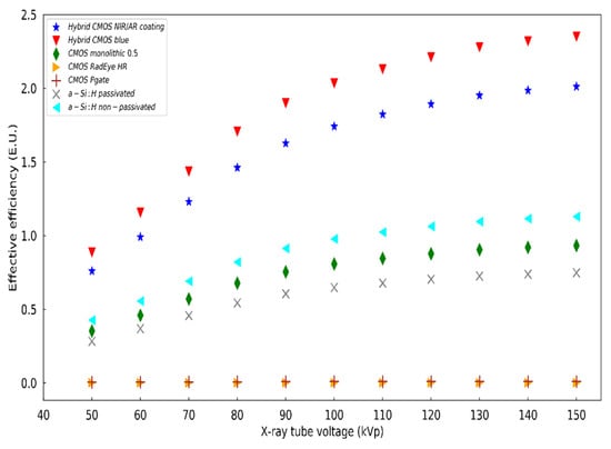
Figure 11.
Effective efficiency of the LaCl3:Ce crystal combined with various complementary metal–oxide semiconductors.
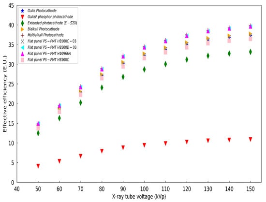
Figure 12.
Effective efficiency of the LaCl3:Ce crystal combined with various photocathodes.
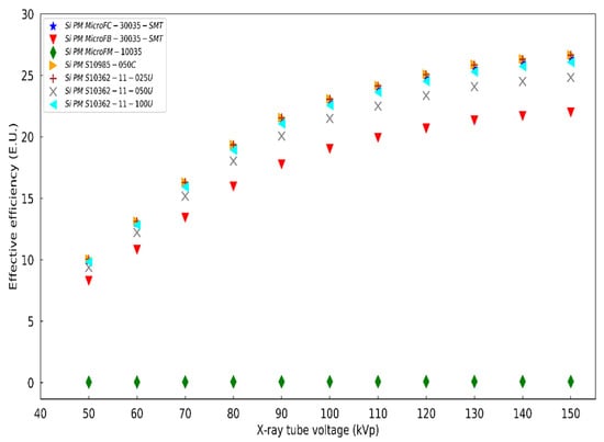
Figure 13.
Effective efficiency of the LaCl3:Ce crystal combined with various silicon photomultipliers.
4. Conclusions
The absolute luminescence efficiency and the spectral matching of a LaCl3:Ce crystal were examined within the tube voltage range (50–150 kVp) employed in X-ray imaging applications. The results were compared with previously published data for LSO, BGO, and CdWO4 single crystals of equal dimensions, commonly utilized in commercial imaging systems. Peak absolute luminescence efficiency of LaCl3:Ce crystal was obtained at 150 kVp (39.9 EU) The luminescence efficiency values of the examined crystal were found to be higher than those of LSO, BGO, and CdWO4 crystals, within the whole X-ray tube voltage range. In terms of EAE, LaCl3:Ce demonstrated reduced performance with respect to LSO and CdWO4 crystals. The spectral compatibility with several commercial optical detectors was also investigated. The emission spectrum of LaCl3:Ce was found to be compatible with various types of photocathodes and silicon photomultipliers (SiPMs).
A high-efficiency detector for hybrid medical imaging systems such as SPECT/CT and PET/CT would require reduced pharmaceutical administered activity for a given signal output. In addition, for the X-ray part of the system, reduced radiation translated to reduced current in the CT generator circuit, would be required. More specifically, if absolute efficiency measurements are considered, for the 140 kV X-ray irradiation conditions, LaCl3:Ce can provide the same output as LSO and BGO with 46% and 10% of radiation exposure, respectively. Thus, a scintillator such as LaCl3:Ce with optimized exposure conditions is expected to reduce patient exposure as well as the aforementioned operational costs of the medical installation.
Considering these properties and the previous studies examining LaCl3:Ce crystal for use in nuclear medicine applications, LaCl3:Ce crystal could be considered suitable for use in hybrid medical imaging systems, such as PET/CT and SPECT/CT scanners.
Author Contributions
Conceptualization, S.T., C.M. and N.K.; methodology, S.T., C.M., I.V., G.F. and N.K.; software, S.T. and I.V.; validation, K.N., G.F. and I.K.; formal analysis, S.T., C.M., I.V. and A.B.; investigation, S.T., I.K., A.B. and K.N.; resources, C.M., I.V. and G.F.; data curation, I.V. and N.K.; writing—original draft preparation, S.T., C.M. and N.K.; writing—review and editing, S.T., C.M., I.K. and N.K.; visualization, C.M. and N.K.; supervision, N.K.; project administration, C.M. and N.K. All authors have read and agreed to the published version of the manuscript.
Funding
This research received no external funding.
Conflicts of Interest
The authors declare no conflict of interest.
References
- Mineev, O.V.; Kudenko, Y.G.; Musienko, Y.V.; Polyansky, I.; Yershov, N. Scintillator detectors with long WLS fibers and multi-pixel photodiodes. J. Instrum. 2011, 6, P12004. [Google Scholar] [CrossRef] [Green Version]
- Vandenbroucke, A.; Foudray, A.M.; Olcott, P.D.; Levin, C.S. Performance characterization of a new high resolution PET scintillation detector. Phys. Med. Biol. 2010, 55, 5895–5911. [Google Scholar] [CrossRef] [PubMed] [Green Version]
- Cha, B.K.; Kim, J.; Kim, Y.; Yun, S.; Cho, G.; Kim, H.; Seo, C.; Jeon, S.; Huh, Y. Design and image-quality performance of high resolution CMOS-based X-ray imaging detectors for digital mammography. J. Instrum. 2012, 7, C04020. [Google Scholar] [CrossRef]
- Nassalski, A.; Kapusta, M.; Batsch, T.; Wolski, D.; Mockel, D.; Enghardt, W.; Moszynski, M. Comparative Study of Scintillators for PET/CT Detectors. IEEE Trans. Nucl. Sci. 2007, 54, 3–10. [Google Scholar] [CrossRef]
- Nikl, M. Scintillation Detectors for X-rays. Meas. Sci. Technol. 2006, 17, R37. [Google Scholar] [CrossRef]
- Valais, I.; Kandarakis, I.; Nikolopoulos, D.; Sianoudis, I.; Dimitropoulos, N.; Cavouras, D.; Nomicos, C.; Panayiotakis, G.S. Luminescence efficiency of Gd2SiO5:Ce scintillator under x-ray excitation. IEEE Trans. Nucl. Sci. 2005, 52, 1830–1835. [Google Scholar] [CrossRef]
- Valais, I.; Kandarakis, I.; Nikolopoulos, D.; Michail, C.; David, S.; Loudos, G.; Cavouras, D.; Panayiotakis, G.S. Luminescence properties of (Lu,Y)2SiO5:Ce and Gd2SiO5:Ce single crystal scintillators under X-ray excitation for use in medical imaging systems. IEEE Trans. Nucl. Sci. 2007, 54, 11–18. [Google Scholar] [CrossRef]
- Valais, I. Systematic Study of the Light Emission Efficiency and the Corresponding Intrinsic Physical Characteristics Of Single Crystal Scintillators, Doped with the Trivalent Cerium (Ce3+) Activator, in Wide Energy Range (from 20 kV–18 MV) for Medical Applications. Ph.D. Thesis, University of Patras, Patras, Greece, 2008. [Google Scholar]
- Alzimami, K.; Abuelhia, E.; Podolyak, Z.; Ioannou, A.; Spyrou, N.M. Characterization of LaBr3:Ce and LaCl3:Ce scintillators for gamma-ray spectroscopy. J. Radioanal. Nucl. Chem. 2008, 278, 755–759. [Google Scholar] [CrossRef]
- Balcerzyk, M.; Moszynski, M.; Kapusta, M. Comparison of LaCl3:Ce and NaI(Tl) scintillators in γ-ray spectrometry. Nucl. Instrum. Methods Phys. Res. A 2005, 537, 50–56. [Google Scholar] [CrossRef]
- Shah, K.S.; Glodo, J.; Klugerman, M.; Cirignano, L.J.; Moses, W.; Derenzo, S.E.; Weber, M.J. LaCl3:Ce scintillator for γ-ray detection. Nucl. Instrum. Methods Phys. Res. Sect. A-Accel. Spectrometers Detect. Assoc. Equip. 2003, 505, 76–81. [Google Scholar] [CrossRef]
- Van Loef, E.V.D.; Dorenbos, P.; Van Eijk, C.W.E.; Kramer, K.; Gudel, H.U. Scintillation properties of LaCl3:Ce3+ crystals: Fast, efficient and high-energy-resolution scintillators. In Proceedings of the 2000 IEEE Nuclear Science Symposium. Conference Record (Cat. No.00CH37149), Lyon, France, 15–20 October 2000; Volume 1, pp. 6/31–6/34. [Google Scholar] [CrossRef]
- Advatech UK. LaCl3:Ce—Lanthanum Chloride (Ce). Available online: https://www.advatech-uk.co.uk/lacl3_ce.html (accessed on 25 June 2021).
- Iltis, A.; Mayhugh, M.R.; Menge, P.; Rozsa, C.M.; Selles, O.; Solovyev, V. Lanthanum halide scintillators: Properties and applications. Nucl. Instrum. Methods Phys. Res. Sect. A Accel. Spectrometers Detect. Assoc. Equip. 2006, 563, 359–363. [Google Scholar] [CrossRef]
- Guillot-Noel, O.; Haas, J.T.; Dorenbos, P.; Eijk, C.W.; Krämer, K.W.; Güdel, H.U. Optical and scintillation properties of cerium-doped LaCl3, LuBr3 and LuCl3. J. Lumin. 1999, 85, 21–35. [Google Scholar] [CrossRef]
- Moszynski, M.; Nassalski, A.; Syntfeld-Każuch, A.; Szczęśniak, T.; Czarnacki, W.; Wolski, D.; Pausch, G.; Stein, J. Temperature dependences of LaBr3(Ce), LaCl3(Ce) and NaI(Tl) scintillators. Nucl. Instrum. Methods Phys. Res. Sect. A Accel. Spectrometers Detect. Assoc. Equip. 2006, 568, 739–751. [Google Scholar] [CrossRef]
- Van Eijk, C.W.E. Inorganic scintillators in medical imaging. Phys. Med. Biol. 2002, 47, R85–R106. [Google Scholar] [CrossRef] [PubMed]
- Van Eijk, C.W.E. Inorganic-scintillator development. Nucl. Instrum. Methods Phys. Res. Sect. A Accel. Spectrometers Detect. Assoc. Equip. 2001, 460, 1–14. [Google Scholar] [CrossRef]
- Antonyak, O.T.; Dorenbos, P.; Rodnyi, P.; Stryganyuk, G.B.; Voloshinovskii, A.S.; Zimmerer, G. Luminescence Spectra of LaCl3-Ce Crystals; Hamburger HASYLAB at DESY: Hamburg, Germany, 2000. [Google Scholar]
- Higgins, W.M.; Glodo, J.; Van Loef, E.V.D.; Klugerman, M.; Gupta, T.; Cirignano, L.; Wong, P.; Shah, K.S. Bridgman growth of LaBr3:Ce and LaCl3:Ce crystals for high-resolution gamma-ray spectrometers. J. Cryst. Growth 2006, 287, 239–242. [Google Scholar] [CrossRef]
- Owens, A.; Bos, A.J.J.; Brandenburg, S.; Dorenbos, P.; Drozdowski, W.; Ostendorf, R.W.; Quarati, F.; Webb, A.; Welter, E. The hard X-ray response of Ce-doped lanthanum halide scintillators. Nucl. Instrum. Methods Phys. Res. A 2007, 574, 158–162. [Google Scholar] [CrossRef] [Green Version]
- Martin, T.; Allier, C.; Bernard, F. Lanthanum Chloride Scintillator for X-ray Detection. AIP Conf. Proc. 2007, 879, 1156–1159. [Google Scholar] [CrossRef]
- Michail, C.; Koukou, V.; Martini, N.; Saatsakis, G.; Kalyvas, N.; Bakas, A.; Kandarakis, I.; Fountos, G.; Panayiotakis, G.; Valais, I. Luminescence Efficiency of Cadmium Tungstate (CdWO4) Single Crystal for Medical Imaging Applications. Crystals 2020, 10, 429. [Google Scholar] [CrossRef]
- Michail, C.; Kalyvas, N.; Bakas, A.; Ninos, K.; Sianoudis, I.; Fountos, G.; Kandarakis, I.; Panayiotakis, G.; Valais, I. Absolute Luminescence Efficiency of Europium-Doped Calcium Fluoride (CaF2:Eu) Single Crystals under X-ray Excitation. Crystals 2019, 9, 234. [Google Scholar] [CrossRef] [Green Version]
- Linardatos, D.; Konstantinidis, A.; Valais, I.; Ninos, K.; Kalyvas, N.; Bakas, A.; Kandarakis, I.; Fountos, G.; Michail, C. On the Optical Response of Tellurium Activated Zinc Selenide ZnSe:Te Single Crystal. Crystals 2020, 10, 961. [Google Scholar] [CrossRef]
- Boone, J. X-ray production, interaction, and detection in diagnostic imaging. In Handbook of Medical Imaging. Volume 1: Physics and Psychophysics; Beutel, J., Kundel, H.L., Van Metter, R.L., Eds.; SPIE Press: Bellingham, WA, USA, 2000; Volume 1, pp. 36–57. ISBN 978-0-8194-7772-9. [Google Scholar]
- Nowotny, R. XMuDat: Photon Attenuation Data on PC (IAEA-NDS-195); International Atomic Energy Agency: Vienna, Austria, 1998. [Google Scholar]
- TASMIP Spectra Calculator. Available online: https://www.solutioinsilico.com/medical-physics/applications/tasmip-app.php?ans=0 (accessed on 14 March 2022).
- Kandarakis, I.; Cavouras, D.; Panayiotakis, G.S.; Nomicos, C.D. Evaluating X-ray detectors for radiographic applications: A comparison of ZnSCdS:Ag with Gd2O2S:Tb and Y2O2S:Tb screens. Phys. Med. Biol. 1997, 42, 1351–1373. [Google Scholar] [CrossRef] [PubMed]
- Kandarakis, I.; Cavouras, D.; Nomicos, C.D.; Panayiotakis, G.S. X-ray luminescence of ZnSCdS:Au, Cu phosphor using X-ray beams for medical applications. Nucl. Instr. Meth. Phys. Res. B 2001, 179, 215–224. [Google Scholar] [CrossRef]
- Hamamatsu Photonics, MPPC (Multi-Pixel Photon Counters). Available online: http://www.hamamatsu.com/us/en/product/category/3100/4004/4113/index.html# (accessed on 3 July 2021).
- Silicon Photomultipliers. Available online: http://sensl.com/products/silicon-photomultipliers/ (accessed on 3 July 2021).
- Magnan, P. Detection of visible photons in CCD and CMOS: A comparative view. Nucl. Instrum. Meth. Phys. Res. A 2003, 504, 199–212. [Google Scholar] [CrossRef]
- Rowlands, J.A.; Yorkston, J. Flat Panel Detectors for Digital Radiography. In Handbook of Medical Imaging Physics and Psychophysics; Beutel, J., Kundel, H., Van Metter, R., Eds.; SPIE: Bellingham, WA, USA, 2000; Volume 1, pp. 223–328. ISBN 978-08-194-7772-9. [Google Scholar]
- Cavouras, D.; Kandarakis, I.; Panayiotakis, G.S.; Bakas, A.; Triantis, D.; Nomicos, C.D. An experimental method to determine the effective efficiency of scintillator-photodetector combinations used in x-ray medical imaging systems. Br. J. Radiol. 1998, 71, 766–772. [Google Scholar] [CrossRef] [PubMed]
- Advatech UK. LSO:Ce—Lutetium Oxyorthosilicate (Ce). Available online: https://www.advatech-uk.co.uk/lso_ce.html (accessed on 25 June 2021).
- Advatech UK. BGO—Bismuth Germanate Scintillator Crystal. Available online: https://www.advatech-uk.co.uk/bgo.html (accessed on 25 June 2021).
- Valais, I.; David, S.; Michail, C.; Nikolopoulos, D.; Vattis, D.; Sianoudis, I.; Cavouras, D.; Nomicos, C.; Kandarakis, I.; Pa-nayiotakis, G. Comparative study of luminescence properties of Lu2SiO5:Ce and YAIO3:Ce single crystal scintillators for use in medical imaging. In Proceedings of the 5th European Symposium on Biomedical Engineering, Patras, Greece, 5–7 July 2006. [Google Scholar]
- Roncali, E.; Stockhoff, M.; Cherry, S.R. An integrated model of scintillator-reflector properties for advanced simulations of optical transport. Phys. Med. Biol. 2017, 62, 4811–4830. [Google Scholar] [CrossRef]
Publisher’s Note: MDPI stays neutral with regard to jurisdictional claims in published maps and institutional affiliations. |
© 2022 by the authors. Licensee MDPI, Basel, Switzerland. This article is an open access article distributed under the terms and conditions of the Creative Commons Attribution (CC BY) license (https://creativecommons.org/licenses/by/4.0/).