1. Introduction
A pearl is a variety of gemstone with a unique nacreous luster forming in mussels, consisting mainly of aragonite (CaCO3) and minor organic materials (usually under 5%). A cultured freshwater nucleated pearl is formed by inserting a selected piece of mantle tissue from a sacrificial mussel, together with a bead nucleus into a partly opened mussel. Currently, about 95% of freshwater pearl production comes from China. In recent years, large freshwater nucleated pearls cultured by some Chinese pearl companies have become sought after in the international jewelry market for their amazing size (diameter above 10 mm) and charming luster.
Luster and surface roughness are two important indicators of the grading and evaluation of pearl appearance quality. However, regarding the new emerging Chinese large freshwater nucleated pearl, the intrinsic incentives to form different appearance quality characteristics and the influence of internal structure on both the luster and surface roughness deserve to be discussed and analyzed. Previous studies mainly focused on the characteristics and formation mechanism of the microstructure in other different varieties of pearls [
1,
2,
3,
4,
5,
6,
7,
8,
9,
10,
11,
12,
13]. Some investigations also discussed the luster formation from the aspect of chemical composition and microstructure [
14,
15,
16,
17,
18,
19]. Nevertheless, pores can be created during the formation of pearls and lead to a change in microstructure. The influence of the nacre layer’s porosity on the visual appearance of Chinese large freshwater nucleated pearls, which determines the quality grade, needs deeper investigation.
In this study, three natural freshwater nucleated pearls were selected as study samples from dozens of pearls. All samples were collected from Zhuji pearl market (Shaoxing, China) which is the largest international trade center of freshwater pearls. Light reflectivity, surface height unevenness parameters and porosity of the nacre layer were measured by chroma meter, laser scanning confocal microscope and X-ray computed tomography (μ-CT), which quantitatively described the characteristics of luster, surface roughness and the structure compactness of the nacre layer. Related results can help establish the quantitative relationship between visual appearance and internal structure, as well as provide a reference for the scientific and quantitative evaluation of pearl quality.
2. Materials and Methods
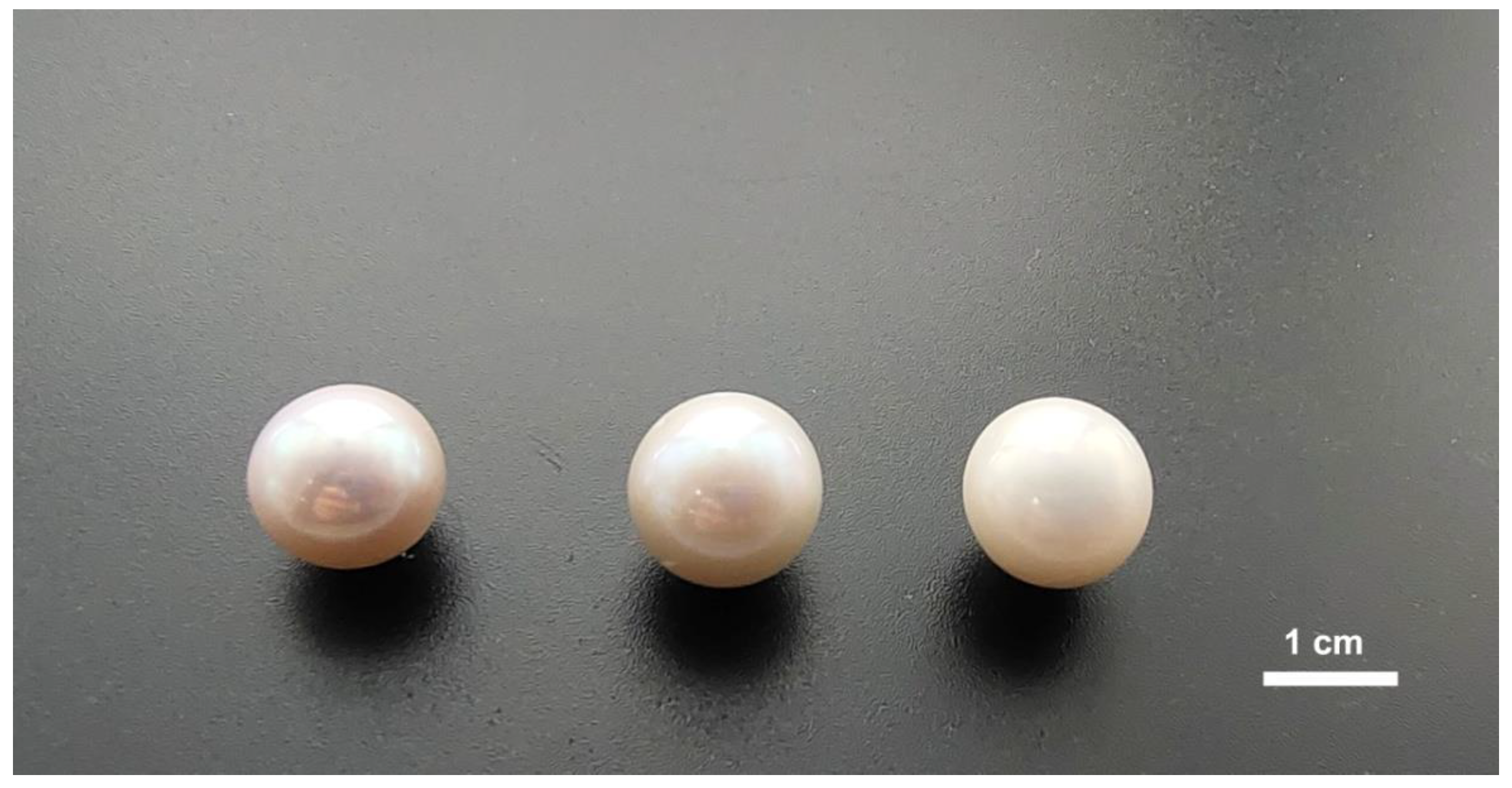
Three white freshwater nucleated pearls of differing quality were selected from dozens of pearls for the investigated samples (
Figure 1). According to the quality grading in trade, which is based on the appearance of pearls, samples with the grades of ‘good’, ‘medium’ and ‘poor’ were named H-1, Z-1 and C-1, respectively. All investigated pearl samples were of the same size (12 mm diameter).
The luster of pearl samples was measured by chroma meter CS-200 under D65 illuminant (simulating sunlight) and reflected by the surface reflectivity. The lightness value of incident light was measured and recorded for reference before the sample test. The brightest spot with 1mm diameter of each sample was selected and measured three times as test values. The surface reflectivity of the pearl samples was calculated by the ratio of the test lightness value and reference value. Laser scanning confocal microscope OLS5000-SAF was used to observe the surface appearance features and calculate roughness parameters, with the resolution of 1 nm and a 405 nm laser light source. The value of surface height unevenness evaluation parameters Sa (the standard deviation of the height distribution) and Sq (arithmetic mean of height difference) were obtained through its analysis software. Porosity of the nacre layer was analyzed by μ-CT scanner (nanoVoxel 1000) and its analysis software, with a spatial resolution of 5 μm.
3. Results
3.1. Luster Measurement
For pearls, the luster mainly depends on the surface reflectivity of incident light [
20]. In this study, average surface reflectivity of the three measurements was used to quantitatively describe the luster of investigated samples. From the results, pearls of different quality grades show clear discrepancy in the surface reflectivity. The values of the ‘good grade’ pearl range between 60% and 70% while those of the ‘poor grade’ fluctuates around 40%. However, the ‘medium grade’ pearl revealed values between 50% and 60%. The results show that the sample of higher quality grade and stronger luster has higher surface reflectivity (
Table 1).
3.2. Surface Roughness
Observed under laser scanning confocal microscope, the three investigated samples showed different surface features (
Figure 2). The higher the quality grade, the fewer the surface blemishes. The surface appearance grade of investigated samples can be described as ‘clean’ (H-1), ‘moderately spotted’ (Z-1) and ‘heavily spotted’ (C-1).
By the calculation and analysis of surface roughness, parameters Sa and Sq both decrease from sample C-1 to sample H-1 (
Table 2). This illustrates that the surface becomes unevener and rougher from ‘good grade’ to ‘poor grade’ pearls, which is consistent with the observation characteristics.
3.3. Porosity of the Nacre Layer
Through X-ray computed tomography, μ-CT images can show nano to micron internal microstructure characteristics of pearl samples. From the cross-section images (
Figure 3), there are pores of various sizes observable in the internal structure. Furthermore, the porosity of the nacre layer can be calculated by using the analysis software of the μ–CT scanner (
Table 3). Comparing the calculation results of porosity with the cross-section images, sample H-1 displays the lowest porosity value while sample C-1 shows the highest porosity value. The porosity difference between samples of adjacent grades (e.g., C-1 and Z-1, Z-1 and H-1) is about an order of magnitude (10
−1).
4. Discussion
A freshwater nucleated pearl is formed by nacre secretions overlying an implanted bead nucleus in the mussel. The gradual accumulation of nacrum leads to a large number of alternating layers of crystalline aragonite (CaCO
3) and biopolymer (e.g., conchiolin) with a brick-and-mortar structure (
Figure 4) [
18,
19].
During the growing process, the adhesion, wrapping and sealing of foreign matter (e.g., inflammatory cells group and tissue fragments) destroys the uniformity of the nacrum and decreases the bonding tightness of aragonite crystal platelets [
21,
22], which can reduce the compactness and increase the porosity of the nacre layer. Moreover, the μ-CT images and data illustrate that there are more pores in the nacre layer of the pearls with lower quality grade. The loosening of the internal microstructure can facilitate the generation of pits/bumps on the surface and increase the surface roughness, which will result in a decrease of light reflectivity on the surface and weakening of the luster. Therefore, it can be inferred that the appearance of pearls is a reflection of their internal microstructure. The porosity degree of the nacre layer bears great influence on the visual appearance quality.
5. Conclusions
Chinese large freshwater nucleated pearls with different quality grades show discrepancies in luster, surface roughness and structure compactness of the nacre layer. Through visual observation, pearls with higher quality grades have stronger luster and fewer surface blemishes. After quantitative measurement, such pearls showed higher light reflectivity, lower surface unevenness parameters and smaller porosity of the nacre layer.
Therefore, structural characteristics of the nacre layer have a significant influence on the optical appearance and surface roughness of Chinese large freshwater nucleated pearls. The higher the porosity of the nacre layer, the more blemishes on the surface (rougher), and the weaker the luster of pearl. This investigation preliminarily discussed and established the relationship between the porosity of the nacre layer, surface roughness and luster of Chinese large freshwater nucleated pearl. Related results can provide reference for the scientific and quantitative evaluation of pearl quality.
Author Contributions
Investigation, D.Z., R.S., T.L.; writing—original draft preparation, D.Z.; writing—review and editing, D.Z., T.L., R.S., J.Z.; visualization, D.Z.; supervision, T.L., J.Z.; project administration, D.Z.; funding acquisition, D.Z., T.L. All authors have read and agreed to the published version of the manuscript.
Funding
This research was funded by the National Science Foundation of China (42073008) and by NGTC Scientific Research Fund (NGTC20210700).
Institutional Review Board Statement
Not applicable.
Informed Consent Statement
Not applicable.
Data Availability Statement
Data available on request due to privacy restrictions. The data provided in this study can be obtained at the request of the corresponding author. As the data needs further research, the data is currently not publicly available.
Acknowledgments
Thanks are given to Weijian Zhan, president of Jiali Pearl Co. Ltd. for providing investigated samples, and Zonglin Wu, president of Sanying Precision Instruments Co. Ltd. for his assistance in the μ-CT measurement. We are also grateful to all reviewers and editors for their constructive and helpful comments, which significantly improved the manuscript.
Conflicts of Interest
The authors declare no conflict of interest.
References
- Scarratt, K.; Moses, T.M.; Akamatsu, S. Characteristics of nuclei in Chinese freshwater cultured pearls. Gems Gemol. 2000, 36, 98–109. [Google Scholar] [CrossRef]
- Zhang, E.; Peng, M.S.; Liang, C.L. Electron microscopic study of the nanometer minerals and microstructure of marine cultured pearls. Acta Mineral. Sin. 2008, 28, 112–116. [Google Scholar]
- Krzemnicki, M.S.; Friess, S.D.; Chalus, P.; Hänni, H.A.; Karampelas, S. X-ray computed microtomography: Distinguishing natural pearls from beaded andnon-beaded cultured pearls. Gems Gemol. 2010, 46, 128–134. [Google Scholar] [CrossRef]
- Yan, X.H. Studies on the Nanostructure of Nacre from Haliotis rufescens, Pinctada maxima and Hyriopsis cumingii. Master’s Thesis, Zhejiang University, Hangzhou, China, 2012. [Google Scholar]
- Fu, F.; Tian, L.G.; Tao, J.B.; Cheng, Y.F.; Hu, X.B.; Xu, X.G. Study on the structure of freshwater cultured pearls with different body colors. J. Synth. Cryst. 2013, 42, 869–874. [Google Scholar]
- Ma, H.Y.; Huang, B.; Zhang, B.L.; Yang, L.X.; Shen, M.D. Some new knowledge of the integrated model of cultured pearl’s internal structure. Acta Mieralogica Sin. 2013, 33, 45–48. [Google Scholar]
- Yan, J.; Tao, J.B.; Ren, Y.Y.; Wang, M.Q.; Hu, X.C.; Wang, X.X. Structure on microstructure and UV-Vis spectra characteristics of natural color golden seawater cultured pearl. Acta Opt. Sin. 2014, 34, 0416005-1–0416005-6. [Google Scholar] [CrossRef]
- Karampelas, S.; Al-Alawi, A.T.; Al-Attawi, A. Real-time microradiography of pearls: A comparison between detectors. Gems Gemol. 2017, 53, 452–456. [Google Scholar] [CrossRef]
- Li, X. Research on Gemological, Mineralogical and Microstructural Characteristics of Beaded Freshwater Cultured Pearls from Zhuji, Zhejiang Province. Master’s Thesis, China University of Geosciences, Beijing, China, 2017. [Google Scholar]
- Yazawa, E.; Zhou, C. 3D Reconstruction of the Internal Structures of Pearls. Gems Gemol. 2018, 54, 294. [Google Scholar]
- Jiang, Q. Analysis of Metal Element Content and Internal Structure Difference of Pearls with Different Colors. Master’s Thesis, Shanghai Ocean University, Shanghai, China, 2019. [Google Scholar]
- Zhang, L.; Liu, Y.X. Comparative study on the structure of freshwater nuclear pearls and non-nuclear pearls. J. Chin. Electron. Microsc. Soc. 2020, 39, 323–330. [Google Scholar]
- Fang, B.; Yan, X.J.; Sun, Q.; Wu, J.Y.; Li, S.H.; Yan, J. Study on the unique mineral microstructure of seawater cultured gray akoya peal by SEM, FTIR and reflection spectroscopy. Rock Miner. Anal. 2021, 40, 42–49. [Google Scholar]
- Cao, L.J.; Guo, S.G.; Shi, L.Y. Study on the relationship between luster and surface structure of pearl. J. Gems Gemmol. 2005, 7, 23–25. [Google Scholar]
- Li, Q.M. New Structural Model of Nacre and Its Photonic Iridescence. Master’s Thesis, Guangxi University, Naning, China, 2012. [Google Scholar]
- Monarumit, N.; Noirawee, N.; Phlayrahan, A.; Promdee, K.; Won-in, K.; Satitkunea, S. Structural analysis of freshwater-cultured pearls with different lusters using the extended X-ray absorption fine structure technique. J. Appl. Spectrosc. 2016, 83, 298–301. [Google Scholar] [CrossRef]
- Li, Q.; Zhang, E.; Tu, X.Q. Relationship Between Luster and Microstructure of Seawater Cultured Pearl. Acta Mineral. Sin. 2016, 36, 225–230. [Google Scholar]
- Snow, M.R.; Pring, A.; Self, P.; Losic, D.; Shapter, J. The origin of the color of pearls in iridescence from nano-composite structures of the nacre. Am. Mineral. 2004, 89, 1353–1358. [Google Scholar] [CrossRef]
- Kwak, Y.; Park, S.M.; Ku, Z.; Urbas, A.; Kim, Y.L. A Pearl Spectrometer. Nanoletters 2021, 21, 921–930. [Google Scholar] [CrossRef] [PubMed]
- Zhang, B.L. Systematic Gemology, 2nd ed.; Geological Publishing House: Beijing, China, 2006. [Google Scholar]
- Xiong, D.R. The quality and its influence factors of pearls. Fish. Educ. 1975, 1, 56–61. [Google Scholar]
- Zhang, C.F.; Liu, Y.; Tong, Y.H.; Yin, G.R. A study on the defective pearls of pearl oyster Pinctada martensii Dunker. J. Aquac. 2015, 36, 20–25. [Google Scholar]
| Publisher’s Note: MDPI stays neutral with regard to jurisdictional claims in published maps and institutional affiliations. |
© 2022 by the authors. Licensee MDPI, Basel, Switzerland. This article is an open access article distributed under the terms and conditions of the Creative Commons Attribution (CC BY) license (https://creativecommons.org/licenses/by/4.0/).


