
2,3-Dihydroquinazolin-4(1H)-one as a New Class of Anti-Leishmanial Agents: A Combined Experimental and Computational Study


 ,
,  ,
,
Abstract
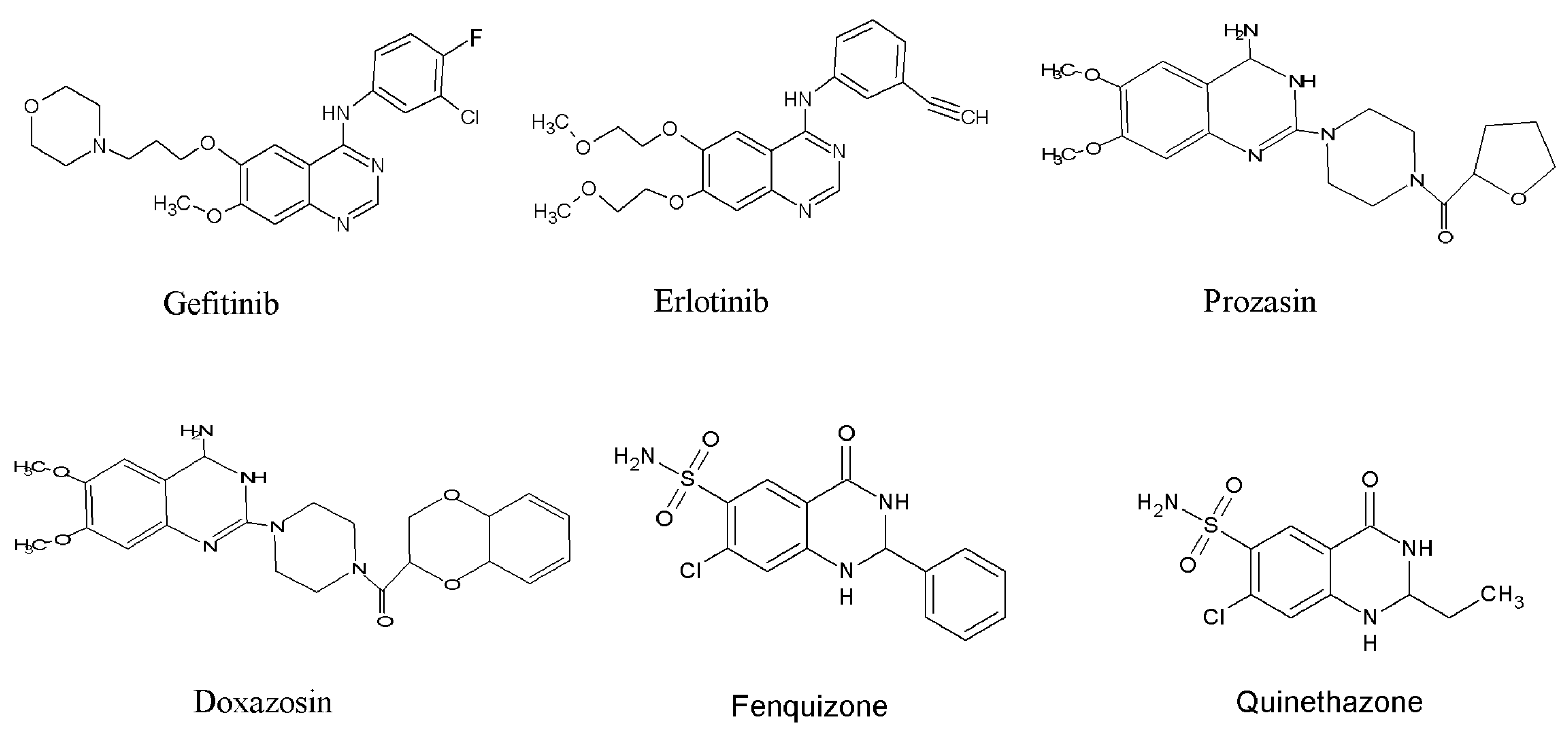
1. Introduction
2. Materials and Methods
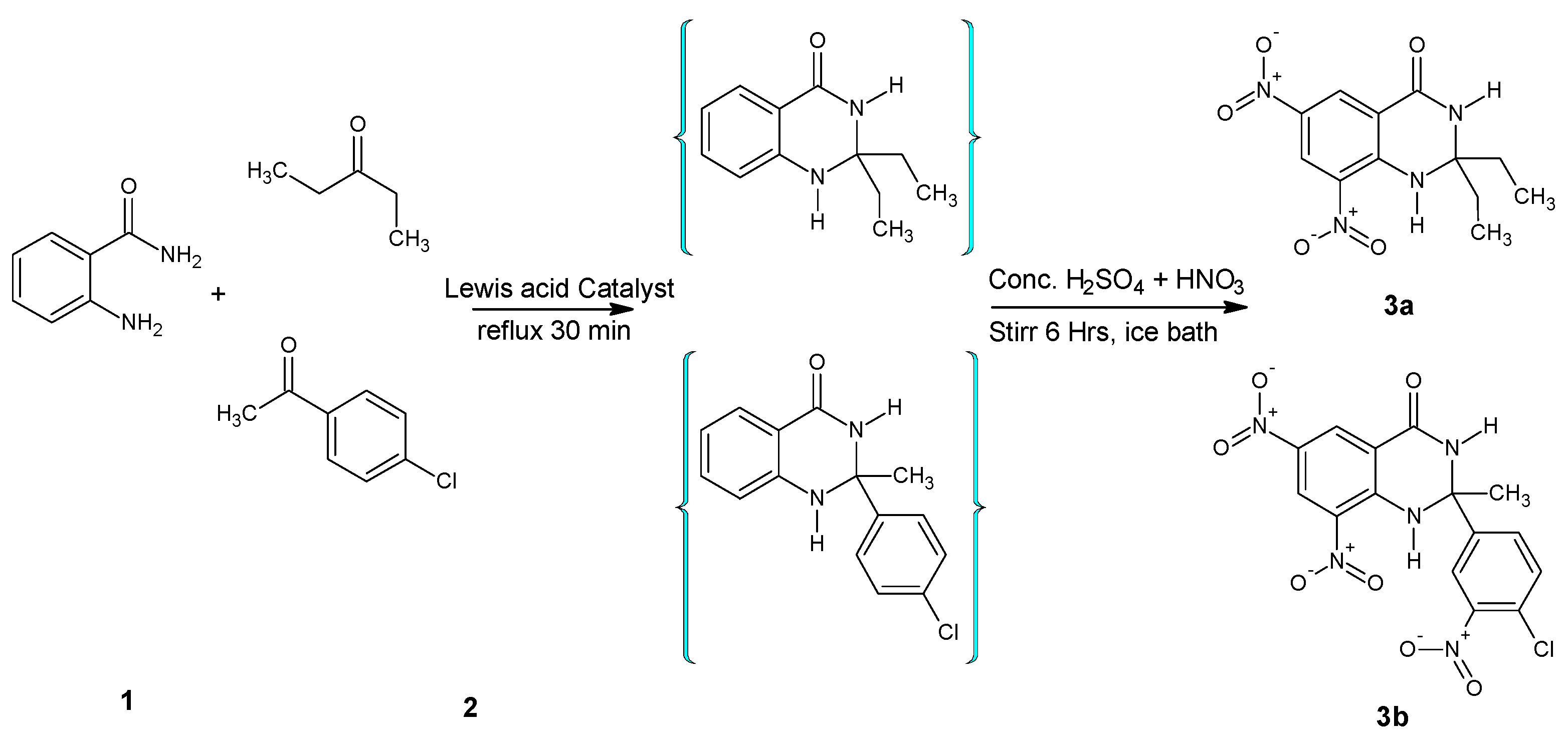
2.1. General Procedure for the Synthesis of 6,8-dinitro-2,2-disubstituted-2,3-dihydroquinazolin-4(1H)-ones (3a and 3b)
2.2. Synthesis of 6,8-dinitro-2,2-diethyl-2,3-dihydroquinazolin-4(1H)-one (3a)
2.3. Synthesis of 2-(4-chloro-3-nitro-phenyl)-2-methyl-6,8-dinitro-2,3-dihydro-1H-quinazolin-4-one (3b)
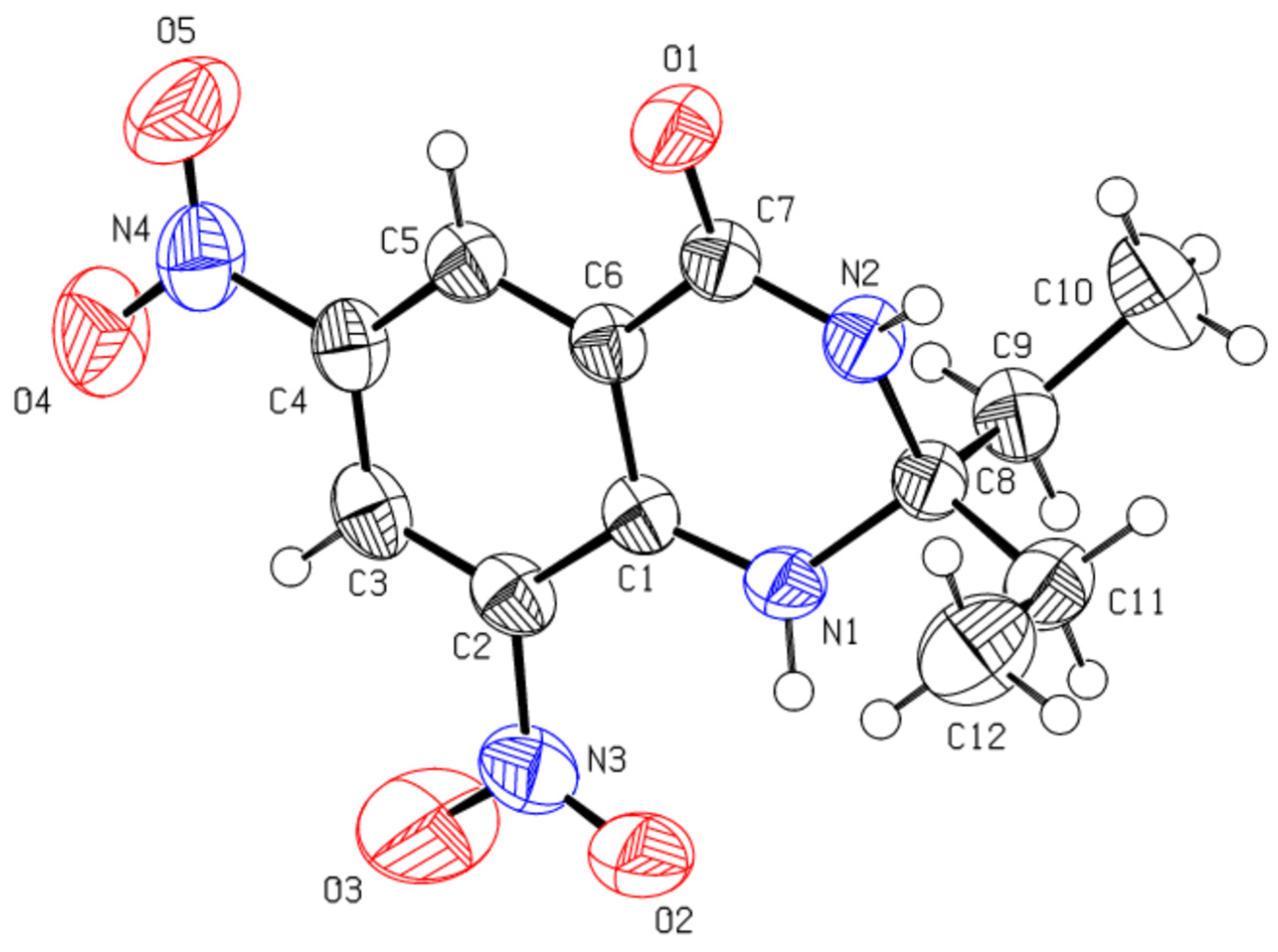
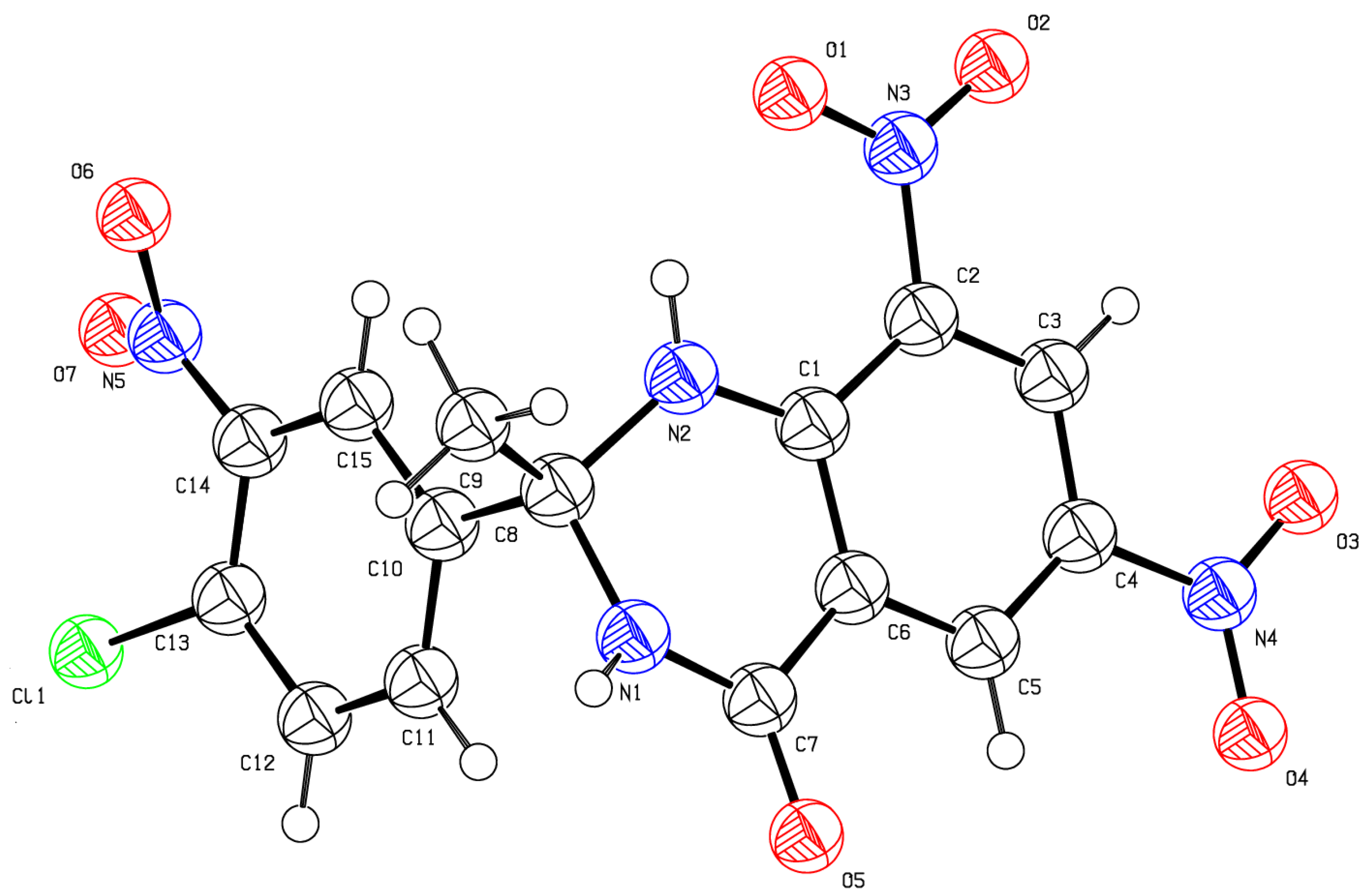
2.4. X-rays Crystallographic Study
2.5. Anti-Leishmanial Activity
2.6. Molecular Docking and Molecular Dynamic Simulations
2.7. Determination of In Silico Pharmacokinetic Properties
3. Results and Discussions
3.1. NMR and XRD Study
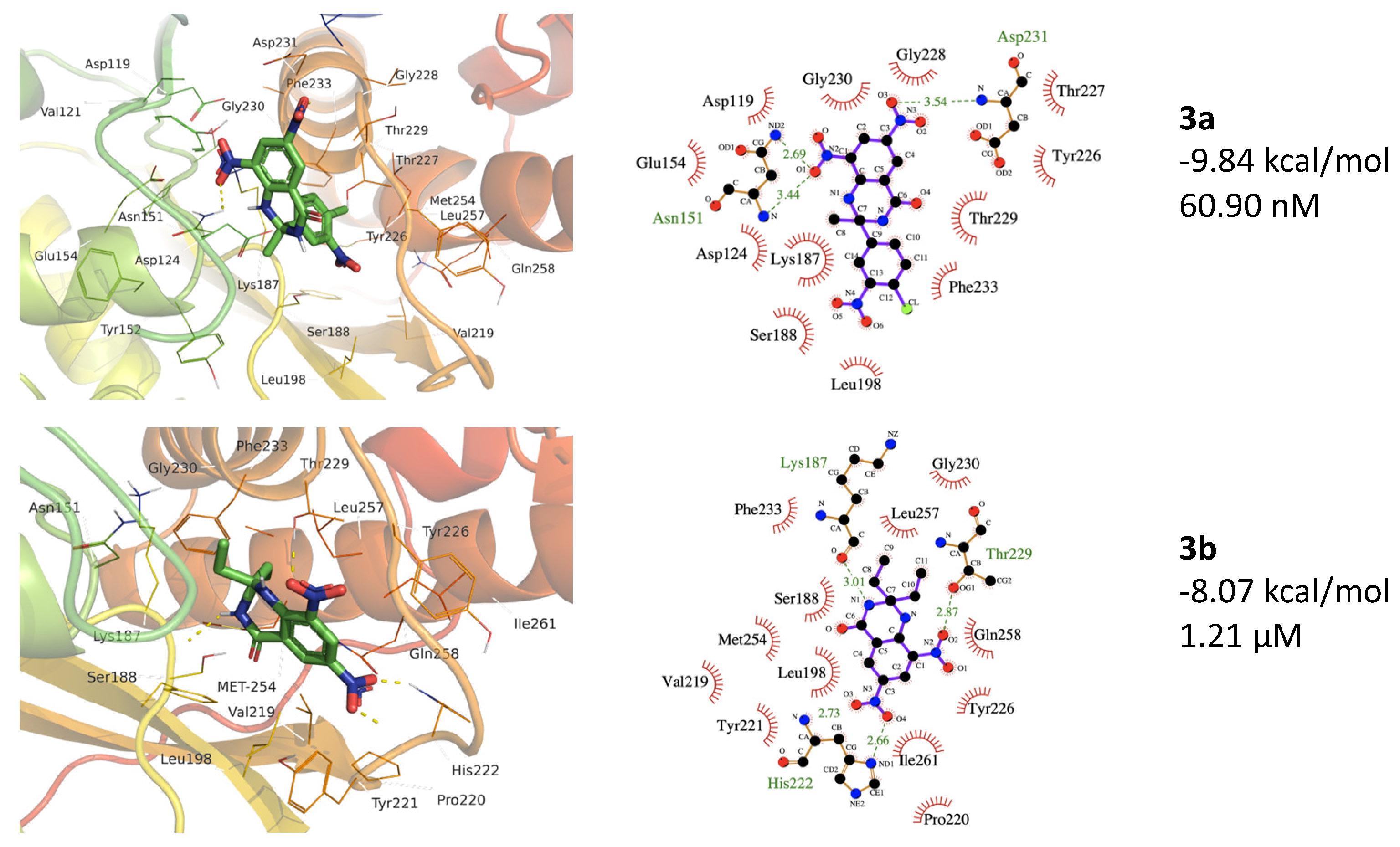
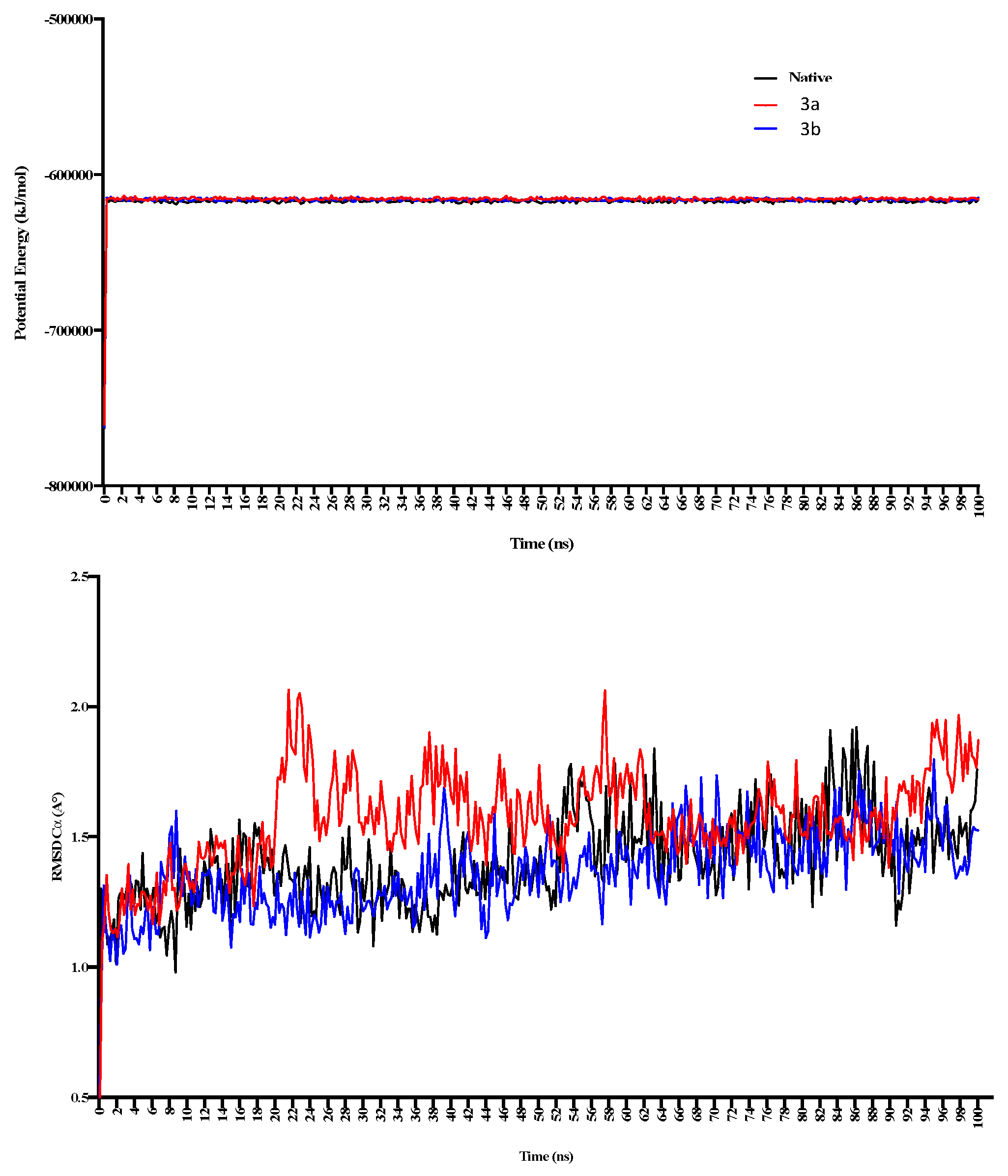
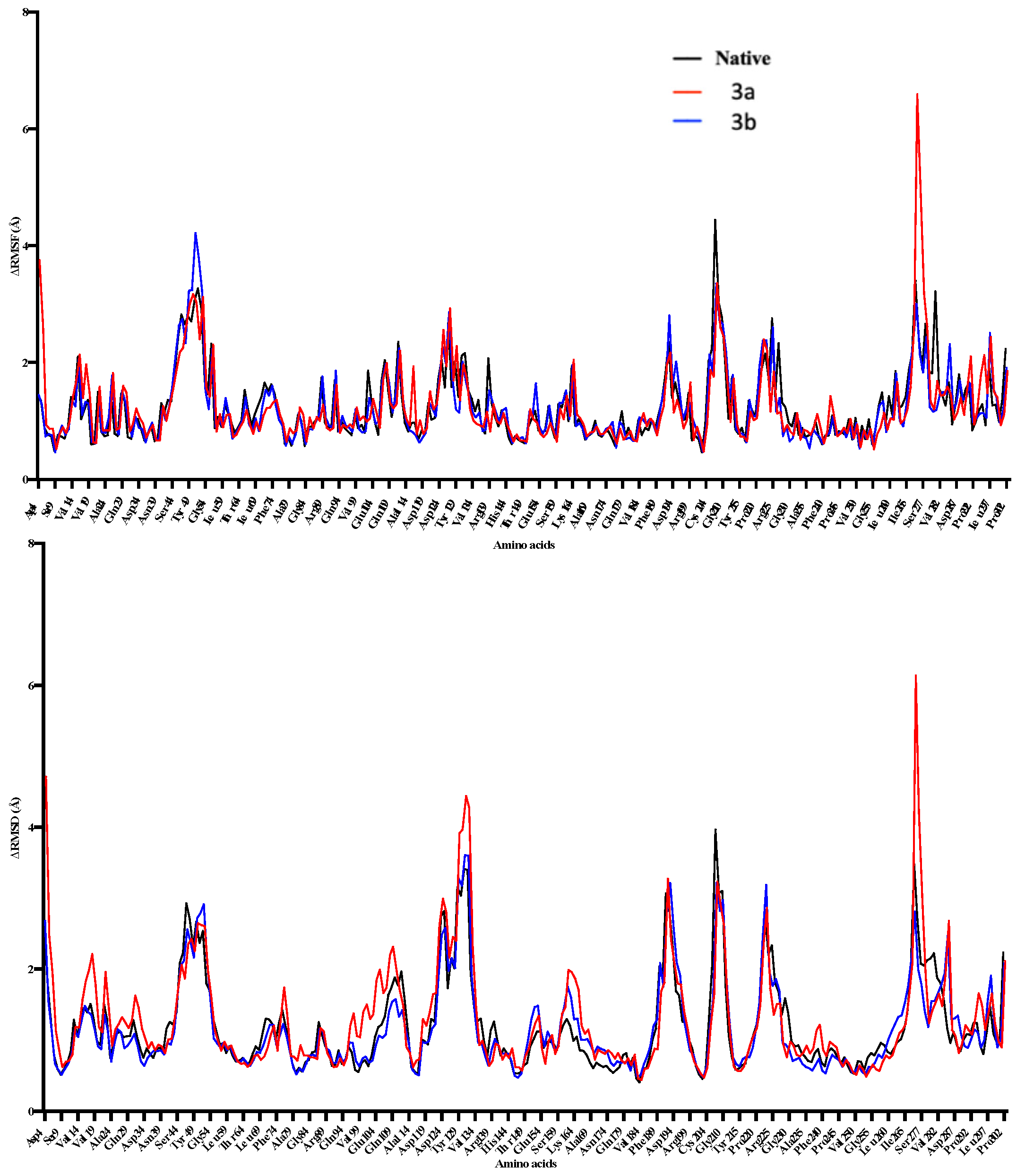
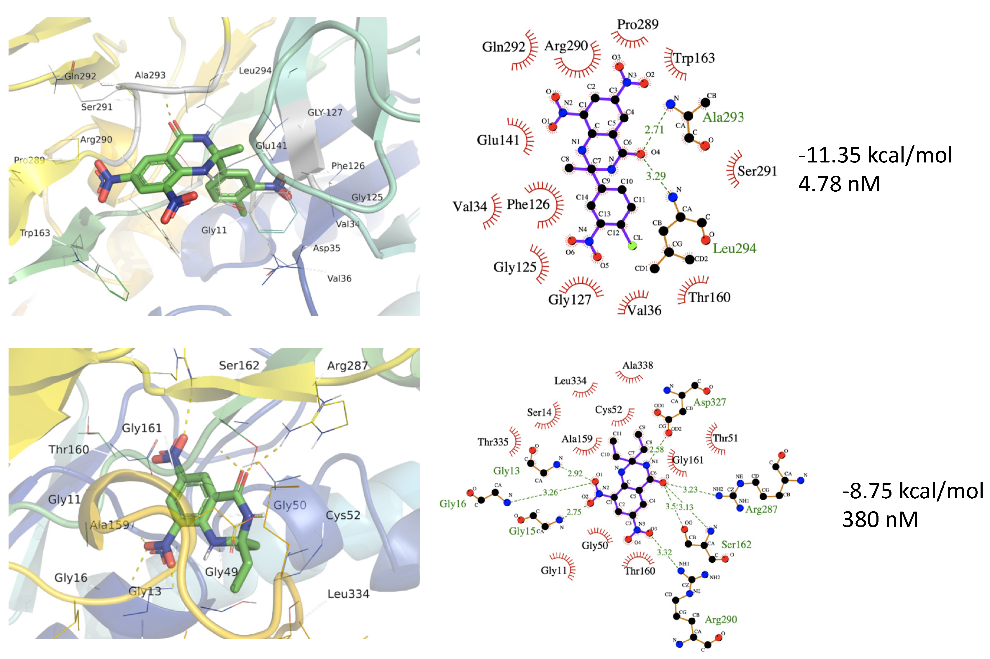
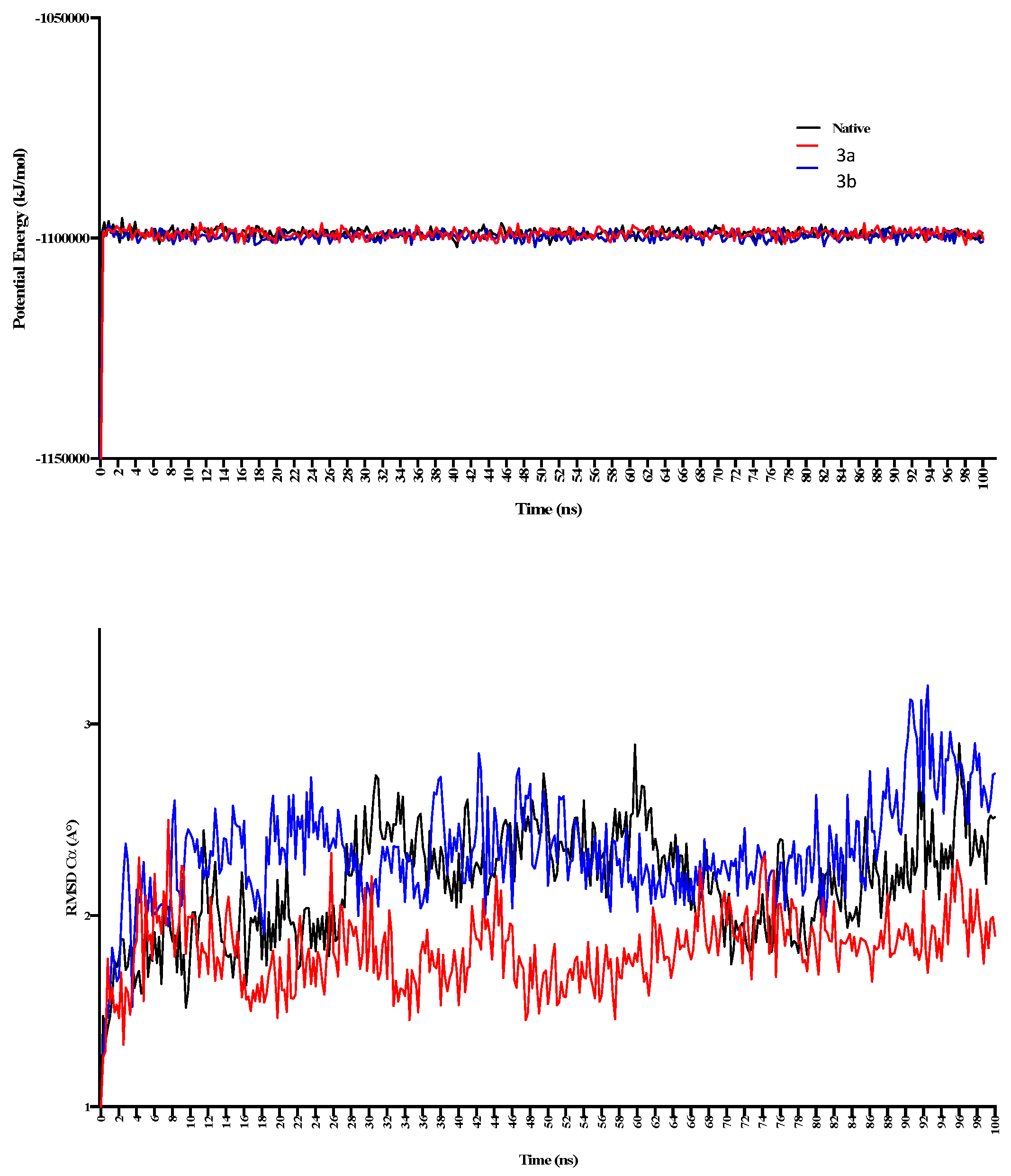
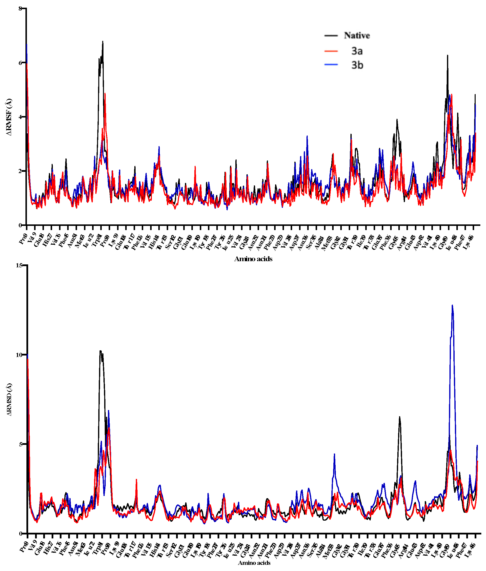
3.2. Docking Studies, MMPBSA Calculations and MD Simaultions
3.3. Anti-Leishmanial Activity
4. Conclusions
Author Contributions
Funding
Conflicts of Interest
References
- Renslo, A.; McKerrow, J.H. Drug discovery and development for neglected parasitic diseases. Nat. Chem. Biol. 2006, 2, 701–710. [Google Scholar] [CrossRef] [PubMed]
- Akopyants, N.S.K.N.; Secundino, N.; Patrick, R.; Peters, N.; Lawyer, P.; Dobson, D.E.; Beverley, S.M.; Sacks, D.L. Demonstration of Genetic Exchange During Cyclical Development of Leishmania in the Sand Fly Vector. Science 2009, 324, 265–268. [Google Scholar] [CrossRef] [PubMed]
- Desjeux, P. Leishmaniasis: Public health aspects and control. Clin. Dermatol. 1996, 14, 417–423. [Google Scholar] [CrossRef]
- WHO World Health Organization Zoonotic Disease: Emerging Public Health Threats in the Region. Available online: http://www.emro.who.int/about-who/rc61/zoonotic-diseases.html (accessed on 18 November 2021).
- Nascimento, M.M.; Queiroz, J.W.; Barroso, A.W.; Araujo, A.F.; Rego, E.F.; Wilson, M.E.; Pearson, R.D.; Jeronimo, S.M. The emergence of concurrent HIV-1/AIDS and visceral leishmaniasis in Northeast Brazil. Trans. R. Soc. Trop. Med. Hyg. 2011, 105, 298–300. [Google Scholar] [CrossRef]
- Aronson, N.; Herwaldt, B.L.; Libman, M.; Pearson, R.; Lopez-Velez, R.; Weina, P.; Carvalho, E.; Ephros, M.; Jeronimo, S.; Magill, A. Diagnosis and Treatment of Leishmaniasis: Clinical Practice Guidelines by the Infectious Diseases Society of America (IDSA) and the American Society of Tropical Medicine and Hygiene (ASTMH). Am. J. Trop. Med. Hyg. 2017, 96, 24–45. [Google Scholar] [CrossRef]
- Leon, L.L.; Carvalho-Paes, L.E.; Grimaldi, J.G. Antigenic differences of Leishmania amazonensis isolate causing diffuse cutaneous leishmaniasis. Trans. R. Soc. Trop. Med. Hyg. 1990, 84, 678–680. [Google Scholar] [CrossRef]
- Chappuis, F.S.; Hailu, A.; Ghalib, H.; Rijal, S.; Peeling, R.W.; Alvar, J.; Boelaert, M. Visceral leishmaniasis: What are the needs for diagnosis, treatment and control? Nat. Rev. Microbiol. 2007, 5, S7–S16. [Google Scholar] [CrossRef]
- Pearson, R.D.S. Clinical spectrum of Leishmaniasis. Clin. Infect. Dis. 1996, 22, 1–13. [Google Scholar] [CrossRef] [PubMed]
- Modabber, F. Leishmaniasis vaccines: Past, present and future. Int. J. Antimicrob. Agents 2010, 36, S58–S61. [Google Scholar] [CrossRef]
- Wise, E.S.; Armstrong, M.; Watson, J.; Lockwood, D.N.J. Monitoring Toxicity Associated with Parenteral Sodium Stibogluconate in the Day-Case Management of Returned Travellers with New World Cutaneous Leishmaniasi. PLoS Negl. Trop. Dis. 2012, 6, e1688. [Google Scholar] [CrossRef]
- Sundar, S.; More, D.K.; Singh, M.K.; Singh, V.P.; Sharma, S.; Makharia, A.; Kumar, P.C.K.; Murray, H.W. Failure of Pentavalent Antimony in Visceral Leishmaniasis in India: Report from the Center of the Indian Epidemic. Clin. Infect. Dis. 2000, 31, 1104–1107. [Google Scholar] [CrossRef]
- Singh, N.; Kumar, M.; Singh, R.K. Leishmaniasis: Current status of available drugs and new potential drug targets. Asian Pac. J. Trop. Med. 2012, 5, 485–497. [Google Scholar] [CrossRef]
- Bray, P.G.; Barrett, M.; Ward, S.; de Koning, H.P. Pentamidine uptake and resistance in pathogenic protozoa: Past, present and future. Trends Parasitol. 2003, 19, 232–239. [Google Scholar] [CrossRef]
- Torres-Guerrero, E.; Ruiz-Esmenjaud, J.; Arenas, R. Leishmaniasis: A review. F1000Res 2017, 6, 1–15. [Google Scholar] [CrossRef] [PubMed]
- Birhan, Y.S.; Bekhit, A.A.; Hymete, A. Synthesis and antileishmanial evaluation of some 2,3-disubstituted-4(3H)-quinazolinone derivatives. Org. Med. Chem. Lett. 2014, 4, 10. [Google Scholar] [CrossRef][Green Version]
- Bonano, V.I.Y.-Y.; Miguel, D.C.; Jones, S.A.; Dodge, J.A.; Uliana, S.R.B. Discovery of Synthetic Leishmania Inhibitors by Screening of a 2-Arylbenzothiophene Library. Chem. Biol. Drug Des. 2014, 81, 658–665. [Google Scholar]
- Coimbra, E.S.A.; Silva, A.D.; Bispo, M.L.F.; Kaiser, C.R.; De Souza, M.V.N. 7-Chloro-4-quinolinyl hydrazones: A promising and potent class of antileishmanial compounds. Chem. Biol. Drug. Des. 2013, 81, 658–665. [Google Scholar] [CrossRef]
- Gómez-Pérez, V.; Manzano, J.I.; García-Hernández, R.; Castanys, S.; Rosa, J.M.C.; Gamarro, F. 4-Amino Bis-Pyridinium Derivatives as Novel Antileishmanial Agents. Antimicrob. Agents Chemother. 2014, 58, 4103–4112. [Google Scholar] [CrossRef]
- Gellis, A.D.; Lanzada, G.; Hutter, S.; Ollivier, E.; Vanelle, P.; Azas, N. Preparation and antiprotozoal evaluation of promising β-carboline alkaloids. Biomed. Pharmacother. 2012, 66, 339–347. [Google Scholar] [CrossRef]
- Machado, P.A.H.; Carvalho, L.O.; Silveira, M.L.T.; Alves, R.B.; Freitas, R.P.; Coimbra, E.S. Effect of 3-Alkylpyridine marine alkaloid analogues in Leishmania species related to American cutaneous lLeishmaniasis. Chem. Biol. Drug Des. 2012, 80, 745–751. [Google Scholar] [CrossRef]
- Roatt, B.M.; De Brito, R.C.F.; Coura-Vital, W.; de Oliveira Aguiar-Soares, R.D.; Reis, A.B. Recent advances and new strategies on leishmaniasis treatment. Appl. Microbiol. Biotechnol. 2020, 104, 8965–8977. [Google Scholar] [CrossRef] [PubMed]
- Tiuman, T.S.; Santos, A.O.; Ueda-Nakamura, T.; Filho, B.P.D.; Nakamura, C.V. Recent advances in leishmaniasis treatment. Int. J. Infect. Dis. 2011, 15, e525–e532. [Google Scholar] [CrossRef] [PubMed]
- Ramsay, R.R.; Tipton, K.F. Assessment of Enzyme Inhibition: A Review with Examples from the Development of Monoamine Oxidase and Cholinesterase Inhibitory Drugs. Molecules 2017, 22, 1192. [Google Scholar] [CrossRef]
- Balbaa, M.; El Ashry, E.S.H. Enzyme Inhibitors as Therapeutic Tools. Biochem. Physiol. Open Access 2012, 1, 13. [Google Scholar] [CrossRef]
- Akram, M.S.; Pery, N.; Butler, L.; Shafiq, M.I.; Batool, N.; Rehman, M.F.U.; Grahame-Dunn, L.G.; Yetisen, A.K. Challenges for biosimilars: Focus on rheumatoid arthritis. Crit. Rev. Biotechnol. 2021, 41, 121–153. [Google Scholar] [CrossRef]
- Sarfraz, M.; Tariq, M.I. Synthesis, in silico study and cholinesterases inhibition activity of 2-substituted 2,3-dihydroquinazolin-4(1H)-one derivatives. Rev. Roum. Chim. 2018, 63, 227–234. [Google Scholar]
- Tyagi, R.; Elfawal, M.A.; Wildman, S.A.; Helander, J.; Bulman, C.A.; Sakanari, J.; Rosa, B.A.; Brindley, P.J.; Janetka, J.W.; Aroian, R.V.; et al. Identification of small molecule enzyme inhibitors as broad-spectrum anthelmintics. Sci. Rep. 2019, 9, 9085. [Google Scholar] [CrossRef]
- Sultana, N.; Sarfraz, M.; Tanoli, S.T.; Akram, M.S.; Sadiq, A.; Rashid, U.; Tariq, M.I. Synthesis, crystal structure determination, biological screening and docking studies of N1-substituted derivatives of 2,3-dihydroquinazolin-4(1H)-one as inhibitors of cholinesterase. Bioorg. Chem. 2017, 72, 256–267. [Google Scholar] [CrossRef]
- Sarfraz, M.; Rashid, U.; Sultana, N.; Tariq, M.I. Synthesis, X-Rays Analysis, Docking Study and Cholinesterase Inhibition Activity of 2, 3-dihydroquinazolin-4 (1H)-one Derivatives. Iran. J. Chem. Chem. Eng. 2019, 38, 213–227. [Google Scholar]
- El-Azab, A.S.E.; Eltahir, K.E.H. Design and synthesis of novel 7-aminoquinazoline derivatives: Antitumor and anticonvulsant activities. Bioorg. Med. Chem. Lett. 2012, 22, 1879–1885. [Google Scholar] [CrossRef]
- Saravanan, G.A.; Prakash, C.R. Design, synthesis and anticonvulsant activities of novel 1-(substituted/unsubstituted benzylidene)-4-(4-(6,8-dibromo-2-(methyl/ phenyl)-4-oxoquinazolin-3(4H)-yl)phenyl) semicarbazide derivatives. Bioorg. Med. Chem. Lett. 2012, 22, 3072–3078. [Google Scholar] [CrossRef] [PubMed]
- Wang, Z.W.; Wang, M.; Yao, X.; Li, Y.; Tan, J.; Wang, L.; Qiao, W.; Geng, Y.; Liu, Y.; Wang, Q. Design, synthesis and antiviral activity of novel quinazolinones. Eur. J. Med. Chem. 2012, 53, 275–282. [Google Scholar] [CrossRef] [PubMed]
- Abbas, S.E.; Awadallah, F.; Ibrahin, N.A.; Said, E.; Kamel, G. New quinazolinone–pyrimidine hybrids: Synthesis, anti-inflammatory, and ulcerogenicity studies. Eur. J. Med. Chem. 2012, 53, 141–149. [Google Scholar] [CrossRef]
- Al-Omary, F.A.; Abou-Zeid, L.A.; Nagi, M.N.; Habib, E.-S.E.; Abdel-Aziz, A.A.-M.; El-Azab, A.S.; Abdel-Hamide, S.G.; Al-Omar, M.A.; Al-Obaid, A.M.; El-Subbagh, H.I. Non-classical antifolates. Part 2: Synthesis, biological evaluation, and molecular modeling study of some new 2,6-substituted-quinazolin-4-ones. Bioorg. Med. Chem. 2010, 18, 2849–2863. [Google Scholar] [CrossRef] [PubMed]
- Amin, K.; Kamel, M.; Anwar, M.; Khedr, M.; Syam, Y. Synthesis, biological evaluation and molecular docking of novel series of spiro [(2H,3H) quinazoline-2,1′- cyclohexan]-4(1H)- one derivatives as anti-inflammatory and analgesic agents. Eur. J. Med. Chem. 2010, 45, 2117–2131. [Google Scholar] [CrossRef]
- Mohameda, M.S.K.M.; Kassem, E.M.M.; Abotaleb, N.; AbdEl-moez, S.I.; Ahmeda, M.F. Novel 6,8-dibromo-4(3H)-quinazolinone derivatives of anti-bacterial and anti-fungal activities. Eur. J. Med. Chem. 2010, 45, 3311–3319. [Google Scholar] [CrossRef]
- Gemma, S.C.; Brindisi, M.; Brogi, S.; Kukreja, G.; Kunjir, S.; Gabellieri, E.; Lucantoni, L.; Habluetzel, A.; Taramelli, D.; Basilico, N.; et al. Mimicking the Intramolecular Hydrogen Bond: Synthesis, Biological Evaluation, and Molecular Modeling of Benzoxazines and Quinazolines as Potential Antimalarial Agents. J. Med. Chem. 2012, 55, 10387–10404. [Google Scholar] [CrossRef]
- Rudolph, J.; O’connor, S.; Coish, P.D.; Wickens, P.L.; Brands, M.; Bierer, D.E.; Bloomquist, B.T.; Bondar, G.; Chen, L.; Chuang, C.Y.; et al. Quinazolinone derivatives as orally available ghrelin receptor antagonists for the treatment of diabetes and obesity. J. Med. Chem. 2007, 50, 5202–5216. [Google Scholar] [CrossRef]
- Rhee, H.-K.; Yoo, J.H.; Lee, E.; Kwon, Y.J.; Seo, H.-R.; Lee, Y.-S.; Choo, H.-Y.P. Synthesis and cytotoxicity of 2-phenylquinazolin-4(3H)-one derivatives. Eur. J. Med. Chem. 2011, 46, 3900–3908. [Google Scholar] [CrossRef]
- Radwan, A.A.; Alanazi, F.K. Biological Activity of Quinazolinones, Quinazolinone and Quinazoline Derivatives; IntechOpen: London, UK, 2020. [Google Scholar]
- Faisal, M.; Saeed, A. Chemical Insights into the Synthetic Chemistry of Quinazolines: Recent Advances. Front. Chem. 2021, 8, 594717. [Google Scholar] [CrossRef]
- Agarwal, K.; Sharma, V.; Shakya, N.; Gupta, S. Design and synthesis of novel substituted quinazoline derivatives as antileishmanial agents. Bioorg. Med. Chem. Lett. 2009, 19, 5474–5477. [Google Scholar] [CrossRef] [PubMed]
- Fleita, D.H.; Mohareb, R.M.; Sakka, O.K. Antitumor and antileishmanial evaluation of novel heterocycles derived from quinazoline scaffold: A molecular modeling approach. Med. Chem. Res. 2013, 22, 2207–2221. [Google Scholar] [CrossRef]
- Tesfahunegn, R.G. Synthesis and Anti-leishmanial Activity Evaluation of Some 2, 3-Disubstituted Quinazoline-4(3H)-Ones Bearing Quinoline and Pyrazole Moieties. EC Pharma. Sci. 2015, 1, 153–164. [Google Scholar]
- Shaterian, H.R.; Ali Reza, O.; Moones, H. Synthesis of 2,3-Dihydroquinazoline-4(1H)-ones. Synth. Commun. 2010, 40, 1231–1242. [Google Scholar] [CrossRef]
- Khattab, S.N.; Haiba, N.S.; Asal, A.M.; Bekhit, A.A.; Guemei, A.A.; Amer, A.; El-Faham, A. Study of antileishmanial activity of 2-aminobenzoyl amino acid hydrazides and their quinazoline derivatives. Bioorg. Med. Chem. Lett. 2017, 27, 918–921. [Google Scholar] [CrossRef]
- Asif, M. Chemical Characteristics, Synthetic Methods, and Biological Potential of Quinazoline and Quinazolinone Derivatives. Int. J. Med. Chem. 2014, 2014, 1–27. [Google Scholar] [CrossRef]
- Badolato, M.; Aiello, F.; Neamati, N. 2,3-Dihydroquinazolin-4(1H)-one as a privileged scaffold in drug design. RSC Adv. 2018, 8, 20894–20921. [Google Scholar] [CrossRef]
- Van Horn, K.S.; Zhu, X.; Pandharkar, T.; Yang, S.; Vesely, B.; Vanaerschot, M.; Dujardin, J.-C.; Rijal, S.; Kyle, D.E.; Wang, M.Z.; et al. Antileishmanial Activity of a Series of N2,N4-Disubstituted Quinazoline-2,4-diamines. J. Med. Chem. 2014, 57, 5141–5156. [Google Scholar] [CrossRef]
- Prinsloo, I.F.; Zuma, N.H.; Aucamp, J.; N’Da, D.D. Synthesis and in vitro antileishmanial efficacy of novel quinazolinone derivatives. Chem. Biol. Drug Des. 2021, 97, 383–398. [Google Scholar] [CrossRef]
- Kshirsagar, U.A. Recent developments in the chemistry of quinazolinone alkaloids. Org. Biomol. Chem. 2015, 13, 9336–9352. [Google Scholar] [CrossRef]
- Prashant, S.A.; Gatish, T.P.G. Recent advances in the pharmacological diversification of quinazoline/quinazolinone hybrids. RSC Adv. 2020, 10, 41353–41392. [Google Scholar]
- Nayyar, P.; Arpana, R.; Mohd, I. An updated review: Newer quinazoline derivatives under clinical trial. Int. J. Pharm. Biol. Arch. 2011, 2, 1651–1657. [Google Scholar]
- Sarfraz, M.; Sultana, N.; Rashid, U.; Akram, M.S.; Sadiq, A.; Tariq, M.I. Synthesis, biological evaluation and docking studies of 2,3-dihydroquinazolin-4(1H)-one derivatives as inhibitors of cholinesterases. Bioorg. Chem. 2017, 70, 237–244. [Google Scholar] [CrossRef] [PubMed]
- Jamil, M.; Sultana, N.; Ashraf, R.; Bashir, M.; Rehman, M.; Kanwal, F.; Ellahi, H.; Lu, C.; Zhang, W.; Tariq, M. Bis (Diamines) Cu and Zn Complexes of Flurbiprofen as Potential Cholinesterase Inhibitors: In Vitro Studies and Docking Simulations. Crystals 2021, 11, 208. [Google Scholar] [CrossRef]
- Tariq, M.I.; Noreen, T.; Tahir, M.N.; Ahmad, S.; Fayyaz-ur-Rehman, M. 2-(2,3-Dimethylphenyl)-1H-isoindole-1,3 (2H)-dione. Acta Cryst. Sect. E Struct. Rep. Online 2010, 66, 2440. [Google Scholar] [CrossRef]
- Lavorato, S.N.; Duarte, M.C.; De Andrade, P.H.R.; Coelho, E.A.F.; Alves, R.J. Synthesis, antileishmanial activity and QSAR studies of 2-chloro- N -arylacetamides. Braz. J. Pharm. Sci. 2017, 53, 16067. [Google Scholar] [CrossRef]
- Krieger, E.; Vriend, G. YASARA View—Molecular graphics for all devices—From smartphones to workstations. Bioinformatics 2014, 30, 2981–2982. [Google Scholar] [CrossRef]
- Bilal, S.; Hassan, M.M.; Rehman, M.F.; Nasir, M.; Sami, A.J.; Hayat, A. An insect acetylcholinesterase biosensor utilizing WO3/g-C3N4 nanocomposite modified pencil graphite electrode for phosmet detection in stored grains. Food Chem. 2021, 346, 128894. [Google Scholar] [CrossRef]
- Rehman, M.F.U.; Akhter, S.; Batool, A.I.; Selamoglu, Z.; Sevindik, M.; Eman, R.; Mustaqeem, M.; Akram, M.S.; Kanwal, F.; Lu, C.; et al. Effectiveness of Natural Antioxidants against SARS-CoV-2? Insights from the In-Silico World. Antibiotics 2021, 10, 1011. [Google Scholar] [CrossRef]
- Laskowski, R.A.; Swindells, M.B. LigPlot+: Multiple ligand-protein interaction diagrams for drug discovery. Chem. Inf. Model. 2011, 51, 2778–2786. [Google Scholar] [CrossRef]
- Fernandez-Poza, S.; Padros, A.; Thompson, R.; Butler, L.; Islam, M.; Mosely, J.; Scrivens, J.H.; Rehman, M.F.; Akram, M.S. Tailor-made recombinant prokaryotic lectins for characterisation of glycoproteins. Anal. Chim. Acta 2021, 1155, 338352. [Google Scholar] [CrossRef]
- Bilal, S.; Sami, A.J.; Hayat, A.; Rehman, M.F.U. Assessment of pesticide induced inhibition of Apis mellifera (honeybee) acetylcholinesterase by means of N-doped carbon dots/BSA nanocomposite modified electrochemical biosensor. Bioelectrochemistry 2021, 144, 107999. [Google Scholar] [CrossRef] [PubMed]
- Motulsky, H. Prism 4 Statistics Guide—Statistical Analyses for Laboratory and Clinical Researchers; GraphPad Software Inc.: San Diego, CA, USA, 2003; pp. 122–126. [Google Scholar]
- Fitzpatrick, T.B.; Amrhein, N.; Kappes, B.; Macheroux, P.; Tews, I.; Raschle, T. Two independent routes of de novo vitamin B6 biosynthesis: Not that different after all. Biochem. J. 2007, 407, 1–13. [Google Scholar] [CrossRef] [PubMed]
- Coelho, A.C.; Boisvert, S.; Mukherjee, A.; Leprohon, P.; Corbeil, J.; Ouellette, M. Multiple Mutations in Heterogeneous Miltefosine-Resistant Leishmania major Population as Determined by Whole Genome Sequencing. PLoS Negl. Trop. Dis. 2012, 6, e1512. [Google Scholar] [CrossRef] [PubMed]
- Are, S.; Gatreddi, S.; Jakkula, P.; Qureshi, I.A. Structural attributes and substrate specificity of pyridoxal kinase from Leishmania donovani. Int. J. Biol. Macromol. 2020, 152, 812–827. [Google Scholar] [CrossRef]
- Alfadhel, S. Virtual screening of Leishmanial pyridoxal kinase enzyme inhibitors by repurposed anti-Trypanosomal libraries reveals two core scaffolds. Latin Am. J. Pharm. 2021, 40, 69–75. [Google Scholar]
- Battista, T.; Colotti, G.; Ilari, A.; Fiorillo, A. Targeting Trypanothione Reductase, a Key Enzyme in the Redox Trypanosomatid Metabolism, to Develop New Drugs against Leishmaniasis and Trypanosomiases. Molecules 2020, 25, 1924. [Google Scholar] [CrossRef]
- Matadamas-Martínez, F.; Hernández-Campos, A.; Téllez-Valencia, A.; Vázquez-Raygoza, A.; Comparán-Alarcón, S.; Yépez-Mulia, L.; Castillo, R. Leishmania mexicana Trypanothione Reductase Inhibitors: Computational and Biological Studies. Molecules 2019, 24, 3216. [Google Scholar] [CrossRef]
- Vargas, J.; López, A.; Vargas, A.; Fidalgo, L.; Froeyen, M. In Vitro Evaluation and Molecular Docking Studies of Aryl-Substituted Imidazoles against Leishmania Amazonensis. Int. J. Trop. Dis. 2021, 4, 50. [Google Scholar]
- Verma, R.K.; Prajapati, V.K.; Verma, G.K.; Chakraborty, D.; Sundar, S.; Rai, M.; Dubey, V.K.; Singh, M.S. Molecular Docking and In Vitro Antileishmanial Evaluation of Chromene-2-thione Analogues. ACS Med. Chem. Lett. 2012, 3, 243–247. [Google Scholar] [CrossRef]


| COMPOUND | 6,8-DINITRO-2,2-DIETHYL-2,3-DIHYDROQUINAZOLIN-4(1H)-ONE | ||
|---|---|---|---|
| Chemical Formula | C12H14N4O5 | ||
| M (g mol−1) | 294.27 | ||
| Temperature (K) | 296(2) | ||
| Crystal system | triclinic | ||
| Space group | P −1 | Cell volume | 668.21(13) |
| A (Å) | 7.2979(9) | α | 74.633(7) |
| B (Å) | 9.3899(10) | β | 82.799(7) |
| C (Å) | 10.2950(11) | γ | 80.358(7) |
| C1 | 0.2542(3) | 0.1917(3) | 0.1182(2) |
| C2 | 0.2105(3) | 0.2055(3) | −0.0153(2) |
| C3 | 0.2138(3) | 0.0835(3) | −0.0662(2) |
| H3 | 0.1823 | 0.0961 | −0.1537 |
| C4 | 0.2638(3) | −0.0568(3) | 0.0133(2) |
| C5 | 0.3167(3) | −0.0776(3) | 0.1418(2) |
| H5 | 0.3553 | −0.1732 | 0.1930 |
| C6 | 0.3121(3) | 0.0430(3) | 0.1936(2) |
| C7 | 0.3904(4) | 0.0215(3) | 0.3242(2) |
| C8 | 0.2366(3) | 0.2765(3) | 0.3250(2) |
| C9 | 0.0347(4) | 0.2546(3) | 0.3813(2) |
| H9A | −0.0464 | 0.3468 | 0.3478 |
| H9B | −0.0019 | 0.1785 | 0.3459 |
| C10 | 0.0027(5) | 0.2103(4) | 0.5336(3) |
| H10A | 0.0830 | 0.1196 | 0.5685 |
| H10B | −0.1250 | 0.1952 | 0.5593 |
| H10C | 0.0297 | 0.2880 | 0.5699 |
| C11 | 0.2977(4) | 0.4066(3) | 0.3615(3) |
| H11A | 0.2146 | 0.4964 | 0.3243 |
| H11B | 0.2831 | 0.3899 | 0.4591 |
| C12 | 0.4944(5) | 0.4334(4) | 0.3133(4) |
| H12A | 0.5788 | 0.3464 | 0.3512 |
| H12B | 0.5204 | 0.5171 | 0.3413 |
| H12C | 0.5100 | 0.4538 | 0.2164 |
| N1 | 0.2466(3) | 0.3024(2) | 0.17796(18) |
| H1 | 0.2475 | 0.3919 | 0.1286 |
| N2 | 0.3645(3) | 0.1397(2) | 0.37514(19) |
| H2 | 0.4267 | 0.1361 | 0.4419 |
| N3 | 0.1546(4) | 0.3500(3) | −0.1038(2) |
| N4 | 0.2658(4) | −0.1854(3) | −0.0407(3) |
| O1 | 0.4806(3) | −0.09969(19) | 0.37765(16) |
| O2 | 0.1690(3) | 0.4622(2) | −0.07018(19) |
| O3 | 0.0948(5) | 0.3551(3) | −0.2099(2) |
| O4 | 0.2167(4) | −0.1651(3) | −0.1536(2) |
| O5 | 0.3161(5) | −0.3090(3) | 0.0304(2) |
| COMPOUND | (2-(4-CHLORO-3-NITRO-PHENYL)-2-METHYL-6,8-DINITRO-2,3-DIHYDRO-1H-QUINAZOLIN-4-ONE | ||
|---|---|---|---|
| Chemical Formula | C15H10N5ClO7 | ||
| M (g mol−1) | 407.5 | ||
| Temperature (K) | 372(2) | ||
| Crystal system | triclinic | ||
| Space group | P −1 | Cell volume | 841.094 |
| A (Å) | 6.8643(8) | α | 75.424(6) |
| B (Å) | 9.2549(9) | β | 87.586(7) |
| C (Å) | 13.7144(15) | γ | 86.231(7) |
| C1 | 0.6656(7) | 0.1751(5) | 0.8254(4) |
| C2 | 0.6775(7) | 0.0369(5) | 0.7961(4) |
| C3 | 0.5281(8) | −0.0597(5) | 0.8182(4) |
| H3 | 0.5382 | -0.1484 | 0.7977 |
| C4 | 0.3649(8) | −0.0247(5) | 0.8705(4) |
| C5 | 0.3486(7) | 0.1037(5) | 0.9045(4) |
| H5 | 0.2388 | 0.1241 | 0.9420 |
| C6 | 0.4959(7) | 0.2013(5) | 0.8826(4) |
| C7 | 0.4912(8) | 0.3295(5) | 0.9310(4) |
| C8 | 0.7650(8) | 0.4278(5) | 0.8170(4) |
| C9 | 0.9567(9) | 0.4859(7) | 0.8365(6) |
| H9A | 0.9337 | 0.5838 | 0.8481 |
| H9B | 1.0433 | 0.4914 | 0.7790 |
| H9C | 1.0148 | 0.4195 | 0.8947 |
| C10 | 0.6525(17) | 0.5296(12) | 0.7192(6) |
| C11 | 0.4757(18) | 0.6030(12) | 0.7359(5) |
| H11 | 0.4210 | 0.5869 | 0.8006 |
| C12 | 0.3807(14) | 0.7005(10) | 0.6557(7) |
| H12 | 0.2624 | 0.7496 | 0.6668 |
| C13 | 0.4624(15) | 0.7246(9) | 0.5589(5) |
| C14 | 0.6392(16) | 0.6512(10) | 0.5422(5) |
| C15 | 0.7342(14) | 0.5537(11) | 0.6224(7) |
| H15 | 0.8525 | 0.5046 | 0.6113 |
| CL1 | 0.3474(18) | 0.8578(13) | 0.4625(7) |
| N1 | 0.6380(6) | 0.4198(4) | 0.9038(3) |
| H1 | 0.6583 | 0.4785 | 0.9415 |
| N2 | 0.8019(7) | 0.2756(4) | 0.8038(3) |
| H2 | 0.9147 | 0.2505 | 0.7814 |
| N3 | 0.8464(7) | −0.0089(5) | 0.7414(4) |
| N4 | 0.2091(8) | −0.1292(5) | 0.8942(4) |
| O1 | 0.9864(6) | 0.0713(4) | 0.7251(3) |
| O2 | 0.8450(6) | −0.1227(5) | 0.7148(4) |
| O3 | 0.2263(6) | −0.2401(5) | 0.8615(4) |
| O4 | 0.0718(7) | −0.1034(5) | 0.9463(4) |
| O5 | 0.3663(6) | 0.3415(4) | 0.9956(3) |
| O6 | 0.914(6) | 0.6698(14) | 0.4572(12) |
| O7 | 0.638(5) | 0.712(3) | 0.3774(13) |
| N5 | 0.703(5) | 0.680(4) | 0.448(3) |
| Docking Scores | |||||
|---|---|---|---|---|---|
| Ligands | Leishmanial Targets | PDB IDs | Binding Energy (kcal/mol) | Dissociation Constant | MM/PBSA |
| 3a | Pyridoxal Kinase | 6K92 | −9.84 | 60.90 nM | −94 KJ/mol |
| Trypanothione reductase | 6T98 | −11.35 | 4.78 nM | −113 KJ/mol | |
| 3b | Pyridoxal Kinase | 6K92 | −8.07 | 1.21 µM | −32 KJ/mol |
| Trypanothione reductase | 6T98 | −8.75 | 380 nM | −61 KJ/mol | |
| Ligands | IC50 (µg/mL) |
|---|---|
| 3a | 1.164 ± 0.123 |
| 3b | 0.085 ± 0.015 |
| Amphotericin B | 0.046 ± 0.101 |
| Miltefosine | 3.191 ± 1.10 |
| 3a | 3b | |
|---|---|---|
| Molecular weight | 294.27 g/mol | 407.5 g/mol |
| Num. H-bond acceptors | 5 | 7 |
| Num. H-bond donors | 2 | 2 |
| Molar Refractivity | 85.58 | 109.33 |
| TPSA | 132.77 Ų | 178.59 Ų |
| Log P (lipophilicity) | 0.55 | 0.56 |
| Log S (solubility) | (soluble) −3.11 | Moderately soluble −4.24 |
| Pharmacokinetics | ||
| GI absorption | High | Low |
| BBB permeant | No | No |
| P-gp substrate | No | Yes |
| CYP1A2 inhibitor | Yes | No |
| CYP2C19 inhibitor | Yes | Yes |
| CYP2C9 inhibitor | No | Yes |
| CYP2D6 inhibitor | No | No |
| CYP3A4 inhibitor | No | No |
| Log Kp (skin permeation) | −6.41 cm/s | −6.74 cm/s |
| Druglikeness | ||
| Lipinski | Yes; 0 violation | Yes; 1 violation: NorO > 10 |
| Ghose | Yes | Yes |
| Veber | Yes | No; 1 violation: TPSA > 140 |
| Egan | No; 1 violation: TPSA > 131.6 | No; 1 violation: TPSA > 131.6 |
| Muegge | Yes | No; 1 violation: TPSA > 150 |
| Bioavailability Score | 0.55 | 0.55 |
Publisher’s Note: MDPI stays neutral with regard to jurisdictional claims in published maps and institutional affiliations. |
© 2021 by the authors. Licensee MDPI, Basel, Switzerland. This article is an open access article distributed under the terms and conditions of the Creative Commons Attribution (CC BY) license (https://creativecommons.org/licenses/by/4.0/).
Share and Cite
Sarfraz, M.; Wang, C.; Sultana, N.; Ellahi, H.; Rehman, M.F.u.; Jameel, M.; Akhter, S.; Kanwal, F.; Tariq, M.I.; Xue, S. 2,3-Dihydroquinazolin-4(1H)-one as a New Class of Anti-Leishmanial Agents: A Combined Experimental and Computational Study. Crystals 2022, 12, 44. https://doi.org/10.3390/cryst12010044
Sarfraz M, Wang C, Sultana N, Ellahi H, Rehman MFu, Jameel M, Akhter S, Kanwal F, Tariq MI, Xue S. 2,3-Dihydroquinazolin-4(1H)-one as a New Class of Anti-Leishmanial Agents: A Combined Experimental and Computational Study. Crystals. 2022; 12(1):44. https://doi.org/10.3390/cryst12010044
Chicago/Turabian StyleSarfraz, Muhammad, Chenxi Wang, Nargis Sultana, Humna Ellahi, Muhammad Fayyaz ur Rehman, Muhammad Jameel, Shahzaib Akhter, Fariha Kanwal, Muhammad Ilyas Tariq, and Song Xue. 2022. "2,3-Dihydroquinazolin-4(1H)-one as a New Class of Anti-Leishmanial Agents: A Combined Experimental and Computational Study" Crystals 12, no. 1: 44. https://doi.org/10.3390/cryst12010044
APA StyleSarfraz, M., Wang, C., Sultana, N., Ellahi, H., Rehman, M. F. u., Jameel, M., Akhter, S., Kanwal, F., Tariq, M. I., & Xue, S. (2022). 2,3-Dihydroquinazolin-4(1H)-one as a New Class of Anti-Leishmanial Agents: A Combined Experimental and Computational Study. Crystals, 12(1), 44. https://doi.org/10.3390/cryst12010044