
Advances in Non-Invasive Screening Methods for Gastrointestinal Cancers: How Continued Innovation Has Revolutionized Early Cancer Detection

 , , , , , and
Simple Summary
Abstract
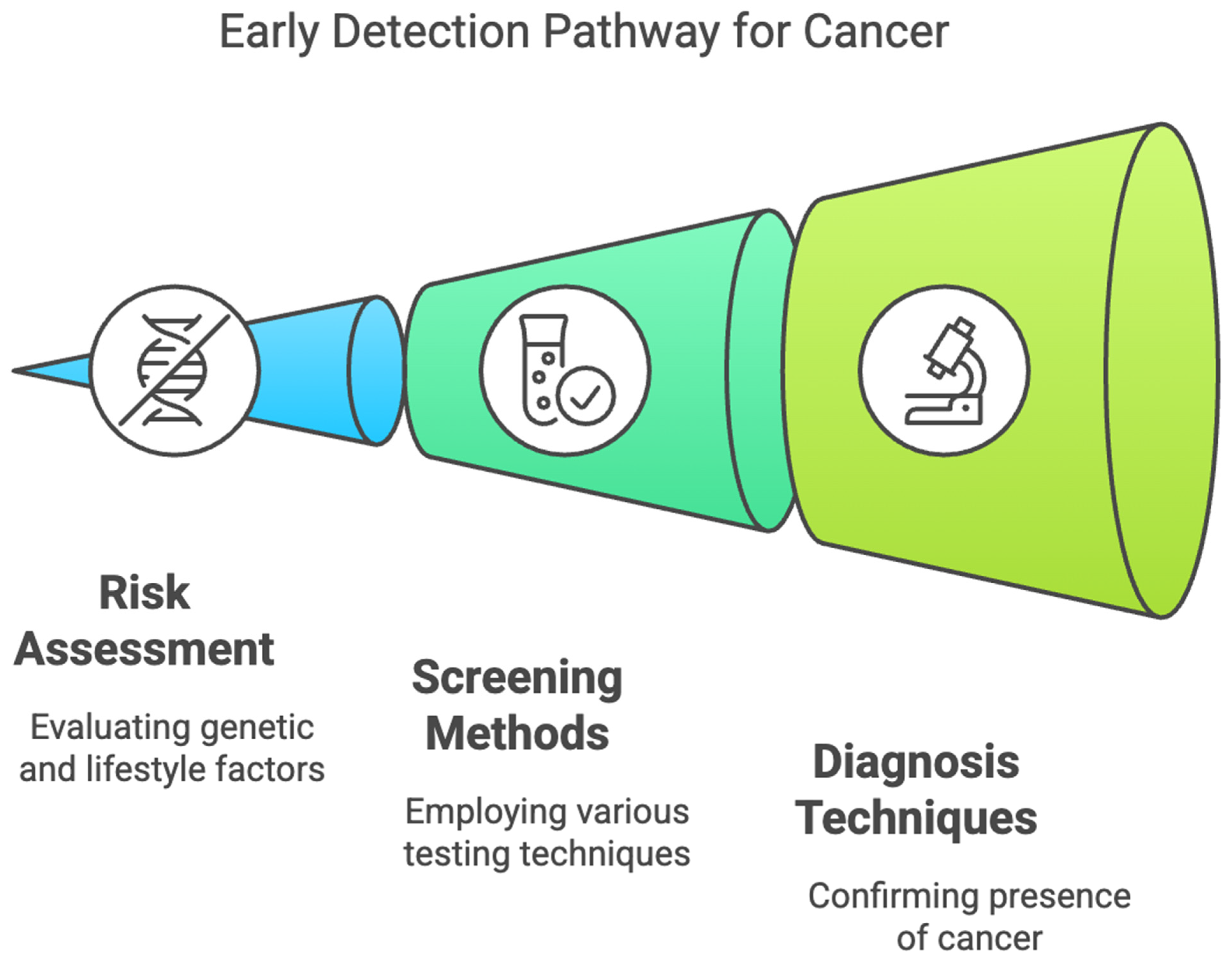
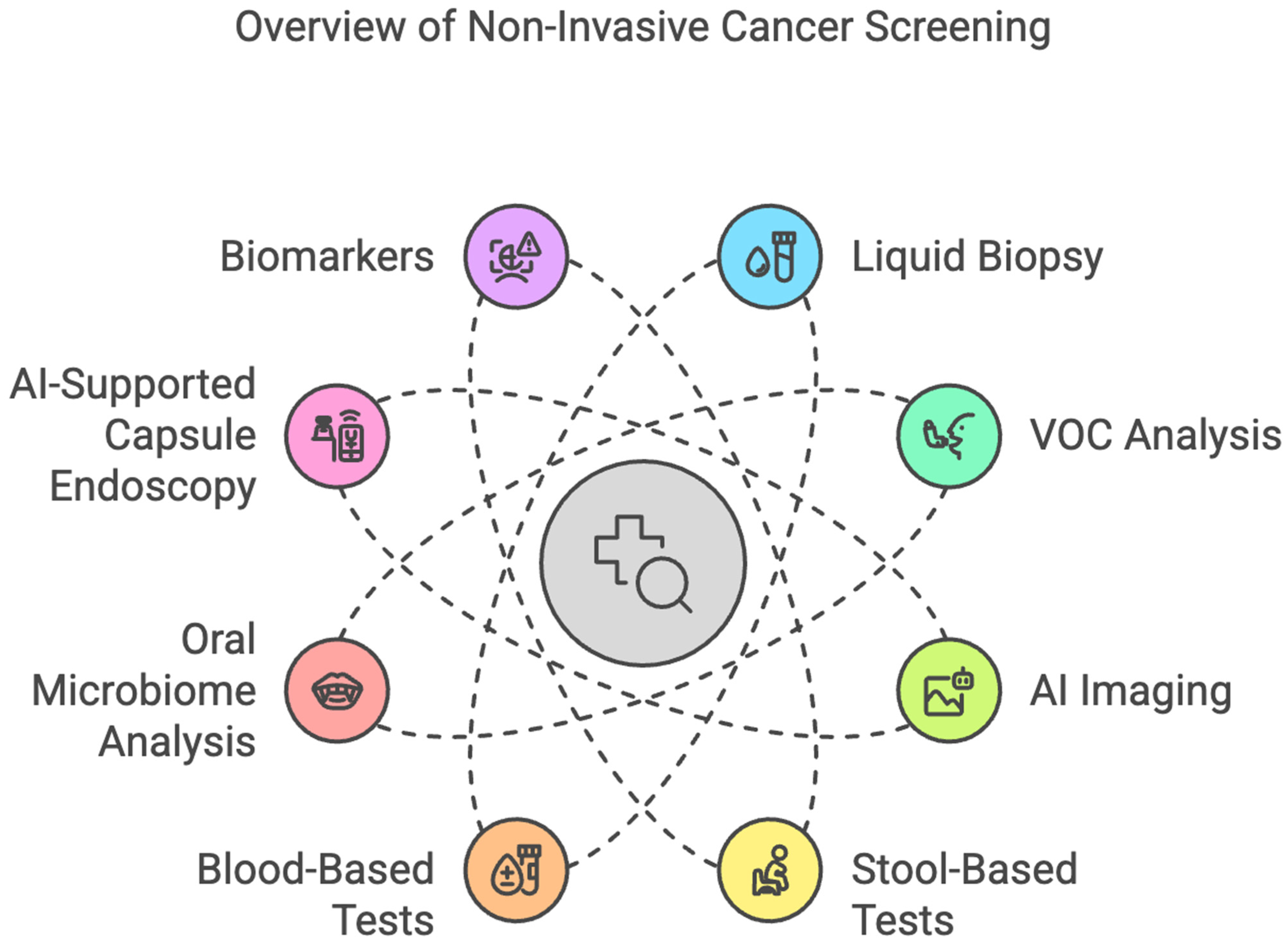
1. Introduction
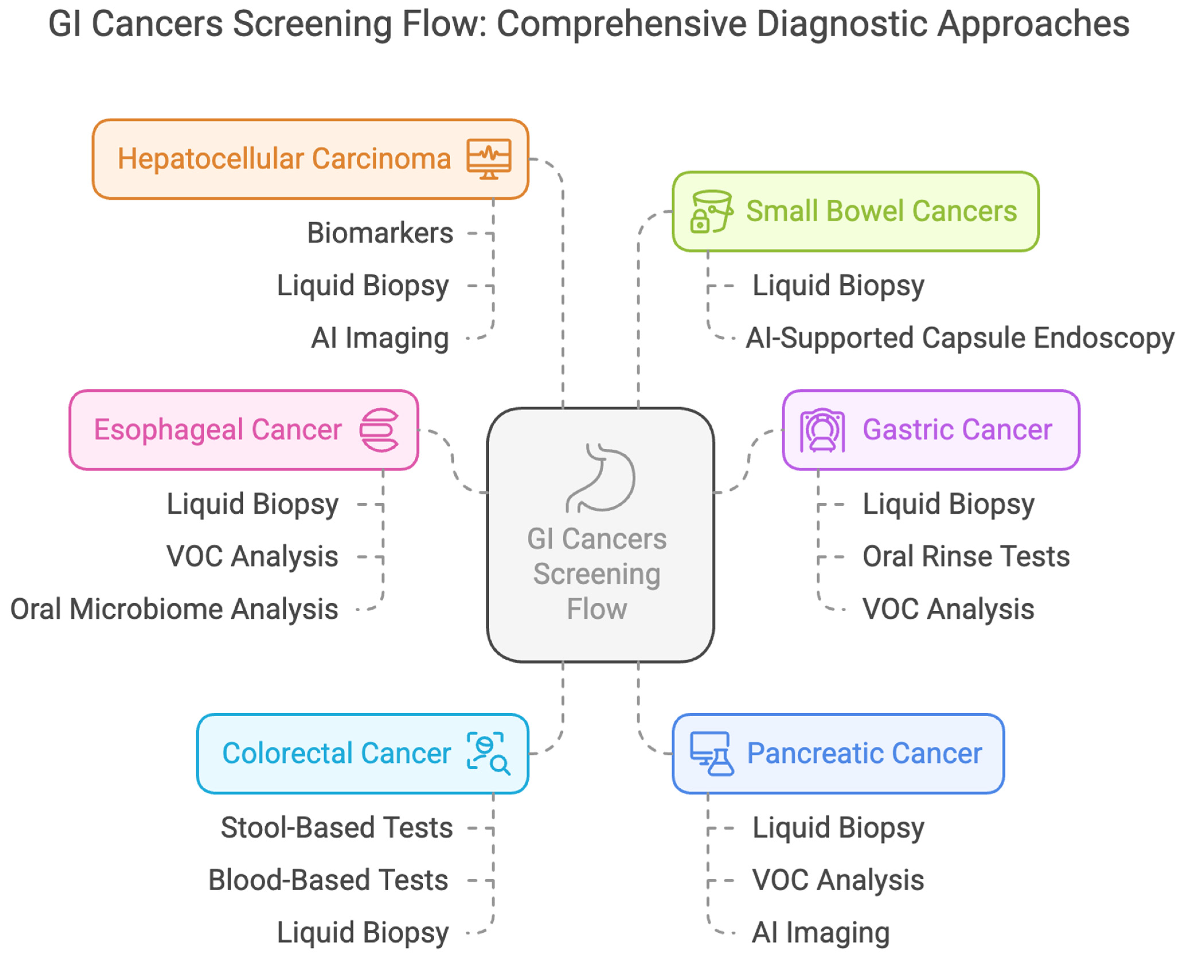
2. Esophageal Cancer
2.1. Liquid Biopsy
2.1.1. Blood Autoantibodies
2.1.2. Circulating MiRNAs
2.1.3. Methylated DNA Markers (MDMs)
2.1.4. Circulating Tumor Cells (CTCs)
2.2. Volatile Organic Compounds (VOCs)
2.3. Esophageal Cytology Specimens Combined with Biomarkers
2.4. Advances in Imaging
2.4.1. Unsedated Transnasal Endoscopy (uTNE)
2.4.2. Capsule-Based Imaging
2.5. Oral Microbiome Analysis
3. Gastric Cancer
3.1. Liquid-Based Tests
3.2. Circulating Tumor Cells
3.3. Circulating Tumor DNA
3.4. Exosomes and Non-Coding RNAs
3.5. Oral Rinse Tests
3.6. VOC Breath Analysis
4. Colorectal Cancer
4.1. Blood-Based Tests
4.2. Stool-Based Tests
4.3. Computed Tomographic (CT) Colonography
4.4. Liquid Biopsy
5. Pancreatic Cancer
5.1. Liquid Biopsy
5.2. VOCs in Breath Analysis
5.3. Advances in Imaging with Artificial Intelligence (AI)
5.4. Multi-Omics Integration
5.5. Future Innovations
6. Hepatocellular Carcinoma
6.1. Biomarkers
6.2. Liquid Biopsy
6.3. Non-Coding RNA Biomarkers
6.4. Metabolomics
6.5. Radiomics
6.6. Combination Approach
7. Small Bowel Cancers
7.1. Liquid Biopsy
7.2. Emerging Approaches
7.3. Complementary Screening Techniques
8. Limitations
9. Conclusions, Challenges, and Considerations
Author Contributions
Funding
Institutional Board Review Statement
Informed Consent Statement
Data Availability Statement
Conflicts of Interest
References
- GBD 2017 Oesophageal Cancer Collaborators. The Global, Regional, and National Burden of Oesophageal Cancer and Its Attributable Risk Factors in 195 Countries and Territories, 1990–2017: A Systematic Analysis for the Global Burden of Disease Study 2017. Lancet Gastroenterol. Hepatol. 2020, 5, 582–597. [Google Scholar] [CrossRef]
- Liu, C.-Q.; Ma, Y.-L.; Qin, Q.; Wang, P.-H.; Luo, Y.; Xu, P.-F.; Cui, Y. Epidemiology of Esophageal Cancer in 2020 and Projections to 2030 and 2040. Thorac. Cancer 2023, 14, 3–11. [Google Scholar] [CrossRef] [PubMed]
- Li, Q.; Xia, C.; Li, H.; Yan, X.; Yang, F.; Cao, M.; Zhang, S.; Teng, Y.; He, S.; Cao, M.; et al. Disparities in 36 Cancers across 185 Countries: Secondary Analysis of Global Cancer Statistics. Front. Med. 2024, 18, 911–920. [Google Scholar] [CrossRef]
- Mulisa, G.; Abebe, T.; Gutema, B.; Mahmuda, J.; Khan, M.A.A.; Gheit, T.; Herceg, Z.; Talukdar, F.R. Exploring Oesophageal Cancer in Ethiopia: Elevated Incidence in Females and Younger Cases. Cancer Rep. 2024, 7, e70048. [Google Scholar] [CrossRef]
- Arnold, M.; Laversanne, M.; Brown, L.M.; Devesa, S.S.; Bray, F. Predicting the Future Burden of Esophageal Cancer by Histological Subtype: International Trends in Incidence up to 2030. Am. J. Gastroenterol. 2017, 112, 1247–1255. [Google Scholar] [CrossRef]
- Pennathur, A.; Gibson, M.K.; Jobe, B.A.; Luketich, J.D. Oesophageal Carcinoma. Lancet 2013, 381, 400–412. [Google Scholar] [CrossRef]
- Liu, M.C.; Oxnard, G.R.; Klein, E.A.; Swanton, C.; Seiden, M.V. CCGA Consortium Sensitive and Specific Multi-Cancer Detection and Localization Using Methylation Signatures in Cell-Free DNA. Ann. Oncol. 2020, 31, 745–759. [Google Scholar] [CrossRef]
- Wong, M.C.S.; Deng, Y.; Huang, J.; Bai, Y.; Wang, H.H.X.; Yuan, J.; Zhang, L.; Yip, H.C.; Chiu, P.W.Y. Performance of Screening Tests for Esophageal Squamous Cell Carcinoma: A Systematic Review and Meta-Analysis. Gastrointest. Endosc. 2022, 96, 197–207.e34. [Google Scholar] [CrossRef]
- Cabalag, C.S.; Yates, M.; Corrales, M.B.; Yeh, P.; Wong, S.Q.; Zhang, B.Z.; Fujihara, K.M.; Chong, L.; Hii, M.W.; Dawson, S.-J.; et al. Potential Clinical Utility of a Targeted Circulating Tumor DNA Assay in Esophageal Adenocarcinoma. Ann. Surg. 2022, 276, e120–e126. [Google Scholar] [CrossRef]
- Fisher, O.M.; Lord, S.J.; Falkenback, D.; Clemons, N.J.; Eslick, G.D.; Lord, R.V. The Prognostic Value of TP53 Mutations in Oesophageal Adenocarcinoma: A Systematic Review and Meta-Analysis. Gut 2017, 66, 399–410. [Google Scholar] [CrossRef]
- Liu, F.; Tian, T.; Xia, L.-L.; Ding, Y.; Cormier, R.T.; He, Y. Circulating MiRNAs as Novel Potential Biomarkers for Esophageal Squamous Cell Carcinoma Diagnosis: A Meta-Analysis Update. Dis. Esophagus 2017, 30, 1–9. [Google Scholar] [CrossRef] [PubMed]
- Moinova, H.R.; LaFramboise, T.; Lutterbaugh, J.D.; Chandar, A.K.; Dumot, J.; Faulx, A.; Brock, W.; De la Cruz Cabrera, O.; Guda, K.; Barnholtz-Sloan, J.S.; et al. Identifying DNA Methylation Biomarkers for Non-Endoscopic Detection of Barrett’s Esophagus. Sci. Transl. Med. 2018, 10, eaao5848. [Google Scholar] [CrossRef]
- Qin, Y.; Wu, C.W.; Taylor, W.R.; Sawas, T.; Burger, K.N.; Mahoney, D.W.; Sun, Z.; Yab, T.C.; Lidgard, G.P.; Allawi, H.T.; et al. Discovery, Validation, and Application of Novel Methylated DNA Markers for Detection of Esophageal Cancer in Plasma. Clin. Cancer Res. 2019, 25, 7396–7404. [Google Scholar] [CrossRef]
- Li, Y.; Wang, Z.; Fu, R.; Wang, S.; Zhang, T.; Tian, X.; Yang, D. Clinical Utility of Circulating Tumor Cells in Patients with Esophageal Cancer. Front. Oncol. 2022, 12, 828368. [Google Scholar] [CrossRef]
- Choi, M.K.; Kim, G.H.; I, H.; Park, S.J.; Lee, M.W.; Lee, B.E.; Park, D.Y.; Cho, Y.-K. Circulating Tumor Cells Detected Using Fluid-Assisted Separation Technique in Esophageal Squamous Cell Carcinoma. J. Gastroenterol. Hepatol. 2019, 34, 552–560. [Google Scholar] [CrossRef] [PubMed]
- Matsumori, S.; Hashimoto, T.; Nasu, M.; Kaga, N.; Taka, H.; Fujimura, T.; Ueno, T.; Miura, Y.; Kajiyama, Y. Development of a Non-Invasive Diagnostic Method for Esophageal Squamous Cell Carcinoma by Gas Chromatographic Analysis of Exhaled Breath. Juntendo Iji Zasshi 2022, 68, 499–504. [Google Scholar] [CrossRef] [PubMed]
- Zhou, J.; Ge, D.; Chu, Y.; Liu, Y.; Lu, Y.; Chu, Y. Distinguish Esophageal Cancer Cells through VOCs Induced by Methionine Regulation. J. Proteome Res. 2024, 23, 2552–2560. [Google Scholar] [CrossRef]
- Zou, X.; Zhou, W.; Lu, Y.; Shen, C.; Hu, Z.; Wang, H.; Jiang, H.; Chu, Y. Exhaled Gases Online Measurements for Esophageal Cancer Patients and Healthy People by Proton Transfer Reaction Mass Spectrometry. J. Gastroenterol. Hepatol. 2016, 31, 1837–1843. [Google Scholar] [CrossRef]
- Huang, J.; Kumar, S.; Abbassi-Ghadi, N.; Spaněl, P.; Smith, D.; Hanna, G.B. Selected Ion Flow Tube Mass Spectrometry Analysis of Volatile Metabolites in Urine Headspace for the Profiling of Gastro-Esophageal Cancer. Anal. Chem. 2013, 85, 3409–3416. [Google Scholar] [CrossRef]
- Moinova, H.R.; Verma, S.; Dumot, J.; Faulx, A.; Iyer, P.G.; Canto, M.I.; Wang, J.S.; Shaheen, N.J.; Thota, P.N.; Aklog, L.; et al. Multicenter, Prospective Trial of Nonendoscopic Biomarker-Driven Detection of Barrett’s Esophagus and Esophageal Adenocarcinoma. Am. J. Gastroenterol. 2024, 119, 2206–2214. [Google Scholar] [CrossRef]
- Iyer, P.G.; Taylor, W.R.; Johnson, M.L.; Lansing, R.L.; Maixner, K.A.; Yab, T.C.; Simonson, J.A.; Devens, M.E.; Slettedahl, S.W.; Mahoney, D.W.; et al. Highly Discriminant Methylated DNA Markers for the Non-Endoscopic Detection of Barrett’s Esophagus. Am. J. Gastroenterol. 2018, 113, 1156–1166. [Google Scholar] [CrossRef] [PubMed]
- Sami, S.S.; Dunagan, K.T.; Johnson, M.L.; Schleck, C.D.; Shah, N.D.; Zinsmeister, A.R.; Wongkeesong, L.-M.; Wang, K.K.; Katzka, D.A.; Ragunath, K.; et al. A Randomized Comparative Effectiveness Trial of Novel Endoscopic Techniques and Approaches for Barrett’s Esophagus Screening in the Community. Am. J. Gastroenterol. 2015, 110, 148–158. [Google Scholar] [CrossRef]
- Bhardwaj, A.; Hollenbeak, C.S.; Pooran, N.; Mathew, A. A Meta-Analysis of the Diagnostic Accuracy of Esophageal Capsule Endoscopy for Barrett’s Esophagus in Patients with Gastroesophageal Reflux Disease. Am. J. Gastroenterol. 2009, 104, 1533–1539. [Google Scholar] [CrossRef]
- Dong, J.; Grant, C.; Vuong, B.; Nishioka, N.; Gao, A.H.; Beatty, M.; Baldwin, G.; Baillargeon, A.; Bablouzian, A.; Grahmann, P.; et al. Feasibility and Safety of Tethered Capsule Endomicroscopy in Patients with Barrett’s Esophagus in a Multi-Center Study. Clin. Gastroenterol. Hepatol. 2022, 20, 756–765.e3. [Google Scholar] [CrossRef] [PubMed]
- Yu, Y.; Xia, L.; Wang, Z.; Zhu, T.; Zhao, L.; Fan, S. A Cross-Cohort Study Identifies Potential Oral Microbial Markers for Esophageal Squamous Cell Carcinoma. iScience 2024, 27, 111453. [Google Scholar] [CrossRef]
- Oh, S.; Kim, J.; Shin, C.M.; Lee, H.-J.; Lee, H.S.; Park, K.U. Metagenomic Characterization of Oral Microbiome Signatures to Predict Upper Gastrointestinal and Pancreaticobiliary Cancers: A Case-Control Study. J. Transl. Med. 2025, 23, 20. [Google Scholar] [CrossRef]
- Shen, S.Y.; Singhania, R.; Fehringer, G.; Chakravarthy, A.; Roehrl, M.H.A.; Chadwick, D.; Zuzarte, P.C.; Borgida, A.; Wang, T.T.; Li, T.; et al. Sensitive Tumour Detection and Classification Using Plasma Cell-Free DNA Methylomes. Nature 2018, 563, 579–583. [Google Scholar] [CrossRef] [PubMed]
- Yamamoto, H.; Watanabe, Y.; Sato, Y.; Maehata, T.; Itoh, F. Non-Invasive Early Molecular Detection of Gastric Cancers. Cancers 2020, 12, 2880. [Google Scholar] [CrossRef]
- Cohen, J.D.; Li, L.; Wang, Y.; Thoburn, C.; Afsari, B.; Danilova, L.; Douville, C.; Javed, A.A.; Wong, F.; Mattox, A.; et al. Detection and Localization of Surgically Resectable Cancers with a Multi-Analyte Blood Test. Science 2018, 359, 926–930. [Google Scholar] [CrossRef]
- Zhang, Z.; Wu, H.; Chong, W.; Shang, L.; Jing, C.; Li, L. Liquid Biopsy in Gastric Cancer: Predictive and Prognostic Biomarkers. Cell Death Dis. 2022, 13, 903. [Google Scholar] [CrossRef]
- Zhang, K.; Shi, H.; Xi, H.; Wu, X.; Cui, J.; Gao, Y.; Liang, W.; Hu, C.; Liu, Y.; Li, J.; et al. Genome-Wide LncRNA Microarray Profiling Identifies Novel Circulating LncRNAs for Detection of Gastric Cancer. Theranostics 2017, 7, 213–227. [Google Scholar] [CrossRef] [PubMed]
- Li, T.; Zuo, X.; Meng, X. Circ_002059 Suppresses Cell Proliferation and Migration of Gastric Cancer via MiR-182/MTSS1 Axis. Acta Biochim. Biophys. Sin. 2021, 53, 454–462. [Google Scholar] [CrossRef] [PubMed]
- In, H.; Perati, S.R.; Usyk, M.; Yang, J.; Sarkar, S.; Rana, B.; Wang, F.; Oh, A.; Adams, A.; Diggs, L.P.; et al. Oral Microbiome Signatures as Potential Biomarkers for Gastric Cancer Risk Assessment. J. Gastrointest. Surg. 2024, 101933. [Google Scholar] [CrossRef]
- Škapars, R.; Gašenko, E.; Broza, Y.Y.; Sīviņš, A.; Poļaka, I.; Bogdanova, I.; Pčolkins, A.; Veliks, V.; Folkmanis, V.; Lesčinska, A.; et al. Breath Volatile Organic Compounds in Surveillance of Gastric Cancer Patients Following Radical Surgical Management. Diagnostics 2023, 13, 1670. [Google Scholar] [CrossRef]
- Markar, S.R.; Wiggins, T.; Antonowicz, S.; Chin, S.-T.; Romano, A.; Nikolic, K.; Evans, B.; Cunningham, D.; Mughal, M.; Lagergren, J.; et al. Assessment of a Noninvasive Exhaled Breath Test for the Diagnosis of Oesophagogastric Cancer. JAMA Oncol. 2018, 4, 970–976. [Google Scholar] [CrossRef] [PubMed]
- Kumar, S.; Huang, J.; Abbassi-Ghadi, N.; Mackenzie, H.A.; Veselkov, K.A.; Hoare, J.M.; Lovat, L.B.; Španěl, P.; Smith, D.; Hanna, G.B. Mass Spectrometric Analysis of Exhaled Breath for the Identification of Volatile Organic Compound Biomarkers in Esophageal and Gastric Adenocarcinoma. Ann. Surg. 2015, 262, 981–990. [Google Scholar] [CrossRef]
- Chen, C.-H.; Wang, W.-L.; Hsu, M.-H.; Mochly-Rosen, D. Alcohol Consumption, ALDH2 Polymorphism as Risk Factors for Upper Aerodigestive Tract Cancer Progression and Prognosis. Life 2022, 12, 348. [Google Scholar] [CrossRef]
- Liu, P.-H.; Pruitt, S.L.; Singal, A.G.; Murphy, C.C. Comparing SEER and NCDB: A Case Study Using Colorectal Cancer. Cancer Causes Control 2024, 35, 1477–1485. [Google Scholar] [CrossRef]
- Chung, D.C.; Gray, D.M.; Singh, H.; Issaka, R.B.; Raymond, V.M.; Eagle, C.; Hu, S.; Chudova, D.I.; Talasaz, A.; Greenson, J.K.; et al. A Cell-Free DNA Blood-Based Test for Colorectal Cancer Screening. N. Engl. J. Med. 2024, 390, 973–983. [Google Scholar] [CrossRef]
- Berger, B.M.; Levin, B.; Hilsden, R.J. Multitarget Stool DNA for Colorectal Cancer Screening: A Review and Commentary on the United States Preventive Services Draft Guidelines. World J. Gastrointest. Oncol. 2016, 8, 450–458. [Google Scholar] [CrossRef]
- Yang, L.; Jiang, Q.; Li, D.-Z.; Zhou, X.; Yu, D.-S.; Zhong, J. TIMP1 MRNA in Tumor-Educated Platelets Is Diagnostic Biomarker for Colorectal Cancer. Aging 2019, 11, 8998–9012. [Google Scholar] [CrossRef] [PubMed]
- Hartwig, C.; Müller, J.; Klett, H.; Kouhestani, D.; Mittelstädt, A.; Anthuber, A.; David, P.; Brunner, M.; Jacobsen, A.; Glanz, K.; et al. Discrimination of Pancreato-Biliary Cancer and Pancreatitis Patients by Non-Invasive Liquid Biopsy. Mol. Cancer 2024, 23, 28. [Google Scholar] [CrossRef]
- Sha, M.; Kunduzi, B.; Froghi, S.; Quaglia, A.; Davidson, B.; Fusai, G.K. Role of Circulating Exosomal Biomarkers and Their Diagnostic Accuracy in Pancreatic Cancer. JGH Open 2023, 7, 30–39. [Google Scholar] [CrossRef] [PubMed]
- Moutinho-Ribeiro, P.; Batista, I.A.; Quintas, S.T.; Adem, B.; Silva, M.; Morais, R.; Peixoto, A.; Coelho, R.; Costa-Moreira, P.; Medas, R.; et al. Exosomal Glypican-1 Is Elevated in Pancreatic Cancer Precursors and Can Signal Genetic Predisposition in the Absence of Endoscopic Ultrasound Abnormalities. World J. Gastroenterol. 2022, 28, 4310–4327. [Google Scholar] [CrossRef]
- Tran, N.H.; Kisiel, J.; Roberts, L.R. Using Cell-Free DNA for HCC Surveillance and Prognosis. JHEP Rep. 2021, 3, 100304. [Google Scholar] [CrossRef] [PubMed]
- David, P.; Mittelstädt, A.; Kouhestani, D.; Anthuber, A.; Kahlert, C.; Sohn, K.; Weber, G.F. Current Applications of Liquid Biopsy in Gastrointestinal Cancer Disease-From Early Cancer Detection to Individualized Cancer Treatment. Cancers 2023, 15, 1924. [Google Scholar] [CrossRef]
- Melo, S.A.; Luecke, L.B.; Kahlert, C.; Fernandez, A.F.; Gammon, S.T.; Kaye, J.; LeBleu, V.S.; Mittendorf, E.A.; Weitz, J.; Rahbari, N.; et al. Glypican-1 identifies cancer exosomes and detects early pancreatic cancer. Nature 2015, 523, 177–182. [Google Scholar] [CrossRef]
- Lamb, Y.N.; Dhillon, S. Epi ProColon® 2.0 CE: A Blood-Based Screening Test for Colorectal Cancer. Mol. Diagn. Ther. 2017, 21, 225–232. [Google Scholar] [CrossRef]
- Qaseem, A.; Crandall, C.J.; Mustafa, R.A.; Hicks, L.A.; Wilt, T.J.; Clinical Guidelines Committee of the American College of Physicians; Forciea, M.A.; Fitterman, N.; Horwitch, C.A.; Kansagara, D.; et al. Screening for Colorectal Cancer in Asymptomatic Average-Risk Adults: A Guidance Statement from the American College of Physicians. Ann. Intern. Med. 2019, 171, 643–654. [Google Scholar] [CrossRef]
- Manoochehri, M.; Wu, Y.; Giese, N.A.; Strobel, O.; Kutschmann, S.; Haller, F.; Hoheisel, J.D.; Moskalev, E.A.; Hackert, T.; Bauer, A.S. SST Gene Hypermethylation Acts as a Pan-Cancer Marker for Pancreatic Ductal Adenocarcinoma and Multiple Other Tumors: Toward Its Use for Blood-Based Diagnosis. Mol. Oncol. 2020, 14, 1252–1267. [Google Scholar] [CrossRef]
- Gupta, D.; Roy, P.; Sharma, R.; Kasana, R.; Rathore, P.; Gupta, T.K. Recent Nanotheranostic Approaches in Cancer Research. Clin. Exp. Med. 2024, 24, 8. [Google Scholar] [CrossRef]
- Brenner, H.; Tao, S. Superior Diagnostic Performance of Faecal Immunochemical Tests for Haemoglobin in a Head-to-Head Comparison with Guaiac Based Faecal Occult Blood Test among 2235 Participants of Screening Colonoscopy. Eur J Cancer 2013, 49, 3049–3054. [Google Scholar] [CrossRef] [PubMed]
- Li, D. Recent Advances in Colorectal Cancer Screening. Chronic Dis. Transl. Med. 2018, 4, 139–147. [Google Scholar] [CrossRef]
- Imperiale, T.F.; Ransohoff, D.F.; Itzkowitz, S.H.; Levin, T.R.; Lavin, P.; Lidgard, G.P.; Ahlquist, D.A.; Berger, B.M. Multitarget Stool DNA Testing for Colorectal-Cancer Screening. N. Engl. J. Med. 2014, 370, 1287–1297. [Google Scholar] [CrossRef]
- US Preventive Services Task Force; Davidson, K.W.; Barry, M.J.; Mangione, C.M.; Cabana, M.; Caughey, A.B.; Davis, E.M.; Donahue, K.E.; Doubeni, C.A.; Krist, A.H.; et al. Screening for Colorectal Cancer: US Preventive Services Task Force Recommendation Statement. JAMA 2021, 325, 1965–1977. [Google Scholar] [CrossRef]
- Porcaro, F.; Voccola, S.; Cardinale, G.; Porcaro, P.; Vito, P. DNA Methylation Biomarkers in Stool Samples: Enhancing Colorectal Cancer Screening Strategies. Oncol. Rev. 2024, 18, 1408529. [Google Scholar] [CrossRef] [PubMed]
- Kadiyska, T.; Nossikoff, A. Stool DNA Methylation Assays in Colorectal Cancer Screening. World J. Gastroenterol. 2015, 21, 10057–10061. [Google Scholar] [CrossRef] [PubMed]
- Coronado, G.D.; Jenkins, C.L.; Shuster, E.; Johnson, C.; Amy, D.; Cook, J.; Sahnow, S.; Zepp, J.M.; Mummadi, R. Blood-Based Colorectal Cancer Screening in an Integrated Health System: A Randomised Trial of Patient Adherence. Gut 2024, 73, 622–628. [Google Scholar] [CrossRef]
- Li, H.; Jing, C.; Wu, J.; Ni, J.; Sha, H.; Xu, X.; Du, Y.; Lou, R.; Dong, S.; Feng, J. Circulating Tumor DNA Detection: A Potential Tool for Colorectal Cancer Management. Oncol. Lett. 2019, 17, 1409–1416. [Google Scholar] [CrossRef]
- Ding, S.; Dong, X.; Song, X. Tumor Educated Platelet: The Novel BioSource for Cancer Detection. Cancer Cell Int. 2023, 23, 91. [Google Scholar] [CrossRef]
- Otsuka, M.; Kotani, A. Recent Advances in Extracellular Vesicles in Gastrointestinal Cancer and Lymphoma. Cancer Sci. 2023, 114, 2230–2237. [Google Scholar] [CrossRef] [PubMed]
- Yang, H.; Li, W.; Ren, L.; Yang, Y.; Zhang, Y.; Ge, B.; Li, S.; Zheng, X.; Liu, J.; Zhang, S.; et al. Progress on Diagnostic and Prognostic Markers of Pancreatic Cancer. Oncol. Res. 2023, 31, 83–99. [Google Scholar] [CrossRef] [PubMed]
- Perets, R.; Greenberg, O.; Shentzer, T.; Semenisty, V.; Epelbaum, R.; Bick, T.; Sarji, S.; Ben-Izhak, O.; Sabo, E.; Hershkovitz, D. Mutant KRAS Circulating Tumor DNA Is an Accurate Tool for Pancreatic Cancer Monitoring. Oncologist 2018, 23, 566–572. [Google Scholar] [CrossRef]
- Hadano, N.; Murakami, Y.; Uemura, K.; Hashimoto, Y.; Kondo, N.; Nakagawa, N.; Sueda, T.; Hiyama, E. Prognostic Value of Circulating Tumour DNA in Patients Undergoing Curative Resection for Pancreatic Cancer. Br. J. Cancer 2016, 115, 59–65. [Google Scholar] [CrossRef] [PubMed]
- Earl, J.; Garcia-Nieto, S.; Martinez-Avila, J.C.; Montans, J.; Sanjuanbenito, A.; Rodríguez-Garrote, M.; Lisa, E.; Mendía, E.; Lobo, E.; Malats, N.; et al. Circulating Tumor Cells (Ctc) and Kras Mutant Circulating Free Dna (Cfdna) Detection in Peripheral Blood as Biomarkers in Patients Diagnosed with Exocrine Pancreatic Cancer. BMC Cancer 2015, 15, 797. [Google Scholar] [CrossRef]
- Choi, M.H.; Mejlænder-Andersen, E.; Manueldas, S.; El Jellas, K.; Steine, S.J.; Tjensvoll, K.; Sætran, H.A.; Knappskog, S.; Hoem, D.; Nordgård, O.; et al. Mutation Analysis by Deep Sequencing of Pancreatic Juice from Patients with Pancreatic Ductal Adenocarcinoma. BMC Cancer 2019, 19, 11. [Google Scholar] [CrossRef]
- Stefanoudakis, D.; Frountzas, M.; Schizas, D.; Michalopoulos, N.V.; Drakaki, A.; Toutouzas, K.G. Significance of TP53, CDKN2A, SMAD4 and KRAS in Pancreatic Cancer. Curr. Issues Mol. Biol. 2024, 46, 2827–2844. [Google Scholar] [CrossRef]
- Markar, S.R.; Brodie, B.; Chin, S.-T.; Romano, A.; Spalding, D.; Hanna, G.B. Profile of Exhaled-Breath Volatile Organic Compounds to Diagnose Pancreatic Cancer. Br. J. Surg. 2018, 105, 1493–1500. [Google Scholar] [CrossRef]
- Hameed, B.S.; Krishnan, U.M. Artificial Intelligence-Driven Diagnosis of Pancreatic Cancer. Cancers 2022, 14, 5382. [Google Scholar] [CrossRef]
- Zhang, M.-M.; Yang, H.; Jin, Z.-D.; Yu, J.-G.; Cai, Z.-Y.; Li, Z.-S. Differential Diagnosis of Pancreatic Cancer from Normal Tissue with Digital Imaging Processing and Pattern Recognition Based on a Support Vector Machine of EUS Images. Gastrointest. Endosc. 2010, 72, 978–985. [Google Scholar] [CrossRef]
- Tonozuka, R.; Itoi, T.; Nagata, N.; Kojima, H.; Sofuni, A.; Tsuchiya, T.; Ishii, K.; Tanaka, R.; Nagakawa, Y.; Mukai, S. Deep Learning Analysis for the Detection of Pancreatic Cancer on Endosonographic Images: A Pilot Study. J. Hepatobiliary Pancreat. Sci. 2021, 28, 95–104. [Google Scholar] [CrossRef]
- Corral, J.E.; Hussein, S.; Kandel, P.; Bolan, C.W.; Bagci, U.; Wallace, M.B. Deep Learning to Classify Intraductal Papillary Mucinous Neoplasms Using Magnetic Resonance Imaging. Pancreas 2019, 48, 805–810. [Google Scholar] [CrossRef] [PubMed]
- Chen, P.-T.; Wu, T.; Wang, P.; Chang, D.; Liu, K.-L.; Wu, M.-S.; Roth, H.R.; Lee, P.-C.; Liao, W.-C.; Wang, W. Pancreatic Cancer Detection on CT Scans with Deep Learning: A Nationwide Population-Based Study. Radiology 2023, 306, 172–182. [Google Scholar] [CrossRef] [PubMed]
- Li, J.; Zhang, H.; Wang, H. N1-Methyladenosine Modification in Cancer Biology: Current Status and Future Perspectives. Comput. Struct. Biotechnol. J. 2022, 20, 6578–6585. [Google Scholar] [CrossRef]
- Tong, C.; Wang, W.; He, C. M1A Methylation Modification Patterns and Metabolic Characteristics in Hepatocellular Carcinoma. BMC Gastroenterol. 2022, 22, 93. [Google Scholar] [CrossRef] [PubMed]
- Diaz, P.M.; Leehans, A.; Ravishankar, P.; Daily, A. Multiomic Approaches for Cancer Biomarker Discovery in Liquid Biopsies: Advances and Challenges. Biomark. Insights 2023, 18, 11772719231204508. [Google Scholar] [CrossRef]
- Omran, M.M.; Farid, K.; Omar, M.A.; Emran, T.M.; El-Taweel, F.M.; Tabll, A.A. A Combination of α-Fetoprotein, Midkine, Thioredoxin and a Metabolite for Predicting Hepatocellular Carcinoma. Ann. Hepatol. 2020, 19, 179–185. [Google Scholar] [CrossRef]
- Chen, X.; Wang, L.; Lou, J. Nanotechnology Strategies for the Analysis of Circulating Tumor DNA: A Review. Med. Sci. Monit. 2020, 26, e921040. [Google Scholar] [CrossRef]
- Cucchiara, F.; Petrini, I.; Romei, C.; Crucitta, S.; Lucchesi, M.; Valleggi, S.; Scavone, C.; Capuano, A.; De Liperi, A.; Chella, A.; et al. Combining Liquid Biopsy and Radiomics for Personalized Treatment of Lung Cancer Patients. State of the Art and New Perspectives. Pharmacol. Res. 2021, 169, 105643. [Google Scholar] [CrossRef]
- Nayak, S.; Blumenfeld, N.R.; Laksanasopin, T.; Sia, S.K. Point-of-Care Diagnostics: Recent Developments in a Connected Age. Anal. Chem. 2017, 89, 102–123. [Google Scholar] [CrossRef]
- Samant, H.; Amiri, H.S.; Zibari, G.B. Addressing the Worldwide Hepatocellular Carcinoma: Epidemiology, Prevention and Management. J. Gastrointest. Oncol. 2021, 12, S361–S373. [Google Scholar] [CrossRef]
- McMahon, B.; Cohen, C.; Brown, R.S.; El-Serag, H.; Ioannou, G.N.; Lok, A.S.; Roberts, L.R.; Singal, A.G.; Block, T. Opportunities to Address Gaps in Early Detection and Improve Outcomes of Liver Cancer. JNCI Cancer Spectr. 2023, 7, pkad034. [Google Scholar] [CrossRef] [PubMed]
- Wang, A.E.; Leven, E.A.; Grinspan, L.T.; Villanueva, A. Novel Biomarkers and Strategies for HCC Diagnosis and Care. Clin. Liver Dis. 2024, 23, e0152. [Google Scholar] [CrossRef]
- Han, Z.; Li, K.; Wu, J.; Wang, K.; Qiu, C.; Ye, H.; Cui, C.; Song, C.; Wang, K.; Shi, J.; et al. Diagnostic Value of RNA for Hepatocellular Carcinoma: A Network Meta-Analysis. Biomark. Med. 2021, 15, 1755–1767. [Google Scholar] [CrossRef] [PubMed]
- Guo, W.; Tan, H.Y.; Wang, N.; Wang, X.; Feng, Y. Deciphering Hepatocellular Carcinoma through Metabolomics: From Biomarker Discovery to Therapy Evaluation. Cancer Manag. Res. 2018, 10, 715–734. [Google Scholar] [CrossRef] [PubMed]
- Chaiteerakij, R.; Ariyaskul, D.; Kulkraisri, K.; Apiparakoon, T.; Sukcharoen, S.; Chaichuen, O.; Pensuwan, P.; Tiyarattanachai, T.; Rerknimitr, R.; Marukatat, S. Artificial Intelligence for Ultrasonographic Detection and Diagnosis of Hepatocellular Carcinoma and Cholangiocarcinoma. Sci. Rep. 2024, 14, 20617. [Google Scholar] [CrossRef]
- Chen, F.; Wang, J.; Wu, Y.; Gao, Q.; Zhang, S. Potential Biomarkers for Liver Cancer Diagnosis Based on Multi-Omics Strategy. Front. Oncol. 2022, 12, 822449. [Google Scholar] [CrossRef]
- Moati, E.; Taly, V.; Garinet, S.; Didelot, A.; Taieb, J.; Laurent-Puig, P.; Zaanan, A. Role of Circulating Tumor DNA in Gastrointestinal Cancers: Current Knowledge and Perspectives. Cancers 2021, 13, 4743. [Google Scholar] [CrossRef]
- Thiis-Evensen, E.; Kjellman, M.; Knigge, U.; Gronbaek, H.; Schalin-Jäntti, C.; Welin, S.; Sorbye, H.; Del Pilar Schneider, M.; Belusa, R. Nordic NET Biomarker Group Plasma Protein Biomarkers for the Detection of Pancreatic Neuroendocrine Tumors and Differentiation from Small Intestinal Neuroendocrine Tumors. J. Neuroendocrinol. 2022, 34, e13176. [Google Scholar] [CrossRef]
- Yamashita, K.; Hosoda, K.; Nishizawa, N.; Katoh, H.; Watanabe, M. Epigenetic Biomarkers of Promoter DNA Methylation in the New Era of Cancer Treatment. Cancer Sci. 2018, 109, 3695–3706. [Google Scholar] [CrossRef]
- Yang, Y.; Li, Y.-X.; Yao, R.-Q.; Du, X.-H.; Ren, C. Artificial Intelligence in Small Intestinal Diseases: Application and Prospects. World J. Gastroenterol. 2021, 27, 3734–3747. [Google Scholar] [CrossRef] [PubMed]

| Cancer Type | Biomarker/Method | Sensitivity (%) | Specificity (%) | Diagnostic Utility | Reference |
|---|---|---|---|---|---|
| Esophageal Cancer (EC) | Circulating miRNAs | 80 | 81 | Early detection of ESCC and EAC | [11] |
| Esophageal Cancer (EC) | 5-MDM Panel (Methylated DNA Markers) | 74 (EAC), 78 (ESCC) | 92 | High accuracy for esophageal cancer detection | [13] |
| Esophageal Cancer (EC) | VOC Breath Analysis (5-VOC model) | 94.1 | 96 | Non-invasive breath test for EC detection | [18,19] |
| Gastric Cancer (GC) | CancerSEEK (ctDNA + proteins) | 70 | >99 | Multianalyte blood test for GC diagnosis | [29] |
| Gastric Cancer (GC) | Oral Microbiome Profiling | 85 (AUC 0.87) | - | Distinguishes GC and pre-malignant lesions | [33] |
| Colorectal Cancer (CRC) | Shield (Blood-based test) | 83 | 90 | Plasma DNA-based screening for CRC | [39] |
| Colorectal Cancer (CRC) | Cologuard (mt-sDNA test) | 92 | 87 | Stool-based multi-target DNA test | [40] |
| Colorectal Cancer (CRC) | TIMP1 mRNA (Tumor-Educated Platelets) | 95.8 (95% CI) | - | Emerging platelet-based biomarker for CRC | [41] |
| Pancreatic Cancer | Methylated cfDNA (Liquid Biopsy) | 85 | 88 | Detects pancreatic cancers with superior accuracy | [42] |
| Pancreatic Cancer | GPC1-Positive Exosomes | 100 | 100 | Early-stage detection with near-perfect accuracy | [43,44] |
| Hepatocellular Carcinoma | GALAD Score | High (exact not specified) | High | Integrates biomarkers and demographics for HCC | [45] |
| Small Bowel Cancers | CTC Detection (Microfluidic) | 85.3 | 90.3 | Liquid biopsy for small bowel tumor detection | [46] |
| Small Bowel Cancers | Exosomal Biomarkers (GPC1) | 97.3 | - | Potential application in small intestinal malignancies | [47] |
| Screening Method | Applicable Cancer(s) | Advantages | Limitations | Recommended Interval |
|---|---|---|---|---|
| Liquid Biopsy (ctDNA, cfDNA, CTCs) | Pancreatic, CRC, EC, HCC | Minimally invasive; dynamic tumor monitoring | Low sensitivity in early stages; cost-intensive | Ongoing research; not routine |
| Stool DNA Testing (mt-sDNA) | Colorectal Cancer (CRC) | High sensitivity; detects non-bleeding lesions | High cost; follow-up colonoscopy for positives | Every 3 years |
| VOC Breath Analysis | Esophageal, Gastric, Pancreatic | Non-invasive; rapid results | Requires further validation; differentiation issues | Under investigation |
| Oral Microbiome Analysis | Gastric, Esophageal Cancer | Easy sample collection; potential early detection | Affected by lifestyle factors; requires standardization | Research stage |
| Capsule Endoscopy | Small Bowel, Esophageal | Non-invasive; good visualization | No biopsy retrieval; labor-intensive interpretation | Every 5 years (if applicable) |
| AI-Enhanced Imaging (CT, MRI, CE) | Pancreatic, HCC, Small Bowel | Improves diagnostic accuracy; early tumor detection | Requires computational infrastructure; costly | Case-specific |
| Exosomal Biomarkers (miRNA, GPC1) | Pancreatic, CRC, HCC | High sensitivity and specificity; dynamic monitoring | Technical complexity; expensive | Ongoing research |
| Fecal Methylation Testing | Colorectal Cancer (CRC) | High specificity; detects DNA changes independent of bleeding | Limited availability; validation needed | Every 3 years |
| AI-Driven Radiomics | Pancreatic, HCC | Predicts tumor aggressiveness; non-invasive | Requires large datasets for validation | Research stage |
Disclaimer/Publisher’s Note: The statements, opinions and data contained in all publications are solely those of the individual author(s) and contributor(s) and not of MDPI and/or the editor(s). MDPI and/or the editor(s) disclaim responsibility for any injury to people or property resulting from any ideas, methods, instructions or products referred to in the content. |
© 2025 by the authors. Licensee MDPI, Basel, Switzerland. This article is an open access article distributed under the terms and conditions of the Creative Commons Attribution (CC BY) license (https://creativecommons.org/licenses/by/4.0/).
Share and Cite
Dahiya, D.S.; Malik, S.; Paladiya, R.; Ahsan, S.; Wasim, H.; Bharadwaj, H.R.; Goel, A.; Jaan, A.; Hayat, U.; Hasan, F.; et al. Advances in Non-Invasive Screening Methods for Gastrointestinal Cancers: How Continued Innovation Has Revolutionized Early Cancer Detection. Cancers 2025, 17, 1085. https://doi.org/10.3390/cancers17071085
Dahiya DS, Malik S, Paladiya R, Ahsan S, Wasim H, Bharadwaj HR, Goel A, Jaan A, Hayat U, Hasan F, et al. Advances in Non-Invasive Screening Methods for Gastrointestinal Cancers: How Continued Innovation Has Revolutionized Early Cancer Detection. Cancers. 2025; 17(7):1085. https://doi.org/10.3390/cancers17071085
Chicago/Turabian StyleDahiya, Dushyant Singh, Sheza Malik, Ruchir Paladiya, Sidra Ahsan, Haniya Wasim, Hareesha Rishab Bharadwaj, Abhishek Goel, Ali Jaan, Umar Hayat, Fariha Hasan, and et al. 2025. "Advances in Non-Invasive Screening Methods for Gastrointestinal Cancers: How Continued Innovation Has Revolutionized Early Cancer Detection" Cancers 17, no. 7: 1085. https://doi.org/10.3390/cancers17071085
APA StyleDahiya, D. S., Malik, S., Paladiya, R., Ahsan, S., Wasim, H., Bharadwaj, H. R., Goel, A., Jaan, A., Hayat, U., Hasan, F., Sonaiya, S., & Ali, H. (2025). Advances in Non-Invasive Screening Methods for Gastrointestinal Cancers: How Continued Innovation Has Revolutionized Early Cancer Detection. Cancers, 17(7), 1085. https://doi.org/10.3390/cancers17071085