Simple Summary
Immune checkpoint inhibitors, which stimulate the patient’s own T-cells to attack tumor cells, have revolutionized the treatment of metastatic melanoma. However, not all melanoma patients respond to therapy possibly due to a lack of T-cells present in or entering tumor tissue. It is presumed that eosinophils could aid T-cell-mediated immune response against tumor cells. In order to describe the local association of eosinophils and T-cells within the tumor microenvironment we investigated specific markers for cell type and activation status using immunofluorescence. Additionally, blood measurements were performed to determine the effects of eosinophil count and their activation status on the efficacy of immune checkpoint inhibition. There was a strong correlation between activated eosinophils and T-cells in melanoma. Furthermore, patients with high blood levels of activated eosinophils showed a delayed tumor progression. In the future, eosinophils may serve as prognostic biomarkers as well as novel therapeutic targets in melanoma.
Abstract
Immune checkpoint inhibition (ICI) has yielded remarkable results in prolonging survival of metastatic melanoma patients but only a subset of individuals treated respond to therapy. Success of ICI treatment appears to depend on the number of tumor-infiltrating effector T-cells, which are known to be influenced by activated eosinophils. To verify the co-occurrence of activated eosinophils and T-cells in melanoma, immunofluorescence was performed in 285 primary or metastatic tumor tissue specimens from 118 patients. Moreover, eosinophil counts and activity markers such as eosinophil cationic protein (ECP) and eosinophil peroxidase (EPX) were measured in the serum before therapy start and before the 4th infusion of ICI in 45 metastatic unresected melanoma patients. We observed a positive correlation between increased tumor-infiltrating eosinophils and T-cells associated with delayed melanoma progression. High baseline levels of eosinophil count, serum ECP and EPX were linked to prolonged progression-free survival in metastatic melanoma. Our data provide first indications that activated eosinophils are related to the T-cell-inflamed tumor microenvironment and could be considered as potential future prognostic biomarkers in melanoma.
Keywords:
melanoma; immune checkpoint inhibition; eosinophils; ECP; EPX; T-cells; inflammation; tumor microenvironment; biomarker 1. Introduction
The discovery of immune checkpoints on T-cells, such as cytotoxic T-lymphocyte-associated antigen 4 (CTLA-4) or programmed cell death protein 1 (PD-1), and using them as therapeutic targets marked a breakthrough in melanoma treatment [1]. The implementation of immune checkpoint inhibition (ICI) via anti-CTLA-4 antibodies (ipilimumab) and anti-PD-1 antibodies (pembrolizumab or nivolumab) resulted in T-cell reactivation and tumor control [2,3]. In particular, anti-PD-1 monotherapy and the combination therapy of ipilimumab and nivolumab were able to prolong both long-term progression-free survival (PFS) and overall survival (OS) in advanced melanoma patients [4,5]. Such outcomes had previously been inconceivable and establish ICI as the gold standard in the first-line treatment of metastatic melanoma.
Unfortunately, not every patient with stage III and IV melanoma (according to the American Joint Committee of Cancer (AJCC) classification) who receives ICI benefits from it. In fact, only 58% of melanoma patients treated with ipilimumab plus nivolumab showed a clinical response and 55% had immune-related adverse events (irAEs) of grade 3 or 4 [6]. The cause of these irAEs is thought to be autoreactive T-cells, which can potentially attack any organ and lead to severe inflammation [7,8]. The most common inflammatory irAEs during combined anti-PD-1 and anti-CTLA therapy were diarrhoea and colitis, which even led to discontinuation of ICI in up to 38% of cases [6]. However, a correlation between the occurrence of irAEs and higher efficacy of anti-PD-1 treatment was found, suggesting a link between inflammatory settings and the effectiveness of ICI [9,10].
A tumor’s particular composition of proinflammatory cytokines and immune cells determines how immunoactive (“hot”) the tumor is. Hot tumors are characterized by a high infiltration of T-cells and an inflammatory tumor microenvironment (TME). They are associated with better response to ICI than tumors without T-cell infiltrate (“cold” tumors) [11,12,13]. On the one hand, understanding which soluble and cellular components characterize a hot tumor could help to predict which patient may respond to ICI. On the other hand, future research could aim at converting cold tumors into hot tumors in order to improve treatment outcomes [12,14].
In search of factors that determine a “hot” T-cell-inflamed TME in melanoma and are crucial for the response to ICI, several T-cell-specific chemokines, such as C-C motif chemokine ligand (CCL) 5, C-X-C motif chemokine ligand (CXCL) 9 or CXCL10, were discovered [15,16,17]. Indeed, these chemokines can be secreted by eosinophils and subsequently lead to a recruitment of cluster of differentiation (CD) 8+ effector T-cells into the tumor, which was recently shown in murine melanoma experiments [18,19]. There are indications that eosinophil activation may be essential to promote the anti-melanoma T-cell response [19]. We hypothesize that activated eosinophils affect disease progression and ICI outcome by stimulating T-cell responses against tumor cells.
Whether activated eosinophils generate an anti-tumoral T-cell response in melanoma and hence influence tumor progression was investigated in the first part of this study in 118 melanoma patients not receiving ICI. Immunofluorescence co-staining of eosinophils and CD8+ effector T-cells in tissue sections of 77 nevi (control), 108 primary melanomas and 177 associated metastases allowed for the analysis of both infiltrating cell populations. For this purpose, we used the established eosinophil marker sialic acid-binding Ig-like lectin 8 (Siglec-8) and the two common activity markers eosinophil cationic protein (ECP) and eosinophil peroxidase (EPX), which were then also tested for their prognostic value [20,21,22,23].
To examine the influence of eosinophils and their activity markers on the effectiveness of ICI, we measured eosinophil, ECP and EPX concentrations in the peripheral blood as the second part of the study. Serum samples of 45 unresected stage III and IV melanoma patients were analyzed before administration (baseline) and during ICI with anti-CTLA-4 and anti-PD-1 antibodies.
The aim of this work was to clarify the impact of eosinophils on tumor progression and the efficacy of ICI in melanoma. We investigated whether the presence of eosinophils can serve as a prognostic marker and whether this could predict the clinical outcome of ICI.
2. Materials and Methods
2.1. Patient Collective and Clinical Data
This study consists of two parts: (1) local expression analyses of tested biomarkers using immunofluorescence co-staining of tissue microarrays (TMA) and (2) systemic investigations of the respective biomarker concentrations in the serum of metastatic melanoma patients based on enzyme-linked immunosorbent assays (ELISA).
(1) The retrospective tissue analyses of nine TMAs included a total of 362 tissue samples from 118 cutaneous melanoma patients of all stages according to the 8th edition of the AJCC melanoma staging system [24]. Among all nine TMAs, 77 melanocytic nevi, 108 primary melanomas and 177 associated metastases were represented (for tissue characteristics see Table 1).

Table 1.
Detailed overview of the TMA characteristics.
Inclusion criteria comprised all histological cutaneous melanoma types. Exclusion criteria were mucosal and uveal melanoma, minority, presence of autoimmune disease, acute viral infections such as human immunodeficiency virus (HIV), hepatitis B or C, pregnancy and concomitant systemic melanoma therapy.
Melanoma patients were treated at the Department of Dermatology, Venereology and Allergology at the University Hospital Mannheim and did not receive any therapy prior tissue biopsy. With the approval of the local ethics committee of the University Medical Center Mannheim (ethics votes 2010-318N-MA and 2014-835R-MA), each patient gave written consent for the tissue biopsies and patient-related data to be collected for research purposes over a seven-year period.
(2) For ELISA measurements, peripheral blood samples from 45 metastatic unresected melanoma patients (AJCC 2017 stage III/IV) receiving ICI at the University Skin Cancer Centre Hamburg (Department of Dermatology and Venereology, UKE) were utilized. All patients provided written informed consent for the usage of any clinical and laboratory data. The Ethics Committee of the Hamburg Medical Association approved this study concept (ethics vote PV5392). In addition, pooled serum from eight healthy volunteers served as controls, matching the average gender and age distribution.
At the Skin Cancer Center Hamburg, patients received either pembrolizumab (10 mg per kg body weight) every 3 weeks, nivolumab (240 mg) every two weeks or a combination of nivolumab (1 mg per kg body weight) and ipilimumab (3 mg per kg body weight) every 3 weeks. Only patients with serum samples available at both measurement time points, before the first ICI administration and before the fourth infusion cycle of ICI, were recruited to the study. The inclusion criteria further included being of legal age, no melanoma-specific therapy within 28 days prior initiation of ICI, and any histological melanoma types, inclusive mucosal and uveal melanoma. Exclusion criteria were the presence of acute viral infections such as HIV, hepatitis B or C, autoimmune disease and pregnancy.
After treatment initiation, response to therapy was monitored every 12 weeks by contrast-enhanced computed tomography (CT) of the whole body, magnetic resonance imaging (MRI) or positron emission tomography (PET-CT). According to the current Response Evaluation Criteria in Solid Tumors (RECIST 1.1), clinical responses were defined radiologically as complete response (CR), partial response (PR), stable disease (SD) or progressive disease (PD) [25,26]. The best overall response determined the classification of patients into responders (CR, PR) and non-responders (SD, PD).
2.2. Immunofluorescence Co-Staining of Eosinophils and Effector T-Cells in Tissue Microarrays
Formalin-fixed paraffin sections of nine TMAs were used for immunofluorescence staining, representing a total of 77 melanocytic nevi, 108 primary tumors and 177 associated metastases. Initially, the paraffin sections (1-µm-thick) underwent rehydration in a descending ethanol series and the antigen epitopes were subsequently unmasked in a citrate buffer (0.01 M and pH = 6) in the microwave at 360 watts. A 0.01% trypsin solution was applied for the final enzymatic unmasking and a protein block (X0909, Dako, Santa Clara, United States) for saturation of the non-specific antibody binding capacity. The following primary antibodies were added and incubated overnight: anti-Siglec-8-antibody (ab198690, Abcam, Cambridge, UK), anti-ECP-antibody (ab207429, Abcam, Cambridge, UK), anti-EPX-antibody (ab238506, Abcam, Cambridge, UK) and anti-CD8-antibody (IR623, Dako, Santa Clara, United States). Alexa Fluor 488 (A11034, Invitrogen, Waltham, United States) was then used as a secondary antibody to visualize the stained eosinophils, and Alexa Fluor 555 (A21422, Invitrogen, Waltham, United States) for the stained effector T-cells. Nuclear staining was conducted in blue with a 0.02% 4′,6-Diamidin-2-phenylindol (DAPI) solution (10236276001, Roche, Mannheim, Germany). To prevent false positive and negative results, a negative control without adding the first antibodies and a positive control were performed.
2.3. Microscopic Analysis of Fluorescent Stained Tissue Samples
In the context of immunofluorescence microscopy, each stained tissue sample was analyzed with a laser microscope (Zeiss Axio Observer.Z1) and its high-resolution 40× oil immersion objective. The nine TMAs were evaluated manually in a blinded fashion by two dermatohistopathologists. Therefore, objective criteria for counting were established in advance, such as the presence of the typical nuclear form, the signal reference to the nucleus, the signal strength standing out from the background and the corresponding signal pattern. Counted cells were divided by the measured area to achieve a standardized comparable cell count.
2.4. Determination of ECP and EPX Serum Levels
Serum was collected the day before the first administration of ICI (baseline) and before the fourth infusion cycle (4th cycle, C4) using gel-coated serum tubes (Sarstedt, Nümbrecht, Germany). After unrestrained centrifugation at 1000× g for 10 min, the serum samples were stored at −80 °C. Serum levels of ECP and EPX were measured in triplicates according to the standards of commercially available sandwich ELISA kits (ECP ELISA kit: 7618E, MEDICAL & BIOLOGICAL LABORATORIES, Nagoya, Japan; EPX ELISA kit: EH2381, Whuan Fine Biotech, Whuan, China). Eosinophil counts were determined during routine clinical laboratory examinations.
2.5. Statistical Analysis
Statistical calculations were performed with R (version 4.1.1) and GraphPad PRISM (version 9). Calculated results with a p-value of less than or equal to 0.05 were defined as statistically significant.
For group comparison of the quantified cells or measured serum concentrations, one-way analysis of variance on ranks was performed, whereby p-values were calculated according to the Kruskal–Wallis test and the Wilcoxon-Mann–Whitney test was used for post hoc comparisons. All correlations were evaluated using Kendall’s rank correlation coefficient (Kendall’s tau). The univariate analyses of survival times were presented in form of Kaplan–Meier curves. For each survival analysis, a cut-off was calculated by optimizing the p-value of the log-rank statistics. Endpoints were progression-free survival (PFS), i.e., the time from study entry to disease progression, and overall survival (OS), which is defined by the time period between study entry and patient death. Patients who did not die or did not experience tumor progression were censored at the last assessment time point.
3. Results
3.1. TMA Expression Analysis—Patient Characteristics
A total of 108 primaries, 77 corresponding melanocytic nevi, and 177 associated metastases from 118 melanoma patients who did not receive systemic therapy were recruited for the local expression analyses. In this cohort, 76 males (64.4%) and 42 females (35.6%) were included and the median age was 65.8 years (standard deviation of 13.5 years). According to the 8th AJCC classification, 17 patients (14.4%) had stage I, 61 patients (51.7%) had stage II, 32 patients (27.1%) had stage III and 8 patients (6.8%) had stage IV melanoma.
3.2. Quantification of Tumor-Infiltrating Eosinophils
It is still not completely elucidated which role eosinophils play in the progression of melanoma and whether they have pro- or anti-tumoral effects. In order to characterize their impact on the course of the disease and to assess whether their counts have prognostic implications, their expression was quantified in three different tissues: melanocytic nevi (as controls), primary tumors and metastases of melanoma patients (Supplementary Table S1). Eosinophils were labelled with an anti-Siglec-8 antibody in the immunofluorescence stainings, while their activity state could be detected using antibodies against degranulated ECP and EPX.
We observed a significantly higher number of Siglec-8+ eosinophils (p < 0.0001, Supplementary Figure S1d), as well as activated ECP+ (p < 0.0001, Figure 1d) and EPX+ (p < 0.0001, Supplementary Figure S2d) eosinophils in the primaries and metastases, compared to the nevi. In contrast, the expression of the three eosinophil markers did not differ significantly between primaries and metastases (Figure 1d, Supplementary Figures S1d and S2d). As an example, there was five times less ECP expressed in nevi (Figure 1a) compared to primaries (Figure 1b) and four and a half times less than in metastases (Figure 1c). Expression of ECP was 8 % higher in primary tumors than in metastases and hence barely differed (Figure 1d).
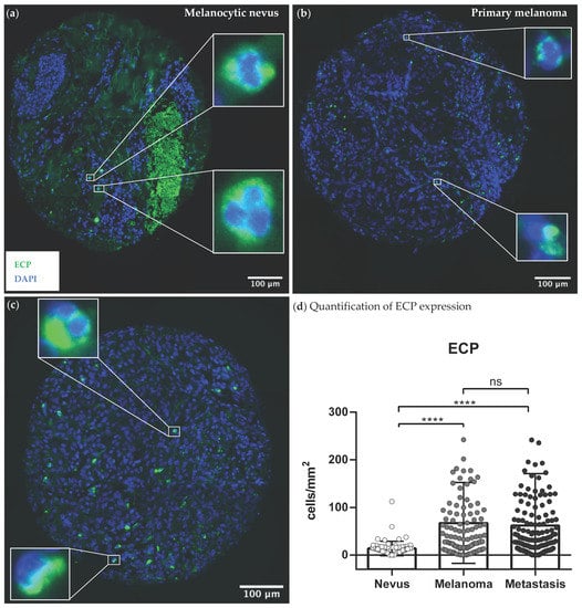
Figure 1.
Representative expression patterns of ECP+ eosinophils in human tumor tissue sections. Paraffin-embedded tissue samples of 73 melanocytic nevi, 105 primaries and 151 metastases from 118 melanoma patients were stained with immunofluorescent anti-ECP antibodies (marked green). Nuclei were stained in blue with DAPI. (a) Exemplary image of a melanocytic nevi stained with anti-ECP antibodies; (b) Exemplary image of a primary melanoma stained with anti-ECP antibodies; (c) Exemplary image of an associated metastasis of melanoma stained with anti-ECP antibodies; (d) Comparison of ECP expression between nevi, primaries and metastases of melanoma patients. Abbreviations: ECP: eosinophil cationic protein; DAPI = 4′,6-Diamidin-2-phenylindol; ns: not significant; ****: p < 0.0001.
In summary, an increased infiltration and activation of eosinophils was seen in melanoma tissues (Figure 1b,c, Supplementary Figures S1b,c and S2b,c). This does not differ significantly between the different stages of progression, such as between primary tumors and metastases in advanced melanoma.
3.3. Quantification of Tumor-Infiltrating Effector T-Cells
To investigate whether the infiltration of eosinophils was related to the infiltration of effector T-cells, nevi (Figure 2a), primaries (Figure 2b) and metastases (Figure 2c) were additionally stained with antibodies against CD8. This allowed an assessment of the amount of T-cells in the same tissue sections, using the mean of the counted T-cells in all three co-stainings (Siglec-8 + CD8, ECP + CD8, EPX + CD8). A significant six-fold reduction in CD8 expression was seen in nevi compared to primaries and a eight-and-a-half-fold reduction in expression compared to metastases. The CD8+ T-cells were 26% more abundant in the metastases than primaries and thus there was no significant difference between primaries and metastases (Figure 2d).
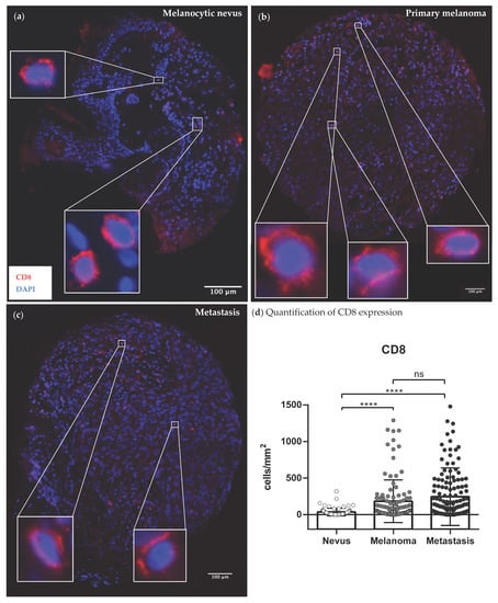
Figure 2.
Representative expression patterns of CD8+ effector T-cells in human tumor tissue sections. Paraffin-embedded tissue samples of 77 melanocytic nevi, 108 primaries and 177 metastases from 118 melanoma patients were stained with immunofluorescent anti-CD8 antibodies (marked red). Nuclei were stained in blue with DAPI. (a) Exemplary image of a melanocytic nevi stained with anti-CD8 antibodies; (b) Exemplary image of a primary melanoma stained with anti-CD8 antibodies; (c) Exemplary image of an associated metastasis of melanoma stained with anti-CD8 antibodies; (d) Comparison of CD8 expression between nevi, primaries and metastases of melanoma patients. Abbreviations: CD: cluster of differentiation; DAPI = 4′,6-Diamidin-2-phenylindol; ns: not significant; ****: p < 0.0001.
3.4. Correlation of Tumor-Infiltrating Eosinophils and Effector T-Cells in Melanoma
Since infiltration of Siglec-8, ECP and EPX expressing eosinophils, as well as CD8+ effector T-cells could previously be observed in the tissue samples of melanoma patients, their correlation was further investigated. In all tissue samples, a positive correlation existed between the amount of infiltrating eosinophils and effector T-cells. The expression of all eosinophil markers (Siglec-8, ECP, EPX) correlated with the level of the expressed T-cell marker (CD8), as shown in Table 2.

Table 2.
Line-up of Kendall-Tau correlations between eosinophil markers (Siglec-8, ECP, EPX) and T-cell marker CD8.
As an illustration, an associated metastasis of melanoma is depicted below, which shows a high infiltration of ECP expressing eosinophils in combination with CD8 positive effector T-cells (Figure 3). Further example images of the coherent expression of the other eosinophil markers (Siglec-8, EPX) and T-cell marker are shown in Supplementary Figures S3 and S4.
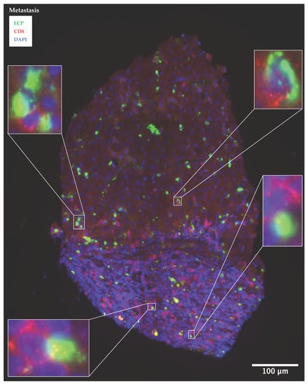
Figure 3.
Melanoma metastasis with a high contiguous infiltration of ECP expressing eosinophils and CD8 expressing effector T-cells. Paraffin-embedded tissue samples of a metastasis are co-stained with immunofluorescent anti-ECP antibodies (marked green) and anti-CD8 antibodies (marked red). Nuclei were stained in blue with DAPI. Abbreviations: ECP: eosinophil cationic protein; CD: cluster of differentiation; DAPI = 4′,6-Diamidin-2-phenylindol.
3.5. Association between the Amount of Tumor-Infiltrated Activated Eosinophils as Well as Effector T-Cells and the Survival of Melanoma Patients
Survival analyses were performed to examine how eosinophil and T-cell infiltration in the primaries affects melanoma progression. Higher counts of ECP+ eosinophils (cut-off 81.26 cells per mm2, Figure 4a) and CD8+ effector T-cells (cut-off 61.89 cells/mm2, Figure 4b) were related to prolonged PFS in primary melanoma. In contrast, the level of expressed Siglec-8 (Supplementary Figure S5a) and EPX (Supplementary Figure S5b) in the primary tumors had no influence on PFS.

Figure 4.
High numbers of ECP expressing eosinophils and CD8 expressing effector T-cells are linked with prolonged PFS in primary melanoma. Kaplan–Meier survival curves for PFS of melanoma patients were stratified by the level (high vs. low infiltration) of ECP+ eosinophils and CD8+ T-cells in the 108 primary melanoma samples. p-values were calculated by the two-sided log-rank test. (a) Survival analysis of melanoma patients with increased (>81.26 cells/mm2) ECP expressing cells in the primaries of melanoma versus those with low ECP expression; (b) Survival analysis for PFS of melanoma patients with increased (>61.89 cells/mm2) CD8 expressing cells in the primaries of melanoma versus those with low CD8 expression. Abbreviations: PFS: progression-free survival; ECP: eosinophil cationic protein; CD: cluster of differentiation; p: p-value.
Similar analyses were conducted on the corresponding nevi (Supplementary Figure S6a–d) and associated metastases (Supplementary Figure S7a–d) of the melanoma patients, where an opposite effect was found. Higher expressions of Siglec-8, ECP, EPX and CD8 were associated with accelerated progression here (Supplementary Figures S6 and S7).
Taken together, the extent of T-cell infiltrates correlates positively with infiltrating eosinophils in melanoma. A high proportion of infiltrated activated (ECP+) eosinophils as well as CD8+ effector T-cells in primary melanomas had a favourable impact on the prognosis of melanoma patients. The way in which eosinophil count and activation status might affect ICI treatment efficacy was then further investigated in the blood-based analysis during ICI.
3.6. Blood-Based Analysis during ICI—Patient Characteristics
For systemic analyses, the peripheral blood of 45 metastatic unresected melanoma patients treated with ICI was analyzed (Table 3). Among them were 32 males (71.1%) and 13 females (28.9%) with a median age of 60.9 (standard deviation of 18.2 years). Based on the current AJCC classification (8th edition), 4 patients were classified as unresectable stage III melanoma and 41 as unresectable stage IV melanoma. Among the cohort were 4 metastatic uveal melanoma and no mucosal melanoma. ICI was administered to all patients; 6 (13.3%) received monotherapy with nivolumab and 11 (24.5%) with pembrolizumab, while 28 (62.2%) received ipilimumab combined with nivolumab. Before the start of ICI, 16 patients received another systemic therapy. 26 patients experienced irAEs, of which 16 (35.6%) were grade I or II and 10 (22.2%) were grade III or IV. Among them, two received a steroid shot together with infliximab, one received two steroid shots and one received carbimazole to alleviate the side effects. Treatment response was assessed by the RECIST 1.1 criteria, with 13 (28.9%) melanoma patients achieving a CR and 16 (35.6%) a PR. These patients were considered as responders, while 5 (11.1%) melanoma patients with a SD and the 11 (24.4%) patients with PD were defined as non-responders.

Table 3.
Patient description of the 45 included advanced stage melanoma patients for the blood analyses.
3.7. High Absolute Eosinophil Count and Elevated ECP Serum Levels Prior to ICI Initiation Are Related to Delayed Progression
To determine whether increased numbers of activated ECP-expressing eosinophils in the primary melanomas translate into increased numbers of degranulated ECP in the serum and which prognostic impact this has on ICI, a second cohort was examined (Table 3). For this purpose, we measured the absolute eosinophil count (AEC) in the peripheral blood as well as the serum level of ECP at two different time points–before ICI (baseline) and before the fourth infusion cycle of ICI (C4)–and performed univariante survival analyses (Figure 5).
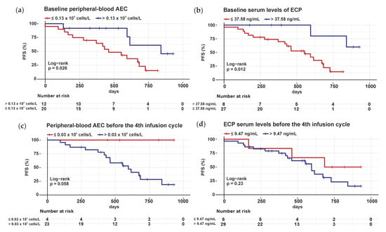
Figure 5.
Elevated baseline peripheral blood AEC and ECP serum levels are associated with extended PFS in stage III and IV melanoma patients treated with ICI. Blood was taken before the start of ICI (baseline) and shortly before the respective infusion cycle of ICI (1st cycle, C1; 2nd cycle C2; 3rd cycle C3, 4th cycle C4; etc.). For our concentration measurements, we used serum at baseline and C4; Kaplan–Meier survival curves for PFS of advanced staged melanoma patients were stratified by the amount (high vs. low) of peripheral-blood AEC and ECP serum levels. p-values were calculated by the two-sided log-rank test; (a) Survival analyses for PFS of advanced melanoma patients with elevated (>0.13 × 10⁹ cells/L) baseline AEC versus those with low peripheral blood AEC; (b) Survival analyses for PFS of advanced melanoma patients with elevated (>37.58 ng/mL) baseline ECP serum levels versus those with low ECP levels; (c) Survival analyses for PFS of advanced melanoma patients with elevated (>0.03 × 10⁹ cells/L) C4 AEC versus those with low peripheral blood AEC; (d) Survival analyses for PFS of advanced melanoma patients with elevated (>9.47 ng/mL) C4 ECP serum levels versus those with low ECP levels. Abbreviations: AEC: absolute eosinophil count; ECP: eosinophil cationic protein; PFS: progression-free survival; ICI: immune checkpoint inhibition; C4: 4th infusion cycle; p: p-value.
Patients with high baseline AEC (>0.13 × 10⁹ cells/L, Figure 5a) and high ECP (>37.58 ng/mL, Figure 5b) serum levels showed significantly prolonged PFS compared to those with low baseline AEC and ECP levels. In contrast, a correlation between increased C4 AEC (>0.03 × 10⁹ cells/L, Figure 5c) and ECP (>9.47 ng/mL, Figure 5d) levels and earlier disease progression was found during therapy, but was not significant.
The same concentration measurements were also carried out with EPX, the second eosiophil activity marker. Melanoma patients with an elevated (>1.07 ng/mL) baseline EPX serum level also showed a significant delay in progression (Supplementary Figure S8a), while the level of EPX serum at C4 had no effect on PFS (Supplementary Figure S8b).
In addition, survival analyses for OS of the 45 metastatic melanoma patients were evaluated using peripheral-blood AEC (Supplementary Figure S9a,b), ECP (Supplementary Figure S9c,d) and EPX (Supplementary Figure S9e,f) serum levels at BE and C4, as shown in Supplementary Figure S9a–f. A significantly prolonged OS was observed for melanoma patients with reduced C4 AEC levels (Supplementary Figure S9b) as well as elevated baseline and C4 EPX serum levels (Supplementary Figure S9e,f).
To summarize, in metastatic melanoma, a high number of eosinophils and strong eosinophil activation in the blood is associated with slower disease progression in patients receiving ICI.
3.8. Constant to Decreasing AEC and ECP Levels between Baseline and the Fourth Infusion Cycle of ICI Are Associated with Later Progression of Metastatic Melanoma
To explore whether the trend in AEC and ECP serum levels during ICI is relevant to treatment outcome, progression analysis was performed. Therefore, the absolute change in AEC and ECP values between C4 and BE was calculated and univariate survival analyses for the PFS of the 45 patients with metastatic melanoma were performed based on the AEC or ECP differences.
Melanoma patients with decreasing (cut-off −0.06 × 10⁹ cells/L) concentrations of AEC between baseline and the fourth infusion cycle of ICI achieved a prolonged PFS compared to patients with increasing peripheral-blood AEC (Figure 6a). Likewise, constant to decreasing (cut-off 3.46 ng/mL) ECP serum levels during ICI were associated with delayed progression in stage III and IV melanoma patients (Figure 6b). The same was done for EPX, but no significant result was obtained (Supplementary Figure S10a).
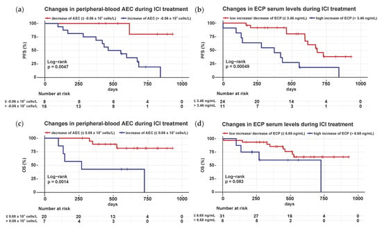
Figure 6.
Decreasing peripheral-blood AEC, as well as constant to decreasing ECP serum levels between baseline and the fourth infusion cycle of ICI correlate with extended PFS and OS in met-astatic melanoma. For the kinetic analyses, the level of AEC and ECP before the fourth infusion cycle of ICI (C4) was subtracted from the corresponding level before ICI was initiated (baseline). Kaplan–Meier survival curves for PFS and OS of patients with advanced melanoma were stratified by the extent of the difference between baseline and C4 values of AEC and ECP in blood. p-values were calculated by the two-sided log-rank test; (a) Survival analyses for PFS of stage III and IV melanoma patients with decreasing (cut-off −0.06 × 10⁹ cells/L) AEC versus those with increasing AEC in peripheral blood between baseline and C4; (b) Survival analyses for PFS of stage III and IV melanoma patients with constant to decreasing (cut-off 3.46 ng/mL) ECP serum levels compared to patients with increasing ECP levels between baseline and C4; (c) Survival analyses for OS of stage III and IV melanoma patients with decreasing (cut-off 0.08 × 10⁹ cells/L) AEC versus those with in-creasing AEC in peripheral blood between baseline and C4; (d) Survival analyses for OS of stage III and IV melanoma patients with low increasing to decreasing (cut-off 6.68 ng/mL) ECP serum levels compared to patients with high increasing ECP levels between baseline and C4. Abbreviations: AEC: absolute eosinophil count; ECP: eosinophil cationic protein; ICI: immune checkpoint inhibition; C4: 4th infusion cycle; PFS: progression-free survival; OS: overall survival; BE: baseline; p: p-value.
Similar survival analyses were conducted for the OS of the metastatic melanoma patients, with decreasing (>0.08 × 10⁹ cells/L) concentrations of AEC during ICI, showing an association with prolonged OS (Figure 6c). Instead, the kinetics of ECP (Figure 6d) and EPX (Supplementary Figure S10b) levels between baseline and C4 had no significant effect on OS in this patient cohort.
3.9. Responders and Non-Responders Do Not Differ in Peripheral Blood AEC, as Well as ECP and EPX Serum Levels
Next, the predictive value of AEC, ECP and EPX was evaluated. We divided the 45 patients with metastatic melanoma into responders (including CR and PR) and non-responders (including SD and PD), and compared baseline AEC, ECP and EPX within both groups. Analysis of variance (one-way ANOVA) revealed no significant difference neither in baseline peripheral-blood AEC (Supplementary Figure S11a) nor ECP (Supplementary Figure S11b) and EPX (Supplementary Figure S11c) serum levels between responders and non-responders.
Corresponding ANOVA analyses were conducted for the C4 time point. During ICI, there were likewise no differences in C4 levels of AEC (Supplementary Figure S12a), ECP (Supplementary Figure S12b) and EPX (Supplementary Figure S12c) between responders and non-responders.
4. Discussion
Melanoma is considered one of the most immunogenic malignancies, which highlights the role of the immune system in tumor control [27,28]. As T-cells represent the effector cells of the immunological tumor defence, their infiltration is crucial for achieving an anti-tumoral response. It is hence of immense prognostic importance [29,30,31,32]. Detailed knowledge of the TME in individual melanoma patients and characterization of cell populations favouring an anti-tumor immune response are still lacking. Recently, it has been reported that eosinophils can facilitate the recruitment and infiltration of T-cells into mice bearing melanoma [18,19].
In the first part of this study, the co-occurrence of eosinophils and T-cells as well as their prognostic value were investigated using co-immunofluorescence staining in human melanoma sections. We observed a strong infiltration of eosinophils in the primary melanomas and associated metastases (Figure 1, Figures S1 and S2), which is consistent with studies in various other solid tumor entities [33]. The expression of eosinophil surface marker protein Siglec-8 correlated positively with CD8 of the effector T-cells (Table 2, Figure S3). Lucarini et al. had previously shown in a mouse model of melanoma that anti-Siglec-F antibody injection was associated with reduced tumoral recruitment of CD8+ effector T-cells, indicating reciprocal promotion of tumor infiltration [18]. The literature suggests that tumor necrosis factor (TNF)- and interferon (INF)γ-activated eosinophils secrete T-cell attracting cytokines such as CCL5, CXCL9 or CXCL10, inducing CD8+ T-cell migration into the TME and resulting in anti-tumoral T-cell response [19,34,35,36]. Accordingly, the activity status of the eosinophils seems particularly relevant for the crosstalk with T-cells. In order to determine whether eosinophils in human melanoma sections were also activated, we co-stained the eosinophil activity markers ECP and EPX with CD8. Again, a positive correlation was found between the infiltrated activated ECP as well as EPX expressing eosinophils and CD8 expressing T-cells (Table 2, Figure 3 and Figure S4). Thus, our study demonstrates a positive association between the infiltration of activated eosinophils and CD8+ T-cells in human melanoma tissue and is in line with a previous report in a small patient cohort [37]. Furthermore, none of the melanoma patients we recruited received systematic therapy before sampling, which renders a therapy-associated effect impossible.
Likewise, with regard to the prognostic potential of the infiltrated eosinophils, their activity state proved to be decisive. Melanoma patients with a high number of ECP expressing eosinophils and CD8+ T-cells in primary melanomas showed prolonged PFS (Figure 4). This observation could be explained by the anti-tumorigenic activity of eosinophils, which has been noted in numerous mice melanoma experiments [18,19,38,39]. Eosinophils mediate anti-tumor responses via direct and indirect mechanisms, e.g., by secreting tumor cytotoxic proteins, such as major basic protein (MBP), ECP, eosinophil-derived neurotoxin (EDN) and granzymes, as well as by limiting tumor cell migration through secretion of interleukin (IL)-12 and IL-10. Additionally, eosinophils are capable of remodelling the TME by expressing natural killer (NK)-cell-associated activation receptors, such as 2B4, NKG2D and LY49, and promoting anti-tumor immunity through release of IFNγ. Besides, they support the polarization of macrophages towards an anti-tumorigenic phenotype and anti-tumor immunity by normalizing the vasculature [33,34].
However, increased amounts of eosinophils and T-cells in metastases of melanoma patients were associated with earlier progression, suggesting an ambivalent role of eosinophils in tumor progression (Figure S7). This opposing trend of eosinophils in primary tumors versus in metastases of melanoma patients need to be re-evaluated in a larger cohort, and at the same time the impact on OS should be considered. Unfortunately, we could not perform survival analyses regarding the OS of the melanoma patients, as a too small proportion of patients died during the observation period. Eosinophils are regarded as heterogeneous immune cells that can polarize in different ways and thus act either pro- or anti-tumoral [22,40]. Therefore, eosinophils could be polarized differently depending on the time phase of tumor growth. More detailed research would be needed to identify the molecular structures in the TME that promote each type of polarization.
The favourable effect of ECP expressing eosinophils in melanoma primaries on prognosis observed in this study could be explained in two ways: in vitro, ECP has been shown to induce tumor lysis through its cytotoxic capacity, which may clarify the delayed progression in primary melanomas with high ECP expression, which we demonstrated [41,42,43]. On the other hand, the activated eosinophils could also have indirectly contributed to a sufficient T-cell response against the melanoma via increased T-cell recruitment, as previously demonstrated in melanoma-bearing mice [18,19]. Our analyses provide a first indication that co-staining of ECP and CD8 in primary melanomas could be used as a prognostic tool to better predict individual melanoma progression.
As melanoma is a rapidly growing tumor entity with a strong tendency to metastasize, such prognostic biomarkers are tremendously important [44]. In this context, biomarkers that can be measured in routine clinical laboratory tests are particularly useful for clinical practice. Consequently, in the second part of this study, we analyzed blood samples from metastatic melanoma patients to determine whether eosinophils and their activity markers ECP and EPX could act as prognostic biomarkers. Assuming that eosinophils contribute to a functioning anti-melanoma T-cell response, they might also enhance the efficiency of ICI, which led to the selection of a second cohort of melanoma patients receiving ICI (Table 3).
Elevated baseline peripheral-blood AEC was found to be associated with improved PFS in 45 unresected stage III and IV melanoma patients (Figure 5a). The baseline level of AEC, however, had no influence on the OS in our cohort (Figure S9a), which contrasts with the study by Martens et al., where a correlation between OS of the 209 stage IV melanoma patients and baseline AEC was confirmed [45]. Analyses of two further large collectives of stage III and IV melanoma patients revealed that relative eosinophil counts correlate with OS [46,47]. As we wanted to test the prognostic effect of eosinophils within a cohort receiving all approved ICI therapy regimens, the discrepancy could be attributed to different patient cohorts and especially to differing follow-up periods. Our results would need to be further monitored over the current follow-up period of approximately 2.5 years. The patient population should be expanded to re-examine the impact of AEC on OS. Concerning a long follow-up period of 12 years, another study of 172 stage IV melanoma patients indicated that regardless of the ICI treatment and timing, any increase in eosinophils resulted in extended survival times [48]. This finding differs from our measurements during ICI, where an increase in AEC between baseline and the fourth infusion cycle was associated with shorter PFS and OS (Figure 6a,c). We are the first research group to observe that a decrease in peripheral-blood eosinophils during ICI led to later disease progression (Figure 6a). All other studies reported a prognostic advantage for melanoma patients with an increase in eosinophil count during ICI treatment [37,49,50]. Indeed, we demonstrated the association of an early increase in AEC with a better outcome after treatment with ipilimumab in previous research analyzing the peripheral blood of 59 stage IV melanoma patients [49]. In addition, higher AEC was measurable in responders, whereas in our analyses the amount of AEC had no influence on treatment response. The inclusion of other immune checkpoint inhibitors, different measurement time points and a varied distribution of responders and non-responders might be responsible for the fact that a predictive value of baseline AEC could not be verified here. Our results support the prognostic value of eosinophils but verification in a larger cohort at all infusion time points of ICI is needed.
To investigate whether activated serum eosinophils have a different effect and their degranulated ECP and EPX can be used as biomarkers, ELISA experiments were per-formed. Higher baseline serum levels of ECP were related to extended PFS in metastatic melanoma patients (Figure 5b). Reduced serum levels of ECP, on the other hand, tended to be associated with a longer OS (Figure S9c). So far, only one other study has analyzed the prognostic effect of ECP serum levels in 56 metastatic melanoma patients, although just 27 patients received ICI. Here, a negative correlation was also seen between high ECP serum levels and the length of OS [51]. Thus, our analyses in a cohort with only melanoma patients receiving ICI provide first evidence that the baseline ECP serum level may be suitable as a prognostic biomarker and that especially a decrease during ICI therapy is prognostically favourable (Figure 6b). Further multicentre studies in metastatic melanoma patients treated with ICI are needed to confirm this finding and, more importantly, to elucidate the underlying mechanisms of the ambivalent ECP effect in vivo. In addition, future studies must be designed to correlate ECP levels in the TME as well as in serum. Only then conclusions can be drawn concerning whether, for example, a high baseline serum ECP level is associated with a lower proportion of infiltrated ECP+ eosinophils in melanoma tissue as these are mobilized into the blood, or instead is linked to a high ECP tissue eosinophilia. With such analyses, it would be possible to explain the different prognostic impact we observed of a high ECP infiltrate in the metastases of the first cohort and elevated baseline ECP level of the second cohort.
Furthermore, our analyses show for the first time an association between increased serum EPX levels and prolonged PFS and OS (Figures S8a and S9e). EPX is considered a very specific eosinophil marker, so it would be particularly desirable to conduct comparative studies demonstrating its prognostic significance [52,53,54].
In summary, our results suggest a correlation of eosinophil and T-cell tumor infiltration. ECP and CD8 co-staining may provide indications for individual prognosis assessment. In addition, high levels of AEC, ECP and EPX in the blood of metastatic melanoma patients showed prognostic value, suggesting their future potential as serological biomarkers.
According to our data, activated eosinophils are part of the tumor-associated inflammatory microenvironment, and both tumor-infiltrating eosinophils and eosinophil blood counts have prognostic significance. In the future, the mechanisms underlying the positive influence of eosinophils on the prognosis of melanoma patients need to be clarified more precisely. There are first studies pointing to the therapeutic potential of eosinophils to affect tumor progression [55,56]. In syngeneic mouse models of hepatocellular carcinoma and breast cancer, additional treatment with an antidiabetic drug, the dipeptidyl peptidase 4 (DPP4) inhibitor sitagliptin, was shown to promote intratumoral recruitment of eosinophils. Interestingly, this phenomenon was associated with accelerated tumor rejection during ICI treatment [56]. To address the current clinical need for new biomarkers in melanoma and to improve the prognosis of metastatic melanoma patients under ICI, eosinophils should be further investigated as a promising cell population.
5. Conclusions
Our data show an association between the extent of eosinophil and T-cell infiltration in melanoma. In particular, a high number of ECP expressing eosinophils and CD8 expressing T-cells in primary melanomas had a favourable impact on prognosis. This suggests that ECP and CD8 as future tissue markers may provide a better prognostic assessment after resection of the primary tumor in melanoma patients based on individual expression patterns. Furthermore, our results reveal the association between elevated AEC, ECP and EPX levels in the peripheral blood and delayed relapse in advanced-stage melanoma patients. For routine clinical practice, measuring blood concentrations of eosinophils, ECP and EPX seem to be relevant prognostic markers.
Overall, eosinophils and their activity markers thus have additional prognostic importance in metastatic melanoma. A deeper understanding of the interaction between eosinophils and T-cells provides the basis for new therapeutic strategies in melanoma patients.
Supplementary Materials
The following supporting information can be downloaded at: https://www.mdpi.com/article/10.3390/cancers14225676/s1, Table S1. Quantification summary of Siglec-8, ECP, EPX and CD8 expressing cells within the human tissue samples represented on the nine TMAs; Figure S1. Representative expression patterns of Siglec-8+ eosinophils in human tumor tissue sections; Figure S2. Representative expression patterns of EPX+ eosinophils in human tumor tissue sections; Figure S3. Primary melanoma with a high contiguous infiltration of Siglec-8 expressing eosinophils and CD8 expressing effector T-cells; Figure S4. Melanoma metastasis with a high contiguous infiltration of EPX expressing eosinophils and CD8 expressing effector T-cells; Figure S5. The number of Siglec-8 and EPX expressing eosinophils has no impact on PFS in primary melanoma; Figure S6. High numbers of Siglec-8, EPX, ECP expressing eosinophils and CD8 expressing effector T-cells are linked with impaired PFS in melanocytic nevi; Figure S7. High numbers of Siglec-8, EPX, ECP expressing eosinophils and CD8 expressing effector T-cells are linked with impaired PFS in melanoma-associated metastases; Figure S8. Elevated baseline EPX serum levels are associated with extended PFS in stage III and IV melanoma patients treated with ICI; Figure S9. Impact of peripheral-blood AEC, ECP and EPX serum levels before and during ICI on OS in metastatic melanoma patients; Figure S10. The progression of serum EPX levels under ICI has no significant impact on the PFS and OS of patients with metastatic melanoma; Figure S11. Responders and non-responders show no difference in their baseline levels of AEC, ECP and EPX; Figure S12. Responders and non-responders show no difference in their C4 levels of AEC, ECP and EPX.
Author Contributions
Conceptualization, C.G. and A.T.B.; methodology, N.L.A., Y.F.S. and C.M.; validation, C.M., C.G., N.L.A. and Y.F.S.; resources, J.U. and K.P.; writing—original draft preparation, N.L.A.; writing—review and editing, Y.F.S., C.M., J.-C.S., G.G., J.K., S.W.S., J.U., K.P., A.T.B. and C.G.; supervision, A.T.B. and C.G.; funding acquisition, C.G. All authors have read and agreed to the published version of the manuscript.
Funding
This research was funded by Hiege-Stiftung gegen Hautkrebs, Germany and the Erich und Gertrud Roggenbuck-Stiftung, Germany.
Institutional Review Board Statement
The study was conducted in accordance with the Declaration of Helsinki, and approved by the Ethics Committee II of Heidelberg University (2010-318N-MA and 2014-835R-MA) and the Hamburg Ethics Committee (PV5392 on 6th December 2016).
Informed Consent Statement
Written informed consent was obtained from all subjects involved in the study.
Data Availability Statement
For original data please contact ch.gebhardt@uke.de.
Acknowledgments
We kindly thank the excellent support by our technicians Ewa Wladykowski and Sabine Vidal-y-Sy. The graphical abstract was created with BioRender.com.
Conflicts of Interest
C.G. is on the advisory board or has received honoraria from Almirall, Amgen, Beiersdorf, BioNTech, Bristol-Myers Squibb, Immunocore, Janssen, MSD Sharp & Dohme, Novartis, Pierre-Fabre Pharma, Roche, Sanofi Genzyme, SUN Pharma and Sysmex/Inostix, research funding from Novartis and Sanofi Genzyme, and travel support from Bristol-Myers Squibb, Pierre Fabre Pharma and SUN Pharma, outside the submitted work. C.G. is co-founder of Dermagnostix and Dermagnostix R&D. J.U. is on the advisory board or has received honoraria and travel support from Amgen, Bristol Myers Squibb, GSK, Immunocore, LeoPharma, Merck Sharp and Dohme, Novartis, Pierre Fabre, Roche, Sanofi outside the submitted work. All of the other authors declare no conflicts of interest.
References
- Pardoll, D.M. The blockade of immune checkpoints in cancer immunotherapy. Nat. Rev. Cancer 2012, 12, 252–264. [Google Scholar] [CrossRef] [PubMed]
- Lipson, E.J.; Drake, C.G. Ipilimumab: An anti-CTLA-4 antibody for metastatic melanoma. Clin. Cancer Res. 2011, 17, 6958–6962. [Google Scholar] [CrossRef] [PubMed]
- Postow, M.A.; Callahan, M.K.; Wolchok, J.D. Immune checkpoint blockade in cancer therapy. J. Clin. Oncol. 2015, 33, 1974–1982. [Google Scholar] [CrossRef] [PubMed]
- Robert, C.; Ribas, A.; Schachter, J.; Arance, A.; Grob, J.J.; Mortier, L.; Daud, A.; Carlino, M.S.; McNeil, C.M.; Lotem, M.; et al. Pembrolizumab versus ipilimumab in advanced melanoma (KEYNOTE-006): Post-hoc 5-year results from an open-label, multicentre, randomised, controlled, phase 3 study. Lancet Oncol. 2019, 20, 1239–1251. [Google Scholar] [CrossRef]
- Larkin, J.; Chiarion-Sileni, V.; Gonzalez, R.; Grob, J.J.; Rutkowski, P.; Lao, C.D.; Cowey, C.L.; Schadendorf, D.; Wagstaff, J.; Dummer, R.; et al. Five-year survival with combined nivolumab and ipilimumab in advanced melanoma. N. Engl. J. Med. 2019, 381, 1535–1546. [Google Scholar] [CrossRef] [PubMed]
- Larkin, J.; Chiarion-Sileni, V.; Gonzalez, R.; Grob, J.J.; Cowey, C.L.; Lao, C.D.; Schadendorf, D.; Dummer, R.; Smylie, M.; Rutkowski, P.; et al. Combined nivolumab and ipilimumab or monotherapy in untreated melanoma. N. Engl. J. Med. 2015, 373, 23–34. [Google Scholar] [CrossRef]
- Postow, M.A.; Sidlow, R.; Hellmann, M.D. Immune-related adverse events associated with immune checkpoint blockade. N. Engl. J. Med. 2018, 378, 158–168. [Google Scholar] [CrossRef]
- Wang, D.Y.; Salem, J.E.; Cohen, J.V.; Chandra, S.; Menzer, C.; Ye, F.; Zhao, S.; Das, S.; Beckermann, K.E.; Ha, L.; et al. Fatal toxic effects associated with immune checkpoint inhibitors: A systematic review and meta-analysis. JAMA Oncol. 2018, 4, 1721–1728. [Google Scholar] [CrossRef]
- Indini, A.; Di Guardo, L.; Cimminiello, C.; Prisciandaro, M.; Randon, G.; De Braud, F.; Del Vecchio, M. Immune-related adverse events correlate with improved survival in patients undergoing anti-PD1 immunotherapy for metastatic melanoma. J. Cancer Res. Clin. Oncol. 2019, 145, 511–521. [Google Scholar] [CrossRef]
- Rogado, J.; Sánchez-Torres, J.M.; Romero-Laorden, N.; Ballesteros, A.I.; Pacheco-Barcia, V.; Ramos-Leví, A.; Arranz, R.; Lorenzo, A.; Gullón, P.; Donnay, O.; et al. Immune-related adverse events predict the therapeutic efficacy of anti-PD-1 antibodies in cancer patients. Eur. J. Cancer 2019, 109, 21–27. [Google Scholar] [CrossRef]
- Duan, Q.; Zhang, H.; Zheng, J.; Zhang, L. Turning cold into hot: Firing up the tumor microenvironment. Trends Cancer 2020, 6, 605–618. [Google Scholar] [CrossRef] [PubMed]
- Gajewski, T.F. The next hurdle in cancer immunotherapy: Overcoming the non-t-cell-inflamed tumor microenvironment. Semin. Oncol. 2015, 42, 663–671. [Google Scholar] [CrossRef] [PubMed]
- Zemek, R.M.; De Jong, E.; Chin, W.L.; Schuster, I.S.; Fear, V.S.; Casey, T.H.; Forbes, C.; Dart, S.J.; Leslie, C.; Zaitouny, A.; et al. Sensitization to immune checkpoint blockade through activation of a STAT1/NK axis in the tumor microenvironment. Sci. Transl. Med. 2019, 11, eaav7816. [Google Scholar] [CrossRef] [PubMed]
- Liu, Z.; Han, C.; Fu, Y.X. Targeting innate sensing in the tumor microenvironment to improve immunotherapy. Cell. Mol. Immunol. 2020, 17, 13–26. [Google Scholar] [CrossRef] [PubMed]
- Mikucki, M.E.; Fisher, D.T.; Matsuzaki, J.; Skitzki, J.J.; Gaulin, N.B.; Muhitch, J.B.; Ku, A.W.; Frelinger, J.G.; Odunsi, K.; Gajewski, T.F.; et al. Non-redundant requirement for CXCR3 signalling during tumoricidal T-cell trafficking across tumour vascular checkpoints. Nat. Commun. 2015, 6, 7458. [Google Scholar] [CrossRef] [PubMed]
- Harlin, H.; Meng, Y.; Peterson, A.C.; Zha, Y.; Tretiakova, M.; Slingluff, C.; McKee, M.; Gajewski, T.F. Chemokine expression in melanoma metastases associated with CD8+ T-cell recruitment. Cancer Res. 2009, 69, 3077–3085. [Google Scholar] [CrossRef] [PubMed]
- Gajewski, T.F.; Corrales, L.; Williams, J.; Horton, B.; Sivan, A.; Spranger, S. Cancer immunotherapy targets based on understanding the t-cell-inflamed versus non-T-cell-inflamed tumor microenvironment. Adv. Exp. Med. Biol. 2017, 1036, 19–31. [Google Scholar] [CrossRef]
- Lucarini, V.; Ziccheddu, G.; Macchia, I.; La Sorsa, V.; Peschiaroli, F.; Buccione, C.; Sistigu, A.; Sanchez, M.; Andreone, S.; D’Urso, M.T.; et al. IL-33 restricts tumor growth and inhibits pulmonary metastasis in melanoma-bearing mice through eosinophils. Oncoimmunology 2017, 6, e1317420. [Google Scholar] [CrossRef]
- Carretero, R.; Sektioglu, I.M.; Garbi, N.; Salgado, O.C.; Beckhove, P.; Hämmerling, G.J. Eosinophils orchestrate cancer rejection by normalizing tumor vessels and enhancing infiltration of CD8(+) T-cells. Nat. Immunol. 2015, 16, 609–617. [Google Scholar] [CrossRef]
- Floyd, H.; Ni, J.; Cornish, A.L.; Zeng, Z.; Liu, D.; Carter, K.C.; Steel, J.; Crocker, P.R. Siglec-8. A novel eosinophil-specific member of the immunoglobulin superfamily. J. Biol. Chem. 2000, 275, 861–866. [Google Scholar] [CrossRef]
- Kikly, K.K.; Bochner, B.S.; Freeman, S.D.; Tan, K.B.; Gallagher, K.T.; D’alessio, K.J.; Holmes, S.D.; Abrahamson, J.A.; Erickson-Miller, C.L.; Murdock, P.R.; et al. Identification of SAF-2, a novel siglec expressed on eosinophils, mast cells, and basophils. J. Allergy Clin. Immunol. 2000, 105, 1093–1100. [Google Scholar] [CrossRef] [PubMed]
- Varricchi, G.; Galdiero, M.R.; Loffredo, S.; Lucarini, V.; Marone, G.; Mattei, F.; Marone, G.; Schiavoni, G. Eosinophils: The unsung heroes in cancer? Oncoimmunology 2017, 7, e1393134. [Google Scholar] [CrossRef] [PubMed]
- Venge, P.; Byström, J.; Carlson, M.; Hâkansson, L.; Karawacjzyk, M.; Peterson, C.; Sevéus, L.; Trulson, A. Eosinophil cationic protein (ECP): Molecular and biological properties and the use of ECP as a marker of eosinophil activation in disease. Clin. Exp. Allergy 1999, 29, 1172–1186. [Google Scholar] [CrossRef] [PubMed]
- Gershenwald, J.E.; Scolyer, R.A.; Hess, K.R.; Sondak, V.K.; Long, G.V.; Ross, M.I.; Lazar, A.J.; Faries, M.B.; Kirkwood, J.M.; McArthur, G.A.; et al. Melanoma staging: Evidence-based changes in the American Joint Committee on Cancer eighth edition cancer staging manual. CA Cancer J. Clin. 2017, 67, 472–492. [Google Scholar] [CrossRef]
- Therasse, P.; Arbuck, S.G.; Eisenhauer, E.A.; Wanders, J.; Kaplan, R.S.; Rubinstein, L.; Verweij, J.; Van Glabbeke, M.; van Oosterom, A.T.; Christian, M.C.; et al. New guidelines to evaluate the response to treatment in solid tumors. European Organization for Research and Treatment of Cancer, National Cancer Institute of the United States, National Cancer Institute of Canada. J. Natl. Cancer Inst. 2000, 92, 205–216. [Google Scholar] [CrossRef] [PubMed]
- Eisenhauer, E.A.; Therasse, P.; Bogaerts, J.; Schwartz, L.H.; Sargent, D.; Ford, R.; Dancey, J.; Arbuck, S.; Gwyther, S.; Mooney, M.; et al. New response evaluation criteria in solid tumours: Revised RECIST guideline (version 1.1). Eur. J. Cancer 2009, 45, 228–247. [Google Scholar] [CrossRef]
- Schumacher, T.N.; Schreiber, R.D. Neoantigens in cancer immunotherapy. Science 2015, 348, 69–74. [Google Scholar] [CrossRef]
- Jacobs, J.F.; Nierkens, S.; Figdor, C.G.; de Vries, I.J.; Adema, G.J. Regulatory T cells in melanoma: The final hurdle towards effective immunotherapy? Lancet Oncol. 2012, 13, e32–e42. [Google Scholar] [CrossRef]
- Dunn, G.P.; Bruce, A.T.; Ikeda, H.; Old, L.J.; Schreiber, R.D. Cancer immunoediting: From immunosurveillance to tumor escape. Nat. Immunol. 2002, 3, 991–998. [Google Scholar] [CrossRef]
- Mlecnik, B.; Tosolini, M.; Kirilovsky, A.; Berger, A.; Bindea, G.; Meatchi, T.; Bruneval, P.; Trajanoski, Z.; Fridman, W.H.; Pagès, F.; et al. Histopathologic-based prognostic factors of colorectal cancers are associated with the state of the local immune reaction. J. Clin. Oncol. 2011, 29, 610–618. [Google Scholar] [CrossRef]
- Kondratiev, S.; Sabo, E.; Yakirevich, E.; Lavie, O.; Resnick, M.B. Intratumoral CD8+ T lymphocytes as a prognostic factor of survival in endometrial carcinoma. Clin. Cancer Res. 2004, 10, 4450–4456. [Google Scholar] [CrossRef] [PubMed]
- Piras, F.; Colombari, R.; Minerba, L.; Murtas, D.; Floris, C.; Maxia, C.; Corbu, A.; Perra, M.T.; Sirigu, P. The predictive value of CD8, CD4, CD68, and human leukocyte antigen-D-related cells in the prognosis of cutaneous malignant melanoma with vertical growth phase. Cancer 2005, 104, 1246–1254. [Google Scholar] [CrossRef] [PubMed]
- Grisaru-Tal, S.; Itan, M.; Klion, A.D.; Munitz, A. A new dawn for eosinophils in the tumour microenvironment. Nat. Rev. Cancer 2020, 20, 594–607. [Google Scholar] [CrossRef] [PubMed]
- Grisaru-Tal, S.; Rothenberg, M.E.; Munitz, A. Eosinophil-lymphocyte interactions in the tumor microenvironment and cancer immunotherapy. Nat. Immunol. 2022, 23, 1309–1316. [Google Scholar] [CrossRef] [PubMed]
- Grisaru-Tal, S.; Dulberg, S.; Beck, L.; Zhang, C.; Itan, M.; Hediyeh-Zadeh, S.; Caldwell, J.; Rozenberg, P.; Dolitzky, A.; Avlas, S.; et al. Metastasis-entrained eosinophils enhance lymphocyte-mediated antitumor immunity. Cancer Res. 2021, 81, 5555–5571. [Google Scholar] [CrossRef]
- Liu, L.Y.; Bates, M.E.; Jarjour, N.N.; Busse, W.W.; Bertics, P.J.; Kelly, E.A. Generation of Th1 and Th2 chemokines by human eosinophils: Evidence for a critical role of TNF-alpha. J. Immunol. 2007, 179, 4840–4848. [Google Scholar] [CrossRef]
- Simon, S.C.S.; Hu, X.; Panten, J.; Grees, M.; Renders, S.; Thomas, D.; Weber, R.; Schulze, T.J.; Utikal, J.; Umansky, V. Eosinophil accumulation predicts response to melanoma treatment with immune checkpoint inhibitors. Oncoimmunology 2020, 9, 1727116. [Google Scholar] [CrossRef]
- Ikutani, M.; Yanagibashi, T.; Ogasawara, M.; Tsuneyama, K.; Yamamoto, S.; Hattori, Y.; Kouro, T.; Itakura, A.; Nagai, Y.; Takaki, S.; et al. Identification of innate IL-5-producing cells and their role in lung eosinophil regulation and antitumor immunity. J. Immunol. 2012, 188, 703–713. [Google Scholar] [CrossRef]
- Mattes, J.; Hulett, M.; Xie, W.; Hogan, S.; Rothenberg, M.E.; Foster, P.; Parish, C. Immunotherapy of cytotoxic T cell-resistant tumors by T helper 2 cells: An eotaxin and STAT6-dependent process. J. Exp. Med. 2003, 197, 387–393. [Google Scholar] [CrossRef]
- Simon, S.C.S.; Utikal, J.; Umansky, V. Opposing roles of eosinophils in cancer. Cancer Immunol. Immunother. 2019, 68, 823–833. [Google Scholar] [CrossRef]
- De Lima, P.O.; Dos Santos, F.V.; Oliveira, D.T.; de Figueiredo, R.C.; Pereira, M.C. Effect of eosinophil cationic protein on human oral squamous carcinoma cell viability. Mol. Clin. Oncol. 2015, 3, 353–356. [Google Scholar] [CrossRef] [PubMed]
- Legrand, F.; Driss, V.; Delbeke, M.; Loiseau, S.; Hermann, E.; Dombrowicz, D.; Capron, M. Human eosinophils exert TNF-α and granzyme A-mediated tumoricidal activity toward colon carcinoma cells. J. Immunol. 2010, 185, 7443–7451. [Google Scholar] [CrossRef]
- Glimelius, I.; Rubin, J.; Fischer, M.; Molin, D.; Amini, R.M.; Venge, P.; Enblad, G. Effect of eosinophil cationic protein (ECP) on Hodgkin lymphoma cell lines. Exp. Hematol. 2011, 39, 850–858. [Google Scholar] [CrossRef] [PubMed]
- Umansky, V.; Utikal, J.; Gebhardt, C. Predictive immune markers in advanced melanoma patients treated with ipilimumab. Oncoimmunology 2016, 5, e1158901. [Google Scholar] [CrossRef] [PubMed]
- Martens, A.; Wistuba-Hamprecht, K.; Geukes Foppen, M.; Yuan, J.; Postow, M.A.; Wong, P.; Romano, E.; Khammari, A.; Dreno, B.; Capone, M.; et al. Baseline peripheral blood biomarkers associated with clinical outcome of advanced melanoma patients treated with ipilimumab. Clin. Cancer Res. 2016, 22, 2908–2918. [Google Scholar] [CrossRef]
- Rosner, S.; Kwong, E.; Shoushtari, A.N.; Friedman, C.F.; Betof, A.S.; Brady, M.S.; Coit, D.G.; Callahan, M.K.; Wolchok, J.D.; Chapman, P.B.; et al. Peripheral blood clinical laboratory variables associated with outcomes following combination nivolumab and ipilimumab immunotherapy in melanoma. Cancer Med. 2018, 7, 690–697. [Google Scholar] [CrossRef]
- Weide, B.; Martens, A.; Hassel, J.C.; Berking, C.; Postow, M.A.; Bisschop, K.; Simeone, E.; Mangana, J.; Schilling, B.; Di Giacomo, A.M.; et al. Baseline biomarkers for outcome of melanoma patients treated with pembrolizumab. Clin. Cancer Res. 2016, 22, 5487–5496. [Google Scholar] [CrossRef]
- Moreira, A.; Leisgang, W.; Schuler, G.; Heinzerling, L. Eosinophilic count as a biomarker for prognosis of melanoma patients and its importance in the response to immunotherapy. Immunotherapy 2017, 9, 115–121. [Google Scholar] [CrossRef]
- Gebhardt, C.; Sevko, A.; Jiang, H.; Lichtenberger, R.; Reith, M.; Tarnanidis, K.; Holland-Letz, T.; Umansky, L.; Beckhove, P.; Sucker, A.; et al. Myeloid cells and related chronic inflammatory factors as novel predictive markers in melanoma treatment with ipilimumab. Clin. Cancer Res. 2015, 21, 5453–5459. [Google Scholar] [CrossRef]
- Delyon, J.; Mateus, C.; Lefeuvre, D.; Lanoy, E.; Zitvogel, L.; Chaput, N.; Roy, S.; Eggermont, A.M.; Routier, E.; Robert, C. Experience in daily practice with ipilimumab for the treatment of patients with metastatic melanoma: An early increase in lymphocyte and eosinophil counts is associated with improved survival. Ann. Oncol. 2013, 24, 1697–1703. [Google Scholar] [CrossRef]
- Krückel, A.; Moreira, A.; Fröhlich, W.; Schuler, G.; Heinzerling, L. Eosinophil-cationic protein—A novel liquid prognostic biomarker in melanoma. BMC Cancer 2019, 19, 207. [Google Scholar] [CrossRef] [PubMed]
- Nair, P.; Ochkur, S.I.; Protheroe, C.; Radford, K.; Efthimiadis, A.; Lee, N.A.; Lee, J.J. Eosinophil peroxidase in sputum represents a unique biomarker of airway eosinophilia. Allergy 2013, 68, 1177–1184. [Google Scholar] [CrossRef] [PubMed]
- Metso, T.; Venge, P.; Haahtela, T.; Peterson, C.G.; Sevéus, L. Cell specific markers for eosinophils and neutrophils in sputum and bronchoalveolar lavage fluid of patients with respiratory conditions and healthy subjects. Thorax 2002, 57, 449–451. [Google Scholar] [CrossRef] [PubMed]
- Saffari, H.; Leiferman, K.M.; Clayton, F.; Baer, K.; Pease, L.F.; Gleich, G.J.; Peterson, K.A. Measurement of inflammation in eosinophilic esophagitis using an eosinophil peroxidase assay. Am. J. Gastroenterol. 2016, 111, 933–939. [Google Scholar] [CrossRef]
- Rafei-Shamsabadi, D.; Lehr, S.; von Bubnoff, D.; Meiss, F. Successful combination therapy of systemic checkpoint inhibitors and intralesional interleukin-2 in patients with metastatic melanoma with primary therapeutic resistance to checkpoint inhibitors alone. Cancer Immunol. Immunother. 2019, 68, 1417–1428. [Google Scholar] [CrossRef]
- Hollande, C.; Boussier, J.; Ziai, J.; Nozawa, T.; Bondet, V.; Phung, W.; Lu, B.; Duffy, D.; Paradis, V.; Mallet, V.; et al. Inhibition of the dipeptidyl peptidase DPP4 (CD26) reveals IL-33-dependent eosinophil-mediated control of tumor growth. Nat. Immunol. 2019, 20, 257–264. [Google Scholar] [CrossRef]
Publisher’s Note: MDPI stays neutral with regard to jurisdictional claims in published maps and institutional affiliations. |
© 2022 by the authors. Licensee MDPI, Basel, Switzerland. This article is an open access article distributed under the terms and conditions of the Creative Commons Attribution (CC BY) license (https://creativecommons.org/licenses/by/4.0/).