Abstract
Algae provide an alternative, sustainable, and environmentally beneficial method of dyetreatment. In this study, algae were successfully used to remove methylene blue (MB) from aqueous solutions. The effects of several parameters, such as initial concentration of MB (5–25 mg L−1), algae dosage (0.02–0.1 g mL−1), temperature (4, 20, and 30 °C), and contact time (24, 48, 72 and 84 h), on MB removal were investigated. In addition, the characterization of MB before and after treatment was achieved using UV-spectrophotometer and Liquid chromatography-mass spectrometry (LC-MS). The experimental data were applied to three kinetic models, namely pseudo-first-order, pseudo-second-order, and Elvoich. Moreover, Langmuir, Freundlich, Dubinin–Raduskevich (D–R), and Temkin isotherm models were tested. The maximum removal efficiency of MB (~96%) was accomplished at optimum conditions at the initial concentration of MB (15 mg L−1), temperature (30 °C), and algae dosage (0.06 g mL−1) after 60 min of contact time. The removal of MB follows the pseudo-second-order kinetic model (R2 > 0.999), and the experimental data is best fitted by the Langmuir isotherm model (R2 > 0.9300).
1. Introduction
The industrial effluents from the textile, cosmetic, paper and printing, rubber, leather, pharmaceutical, food, and plastic industries contain many toxic pollutants, such as dyes, which harm the environment [1]. Dyes can be classified into two categories: non-ionic (vat and disperse dyes) and ionic (cationic (basic) and anionic dyes (reactive, direct, and acidic)) dyes [2]. The presence of dyes in aquatic ecosystems reduces light penetration and thus decreases the photosynthesis necessary for living organisms [3]. In addition, industrial dyes are mutagenic, toxic, and dangerous to humans and animals [4]. Therefore, the removal of dyes from aquatic environments is a necessary process to prevent water pollution [5]. Several processes, including mechanical (e.g., filtration and reverse osmosis), physical (e.g., adsorption, extraction, and flocculation), chemical (e.g., precipitation, oxidation, ion exchange, and ozonolysis), and thermal (e.g., evaporation and distillation) methods, have been used to remediate the contaminated water by dyes [6]. However, these methods have clear disadvantages, such as high cost, secondary pollution, not being effective on a large scale (e.g., remediation of polluted soil), and low efficiency [7]. Therefore, bioremediation using microbial species is a favored alternative process for being highly effective with low cost and environmentally friendly [8].
Algae contain main groups, such as polysaccharides, polyunsaturated fatty acids, pigments, proteins, lipids, vitamins, phycobiliproteins, and enzymes [9]. Consequently, the bio-removal of dyes by algae occurs via the interactions between the functional groups in algae (e.g., carboxylate, hydroxyl, sulfate, phosphate, and amino) with dye molecules [10]. Algae are highly available, and their growth is easy and fast [11]; thus, algae are widely used in environmental remediation for wastewater decolorization [12]. For example, the decolorization of tartrazine by Scenedesmus bijugatus (green algae) [13], direct brown NM dye by immobilized Chlorellaalgae [14], methyl red, orange II, G-Red (FN-3G), basic cationic, and basic fuchsin by Chlorella vulgaris, Lyngbyalagerlerimi, Nostoclincki, Oscillatoria rubescens, Elkatothrixviridis and Volvox aureus [15] were successfully achieved in previous studies. In addition, malachite green was highly removed by Chlorella algae [16] and freshwater algae Pithophora sp. [17].
Methylene blue is a common dye that is largely utilized in industry. Dye leftovers can be found in effluent discharged by such enterprises. As a result, the presence of very low quantities in the effluent is readily apparent. The discharge of methylene blue can produce a variety of issues, including increased COD by the water body, increased toxicity, decreased light penetration and photosynthesis, and damage to the aesthetic character of the water surface. Furthermore, the products of their degradation may be mutagenic and carcinogenic.
The ability of green algae of various types to break down or decolorize large concentrations of poisonous azo dyesis accomplished through many techniques.
Much researchremains to be completed in order to fully comprehend the entire mechanism(s) of deterioration.
To the best of our knowledge, this study is the first to optimize experimental parameters for removing MB from an aqueous solution using Bracteacoccus sp. isolated from northern Jordan and attempt to explain the removal mechanism through kinetic and equilibrium studies. Consequently, the main objectives of the current study were to estimate the ability of Bracteacoccus sp.to remove MB from an aqueous solution where MB dye was selected as a model compound of cationic days, to determine the optimum conditions for obtaining the highest removal of MB, and to illustrate the mechanism for MB removal by applying the kinetic and the equilibrium isotherm models to the experimental data.
2. Materials and Methods
2.1. Materials
All chemicals, such as methylene blue, which is used in the current study, are of analytical grade and used as received without further purification.
The MB used in this study was chosen as the model compound of cationic dyes, and its properties are listed in Table 1. A stock solution of MB 1000 mg L−1 was prepared by dissolving 1.000 g of MB in 50 mL of deionized water and then diluted to 1000 mL. This stock solution was used to prepare the diluted solutions of MB in the range from 5 to 25 mg L−1.

Table 1.
Properties of methylene blue (MB) dye [18].
2.2. Microalgal Culture
Green microalgae were collected from a water stream originating from a water spring located west of Mafraq city in north Jordan (location: 32°12′17.6″ N 36°00′05.1″ E). The samples were directly transferred to the laboratory.
2.3. Isolation, Cultivation, and Classification of Microalgae
Green microalgal samples were cultivated in flasks containing Bold Basal Medium (BBM). The isolation of algal colonies was conducted by a series of subcultures on BBM agar plates. Once algal colonies were separated, a pure culture was prepared and microscopically examined. Routine cultivation was conducted at 25 ± 2 °C under a light intensity of 20.25 μEm−2S−1 for 25 days. A pure culture was chosen and was identified as Bracteacoccus sp.
2.4. Characterization of Dye Aqueous Solutions
The treated and control samples of MB were characterized using the following methods. (a) UV–vis spectroscopy (Specord S 600-Molecular Spectroscopy-UV Vis Diode-array Spectrophotometers, Germany). In this method, the treated and control samples of MB were measured by a UV-visible spectrum at a scanning speed of 210–790 nm with a quartz cuvette. (b)High-resolution TOF-MS/MS (impact II-Bruker) (A Bruker Daltonik (Bremen, Germany) Impact II ESI-Q-TOF System equipped with Bruker Dalotonik Elute UPLC system (Bremen, Germany) was used for screening compounds of the treated and control samples of MB.
- a.
- Immobilization of Bracteacoccus sp.
Immobilization of Bracteacoccus sp. cells in a sodium alginate (Na-alginate) solution was prepared by the method described by [14]. A total of 3% of the Na-alginate solution was mixed with the algal suspension to give a 2:1 ratio (alginate to algal suspension (v/v)). To create blank beads (alginate without algae), 3% sodium alginate was mixed with deionized water. The 3% alginate–algae mixture, or alginate suspension, was then introduced dropwise (8–10 cm away from CaCl2 solution) into 0.03 M CaCl2 to produce the beads. The beads started to harden almost immediately when dropped into the CaCl2 solution and were left in the solution to fully harden. The beads were then washed three times in deionized water and stored at 4 °C until used.
- b.
- Batch experiments
The effects of the initial MB concentration, temperature, immobilization, algae dosage, and contact time on the removal of MB from the aqueous solution were investigated. In detail, fresh algae biomass (equivalent to 0.03 g mL−1 dry weight) was added to 20 mL of different MB solution (5–25 mg L−1) in a 50 mL glass test tube, and then the batch temperature was adjusted to 4, 20, and 30 °C. The initial pH of the MB solution and the initial pH of the MB with the algae mixture was measured, and it was approximately constant (~7), which was reported in the literature as the favored pH value for obtaining the highest removal efficiency (>94%) [19].
Subsequently, the test tube with the MB and algae mixture was kept in the dark for different time intervals (24, 48, 72, and 84 h). After a specific interval of time, the mixture was centrifuged at 3500 rpm for 10 min. Then the absorbance of the cell-free supernatant was measured at the maximum absorption wavelength (λ = 664 nm) using a UV spectrophotometer. A calibration curve of standard MB solutions in the range from 2 to 30 mg L−1was also prepared with a good correlation factor (R2 = 0.998). All experiments in this study were conducted in triplicate. The removal percentage (R%) of MB and the removed quantity of MB from the aqueous solution were calculated using Equations (1) and (2), respectively [20].
where Co and Ct are the initial concentration of MB and the MB concentration at time t, respectively. The terms qt is the quantity of MB removed in mg per gram of algae, V is the volume in L of the solution, and W is the mass of algae in grams. In immobilization experiments, the difference between blank beads and beads with microalgae in dye concentration was considered.
- c.
- Removal Kinetics
To understand the mechanism of MB removal using algae, three kinetic models, namely the pseudo-first-order [21], pseudo-second-order [22], and Elovich [23] models were tested. These models are expressed in Equations (3)–(5), respectively:
where qe (mg g−1) is the amount of MB adsorbed at equilibrium, qt (mg g−1) is the amount of MB adsorbed at time t (min), and k1is the rate constant (min−1). The linear plot between ln (qe − qt) versus t can be used to calculate the k1 (slope) and qe from intercept (lnqe). k2 is the rate constant of pseudo-second-order adsorption (g mg−1 min−1), β is the extent of surface coverage (min g mg−1), α is the rate of adsorption (mg g−1 min−1), and t is time (5–360 min).
- d.
- Removal isotherms
The isotherm adsorption equilibrium was studied using three isotherm models, namely the Langmuir [24], Freundlich [25], Temkin [26], and Dubinin–Raduskevich (D–R) [27] models. According to the Langmuir isotherm model, a monolayer of adsorbate molecules is formed on the homogenous adsorbent surface; therefore, the adsorption occurs on specific equal sites available onto adsorbate while ignoring the side interactions and steric hindrance. The equation of the Langmuir isotherm model is given in Equation (6):
where Ce is MB concentration at equilibrium (mg L−1), qmax is the maximum adsorption capacity (mg g−1), and KL is the Langmuir constant. The nature of MB adsorption can be predicted by the term RL, which represents a significant characteristic of the Langmuir model and can be calculated from Equation (7):
where Co is the initial MB concentration (mg L−1). RL is favored if its value is located between 0 and 1. Based on RL values, equilibrium is linear at RL = 1, unfavored at RL > 1, and irreversible at RL = 0. The plot of Ce/qe versus Ce gives a straight line with the slope 1/qmax and the intercept (1/qmax) KL.
In addition, the adsorption process onto heterogeneous surfaces forms multilayers and can be described by the Freundlich adsorption isotherm model. The Freundlich adsorption isotherm model is given in Equation (8):
The plot of log qe versus log Ce gives a straight line with slope 1/n (the heterogeneity factor) and intercept log Qf, where Qf is the Freundlich constant. The Temkin isotherm model explains the interactions between adsorbate and adsorbent and determines whether the adsorption process is physisorption or chemisorption. The Temkin isotherm model is given in Equation (9):
where KT is the equilibrium constant for binding energy (L mol−1), β1 is a constant related to adsorption heat (β1 = RT/b), R is the universal gas constant (8.314 J K−1 mol−1), and T is the temperature (K). Based on the b value, the sorption can be physisorption (8.0 kJ mol−1) or chemisorption (between 8.0 and 16.0 kJ mol−1). The plot qe versus ln Ce gives a straight line with the slope β1 and the intercept β1 ln KT.
Dubinin–Raduskevich (D–R) isotherm model is used to determine whether adsorption is physisorption or chemisorption. The D–R isotherm model is expressed by Equation (10):
where qm is the theoretical isotherm saturation capacity (mg g−1), KD is constant related to the mean free adsorption energy (mol2.kJ−2), ε is the Polanyi potential (KJ mol−1), and it is expressed in Equation (11):
where R is the universal gas constant (8.314 × 10⁻³ kJ mol−1K−1), and T is the temperature (K). The mean free energy of sorption E (KJ mol−1) represents the change in Gibbs free energy per mole ion transferred to the surface of the solid from the solution, which is equal to . The plot of lnqe versus ε2 gives a straight line with the slope K and the intercept lnqD.
lnqe = lnqm − KDε2
3. Results and Discussion
3.1. Characterization of Dye Aqueous Solutions
At the end of time terms, the adsorbent was removed by centrifugation and the supernatant was analyzed by UV-Visible spectroscopy for the residual of MB with a measurement range from 200 to 800 nm and a data interval of 10.0 nm (Figure 1). The UV-visible spectra of MB before and after treatment with Bracteacoccus sp. were determined after 84 h of exposure; the results in Figure 1 display the UV-visible spectra of MB before and after treatment with algal biomass. λmax was found to be around 664 nm before treatment, and the spectra were different. Some peaks disappeared after treatment, which means that the MB was removed from the solution, and this can be confirmed by visual observation, as shown in Figure 2.
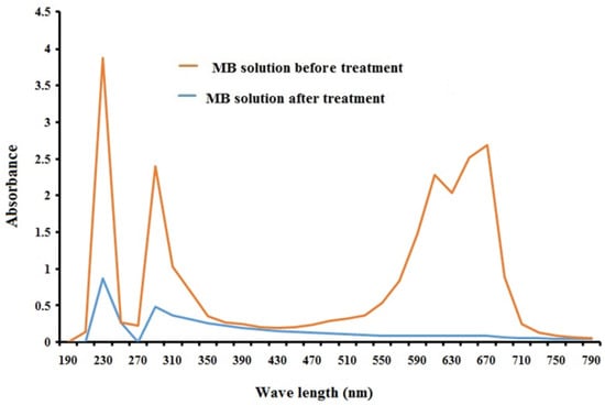
Figure 1.
UV-Vis absorption spectra of the MB solution before and after treatment with algal biomass after 84h of incubation.
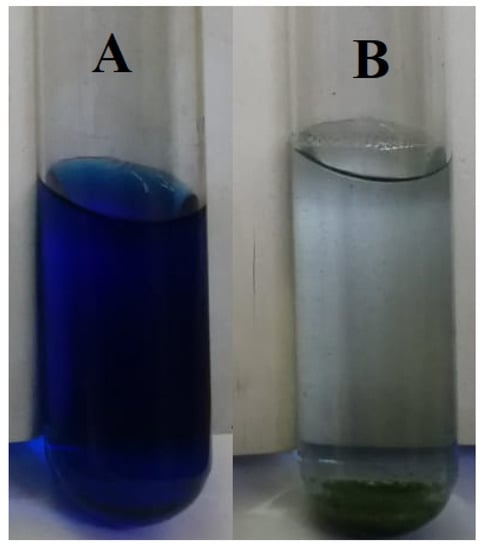
Figure 2.
Removal of MB using algae biomass. (A) MB control and (B) MB with algae after 84 h of incubation.
As shown in Figure 1, methylene blue before treatment has two absorbance peaks at 610 and 670 nm; the peak at 610 nm is not a band but a shoulder, and it appears because of a transition between the two levels: the ground state and the excited state. The two peaks disappeared after treatment with algae. The reason behind color disappearance is the binding process that occurs on the surface of algal biomass. The sorption of MB into algae may occur through the interactions between the functional groups on the algal surface and MB. After adding algal cells to MB, the solution color became dark blue and gradually disappeared and changed to nearly colorless after the first 24 and 84 h, as shown in Figure 2. Hence, MB decolorization may be due to both the sorption and degradation of MB [28].
3.2. Optimization of the Removal of MB
3.2.1. Effects of Incubation Temperatures, Initial MB Concentrations, and Contact Time
Optimizing the experimental parameters, such as initial MB concentration, incubation temperature, contact time, adsorbent dose, and immobilization, is a key point for obtaining the maximum methylene blue removal from aqueous solutions.
Initial dye concentration, incubation temperature, and contact time are important parameters to determine the optimal removal of methylene blue. Dye removal studies were conducted with different MB concentrations (5–25 mg L−1) and three incubation temperatures (4, 20, and 30 °C) for 84 h (Figure 3), and it was observed that decolorization of MB was found to increase rapidly in the first 24 h. Removal percentages in Figure 3, Figure 4 and Figure 5 increased as time increased, but removal percentages decreased as methylene blue increased. Figure 3a–c show the effects of incubation temperatures (4, 20, and 30 °C) on the removal percentages of MB; it was observed that as the temperature increased, the removal percentage increased. The best removal percentage was observed atconcentrationsof5 and 10 mg L−1and at incubation temperatures of 20 and 30 °C after 84 h.
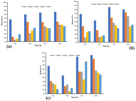
Figure 3.
(a) Effects of initial MB concentration and contact time on MB removal percentage using Bracteacoccus sp. (a) incubated at 4 °C (b) at 20 °C and (c) at 30 °C in all temperatures (algae dosage = 0.03 g mL−1). All experiments were conducted in triplicate.
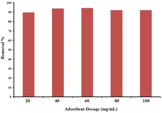
Figure 4.
Effects of adsorbent dosage on MB (Co = 15 mg L−1) removal efficiency (%) using Bracteacoccus sp. incubated at 30 °C for 24 h.
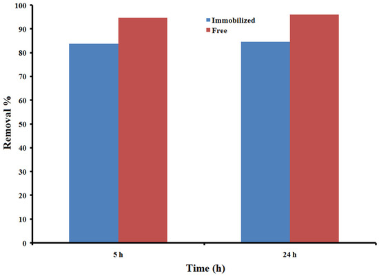
Figure 5.
Effects of Immobilization on MB removal efficiency (%) (Co = 15 mg L−1, algae dosage = 60 mg/mL, and T = 30 °C after 5 and 24 h).
Bracteacoccus sp. is green microalgae, and its adsorption properties are due to different types of proteins found in the algae cell wall. From the results in Figure 3, Figure 4 and Figure 5, the contact time shows the gradual effects on the decolorization of MB. This behavior may be because the decolorization process occurs gradually, and the MB molecules need time to be adsorbed into algae sorption sites. The decrease in methylene blue removal happened because of the saturation of the biosorbent site on the cell surface of the algae [29,30]. Temperature is another parameter that affects the decolorization process. The removal percentages increased with increasing temperature from 4 °C to 30 °C due to the increase of sorption sites on the surface of the Bracteacoccus sp. cell wall, and these results are in agreement with several previous reports [31]. Several reports have shown that as the temperature increases above 25 °C, the percentage of removal increases as the temperature increases due to the high temperatures inducing dye molecule diffusion in the porous interior structure of the sorbent [32].
3.2.2. Effects of Adsorbent Dose
Decolorization of methylene blue dye solutions was performed to fix the optimal algal dose required to decolorize MB from the aqueous solutions (Figure 4). From an economic standpoint, determining the amount of adsorbent is very important for selecting the optimum adsorbent dosage for industrial applications [32]. The effects of different algae doses (0.02–0.1 g mL−1) are shown in Figure 4. Results showed that when the dosage of algae changed from 0.02 to 0.06 g, the dye removal increased, and the largest quantity of dye removal was attained with adsorbent masses of 0.06 g, with a percentage removal of 94.3%, after that, the removal percentage was decreased (91.9 and 92.0 %) when algae doses were 0.08 and 0.1 g, respectively. Algae dosage affected the decolorization process using active sites on the algae cell surface and may be attributed to an increase in the biosorption surface area and the number of effective sites on the adsorbent surface. Similar results were reported by other authors [29,31,32]. However, the removal percentages decreased at the higher adsorbent dosage (80 and 100 mg). This may be due to the partial aggregation of Bracteacoccus sp. in an aqueous solution, which restricts the number of accessible adsorption sites, or high algal biomass amounts are known to cause aggregation of cells, which will reduce the distance between cells leading to protecting functional groups from MB, as previously reported [32,33].
3.2.3. Effects of Immobilization
Most previous reports that used the immobilization of algae focused on the removal of heavy metals; this study supports the use of immobilization for the decolorization process [34]. The percentage of removal in MB dye solution with a 15 mg L−1 concentration using immobilized Bracteacoccus sp. incubated at 30 °C for 5 and 24 h can be seen in Figure 5. Based on Figure 5, the decolorization percentage of MB by Bracteacoccus sp. immobilized in alginate beads at incubation times of 5 h and 24 h, respectively, are 83.8% and 84.7%. The percentage of decolorization of MB by free Bracteacoccus sp. at incubation times of 5h and 24h, respectively, are 94.8% and 96.1%. Free and immobilized states of Bracteacoccus sp. were able to decolorize methylene blue from aqueous solutions with high capacity reaching 94.8% MB removal. For both incubation times, free Bracteacoccus sp. shows the best decolorization efficiency compared with the immobilized state; there was a slight decrease in MB color after 24h when the blank beads (beads without Bracteacoccus sp.) were used as biosorbents compared with a clear solution when Bracteacoccus sp. was used. Immobilization has many advantages, such as the reusability of cells and its ability for desorption. Immobilization with alginate beads has a semipermeable structure that allows materials with small sizes and water-soluble materials to diffuse in and out of the beads [35]. The removal percentage of free Bracteacoccus sp. was slightly higher than immobilized algae beads, and this may be due to the super-concentrated algal cells in alginate beads, which may affect the decolorization process. Immobilization of Bracteacoccus sp. in alginate beads is a very efficient method of cell entrapment because immobilization allows high cell loading and light penetration, and it can also be used many times in bioreactors. Alginate beads will protect algal cells from pollutants, such as methylene blue [36].
Kinetic Models
According to the results from applying pseudo-first-order, pseudo-second-order, and Elovich kinetic models in Equations (1)–(3), the removal of MB using algae is best fitted with the pseudo-second-order model with the highest correlation coefficient (R2 = 0.9997), and the lowest RSME is considered the best fitted for the removal of MB using algae. The parameters and their values related to these kinetic models are listed in Table 1. The order of these kinetic models based on the values of correlation coefficients from high to low are as follows:
pseudo-second-order > Elvoich > pseudo-first-order
These findings agree with the results of previous studies reported in the literature. According to the values of the correlation factors (R2) listed in Table 1, the pseudo-second-order model has the highest R2 value (R2 = 0.9997), followed by the Elvoich and pseudo-first-order models. Therefore, the pseudo-second-order kinetic model is the best to describe the removal of MB using algae.
3.3. Isotherm Models
The isotherm models, such as the Langmuir, Freundlich, Temkin, and D–R, help to explain the nature of the interaction between adsorbate (e.g., MB) and adsorbent surface (e.g., algae) [37]. The values of parameters related to the isotherm models are listed in Table 2. The order of the isotherm models from high to low R2 is as follows:
Langmuir > Freundlich ≈ D–R > Temkin

Table 2.
Langmuir, Freundlich, and Temkin isotherm model constants, correlation coefficients, and error function parameters of MB adsorption on algae.
Consequently, the experimental data are best fitted by the Langmuir model and least fitted by the Temkin model. Thus, a monolayer of MB is formed on the algae surface. This finding agrees with some previous studies [19,38,39].
Comparison between Different Types of Algae
Table 3 shows a comparison between some types of algae used in previous studies as adsorbents for MB removal with the main result obtained from the current study. Among these adsorbents, Immobilized Bracteacoccus sp., Algae (Ulva Lactuca and Sargassum), and the algae used in the current study show the highest removal efficiency of MB (>95%), followed by giant duckweed (Spirodelapolyrrhiza). While other bioadsorbents, such as pretreated dead Streptomyces rimosus and dead algal biomass, achieve acceptable removal percentages (<70%). Therefore, algae are still effective bioadsorebnets to decolorize contaminated water by MB.

Table 3.
Maximum uptake capacity of MB on several biosorbents.
Different low-cost adsorbents (such as Azolla pinnata and GigantochloaBamboo derived biochar) and their adsorption capacity of methylene blue dye from aqueous solutions were discussed in previous studies, which showed high adsorption performance [49,50].
4. Conclusions
Algae are effective biosorbents for MB removal from aqueous solutions. In addition, algae have clear advantages, such as fast growth, availability, high efficiency for MB removal, low cost, and environment friendliness, which make the use of algae a promising route to protect aquatic environments from pollution by industrial effluents, including dyes. Optimization of the experimental parameters, such as initial MB concentration, algae dosage, temperature, and contact time, is an essential step in MB removal from an aqueous solution to obtain the maximum removal efficiency of MB. Based on the decolorization of MB using algae, a potential mechanism of MB removal may include sorption and degradation. Therefore, further studies, including analysis of algae with MB mixture, will be interesting to exactly depict the removal mechanisms of MB using algae.
Author Contributions
Conceptualization, A.T.A.-F. and E.E.; data curation, A.A.S., A.T.A.-F. and E.E.; formal analysis, A.T.A.-F. and A.A.S.; methodology, A.T.A.-F., A.A.S. and E.E.; resources, A.T.A.-F., E.E. and A.A.S.; writing—original draft, A.T.A.-F.; writing—review and editing, A.T.A.-F. and E.E. All authors have read and agreed to the published version of the manuscript.
Funding
This research received no external funding.
Institutional Review Board Statement
Not applicable.
Informed Consent Statement
Not applicable.
Data Availability Statement
Not applicable.
Acknowledgments
The authors would like to thank the Department of Biological Sciences, Al al-Bayt University, for providing administrative and research support.
Conflicts of Interest
The authors declare no conflict of interest.
References
- Yu, K.L.; Lee, X.J.; Ong, H.C.; Chen, W.H.; Chang, J.S.; Lin, C.S.; Show, P.L.; Ling, T.C. Adsorptive removal of cationic methylene blue and anionic Congo red dyes using wet-torrefied microalgal biochar: Equilibrium, kinetic and mechanism modeling. Environ. Pollut. 2021, 272, 115986. [Google Scholar] [CrossRef] [PubMed]
- Homaeigohar, S. The nanosized dye adsorbents for water treatment. Nanomaterials 2020, 10, 295. [Google Scholar] [CrossRef] [PubMed]
- Krishna Moorthy, A.; Govindarajan Rathi, B.; Shukla, S.P.; Kumar, K.; Shree Bharti, V. Acute toxicity of textile dye Methylene blue on growth and metabolism of selected freshwater microalgae. Environ. Toxicol. Pharmacol. 2021, 82, 103552. [Google Scholar] [CrossRef] [PubMed]
- Lellis, B.; Fávaro-Polonio, C.Z.; Pamphile, J.A.; Julio, C.P. Effects of textile dyes on health and the environment and bioremediation potential of living organisms. Biotechnol. Res. Innov. 2019, 3, 275–290. [Google Scholar] [CrossRef]
- Omar, H.; El-Gendy, A.; Al-Ahmary, K. Bioremoval of toxic dye by using different marine macroalgae. Turk. J. Bot. 2018, 42, 15–27. [Google Scholar] [CrossRef]
- Elgarahy, A.M.; Elwakeel, K.Z.; Mohammad, S.H.; Elshoubaky, G.A. A critical review of biosorption of dyes, heavy metals and metalloids from wastewater as an efficient and green process. Clean. Eng. Technol. 2021, 4, 100209. [Google Scholar] [CrossRef]
- Chen, Z.; Chen, H.; Pan, X.; Lin, Z.; Guan, X. Investigation of Methylene Blue Biosorption and Biodegradation by Bacillus thuringiensis 016. Water Air Soil Pollut. 2015, 3, 226. [Google Scholar] [CrossRef]
- Elnahas, M.O.; Hou, L.; Wall, J.D.; Majumder, E.W. Bioremediation Potential of Streptomyces sp. MOE6 for Toxic Metals and Oil. Polysaccharides 2021, 2, 47–68. [Google Scholar] [CrossRef]
- Dolganyuk, V.; Belova, D.; Babich, O.; Prosekov, A.; Ivanova, S.; Katserov, D.; Patyukov, N.; Sukhikh, S. Microalgae: A promising source of valuable bioproducts. Biomolecules 2020, 10, 1153. [Google Scholar] [CrossRef]
- Doǧar, Ç.; Gürses, A.; Açikyildiz, M.; Özkan, E. Thermodynamics and kinetic studies of biosorption of a basic dye from aqueous solution using green algae Ulothrix sp. Colloids Surf. B Biointerfaces 2010, 76, 279–285. [Google Scholar] [CrossRef]
- Aravindhan, R.; Rao, J.R.; Nair, B.U. Removal of basic yellow dye from aqueous solution by sorption on green alga Caulerpa scalpelliformis. J. Hazard. Mater. 2007, 142, 68–76. [Google Scholar] [CrossRef] [PubMed]
- Mahalakshmi, S.; Lakshmi, D.; Menaga, U. Biodegradation of different concentration of dye (Congo red dye) by using green and blue green algae. Int. J. Environ. Res. 2015, 9, 735–744. [Google Scholar]
- Omar, H.H. Algal decolorization and degradation of monoazo and diazo dyes. Pak. J. Biol. Sci. 2008, 11, 1310–1316. [Google Scholar] [CrossRef]
- Guolan, H.; Hongwen, S.; Li, C.L. Study on the physiology and degradation of dye with immobilized algae. Artif. Cells Blood Substit. Biotechnol. 2000, 28, 347–363. [Google Scholar] [CrossRef] [PubMed]
- El-Sheekh, M.M.; Gharieb, M.M.; Abou-El-Souod, G.W. Biodegradation of dyes by some green algae and cyanobacteria. Int. Biodeterior. Biodegrad. 2009, 63, 699–704. [Google Scholar] [CrossRef]
- Daneshvar, N.; Khataee, A.R.; Rasoulifard, M.H.; Pourhassan, M. Biodegradation of dye solution containing Malachite Green: Optimization of effective parameters using Taguchi method. J. Hazard. Mater. 2007, 143, 214–219. [Google Scholar] [CrossRef]
- Vasanth Kumar, K.; Sivanesan, S.; Ramamurthi, V. Adsorption of malachite green onto Pithophora sp., a fresh water algae: Equilibrium and kinetic modelling. Process Biochem. 2005, 40, 2865–2872. [Google Scholar] [CrossRef]
- Khan, I.; Saeed, K.; Zekker, I.; Zhang, B.; Hendi, A.H.; Ahmad, A.; Ahmad, S.; Zada, N.; Ahmad, H.; Shah, L.A.; et al. Review on Methylene Blue: Its Properties, Uses, Toxicity and Photodegradation. Water 2022, 14, 242. [Google Scholar] [CrossRef]
- Cengiz, S.; Cavas, L. Removal of methylene blue by invasive marine seaweed: Caulerpa racemosa var. cylindracea. Bioresour. Technol. 2008, 99, 2357–2363. [Google Scholar] [CrossRef]
- Omeodisemi, D. Experimental Modelling Studies on the removal of crystal violet, methylene blue and malachite green dyes using Theobroma cacao (Cocoa Pod Powder). J. Chem. Lett. 2021, 2, 9–24. [Google Scholar]
- Jabar, J.M.; Odusote, Y.A.; Alabi, K.A.; Ahmed, I.B. Kinetics and mechanisms of congo-red dye removal from aqueous solution using activated Moringa oleifera seed coat as adsorbent. Appl. Water Sci. 2020, 10, 136. [Google Scholar] [CrossRef]
- Boulaiche, W.; Hamdi, B.; Trari, M. Removal of heavy metals by chitin: Equilibrium, kinetic and thermodynamic studies. Appl. Water Sci. 2019, 9, 39. [Google Scholar] [CrossRef]
- Botelho Junior, A.B.; Pinheiro, É.F.; Espinosa, D.C.R.; Soares Tenório, J.A.; Galluzzi Baltazar, M.D.P. Adsorption of lanthanum and cerium on chelating ion exchange resins: Kinetic and thermodynamic studies. Sep. Sci. Technol. 2022, 57, 60–69. [Google Scholar] [CrossRef]
- Saxena, M.; Sharma, N.; Saxena, R. Highly efficient and rapid removal of a toxic dye: Adsorption kinetics, isotherm, and mechanism studies on functionalized multiwalled carbon nanotubes. Surf. Interfaces 2020, 21, 100639. [Google Scholar] [CrossRef]
- Kumar, V.; Saharan, P.; Sharma, A.K.; Umar, A.; Kaushal, I.; Mittal, A.; Al-Hadeethi, Y.; Rashad, B. Silver doped manganese oxide-carbon nanotube nanocomposite for enhanced dye-sequestration: Isotherm studies and RSM modelling approach. Ceram. Int. 2020, 46, 10309–10319. [Google Scholar] [CrossRef]
- Munagapati, V.S.; Wen, H.Y.; Wen, J.C.; Gutha, Y.; Tian, Z.; Reddy, G.M.; Garcia, J.R. Anionic congo red dye removal from aqueous medium using Turkey tail (Trametes versicolor) fungal biomass: Adsorption kinetics, isotherms, thermodynamics, reusability, and characterization. J. Dispers. Sci. Technol. 2021, 42, 1785–1798. [Google Scholar] [CrossRef]
- Li, Z.; Gou, M.; Yue, X.; Tian, Q.; Yang, D.; Qiu, F.; Zhang, T. Facile fabrication of bifunctional ZIF-L/cellulose composite membrane for efficient removal of tellurium and antibacterial effects. J. Hazard. Mater. 2021, 416, 125888. [Google Scholar] [CrossRef]
- Tang, W.; Xu, X.; Ye, B.C.; Cao, P.; Ali, A. Decolorization and degradation analysis of Disperse Red 3B by a consortium of the fungus: Aspergillus sp. XJ-2 and the microalgae Chlorella sorokiniana XJK. RSC Adv. 2019, 9, 14558–14566. [Google Scholar] [CrossRef]
- Bonyadi, Z.; Nasoudari, E.; Ameri, M.; Ghavami, V.; Shams, M.; Sillanpää, M. Biosorption of malachite green dye over Spirulina platensis mass: Process modeling, factors optimization, kinetic, and isotherm studies. Appl. Water Sci. 2022, 12, 167. [Google Scholar] [CrossRef]
- Mohebbrad, B.; Bonyadi, Z.; Dehghan, A.A.; Rahmat, M.H. Arsenic removal from aqueous solutions using Saccharomyces cerevisiae: Kinetic and equilibrium study. Environ. Prog. Sustain. Energy 2019, 38, S398–S402. [Google Scholar]
- Gül, D.; Taştan, B.E.; Bayazıt, G. Assessment of algal biomasses having different cell structures for biosorption properties of acid red P-2BX dye. S. Afr. J. Bot. 2019, 127, 147–152. [Google Scholar] [CrossRef]
- Mansour, A.T.; Alprol, A.E.; Abualnaja, K.M.; El-Beltagi, H.S.; Ramadan, K.M.A.; Ashour, M. The Using of Nanoparticles of Microalgae in Remediation of Toxic Dye from Industrial Wastewater: Kinetic and Isotherm Studies. Materials 2022, 15, 3922. [Google Scholar] [CrossRef]
- Shi, H.; Li, W.; Zhong, L.; Xu, C. Methylene blue adsorption from aqueous solution by magnetic cellulose/graphene oxide composite: Equilibrium, kinetics, and thermodynamics. Ind. Eng. Chem. Res. 2014, 53, 1108–1118. [Google Scholar] [CrossRef]
- Majhi, P.K.; Kothari, R.; Pandey, A.; Tyagi, V.V. Adsorptive behavior of free and immobilized Chlorella pyrenoidosa for decolorization. Biomass Convers. Biorefinery 2021, 11, 3023–3036. [Google Scholar] [CrossRef]
- Dewi, R.S.; Mumpuni, A.; Tsabitah, N.I. Batik Dye Decolorization by Immobilized Biomass of Aspergillus sp. IOP Conf. Ser. Earth Environ. Sci. 2020, 550, 012020. [Google Scholar] [CrossRef]
- Kumar, S.D.; Santhanam, P.; Nandakumar, R.; Anath, S.; Balaji Prasath, B.; Shenbaga Devi, A.; Jeyanthi, S.; Jayalakshima, T.; Ananthi, P. Preliminary study on the dye removal efficacy of immobilized marine and freshwater microalgal beads from textile wastewater. Afr. J. Biotechnol. 2014, 13, 2288–2294. [Google Scholar]
- Coates, J. Interpretation of Infrared Spectra, A Practical Approach. Encycl. Anal. Chem. 2006, 1–23. [Google Scholar] [CrossRef]
- Marungrueng, K.; Pavasant, P. High performance biosorbent (Caulerpa lentillifera) for basic dye removal. Bioresour. Technol. 2007, 98, 1567–1572. [Google Scholar] [CrossRef]
- Afshariani, F.; Roosta, A. Experimental study and mathematical modeling of biosorption of methylene blue from aqueous solution in a packed bed of microalgae Scenedesmus. J. Clean. Prod. 2019, 225, 133–142. [Google Scholar] [CrossRef]
- El Sikaily, A.; Khaled, A.; El Nemr, A.; Abdelwahab, O. Removal of Methylene Blue from aqueous solution by marine green alga Ulva lactuca. Chem. Ecol. 2006, 22, 149–157. [Google Scholar] [CrossRef]
- Nacèra, Y.; Aicha, B. Equilibrium and kinetic modelling of methylene blue biosorption by pretreated dead streptomyces rimosus: Effect of temperature. Chem. Eng. J. 2006, 119, 121–125. [Google Scholar] [CrossRef]
- Rubin, E.; Rodriguez, P.; Herrero, R. Removal of Methylene Blue from aqueous solutions using as biosorbent Sargassum muticum: An invasive macroalga in Europe. J. Chem. Technol. Biotechnol. 2005, 80, 291–298. [Google Scholar] [CrossRef]
- Maurya, N.S.; Mittal, A.K.; Cornel, P.; Rother, E. Biosorption of dyes using dead macro fungi: Effect of dye structure, ionic strength and pH. Bioresour. Technol. 2006, 97, 512–521. [Google Scholar] [CrossRef]
- Vilar, V.J.P.; Botelho, C.M.S.; Boaventura, R.A.R. Methylene blue adsorption by algal biomass based materials: Biosorbents characterization and process behaviour. J. Hazard. Mater. 2007, 147, 120–132. [Google Scholar] [CrossRef]
- Waranusantigul, P.; Pokethitiyook, P.; Kruatrachue, M.; Upatham, E.S. Kinetics of basic dye (methylene blue) biosorption by giant duckweed (Spirodela polyrrhiza). Environ. Pollut. 2003, 125, 385–392. [Google Scholar] [CrossRef] [PubMed]
- Caparkaya, D.; Cavas, L. Biosorption of methylene blue by a brown alga Cystoseira barbatula Kützing. Acta Chim. Slov. 2008, 55, 547–553. [Google Scholar]
- Tahir, H.; Sultan, M.; Jahanzeb, Q. Removal of basic dye methylene blue by using bioabsorbents Ulva lactuca and Sargassum. Afr. J. Biotechnol. 2008, 7, 2649–2655. [Google Scholar]
- Al-Fawwaz, A.T.; Mufida, A. Decolorization of Methylene Blue and Malachite Green by Immobilized Desmodesmus sp. Isolated from North Jordan. Int. J. Environ. Sci. Dev. 2016, 7, 95–99. [Google Scholar] [CrossRef]
- Rahimi Kooh, M.R.; Thotagamuge, R.; Chou Chau, Y.F.; Mahadi, A.; Lim, C.M. Machine learning approaches to predict adsorption capacity of Azolla pinnata in the removal of methylene blue. J. Taiwan Inst. Chem. Eng. 2022, 132, 104134. [Google Scholar] [CrossRef]
- Nabilah Suhaimi, N.; Rahimi Kooh, M.R.; Lim, C.M.; Chou Chao, C.T.; Chou Chau, Y.F.; Mahadi, A.; Chiang, H.P.; Haji Hassan, N.H.; Thotagamuge, R. The Use of Gigantochloa Bamboo-Derived Biochar for the Removal of Methylene Blue from Aqueous Solution. Adsorpt. Sci. Technol. 2022, 2022, 8245797. [Google Scholar] [CrossRef]
Disclaimer/Publisher’s Note: The statements, opinions and data contained in all publications are solely those of the individual author(s) and contributor(s) and not of MDPI and/or the editor(s). MDPI and/or the editor(s) disclaim responsibility for any injury to people or property resulting from any ideas, methods, instructions or products referred to in the content. |
© 2023 by the authors. Licensee MDPI, Basel, Switzerland. This article is an open access article distributed under the terms and conditions of the Creative Commons Attribution (CC BY) license (https://creativecommons.org/licenses/by/4.0/).
