Next Issue
Volume 11, December
Previous Issue
Volume 11, June


Cardiogenetics, Volume 11, Issue 3 (September 2021) – 7 articles

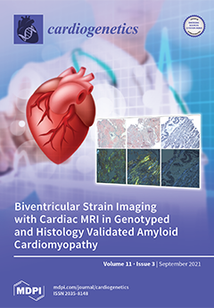
Cover Story (view full-size image): A diagnosis of cardiac amyloidosis is becoming increasingly prevalent due to greater awareness of the disease process among clinicians. The gold standard for diagnosis is invasive endomyocardial biopsy. In this article, we present a novel use of non-invasive imaging, in particular cardiac magnetic resonance (CMR), to identify CMR-based strain patterns associated with biopsy proven cardiac amyloidosis. Our findings provide the initial groundwork for further utilization of non-invasive modalities in the diagnosis of cardiac amyloid. View this paper.
  • Issues are regarded as officially published after their release is announced to the table of contents alert mailing list.
  • You may sign up for e-mail alerts to receive table of contents of newly released issues.
  • PDF is the official format for papers published in both, html and pdf forms. To view the papers in pdf format, click on the "PDF Full-text" link, and use the free Adobe Reader to open them.
Order results
Result details
Section
Select all
Export citation of selected articles as:
16 pages, 797 KiB  
Study Protocol
A Novel Human Biospecimen Repository for Clinical and Molecular Investigation of Thoracic Aortopathy
by Courtney E. Vujakovich and Benjamin J. Landis
Cardiogenetics 2021, 11(3), 148-163; https://doi.org/10.3390/cardiogenetics11030017 - 18 Sep 2021
Cited by 2 | Viewed by 3193
Abstract
Thoracic aortic aneurysm (TAA) is a heritable aortopathy with significant morbidity and mortality, affecting children and adults. Genetic causes, pathobiological mechanisms, and prognostic markers are incompletely understood. In 2015, the Collaborative Human Aortopathy Repository (CHAR) was created to address these fundamental gaps. Patients [...] Read more.
Thoracic aortic aneurysm (TAA) is a heritable aortopathy with significant morbidity and mortality, affecting children and adults. Genetic causes, pathobiological mechanisms, and prognostic markers are incompletely understood. In 2015, the Collaborative Human Aortopathy Repository (CHAR) was created to address these fundamental gaps. Patients with thoracic aortopathy, associated genetic diagnoses, or aortic valve disease are eligible for prospective enrollment. Family members and controls are also enrolled. Detailed clinical and family data are collected, and blood and aortic tissue biospecimens are processed for broad usage. A total of 1047 participants were enrolled. The mean age in 834 affected participants was 47 ± 22 (range <1 to 88) years and 580 were male (70%). A total of 156 (19%) were under the age of 21 years. Connective tissue diagnoses such as Marfan syndrome were present in 123 (15%). Unaffected participants included relatives (N = 176) and healthy aorta tissue controls (N = 37). Aortic or aortic valve biospecimens were acquired from over 290 and 110 participants, respectively. RNA and protein were extracted from cultured aortic smooth muscle cells (SMCs) for 90 participants. Over 1000 aliquots of aortic SMCs were cryopreserved. The CHAR’s breadth, robust biospecimen processing, and phenotyping create a unique, multipronged resource to accelerate our understanding of human aortopathy. Full article
(This article belongs to the Special Issue Cardiogenetics: Feature Papers 2021)
Show Figures

Figure 1

10 pages, 1131 KiB  
Article
Mutations in MYBPC3 and MYH7 in Association with Brugada Type 1 ECG Pattern: Overlap between Brugada Syndrome and Hypertrophic Cardiomyopathy?
by Marianna Farnè, Cristina Balla, Alice Margutti, Rita Selvatici, Martina De Raffele, Assunta Di Domenico, Paola Imbrici, Elia De Maria, Mauro Biffi, Matteo Bertini, Claudio Rapezzi, Alessandra Ferlini and Francesca Gualandi
Cardiogenetics 2021, 11(3), 139-147; https://doi.org/10.3390/cardiogenetics11030016 - 9 Sep 2021
Cited by 5 | Viewed by 4213
Abstract
Brugada syndrome (BrS) is an inherited disorder with high allelic and genetic heterogeneity clinically characterized by typical coved-type ST segment elevation at the electrocardiogram (ECG), which may occur either spontaneously or after provocative drug testing. BrS is classically described as an arrhythmic condition [...] Read more.
Brugada syndrome (BrS) is an inherited disorder with high allelic and genetic heterogeneity clinically characterized by typical coved-type ST segment elevation at the electrocardiogram (ECG), which may occur either spontaneously or after provocative drug testing. BrS is classically described as an arrhythmic condition occurring in a structurally normal heart and is associated with the risk of ventricular fibrillation and sudden cardiac death (SCD). We studied five patients with spontaneous or drug-induced type 1 ECG pattern, variably associated with symptoms and a positive family history through a Next Generation Sequencing panels approach, which includes genes of both channelopathies and cardiomyopathies. We identified variants in MYBPC3 and in MYH7, hypertrophic cardiomyopathy (HCM) genes (MYBPC3: p.Lys1065Glnfs*12 and c.1458-1G > A, MYH7: p.Arg783His, p.Val1213Met, p.Lys744Thr). Our data propose that Brugada type 1 ECG may be an early electrocardiographic marker of a concealed structural heart disease, possibly enlarging the genotypic overlap between Brugada syndrome and cardiomyopathies. Full article
(This article belongs to the Section Cardiovascular Genetics in Clinical Practice)
Show Figures

Figure 1

7 pages, 598 KiB  
Case Report
Human SMAD4 Genomic Variants Identified in Individuals with Heritable and Early-Onset Thoracic Aortic Disease
by Shreyas A. Bhave, Dongchuan Guo, Stoyan N. Angelov, Michael J. Bamshad, Deborah A. Nickerson, Dianna M. Milewicz and Mary C. Wallingford
Cardiogenetics 2021, 11(3), 132-138; https://doi.org/10.3390/cardiogenetics11030015 - 18 Aug 2021
Cited by 1 | Viewed by 3878
Abstract
Thoracic aortic aneurysms (TAAs) that progress to acute thoracic aortic dissections (TADs) are life-threatening vascular events that have been associated with altered transforming growth factor (TGF) β signaling. In addition to TAA, multiple genetic vascular disorders, including hereditary hemorrhagic telangiectasia (HHT), involve altered [...] Read more.
Thoracic aortic aneurysms (TAAs) that progress to acute thoracic aortic dissections (TADs) are life-threatening vascular events that have been associated with altered transforming growth factor (TGF) β signaling. In addition to TAA, multiple genetic vascular disorders, including hereditary hemorrhagic telangiectasia (HHT), involve altered TGFβ signaling and vascular malformations. Due to the importance of TGFβ, genomic variant databases have been curated for activin receptor-like kinase 1 (ALK1) and endoglin (ENG). This case report details seven variants in SMAD4 that are associated with either heritable or early-onset aortic dissections and compares them to pathogenic exon variants in gnomAD v2.1.1. The TAA and TAD variants were identified through whole exome sequencing of 346 families with unrelated heritable thoracic aortic disease (HTAD) and 355 individuals with early-onset (age ≤ 56 years old) thoracic aortic dissection (ESTAD). An allele frequency filter of less than 0.05% was applied in the Genome Aggregation Database (gnomAD exome v2.1.1) with a combined annotation-dependent depletion score (CADD) greater than 20. These seven variants also have a higher REVEL score (>0.2), indicating pathogenic potential. Further in vivo and in vitro analysis is needed to evaluate how these variants affect SMAD4 mRNA stability and protein activity in association with thoracic aortic disease. Full article
(This article belongs to the Special Issue Cardiogenetics: Feature Papers 2021)
Show Figures

Figure 1

3 pages, 566 KiB  
Case Report
Spontaneous Coronary Artery Dissection as Presenting Feature of Vascular Ehlers-Danlos Syndrome
by J. Bos, E. Overwater, M.T. Dirksen, S. Simsek, S. Demirdas and A.C. Houweling
Cardiogenetics 2021, 11(3), 129-131; https://doi.org/10.3390/cardiogenetics11030014 - 13 Aug 2021
Viewed by 3135
Abstract
A spontaneous coronary artery dissection as the sole presenting feature of vascular Ehlers-Danlos syndrome is an uncommon finding. We present a 33-year-old woman with sudden onset chest pain caused by a spontaneous coronary artery dissection. Genetic testing revealed vascular Ehlers-Danlos syndrome as the [...] Read more.
A spontaneous coronary artery dissection as the sole presenting feature of vascular Ehlers-Danlos syndrome is an uncommon finding. We present a 33-year-old woman with sudden onset chest pain caused by a spontaneous coronary artery dissection. Genetic testing revealed vascular Ehlers-Danlos syndrome as the underlying cause. Specifically, we show the value of genetic testing, which in some patients may be the only way of establishing a diagnosis. Full article
(This article belongs to the Section Cardiovascular Genetics in Clinical Practice)
Show Figures

Figure 1

7 pages, 6107 KiB  
Case Report
Reassessment of Gene-Elusive Familial Dilated Cardiomyopathy Leading to the Discovery of a Homozygous AARS2 Variant—The Importance of Regular Reassessment of Genetic Findings
by Priya Bhardwaj, Christoffer Rasmus Vissing, Niels Kjær Stampe, Kasper Rossing, Alex Hørby Christensen, Thomas Hartvig Lindkær Jensen and Bo Gregers Winkel
Cardiogenetics 2021, 11(3), 122-128; https://doi.org/10.3390/cardiogenetics11030013 - 23 Jul 2021
Viewed by 3797
Abstract
Background: AARS2 encodes the mitochondrial protein alanyl-tRNA synthetase 2 (MT-AlaRS), an important enzyme in oxidative phosphorylation. Variants in AARS2 have previously been associated with infantile cardiomyopathy. Case summary: A 4-year-old girl died of infantile-onset dilated cardiomyopathy (DCM) in 1996. Fifteen years later, her [...] Read more.
Background: AARS2 encodes the mitochondrial protein alanyl-tRNA synthetase 2 (MT-AlaRS), an important enzyme in oxidative phosphorylation. Variants in AARS2 have previously been associated with infantile cardiomyopathy. Case summary: A 4-year-old girl died of infantile-onset dilated cardiomyopathy (DCM) in 1996. Fifteen years later, her 21-year-old brother was diagnosed with DCM and ultimately underwent heart transplantation. Initial sequencing of 15 genes discovered no pathogenic variants in the brother at the time of his diagnosis. However, 9 years later re-screening in an updated screening panel of 129 genes identified a homozygous AARS2 (c.1774C > T) variant. Sanger sequencing of the deceased girl confirmed her to be homozygous for the AARS2 variant, while both parents and a third sibling were all found to be unaffected heterozygous carriers of the AARS2 variant. Discussion: This report underlines the importance of repeated and extended genetic screening of elusive families with suspected hereditary cardiomyopathies, as our knowledge of disease-causing mutations continuously grows. Although identification of the genetic etiology in the reported family would not have changed the clinical management, the genetic finding allows genetic counselling and holds substantial value in identifying at-risk relatives. Full article
(This article belongs to the Special Issue Genetic Diagnostics in Inherited Cardiomyopathies)
Show Figures

Figure 1

11 pages, 2201 KiB  
Review
Cardiac Amyloidosis: Diagnostic Tools for a Challenging Disease
by Marco Giuseppe Migliaccio, Franco Iodice, Marco Di Mauro, Angela Iannuzzi, Roberta Pacileo, Martina Caiazza and Augusto Esposito
Cardiogenetics 2021, 11(3), 111-121; https://doi.org/10.3390/cardiogenetics11030012 - 23 Jul 2021
Viewed by 6203
Abstract
Amyloidosis is a group of diseases in which amyloid fibrils build up in tissues, leading to organ dysfunction. Cardiac involvement is observed in immunoglobulin light chain amyloidosis (AL) and transthyretin amyloidosis (ATTR) and, when it occurs, the prognosis worsens. Cardiac tissue infiltration can [...] Read more.
Amyloidosis is a group of diseases in which amyloid fibrils build up in tissues, leading to organ dysfunction. Cardiac involvement is observed in immunoglobulin light chain amyloidosis (AL) and transthyretin amyloidosis (ATTR) and, when it occurs, the prognosis worsens. Cardiac tissue infiltration can lead to restrictive cardiomyopathy with clinical signs of diastolic heart failure, without reduction of ejection fraction (HFpEF). The aim of multiple and less invasive diagnostic tests is to discern peculiar characteristics and reach the diagnosis without performing an invasive endomyocardial biopsy. These diagnostic tools allow early diagnosis, and they are crucial to best benefit from target therapy. In this review article, we describe the mechanism behind amyloid fibril formation, infiltration of tissues, and consequent clinical signs, focusing on the diagnostic tools and the red flags to obtain a diagnosis. Full article
(This article belongs to the Section Rare Disease-Metabolic Diseases)
Show Figures

Figure 1

13 pages, 3557 KiB  
Article
Biventricular Strain Imaging with Cardiac MRI in Genotyped and Histology Validated Amyloid Cardiomyopathy
by Abhinay Reddy, Vasvi Singh, Badri Karthikeyan, Leyi Jiang, Silva Kristo, Sharma Kattel, Ram Amuthan, Saraswati Pokharel and Umesh C. Sharma
Cardiogenetics 2021, 11(3), 98-110; https://doi.org/10.3390/cardiogenetics11030011 - 30 Jun 2021
Cited by 8 | Viewed by 4368
Abstract
Cardiac amyloidosis (CA) is a common and potentially fatal infiltrative cardiomyopathy. Contrast-enhanced cardiac MRI (CMR) is used as a diagnostic tool. However, utility of CMR for the comprehensive analysis of biventricular strains and strain rates is not reported as extensively as echocardiography. In [...] Read more.
Cardiac amyloidosis (CA) is a common and potentially fatal infiltrative cardiomyopathy. Contrast-enhanced cardiac MRI (CMR) is used as a diagnostic tool. However, utility of CMR for the comprehensive analysis of biventricular strains and strain rates is not reported as extensively as echocardiography. In addition, RV strain analysis using CMR has not been described previously. Objectives: We sought to study the global and regional indices of biventricular strain and strain rates in endomyocardial biopsy (EMB)-proven, genotyped cases of CA. Methods: A database of 80 EMBs was curated from 2012 to 2019 based on histology. A total of 19 EMBs positive for CA were subjected to further tissue-characterization with histology, and compared with four normal biopsy specimens. Samples were genotyped for ATTR- or AL-subtypes. Five patients, with both echocardiography and contrast-enhanced CMR performed 72-h apart, were subjected to comprehensive analysis of biventricular strain and strain-rates. Results: Histology confirmed that the selected samples were indeed positive for cardiac amyloid. Echocardiography showed reduced global and regional left-ventricular (LV) longitudinal strain indices. CMR with tissue-characterization of LV showed global reductions in circumferential, radial and longitudinal strains and strain-rates, following a general trend with the echocardiographic findings. The basal right-ventricular (RV) segments had reduced circumferential strains with no changes in longitudinal strain. Conclusions: In addition to providing a clinical diagnosis of CA based on contrast clearance-dynamics, CMR can be a potent tool for accurate functional assessment of global and regional changes in strain and strain-rates involving both LV and RV. Further studies are warranted to validate and curate the strain imaging capacity of CMR in CA. Full article
(This article belongs to the Section Cardiovascular Genetics in Clinical Practice)
Show Figures

Figure 1

Previous Issue
Next Issue
Back to TopTop