Review

Cardiac Amyloidosis: Diagnostic Tools for a Challenging Disease

Department of Translational Medical Sciences, University of Campania “Luigi Vanvitelli”, 80131 Naples, Italy
*
Author to whom correspondence should be addressed.
Cardiogenetics 2021, 11(3), 111-121; https://doi.org/10.3390/cardiogenetics11030012
Submission received: 7 January 2021 / Revised: 23 June 2021 / Accepted: 27 June 2021 / Published: 23 July 2021
(This article belongs to the Section Rare Disease-Metabolic Diseases)

Abstract

:
Amyloidosis is a group of diseases in which amyloid fibrils build up in tissues, leading to organ dysfunction. Cardiac involvement is observed in immunoglobulin light chain amyloidosis (AL) and transthyretin amyloidosis (ATTR) and, when it occurs, the prognosis worsens. Cardiac tissue infiltration can lead to restrictive cardiomyopathy with clinical signs of diastolic heart failure, without reduction of ejection fraction (HFpEF). The aim of multiple and less invasive diagnostic tests is to discern peculiar characteristics and reach the diagnosis without performing an invasive endomyocardial biopsy. These diagnostic tools allow early diagnosis, and they are crucial to best benefit from target therapy. In this review article, we describe the mechanism behind amyloid fibril formation, infiltration of tissues, and consequent clinical signs, focusing on the diagnostic tools and the red flags to obtain a diagnosis.

1. Introduction

Amyloidosis is a rare protein deposition disease that occurs when the amyloid, an abnormal protein formed from other precursors’ misfolding, assembles into oligomers and fibrils that deposit in the organs, interfering with their normal functions. Amyloid fibrils show typical features on electron microscopy and a pathognomonic “apple-green” birefringence on polarized light microscopy (related to Congo red staining’s affinity for ß-pleated sheets) [1,2,3].
Among several types of amyloid diseases, two of them account for almost all cardiac amyloidosis (CA): transthyretin amyloidosis (ATTR) and immunoglobulin light chain amyloidosis (AL) [4].
AL, also named primary amyloidosis, is related to the deposit of immunoglobulin light chain fragments. It can be defined as a rare disease [5], with an estimated incidence and prevalence of about 3–12 cases per million persons per year and 30,000–45,000, respectively [6]. It is strongly associated with multiple myeloma (MM), and in about 10% of cases, the two diseases are concomitant. The median age at diagnosis is 63 years old, although it can also present in patients aged between 30 and 40. It is an aggressive disease (more so than ATTR) that can affect all organs, in particular the kidneys, heart, and nervous system. The heart’s involvement decreases survival, especially if heart failure is present [7]. For this reason, an early diagnosis of cardiac disease is essential in order to start treatment and reduce the otherwise high mortality.
In ATTR amyloidosis, there is a misfolding of transthyretin (TTR), a liver-derived protein involved in the carriage of thyroid hormone and vitamin A in the blood. There is an acquired wild-type variant (ATTRwt) and a hereditary mutant variant (ATTRm or ATTRv), differentiated by genetic testing for mutations of the TTR gene [8].
ATTRwt is also named “senile CA” because it typically occurs in older age. The most common clinical manifestations are carpal tunnel syndrome (almost always bilateral), spinal stenosis, and—if there is extensive heart involvement—hypertrophic restrictive cardiomyopathy with diastolic heart failure, often unrecognized in the elderly [9]. Considering the aging population, the ATTRwt variant will become the most common form of amyloidosis. It is much more common in males than in females.
The ATTRm variant is related to TTR gene point mutations. The clinical manifestations are mainly related to heart and nervous system involvement, and usually both are implicated. In some cases, there is a clear prevalence of one of them, with a more evident vertical transmission, as in familial amyloid polyneuropathy (FAP) [10] and familial amyloid cardiomyopathy. The most common of the more than 100 possible gene mutations is V122I, in which an isoleucine substitutes a valine at the 122nd amino acid position. This mutation causes extensive heart involvement with late-onset restrictive cardiomyopathy, often unrecognized and confused with hypertensive heart disease, and with minimal neuropathy. Although the prognosis is usually better than that of AL, without treatment, the median survival for V122I ATTRm-CA is about 2 years.

2. Pathophysiology

Regardless of which type of precursor protein misfolds, in both AL-CA and ATTR-CA, there is a large amyloid fibril organ deposition. This causes the thickening of both ventricles’ walls, with a different pattern in the two types of CA; in AL-CA, the involvement is more disseminate and usually subendocardial, while in ATTR-CA, the deposits can involve more the interventricular septum, mimicking hypertrophic cardiomyopathy, with more transmural involvement [11]. CA leads to an increased ventricular wall thickness and stiffness in both ventricles, trademarks of restrictive cardiomyopathy. In CA, the amyloid deposits are usually interstitial, surrounding the myocytes, but they can also be intramural, causing stenosis of coronary arteries and angina or even myocardial infarction. Amyloid deposits can be found by taking a biopsy of myocardial tissue (endomyocardial biopsy, EMB), which is the most sensitive diagnostic tool for CA diagnosis (almost 100%). This is the main difference from other deposition diseases such as sarcoidosis, in which a biopsy cannot give any diagnostic information. In AL-CA, other than the damage related to fibril deposition, cardiac dysfunction may also be related to direct cell damage mediated by light chains [12].

3. Clinical Features

The different types of CA can have various clinical manifestations and different prognoses and treatments. Key “red flags” for possible systemic amyloidosis can be evaluated (Figure 1) [4].
Cardiac involvement is usually asymptomatic at diagnosis. The most typical cardiac manifestation in CA is heart failure with preserved ejection fraction (HFpEF—diastolic heart failure). Symptoms related to low cardiac output (dyspnea on exertion, fatigue, and weakness) are common and are often the cause of initial misdiagnosis. In other cases, the first manifestation could be atrial fibrillation or cardioembolic stroke. Atrial fibrillation has a high prevalence and is more common in ATTRwt-CA [13]. For years, it was not considered a sign of CA. The risk of thromboembolism can also be increased by electromechanical dissociation secondary to atrial infiltration, even without atrial fibrillation. More so in ATTR-CA than in AL-CA, amyloid infiltration can lead to bundle branch block and third-degree atrioventricular block. Some patients develop symptoms such as ascites and lower extremity edema related to right-sided heart failure [14]. Simil-ischemic manifestations (angina with normal coronary arteries, cardiogenic shock) and low flow, low gradient aortic stenosis are less common. The normalization of blood pressure values in previously hypertensive patients can be a sign of possible CA. Other symptoms can raise suspicion of CA if considered in a specific clinical context. Carpal tunnel syndrome, almost always bilateral, and spinal stenosis can precede for years the symptoms of heart failure. They can be present in about 50% of patients with ATTRwt [7], with a higher specificity than in AL-CA. Peripheral and autonomic neuropathy can occur in both forms, but they are uncommon in ATTRwt. Nevertheless, some point mutations in the TTR gene can be associated with predominant neurological symptoms. This is the case with the Val30Met mutation, a cause of prevailing peripheral nervous system involvement (hereditary amyloid transthyretin amyloidosis with polyneuropathy). Other signs and symptoms typical of AL are macroglossia and periorbital purpura, which are pathognomonic when occurring together but infrequent, and signs of renal and gastrointestinal involvement, such as proteinuria, diarrhea, and weight loss.
ATTRv amyloidosis, related to gene mutations, is differentiated into early- or late-onset if symptoms occur before or after 50 years old. Late-onset is more frequently sporadic and aggressive, with predominant peripheral neuropathy [15].

4. Diagnosis and Evaluation of Cardiac Amyloidosis

It is often difficult to obtain an early diagnosis of CA, which has consequences for the prognosis. This is mainly related to the different possible clinical manifestations of the disease and the need for a histological demonstration requiring endomyocardial biopsy. A complete evaluation of CA includes consideration of clinical symptoms, cardiac involvement, and systemic amyloidosis, followed by the differentiation of the amyloid deposits into AL or ATTR; last, in the case of ATTR, there is the need to find the specific genetic mutation.
Endomyocardial biopsy could be a gold standard considering its high sensitivity (100%), but it is not practical as a screening test for CA because of the procedural risk and the requirement of high procedure and disease knowledge. Furthermore, because it samples only some heart areas, it cannot quantify whole-heart involvement or the extra-cardiac burden, and it has a limited ability to evaluate the response to therapy.
Thus, there is a need for a multi-imaging approach with contemporary imaging techniques, including CMR, radionuclide imaging, and echocardiography with longitudinal strain quantification. These are now becoming the main features to diagnose and manage CA [16].

5. Biomarkers

A diagnostic approach using biomarkers is possible only for AL-CA, in which immunofixation has a high sensitivity to detect and quantify free light chains. By contrast, there is no blood test that can actually diagnose ATTR-CA by identifying TTR oligomers. However, it has to be considered that abnormal levels of free light chains alone are not specific for the diagnosis of AL amyloidosis, considering the incidence in older age groups of monoclonal gammopathy of undetermined significance (MGUS)—up to 5% of the population over 65 years old. Elderly patients with ATTR-CA and MGUS could show high levels of light chains, leading to a misdiagnosis of AL-CA. Patients with chronic kidney failure could have increased serum concentrations of free light chains filtered by the renal glomeruli. Natriuretic peptides are usually observed in AL-CA, often disproportionate to the symptoms [17,18]. Elevated levels of troponins are also common, related to a toxic effect of the amyloid, and this can lead to false diagnoses of acute coronary syndrome. Laboratory tests can also predict the prognosis in CA. In AL-CA, a combination of NT-pro-BNP, Troponin T, and the difference between kappa and lambda free light chains has been used for a staging system by the Mayo Clinic; patients with a marked elevation of one or more of these parameters tend to have a worse prognosis. At the same time, NT-pro-BNP reduction predicts the clinical outcome and survival independently of the type of therapy.

6. Genetic Testing

Gene sequencing is essential to differentiate acquired from hereditary amyloidosis, and it is recommended in all clinical settings when there is a high suspicion of TTR-related amyloidosis. For incomplete and late penetrance, it is unusual to find a family history indicating autosomal dominant inheritance. DNA sequencing is a valuable approach to confirm or exclude ATTRv diagnosis in these cases when ATTR variants alone cannot confirm the diagnosis. Thus, transthyretin gene sequencing is recommended in cases in which mass spectroscopy is positive for hereditary TTR or negative but with a high probability of disease. Gene sequencing is also essential for the diagnosis of ATTR with polyneuropathy to search for specific TTR gene amyloidogenic variants (Val30Met).

7. Electrocardiography

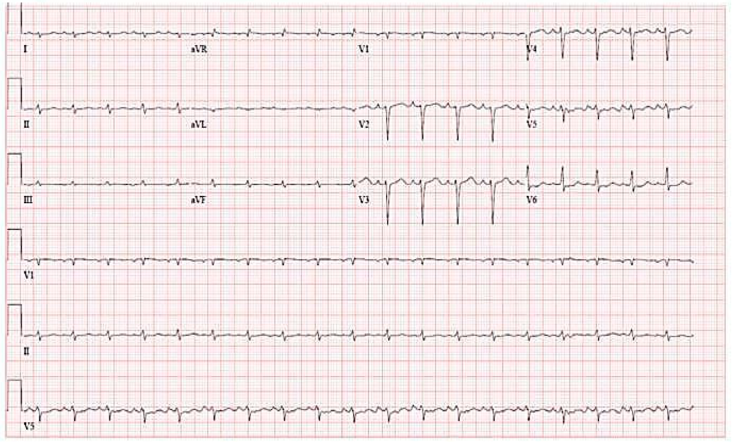
Several electrocardiographic patterns can be present in CA. The most typical is related to low QRS voltages (height <5 mm in all limb leads). In almost 50% of patients with AL-CA, there is a pseudoinfarction pattern associated with poor R wave progression in the chest leads (Figure 2).
As mentioned, one of the most common arrhythmias in CA is atrial fibrillation (about 20% of patients), but amyloid infiltrations can also involve the conduction system in different degrees of severity, from first-degree atrioventricular block (about 20%) to third-degree atrioventricular block. Other less common manifestations are left and right bundle branch block and ventricular tachycardia [19,20]. Holter ECG monitoring helps to identify asymptomatic arrhythmias in more than 75% of AL-CA patients, mainly supraventricular tachyarrhythmias and some nonsustained ventricular tachycardias. Some ECG patterns are more frequent in AL than in ATTR-CA, such as low QRS voltages. Conversely, atrial fibrillation, left bundle branch block, and complete heart block are more common in ATTR-CA. Conflicting data between ECG and echocardiography could sometimes raise the suspicion of CA. For instance, a left ventricular wall thickening can usually be associated with high QRS voltages. If there are low or normal QRS voltages, CA should be considered. A useful instrument to help diagnose CA is the ratio of ECG voltage to LV wall thickness [21].

8. Echocardiography

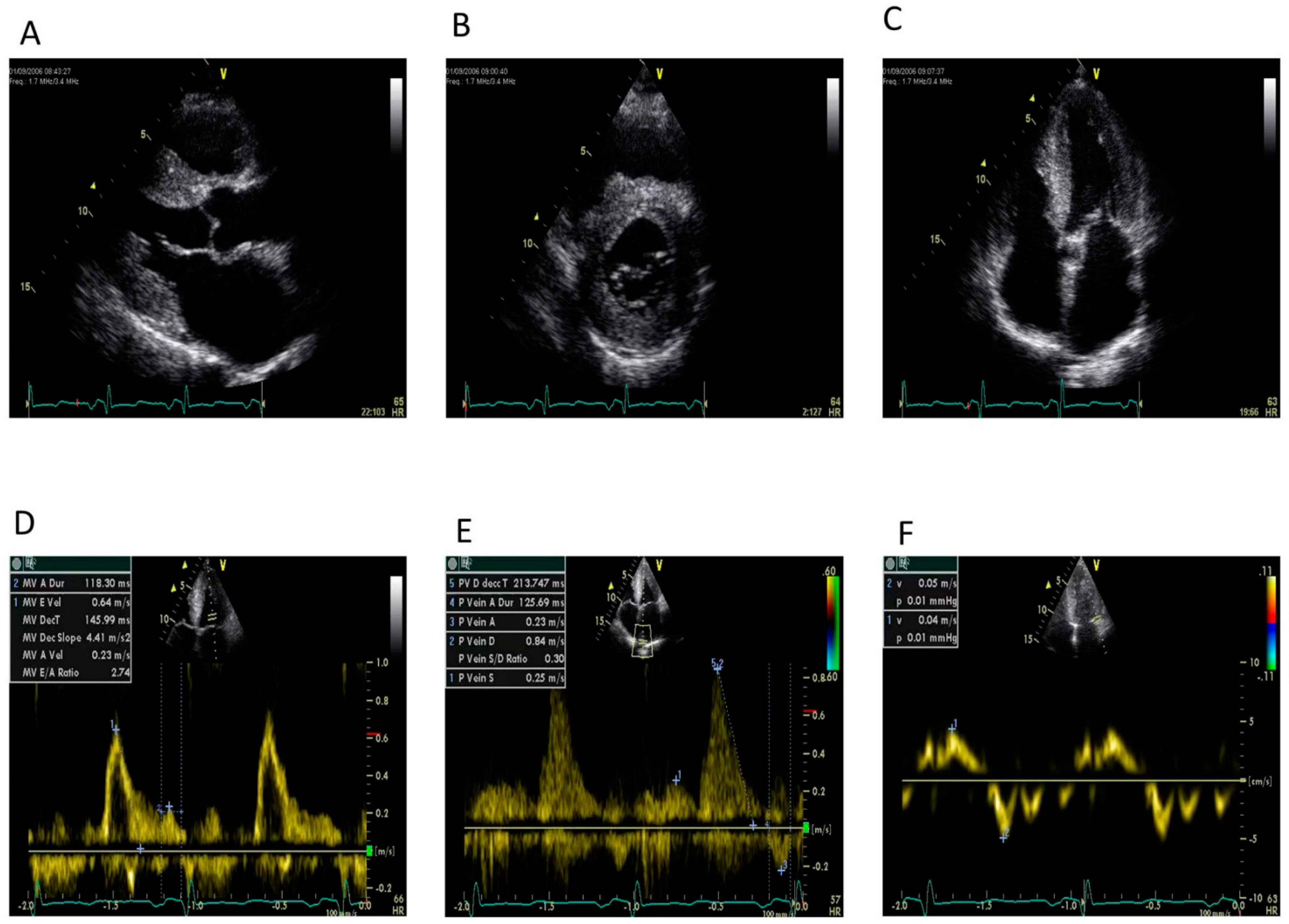
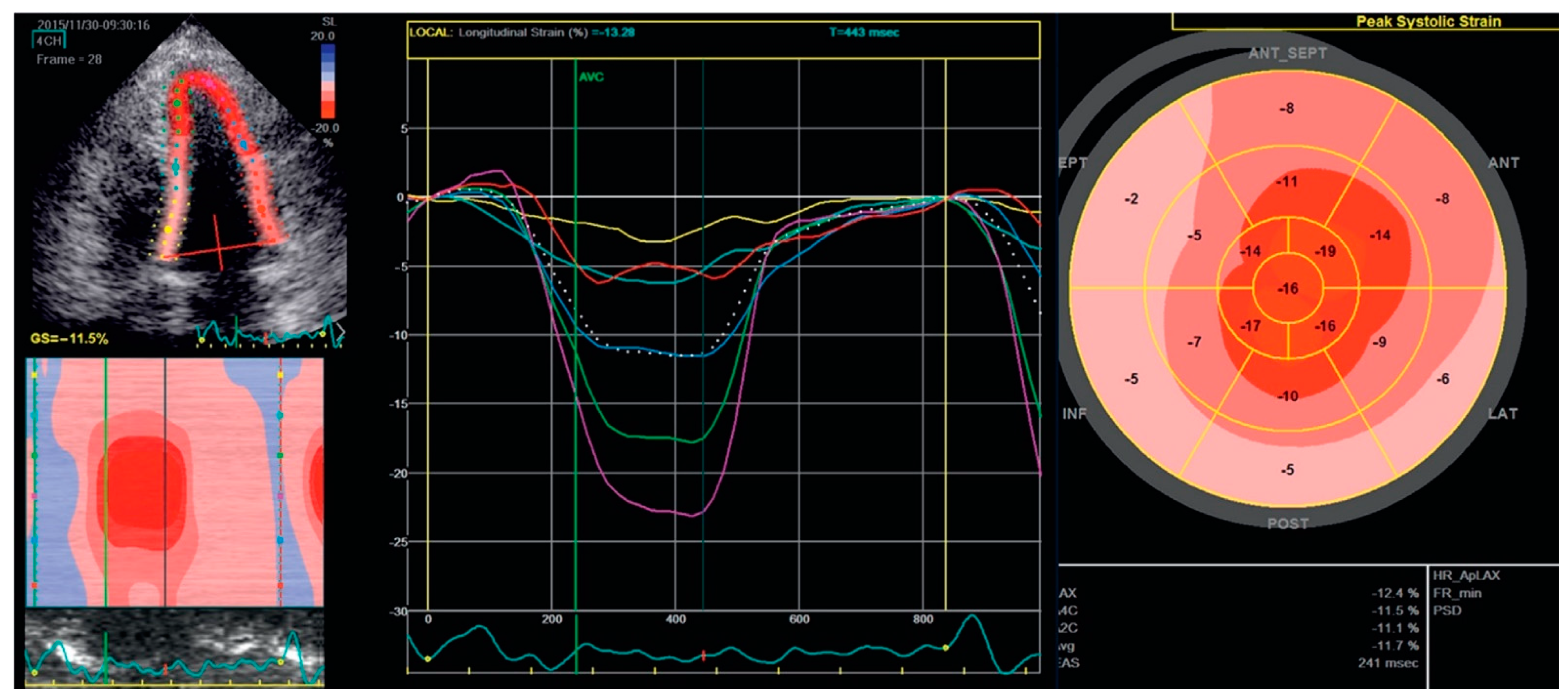
Echocardiography plays a fundamental role in the non-invasive diagnosis of CA, studying heart function and structure in patients with cardiac symptoms or even before symptoms appear. The main focus in CA is to evaluate the thickening of LV walls and the exclusion of other likely causes of LV hypertrophy, such as severe hypertension and moderate to severe aortic stenosis (Figure 3) [22]. However, a clear distinction could be challenging. The suspicion of CA can be increased by other echocardiographic features: biatrial enlargement with a small or normal LV cavity size, presence of thrombi in the left atrium or left atrial appendage, thickening of the cardiac valves, thickening of the right ventricular wall, pericardial effusion, and a restrictive transmitral Doppler filling pattern. In particular, the typical increased wall thickness (>12 mm) with a reduced fractional shortening (<30%) has a significant impact on diagnosis and prognosis [23]. A granular sparkling of the myocardial walls can also be recognized, especially in the interventricular septum, but it does not have a high specificity. Other than the classical evaluation of left ventricular systolic function with LV ejection fraction (LVEF), tissue Doppler imaging (TDI) and speckle-tracking echocardiography (STE) have refined the evaluation of longitudinal systolic function and consequently have increased the probability of an early diagnosis of CA [24,25]. The study of cardiac function with global longitudinal strain (GLS) can help in the diagnosis of CA; despite a preserved LVEF, there is an early reduction of longitudinal shortening, absent in other causes of increased LV wall thickness. In both AL-CA and ATTR-CA, there is a typical apical sparing pattern in STE-derived longitudinal strain; apical LV segments are commonly unaffected, while there is severe impairment in basal and mid-cavity segments (Figure 4). This pattern does not affect patients with other causes of LV hypertrophy, in which the areas with the impairment of the LV longitudinal strain are those with maximal hypertrophy. Despite this peculiarity, it could be difficult to exclude or confirm a diagnosis of amyloidosis in patients with increased heart wall thickness [26]. A multiparametric echocardiographic approach has been proposed, focusing on relative wall thickness, global longitudinal strain (with apical sparing pattern), TAPSE, and E/E’. To guide the diagnostic algorithm, it is often necessary to use highly specific or sensitive cutoffs in patients with a hypertrophic phenotype to avoid unnecessary tests and to limit the time to diagnosis. This approach could be useful to restrict the use of other imaging techniques, such as CMR or endomyocardial biopsy, to patients for whom there is an intermediate to high probability of disease, despite the uncertainty of the first-level diagnostic modalities [27]. Other echocardiographic parameters can be useful. The myocardial contraction fraction (MCF) is the ratio of stroke volume to myocardial volume, and it is helpful in the evaluation of volumetric shortening (correlated with LV longitudinal strain) independent of LV size. Abnormalities beyond the left ventricle can also suggest CA [28]. Stroke volume index has prognostic value in predicting survival in AL-CA; similar to LV strain, it is routinely calculated and easier to achieve than STE. Left atrial dysfunction can also be documented, resulting in both reservoir and pump function impairment with strain because of the increased LV filling pressure and direct amyloid infiltration. This dysfunction may cause the formation of thrombi directly in the atrium or in the appendages (mostly the left appendage), increasing cardioembolic stroke risk even if the patient maintains sinus rhythm [29,30]. The right chambers’ involvement, as documented by CMR studies, contributes to predicting the prognosis in CA. The right ventricle is often affected because of direct amyloid infiltration (as with the left atrium) and the increased afterload from pulmonary hypertension. The result is an impairment of right ventricle systolic function, measured as reduced tricuspid annular plane systolic excursion (TAPSE), tissue Doppler systolic velocity (Sm wave), and longitudinal strain [31,32]. Even with the most modern echocardiographic techniques, the diagnosis of the right chambers’ involvement in CA often depends on the tissue characterization provided by CMR. The combination of echocardiographic parameters with other findings (clinical, biomarkers, and electrocardiography) can increase diagnostic accuracy [33].

9. Cardiac Magnetic Resonance

Cardiac magnetic resonance (CMR) has a central role in the non-invasive diagnosis of CA because of its capacity to give a precise tissue characterization and to differentiate CA from other causes of LV thickening. An extensive CMR evaluation of the four chambers is essential to provide information on ventricle thickness and function, as well as for the study of the atria and the research of potential thrombi. An extensive evaluation with CMR involves cine imaging, assessment of8 native T1 signal, late gadolinium enhancement (LGE), and extracellular volume (ECV) [34,35]. Amyloid deposition has a typical appearance of global and subendocardial LGE, and the distribution strictly correlates with prognosis [36]. However, the enhancement can sometimes be more transmural or, conversely, localized and spotty. To make scans less operator-dependent and to reduce false negatives, it is possible to use phase-sensitive inversion recovery (PSIR) sequences. T1 mapping, which compares scans after contrast administration with the basal images (native T1), is a useful technique to take a quantitative approach in the evaluation of myocardial involvement. In CA, there is an increase in native T1 in both AL and ATTR-CA (higher in AL-CA). In the case of renal impairment, it is possible to use only native T1, requiring no contrast administration, and it may be abnormal before the thickening of the left ventricular wall becomes evident [37]. ECV evaluation before and after contrast administration and combined with native T1 mapping is useful to measure the amyloid burden and myocardial edema—it is usually higher in ATTR-CA. This approach is also helpful in following the progression of the disease and the response to therapy [38,39,40,41,42].

10. Radionuclide Imaging

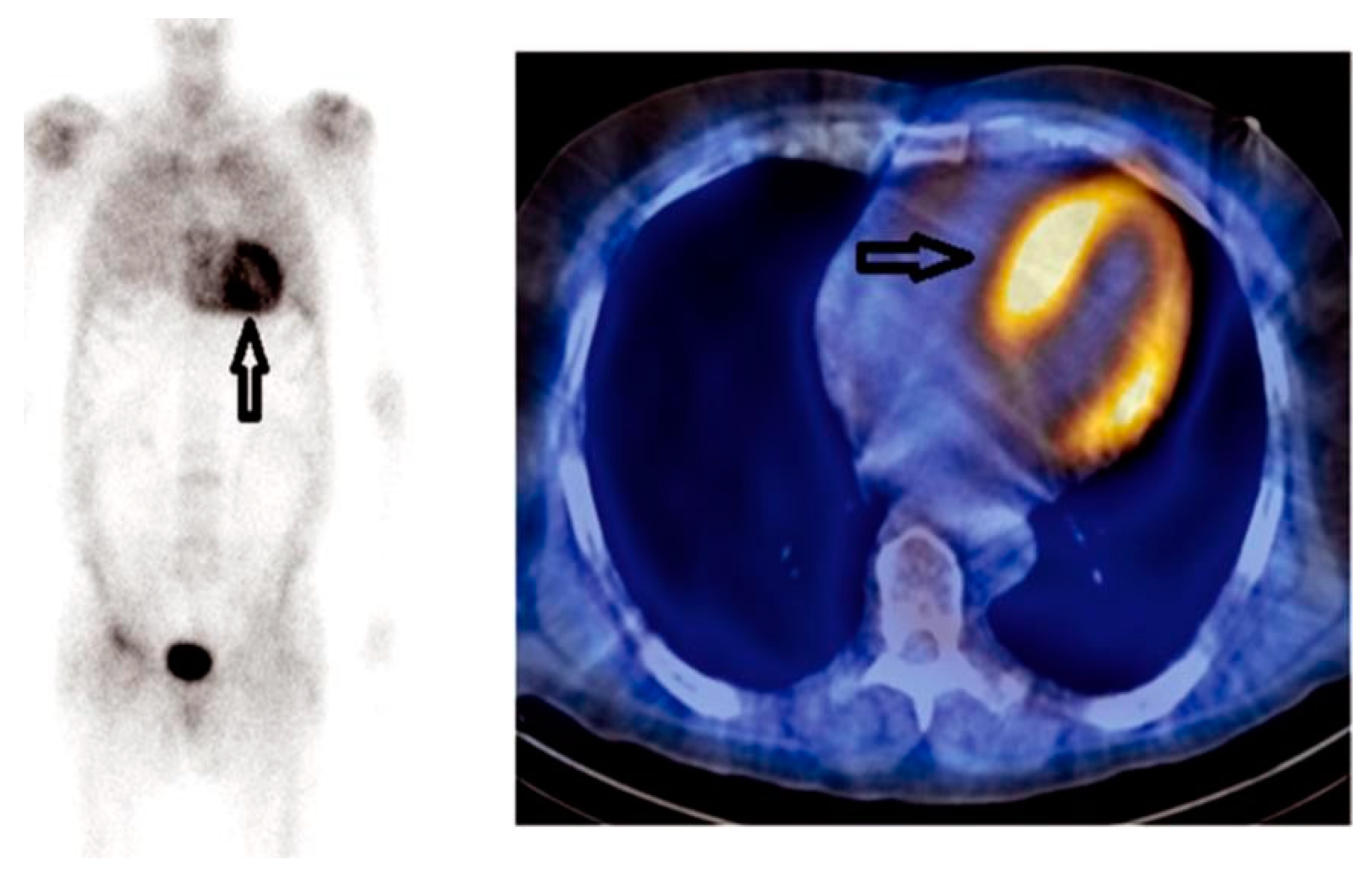
Radionuclide imaging plays a key role in the non-invasive diagnosis of CA. Cardiac involvement is measured by the evaluation of the uptake of diphosphonate radiotracers, such as 99mTechnetium-pyrophosphate (99mTc-PYP), 99mTechnetium-3,3-diphosphono-1,2propanodicarboxy-lic (99mTc-DPD), and 99mTechnetium-hydroxymethylene diphosphonate (99mTc-HMDP). Myocardial uptake for these tracers is remarkably sensitive for ATTR-CA, although not completely specific (Figure 5) [43]. Collectively, uptake in AL-CA is almost absent, and this is the most significant difference from ATTR-CA, which has a high affinity for bone tracers. This differential uptake seems to be related to preferential binding to ATTR because of a higher calcium content [44]. The affinity of the bone tracers in ATTR-CA is useful for the early identification of the disease, and it can help to identify cardiac ATTR deposits in asymptomatic patients at an early stage when the other diagnostic findings might still be absent. Nevertheless, the uptake of 99mTc-DPD can occur in some patients with AL-CA (about 30%); in these cases, the use of 99mTc-DPD SPECT-CT can help to distinguish the two types of CA, having high sensitivity for ATTR-CA. Its tracing has also been evaluated as a potential target for diagnosis and screening [45,46].
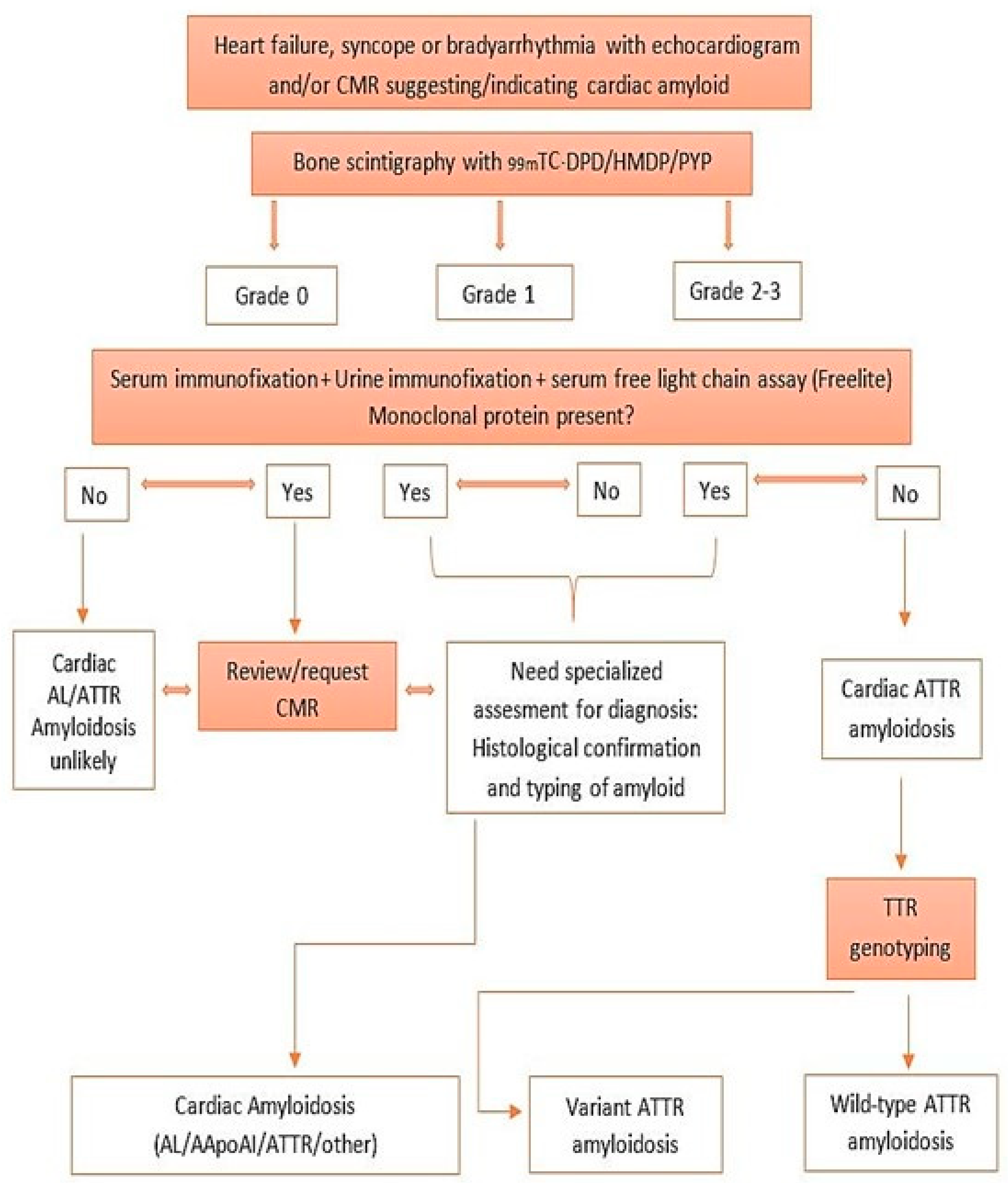
The role of 99mTc-YP/DPD/HMDP cardiac scintigraphy was highlighted in the recently developed consensus algorithm for the non-invasive diagnosis of CA (Figure 6) [47].
This algorithm gives an approach to the evaluation of patients with CA considering the probability of CA. The diagnosis of ATTR-CA can be highly suspected, avoiding the need for endomyocardial biopsy, if there are not clonal abnormalities and there is a high level of uptake of 99mTc-PYP/DPD/HMDP in the radionuclide imaging.
On the other side, patients with biomarker abnormalities, such as high light chain levels in serum/urine immunofixation, have a high probability of AL-CA, and they should be evaluated by hematologists. Cardiac involvement can be evaluated with the aforementioned non-invasive imaging techniques or with endomyocardial biopsy.

11. Tissue Biopsy

Endomyocardial biopsy (EMB) was previously considered the diagnostic gold standard to evaluate cardiac amyloid deposition. Typically, the pathognomonic apple-green birefringence under cross-polarized light microscopy confirms the presence of amyloid fibrils. Today, it cannot be considered the optimal diagnostic choice to obtain a diagnosis of CA because of the need for an invasive approach and the potential risks. For the evaluation of systemic burden, the diagnostic accuracy of an extra-cardiac biopsy, i.e., the abdominal fat pad, depends on the examined tissue and the type of amyloidosis. The accuracy is higher for AL-CA, in which the yield of a fat pad biopsy is >70%, and it is strongly associated with whole-body amyloid load [48]. For the relatively low invasiveness, a fat pad biopsy is the preferred initial site, but a negative result is not sufficient to rule out a diagnosis of CA. If there is a high suspicion of disease, an EMB should be performed despite a negative extra-cardiac biopsy.

12. Conclusions

Cardiac involvement is variable in the different types of amyloidosis, but it has a major impact on prognosis [49]. Cardiac amyloidosis has been revalued as a more treatable and possibly curable condition thanks to the recent improvements in diagnostic and therapeutic strategies. Nevertheless, morbidity and mortality remain high. For this reason, more advancements are needed to obtain an early diagnosis and to enhance prognoses in these patients.

Author Contributions

Conceptualization, M.G.M. and F.I.; methodology, M.D.M.; software, A.I.; validation, M.C. and A.E.; formal analysis, R.P.; investigation, F.I.; resources, M.D.M.; data curation, A.I.; writing—original draft preparation, M.G.M.; writing—review and editing, A.E.; visualization, M.C.; supervision, A.E.; project administration, R.P.; funding acquisition, A.I. All authors have read and agreed to the published version of the manuscript.

Funding

This research received no external funding.

Conflicts of Interest

The authors declare no conflict of interest.

References

  1. Merlini, G.; Bellotti, V. Molecular mechanisms of amyloidosis. N. Engl. J. Med. 2003, 349, 583–596. [Google Scholar] [CrossRef] [Green Version]
  2. Richards, D.B.; Cookson, L.M.; Berges, A.C.; Barton, S.V.; Lane, T.; Ritter, J.M.; Fontana, M.; Moon, J.C.; Pinzani, M.; Gillmore, J.D.; et al. Therapeutic Clearance of Amyloid by Antibodies to Serum Amyloid P Component. N. Engl. J. Med. 2015, 373, 1106–1114. [Google Scholar] [CrossRef] [Green Version]
  3. Sipe, J.D.; Benson, M.D.; Buxbaum, J.N.; Ikeda, S.; Merlini, G.; Saraiva, M.J.; Westermark, P. Nomenclature 2014: Amyloid fibril proteins and clinical classification of the amyloidosis. Amyloid 2014, 21, 221–224. [Google Scholar] [CrossRef] [PubMed]
  4. Maurer, M.S.; Elliott, P.; Comenzo, R.; Semigran, M.; Rapezzi, C. Addressing Common Questions Encountered in the Diagnosis and Management of Cardiac Amyloidosis. Circulation 2017, 135, 1357–1377. [Google Scholar] [CrossRef] [Green Version]
  5. Kyle, R.A.; Linos, A.; Beard, C.M.; Linke, R.P.; Gertz, M.A.; O’Fallon, W.M.; Kurland, L.T. Incidence and natural history of primary systemic amyloidosis in Olmsted County, Minnesota, 1950 through 1989. Blood 1992, 79, 1817–1822. [Google Scholar] [CrossRef] [Green Version]
  6. Quock, T.P.; Yan, T.; Chang, E.; Guthrie, S.; Broder, M.S. Epidemiology of AL amyloidosis: A real-world study using US claims data. Blood Adv. 2018, 2, 1046–1053. [Google Scholar] [CrossRef] [Green Version]
  7. Sperry, B.W.; Ikram, A.; Hachamovitch, R.; Valent, J.; Vranian, M.N.; Phelan, D.; Hanna, M. Efficacy of Chemotherapy for Light-Chain Amyloidosis in Patients Presenting With Symptomatic Heart Failure. J. Am. Coll. Cardiol. 2016, 67, 2941–2948. [Google Scholar] [CrossRef]
  8. Donnelly, J.P.; Hanna, M. Cardiac amyloidosis: An update on diagnosis and treatment. Cleve Clin. J. Med. 2017, 84 (Suppl. 3), 12–26. [Google Scholar] [CrossRef] [Green Version]
  9. Dubrey, S.W.; Cha, K.; Anderson, J.; Chamarthi, B.; Reisinger, J.; Skinner, M.; Falk, R.H. The clinical features of immunoglobulin light-chain (AL) amyloidosis with heart involvement. QJM 1998, 91, 141–157. [Google Scholar] [CrossRef] [PubMed] [Green Version]
  10. Sekijima, Y.; Yoshida, K.; Tokuda, T.; Ikeda, S.-I. Familial Transthyretin Amyloidosis; Adam, M.P., Ardinger, H.H., Pagon, R.A., Eds.; University of Washington, Seattle: Seattle, WA, USA, 1993–2017; Updated 26 January 2012. Available online: https://www-ncbi-nlm-nih-gov.ccmain.ohionet.org/books/NBK1194/ (accessed on 16 November 2017).
  11. Vermeer, A.M.C.; Janssen, A.; Boorsma, P.C.; Mannens, M.M.A.M.; Wilde, A.A.M.; Christiaans, I. Transthyretin amyloidosis: A phenocopy of hypertrophic cardiomyopathy. Amyloid 2017, 24, 87–91. [Google Scholar] [CrossRef] [PubMed] [Green Version]
  12. Siddiqi, O.K.; Ruberg, F.L. Cardiac amyloidosis: An update on pathophysiology, diagnosis, and treatment. Trends Cardiovasc. Med. 2018, 28, 10–21. [Google Scholar] [CrossRef] [PubMed]
  13. Longhi, S.; Quarta, C.C.; Milandri, A.; Lorenzini, M.; Gagliardi, C.; Manuzzi, L.; Bacchi-Reggiani, M.L.; Leone, O.; Ferlini, A.; Russo, A.; et al. Atrial fibrillation in amyloidotic cardiomyopathy: Prevalence, incidence, risk factors and prognostic role. Amyloid 2015, 22, 147–155. [Google Scholar] [CrossRef]
  14. Mesquita, E.T.; Jorge, A.J.L.; Souza CVJunior Andrade, T.R. Cardiac amyloidosis and its new clinical phenotype: Heart failure with preserved ejection fraction. Arq. Bras. Cardiol. 2017, 109, 71–80. [Google Scholar] [CrossRef]
  15. Adams, D.; Ando, Y.; Beirão, J.M.; Coelho, T.; Gertz, M.A.; Gillmore, J.D.; Hawkins, P.N.; Lousada, I.; Suhr, O.B.; Merlini, G. Expert consensus recommendations to improve diagnosis of ATTR amyloidosis with polyneuropathy. J Neurol. 2021, 268, 2109–2122. [Google Scholar] [CrossRef] [Green Version]
  16. Dorbala, S.; Ando, Y.; Bokhari, S.; Dispenzieri, A.; Falk, R.H.; Ferrari, V.A.; Fontana, M.; Gheysens, O.; Gillmore, J.D.; Glaudemans, A.W.J.M.; et al. ASNC/AHA/ASE/EANM/HFSA/ISA/SCMR/SNMMI expert consensus recommendations for multimodality imaging in cardiac amyloidosis: Part 1 of 2-evidence base and standardized methods of imaging. J. Nucl. Cardiol. 2019, 26, 2065–2123. [Google Scholar] [CrossRef] [PubMed]
  17. Palladini, G.; Campana, C.; Klersy, C.; Balduini, A.; Vadacca, G.; Perfetti, V.; Perlini, S.; Obici, L.; Ascari, E.; d’Eril, G.M.; et al. Serum N-terminal probrain natriuretic peptide is a sensitive marker of myocardial dysfunction in AL amyloidosis. Circulation 2003, 107, 2440–2445. [Google Scholar] [CrossRef] [PubMed]
  18. Wechalekar, A.D.; Gillmore, J.D.; Wassef, N.; Lachmann, H.J.; Whelan, C.; Hawkins, P.N. Abnormal N-terminal fragment of brain natriuretic peptide in patients with light chain amyloidosis without cardiac involvement at presentation is a risk factor for development of cardiac amyloidosis. Haematologica 2011, 96, 1079–1080. [Google Scholar] [CrossRef] [PubMed] [Green Version]
  19. Cheng, Z.W.; Tian, Z.; Kang, L.; Chen, T.B.; Fang, L.G.; Cheng, K.A.; Zeng, Y.; Fang, Q. Electrocardiographic and echocardiographic features of patients with primary cardiac amyloidosis. Zhonghua Xin Xue Guan Bing Za Zhi 2010, 38, 606–609. [Google Scholar] [PubMed]
  20. Murtagh, B.; Hammill, S.C.; Gertz, M.A.; Kyle, R.A.; Tajik, A.J.; Grogan, M. Electrocardiographic findings in primary systemic amyloidosis and biopsy- proven cardiac involvement. Am. J. Cardiol. 2005, 95, 535–537. [Google Scholar] [CrossRef] [PubMed]
  21. Carroll, J.D.; Gaasch, W.H.; McAdam, K.P. Amyloid cardiomyopathy: Characterization by a distinctive voltage/mass relation. Am. J. Cardiol. 1982, 49, 9–13. [Google Scholar] [CrossRef]
  22. Gertz, M.A.; Comenzo, R.; Falk, R.H.; Fermand, J.P.; Hazenberg, B.P.; Hawkins, P.N.; Merlini, G.; Moreau, P.; Ronco, P.; Sanchorawala, V.; et al. Definition of organ involvement and treatment response in immunoglobulin light chain amyloidosis (AL): A consensus opinion from the 10th International Symposium on Amyloid and Amyloidosis, Tours, France, 18-22 April 2004. Am. J. Hematol. 2005, 79, 319–328. [Google Scholar] [CrossRef]
  23. Cueto-Garcia, L.; Reeder, G.S.; Kyle, R.A.; Wood, D.L.; Seward, J.B.; Naessens, J.; Offord, K.P.; Greipp, P.R.; Edwards, W.D.; Tajik, A.J. Echocardiographic findings in systemic amyloidosis: Spectrum of cardiac involvement and relation to survival. J. Am. Coll. Cardiol. 1985, 6, 737–743. [Google Scholar] [CrossRef] [Green Version]
  24. Buss, S.J.; Emami, M.; Mereles, D.; Korosoglou, G.; Kristen, A.V.; Voss, A.; Schellberg, D.; Zugck, C.; Galuschky, C.; Giannitsis, E.; et al. Longitudinal left ventricular function for prediction of survival in systemic light-chain amyloidosis: Incremental value compared with clinical and biochemical markers. J Am. Coll. Cardiol. 2012, 60, 1067–1076. [Google Scholar] [CrossRef] [PubMed] [Green Version]
  25. Koyama, J.; Ray-Sequin, P.A.; Falk, R.H. Longitudinal myocardial function assessed by tissue velocity, strain, and strain rate tissue Doppler echocardiography in patients with AL (primary) cardiac amyloidosis. Circulation 2003, 107, 2446–2452. [Google Scholar] [CrossRef] [PubMed]
  26. Phelan, D.; Collier, P.; Thavendiranathan, P.; Popović, Z.B.; Hanna, M.; Plana, J.C.; Marwick, T.H.; Thomas, J.D. Relative apical sparing of longitudinal strain using two-dimensional speckle-tracking echocardiography is both sensitive and specific for the diagnosis of cardiac amyloidosis. Heart 2012, 1442–1448. [Google Scholar] [CrossRef] [PubMed]
  27. Boldrini, M.; Cappelli, F.; Chacko, L.; Restrepo-Cordoba, M.A.; Lopez-Sainz, A.; Giannoni, A.; Aimo, A.; Baggiano, A.; Martinez-Naharro, A.; Whelan, C.; et al. Multiparametric Echocardiography Scores for the Diagnosis of Cardiac Amyloidosis. JACC Cardiovasc. Imaging 2020, 13, 909–920. [Google Scholar] [CrossRef] [PubMed]
  28. Tendler, A.; Helmke, S.; Teruya, S.; Alvarez, J.; Maurer, M.S. The myocardial contraction fraction is superior to ejection fraction in predicting survival in patients with AL cardiac amyloidosis. Amyloid 2015, 22, 61–66. [Google Scholar] [CrossRef] [PubMed]
  29. Martinez-Naharro, A.; Gonzalez-Lopez, E.; Corovic, A.; Mirelis, J.G.; Baksi, A.J.; Moon, J.C.; Garcia-Pavia, P.; Gillmore, J.D.; Hawkins, P.N.; Fontana, M. High Prevalence of Intracardiac Thrombi in Cardiac Amyloidosis. J. Am. Coll. Cardiol. 2019, 73, 1733–1734. [Google Scholar] [CrossRef]
  30. Feng, D.; Edwards, W.D.; Oh, J.K.; Chandrasekaran, K.; Grogan, M.; Martinez, M.W.; Syed, I.S.; Hughes, D.A.; Lust, J.A.; Jaffe, A.S.; et al. Intracardiac thrombosis and embolism in patients with cardiac amyloidosis. Circulation 2007, 116, 2420–2426. [Google Scholar] [CrossRef] [PubMed] [Green Version]
  31. Bellavia, D.; Pellikka, P.A.; Dispenzieri, A.; Scott, C.G.; Al-Zahrani, G.B.; Grogan, M.; Pitrolo, F.; Oh, J.K.; Miller, F.A., Jr. Comparison of right ventricular longitudinal strain imaging, tricuspid annular plane systolic excursion, and cardiac biomarkers for early diagnosis of cardiac involvement and risk stratification in primary systematic (AL) amyloidosis: A 5-year cohort study. Eur. Heart J. Cardiovasc. Imaging 2012, 13, 680–689. [Google Scholar] [CrossRef] [Green Version]
  32. Rapezzi, C.; Lorenzini, M.; Longhi, S.; Milandri, A.; Gagliardi, C.; Bartolomei, I.; Salvi, F.; Maurer, M.S. Cardiac amyloidosis: The great pretender. Heart Fail. Rev. 2015, 20, 117–124. [Google Scholar] [CrossRef] [PubMed]
  33. Damy, T.; Maurer, M.S.; Rapezzi, C.; Planté-Bordeneuve, V.; Karayal, O.N.; Mundayat, R.; Suhr, O.B.; Kristen, A.V. Clinical, ECG and echocardiographic clues to the diagnosis of TTR-related cardiomyopathy. Open Heart 2016, 3, e000289. [Google Scholar] [CrossRef] [Green Version]
  34. Maceira, A.M.; Joshi, J.; Prasad, S.K.; Moon, J.C.; Perugini, E.; Harding, I.; Sheppard, M.N.; Poole-Wilson, P.A.; Hawkins, P.N.; Pennell, D.J. Cardiovascular magnetic resonance in cardiac amyloidosis. Circulation 2005, 111, 186–193. [Google Scholar] [CrossRef] [Green Version]
  35. Pandey, T.; Jambhekar, K.; Shaikh, R.; Lensing, S.; Viswamitra, S. Utility of the inversion scout sequence (TI scout) in diagnosing myocardial amyloid infiltration. Int. J. Cardiovasc. Imaging 2013, 29, 103–112. [Google Scholar] [CrossRef] [PubMed] [Green Version]
  36. Fontana, M.; Pica, S.; Reant, P.; Abdel-Gadir, A.; Treibel, T.A.; Banypersad, S.M.; Maestrini, V.; Barcella, W.; Rosmini, S.; Bulluck, H.; et al. Prognostic Value of Late Gadolinium Enhancement Cardiovascular Magnetic Resonance in Cardiac Amyloidosis. Circulation 2015, 132, 1570–1579. [Google Scholar] [CrossRef]
  37. Vogelsberg, H.; Mahrholdt, H.; Deluigi, C.C.; Yilmaz, A.; Kispert, E.M.; Greulich, S.; Klingel, K.; Kandolf, R.; Sechtem, U. Cardiovascular magnetic resonance in clinically suspected cardiac amyloidosis: Noninvasive imaging compared to endomyocardial biopsy. J. Am. Coll. Cardiol. 2008, 51, 1022–1030. [Google Scholar] [CrossRef] [PubMed] [Green Version]
  38. Syed, I.S.; Glockner, J.F.; Feng, D.; Araoz, P.A.; Martinez, M.W.; Edwards, W.D.; Gertz, M.A.; Dispenzieri, A.; Oh, J.K.; Bellavia, D.; et al. Role of cardiac magnetic resonance imaging in the detection of cardiac amyloidosis. JACC Cardiovasc. Imaging 2010, 3, 155–164. [Google Scholar] [CrossRef]
  39. White, J.A.; Kim, H.W.; Shah, D.; Fine, N.; Kim, K.Y.; Wendell, D.C.; Al-Jaroudi, W.; Parker, M.; Patel, M.; Gwadry-Sridhar, F.; et al. CMR imaging with rapid visual T1 assessment predicts mortality in patients suspected of cardiac amyloidosis. JACC Cardiovasc. Imaging 2014, 7, 143–156. [Google Scholar] [CrossRef] [Green Version]
  40. Ruberg, F.L.; Appelbaum, E.; Davidoff, R.; Ozonoff, A.; Kissinger, K.V.; Harrigan, C.; Skinner, M.; Manning, W.J. Diagnostic and prognostic utility of cardiovascular magnetic resonance imaging in light-chain cardiac amyloidosis. Am. J. Cardiol. 2009, 103, 544–549. [Google Scholar] [CrossRef] [Green Version]
  41. Austin, B.A.; Tang, W.H.; Rodriguez, E.R.; Tan, C.; Flamm, S.D.; Taylor, D.O.; Starling, R.C.; Desai, M.Y. Delayed hyper-enhancement magnetic resonance imaging provides incremental diagnostic and prognostic utility in suspected cardiac amyloidosis. JACC Cardiovasc. Imaging 2009, 2, 1369–1377. [Google Scholar] [CrossRef] [Green Version]
  42. Fontana, M. Systemic Amyloidosis by Cardiovascular Magnetic Resonance. Ph.D. Thesis, UCL University College London, London, UK, 2016. [Google Scholar]
  43. Banypersad, S.M.; Moon, J.C.; Whelan, C.; Hawkins, P.N.; Wechalekar, A.D. Updates in cardiac amyloidosis: A review. J. Am. Heart Assoc. 2021. [Google Scholar] [CrossRef] [PubMed] [Green Version]
  44. Hutt, D.F.; Quigley, A.M.; Page, J.; Hall, M.L.; Burniston, M.; Gopaul, D.; Lane, T.; Whelan, C.J.; Lachmann, H.J.; Gillmore, J.D.; et al. Utility and limitations of 3,3-diphosphono-1,2- propanodicarboxylic acid scintigraphy in systemic amyloidosis. Eur Heart J. Cardiovasc. Imaging 2014, 15, 1289–1298. [Google Scholar] [CrossRef] [PubMed]
  45. Cappelli, F.; Gallini, C.; Di Mario, C.; Costanzo, E.N.; Vaggelli, L.; Tutino, F.; Ciaccio, A.; Bartolini, S.; Angelotti, P.; Frusconi, S.; et al. Accuracy of 99mTc-Hydroxymethylene diphosphonate scintigraphy for diagnosis of transthyretin cardiac amyloidosis. J. Nucl. Cardiol. 2019, 26, 497–504. [Google Scholar] [CrossRef] [PubMed]
  46. Galat, A.; Rosso, J.; Guellich, A.; Van Der Gucht, A.; Rappeneau, S.; Bodez, D.; Guendouz, S.; Tissot, C.M.; Hittinger, L.; Dubois-Randé, J.L.; et al. Usefulness of (99m)Tc-HMDP scintigraphy for the etiologic diagnosis and prognosis of cardiac amyloidosis. Amyloid 2015, 22, 210–220. [Google Scholar] [CrossRef]
  47. Gillmore, J.D.; Maurer, M.S.; Falk, R.H.; Merlini, G.; Damy, T.; Dispenzieri, A.; Wechalekar, A.D.; Berk, J.L.; Quarta, C.C.; Grogan, M.; et al. Nonbiopsy Diagnosis of Cardiac Transthyretin Amyloidosis. Circulation 2016, 133, 2404–2412. [Google Scholar] [CrossRef]
  48. Fine, N.M.; Arruda-Olson, A.M.; Dispenzieri, A.; Zeldenrust, S.R.; Gertz, M.A.; Kyle, R.A.; Swiecicki, P.L.; Scott, C.G.; Grogan, M. Yield of noncardiac biopsy for the diagnosis of transthyretin cardiac amyloidosis. Am. J. Cardiol. 2014, 113, 1723–1727. [Google Scholar] [CrossRef]
  49. Van Der Gucht, A.; Cottereau, A.S.; Abulizi, M.; Guellich, A.; Blanc-Durand, P.; Israel, J.M.; Galat, A.; Plante-Bordeneuve, V.; Dubois-Randé, J.L.; Bodez, D.; et al. Apical sparing pattern of left ventricular myocardial 99mTc-HMDP uptake in patients with transthyretin cardiac amyloidosis. J. Nucl. Cardiol. 2017, 25, 2072–2079. [Google Scholar] [CrossRef] [PubMed]
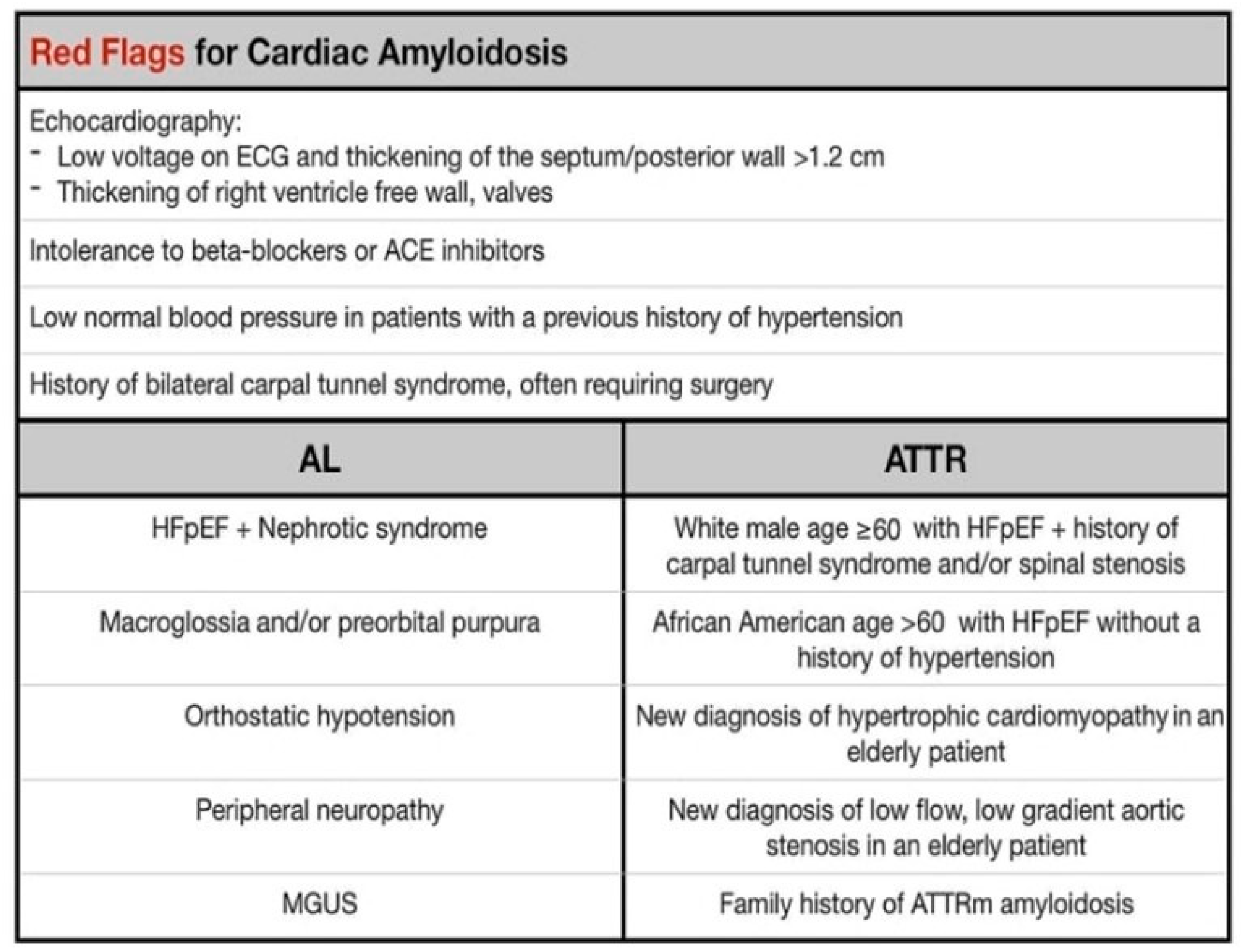
Figure 1. Red Flags for cardiac amyloidosis.
Cardiogenetics 11 00012 g001
Figure 2. ECG of a patient with cardiac AL amyloidosis showing the pseudoinfarction pattern in anterior leads and small QRS voltages predominantly in the limb leads.
Cardiogenetics 11 00012 g002
Figure 3. Sequence of still images showing typical echocardiographic features of cardiac amyloidosis. (A): Two-dimensional (2-D) parasternal long axis showing an increase of left ventricular wall thickness—in both the interventricular septum and the posterior wall (concentric hypertrophy)—and left atrial dilatation. (B): Two-dimensional parasternal short axis. (C): Two-dimensional apical four-chamber view. In B and C, it is possible to highlight not only the left ventricular hypertrophy but also the biatrial dilatation. (D): Pulsed wave Doppler of mitral inflow showing a restrictive pattern at transmitral flow: increase in E/A ratio and normal E wave deceleration time with a marked reduction in transmitral A wave velocity. (E): Pulsed wave Doppler of pulmonary vein inflow showing marked diastolic prominence and increased duration and peak velocity of atrial reversal compared with the transmitral signal. (F): Pulsed tissue Doppler of the lateral mitral annulus showing marked reduction in apical systolic and diastolic velocities (normal velocities: >6 cm/s and >8 cm/s, respectively). Images courtesy of Professor Elliott, University College London, UK.
Cardiogenetics 11 00012 g003
Figure 4. (Left panel) Segmental color coding in the apical four-chamber view showing the typical apical sparing pattern, with the more negative strain segment in the apex (darker red), compared with a lesser negative strain in the basal segments (pink). (Middle panel) Individual segmental strain for the same view. (Right panel) A bull’s-eye plot derived from the three apical views, showing sparing of apical strain (center of plot) with impaired mid-cavity and basal strain.
Cardiogenetics 11 00012 g004
Figure 5. A positive 99mTc-DPD scan for TTR cardiac amyloid (left) showing uptake in the heart (arrow) and reduced bone uptake. The right-hand panel shows a fused CT/SPECT image showing myocardial uptake, with greater uptake in the septum.
Cardiogenetics 11 00012 g005
Figure 6. Consensus algorithm for non-invasive diagnosis of cardiac amyloidosis.
Cardiogenetics 11 00012 g006
Publisher’s Note: MDPI stays neutral with regard to jurisdictional claims in published maps and institutional affiliations.