Abstract
Background: AARS2 encodes the mitochondrial protein alanyl-tRNA synthetase 2 (MT-AlaRS), an important enzyme in oxidative phosphorylation. Variants in AARS2 have previously been associated with infantile cardiomyopathy. Case summary: A 4-year-old girl died of infantile-onset dilated cardiomyopathy (DCM) in 1996. Fifteen years later, her 21-year-old brother was diagnosed with DCM and ultimately underwent heart transplantation. Initial sequencing of 15 genes discovered no pathogenic variants in the brother at the time of his diagnosis. However, 9 years later re-screening in an updated screening panel of 129 genes identified a homozygous AARS2 (c.1774C > T) variant. Sanger sequencing of the deceased girl confirmed her to be homozygous for the AARS2 variant, while both parents and a third sibling were all found to be unaffected heterozygous carriers of the AARS2 variant. Discussion: This report underlines the importance of repeated and extended genetic screening of elusive families with suspected hereditary cardiomyopathies, as our knowledge of disease-causing mutations continuously grows. Although identification of the genetic etiology in the reported family would not have changed the clinical management, the genetic finding allows genetic counselling and holds substantial value in identifying at-risk relatives.
1. Introduction
Genetic variants in the nuclear gene AARS2 have previously been associated with recessively inherited infantile mitochondrial cardiomyopathy [1,2,3,4]. AARS2 encodes the mitochondrial protein alanyl-tRNA synthetase 2 (MT-AlaRS), an important enzyme in oxidative phosphorylation contributing to normal functioning of respiratory chain complexes I, III, and IV1.
Typically, genetic variants in AARS2 have been identified in one tissue-specific disease, most commonly in patients with infantile-onset cardiomyopathy and in patients with childhood to adulthood-onset leukoencephalopathy [5].
1.1. Patient 1
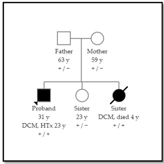
A 21-year-old Caucasian male diagnosed with Asperger Syndrome, was admitted (2011) to the emergency department with progressive dyspnea and lower limb edemas. The patient was the second of three children from healthy nonconsanguineous Danish parents. Due to a family history of dilated cardiomyopathy (Figure 1), an echocardiography at age 6 years (1996) had been performed with no abnormal findings, and no follow-up was planned at that point.

Figure 1.
Pedigree showing recessive inheritance of dilated cardiomyopathy (DCM). Individuals with DCM are illustrated by black filling.
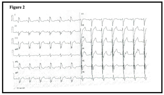
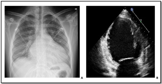
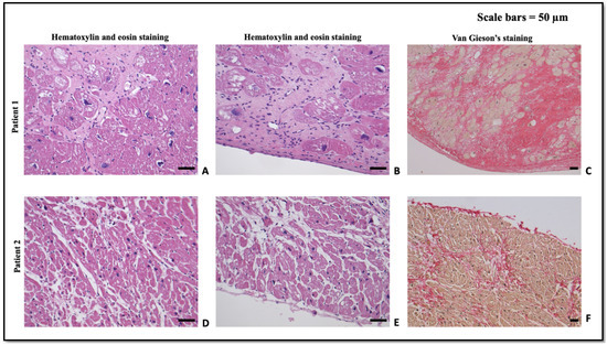
At the time of admission in 2011, the patient was hemodynamically stable (blood pressure 116/69, heart rate 100) and presented with a fever leading to initiation of antibiotic treatment due to suspicion of pneumonia. Routine blood testing including cardiac troponins, hematological, and kidney biomarkers were unremarkable. However, an ECG showed intraventricular conduction defect with a QRS-duration of 150 ms (Figure 2). A chest radiograph revealed cardiomegaly (Figure 3A). Subsequently, an echocardiography demonstrated severe left ventricular (LV) dilation with an end-diastolic diameter of 82 mm (indexed 41 mm/m2) and a LV ejection fraction (EF) of 10% (Figure 3B). These findings led to the initiation of anti-congestive medical treatment and later implantation of a cardiac resynchronization therapy-defibrillator. Further diagnostic work-up included genetic screening, right heart catheterization, and coronary angiography. The right heart catheterization showed a reduced cardiac index (1.8 L/min/m²) and elevated cardiac chamber filling pressures (right atrial pressure of 12 mmHg, mean pulmonary artery pressure of 47 mmHg, and wedge pressure of 31 mmHg). The coronary angiogram was normal, and the myocardial biopsy showed histologic features consistent with dilated cardiomyopathy with severely hypertrophic cardiomyocytes (Figure 4A–C).

Figure 2.
ECG of patient 1 at admission time showing an intraventricular conduction defect.

Figure 3.
(A) Chest radiograph of patient 1 at admission time showing cardiomegaly. (B) Echocardiography of patient 1 at admission time showing severe left ventricular dilation.

Figure 4.
(A–C) Histological study of the myocardial biopsy of patient 1. In both deep (A) and subendocardial myocardium (B) substantial numbers of cardiomyocytes are replaced with fibrosis consisting of irregularly arranged fine collagen fibrils with entrapped myocytes in the periphery. Severe hypertrophy (myocyte diameter up to 50 µm) as well as irregularly shaped and enlarged nuclei (diameter typically 15 µm) are observed in the remaining myocytes. In addition, fragmentation and loss of central myofibrils occurs focally. No obvious myocyte disarray was observed. The features are consistent with cardiomyopathy and compatible with the clinically observed dilated cardiomyopathy, although hypertrophic cardiomyopathy is a histological differential diagnosis. (D–F) Histological study of the myocardial biopsy of patient 2. Appearance of variably solidified fibrotic areas in both central (D) and subendocardial myocardium (E) replacing few cardiomyocytes. The caliber is lightly variably with a diameter around 20 µm and nuclei appearing slightly enlarged (typically 10 µm in diameter) and often slightly irregular in shape. Focally perinuclear myofibrils seem fragmented and diminished in occurrence. The features are similar to those observed in patient 1, although less severely manifested, and consistent with cardiomyopathy compatible with the clinically observed dilated cardiomyopathy.
The patient was hospitalized for two weeks, and subsequently followed closely as an outpatient in the heart failure clinic. Despite heart failure medication and cardiac resynchronization therapy, the patient remained severely symptomatic with no signs of left ventricular reverse remodeling or recovery of LVEF. Five months later, a left ventricular assist device was therefore implanted as a bridge to heart transplantation. Transplantation was performed without complications 40 months after the initial diagnosis. Today, eight years post-transplantation the patient is alive and doing well.
1.2. Patient 2
Fifteen years prior (1996) to the events described above, the patient’s 4-year old sister was admitted with dyspnea and fever to a local hospital. Initially, the patient was suspected to have pneumonia. The patient’s mother had noticed a reduced appetite and onset of mild dyspnea a week prior to admission and prior to admission the girl was described to have a short stature and physically weak compared to her peers with some circumstantial evidence of delayed neuro-cognitive development. Symptoms of dyspnea progressed and was accompanied by fever (40 °C) and incessant coughing with intermittent mucous vomit on the day of admission. On clinical examination the patient was found to be cyanotic, have massive jugular venous distension (10–12 cm), and peripheral edema. A chest X-ray did not identify pneumonia, but cardiac enlargement was observed. An echocardiography showed severe LV dilation, a LVEF of 5% and severe universal hypokinesia of the LV. At this point, the patient was suspected to suffer from acute fulminant myocarditis and was transferred to a tertiary hospital. During transport, the patient developed cardiogenic shock with pulmonary edema, respiratory acidosis and was admitted directly to an intensive care unit (ICU). At the ICU, the patient received invasive mechanical ventilation along with vasopressors, intravenous immunoglobulin, and other supportive care. Treatment led to some initial clinical improvement although development of kidney failure required hemodialysis. During the admission, no subsequent improvement in systolic functioning was observed and the patient died of terminal heart failure after four weeks of hospitalization. An autopsy was performed with histologic features strongly suggestive of cardiomyopathy compatible with dilated cardiomyopathy (Figure 4D–F).
2. Genetic Investigations
2.1. Patient 1
Initially, genome based genetic sequencing was performed in a standard screening panel, which in 2011 consisted of 15 genes (MYH7, MYBPC3, TNNT2, TNNI3, TPM1, ACTC, MYL2, MYL3, CSRP3, SCN5A, LAMP2, PRKAG2, GLA, LMNA, and SLC225A). No disease-associated genetic variants were discovered. At a consultation nine years later in the outpatient clinic, re-screening with an updated screening panel including 129 genes was requested. Two genetic variants were identified: a homozygous variant in AARS2 (NM_020745.3:c.1774C>T, p.(Arg592Trp)) classified as pathogenic, and a heterozygous variant in ACTN2 (NM:001103.3:c.1445G>A, p.(Arg482Gln)) classified as a variant of unknown significance.
2.2. Patient 2
The timing of the patient’s disease (1996) predated the era of genetic screening. Following the genetic findings in patient 1, the patient’s Guthrie card (routinely collected in all infants in Denmark since 1981) [6] was analyzed. DNA was extracted, and Sanger sequencing confirmed that patient 2 was a homozygous carrier of the AARS2 variant but did not carry the ACTN2 variant.
2.3. Family Screening and Variant Classification
Sanger sequencing of both parents (with no familial relation) and the third sibling found all to be unaffected, heterozygous carriers of the AARS2 variant (Figure 1). Only the mother carried the ACTN2 variant. The AARS2 variant is classified as pathogenic in homozygous carriers, causing infantile mitochondrial cardiomyopathy in homozygous or compound heterozygous carriers, however the genetic causes are poorly known Ref. [1]. A significant reduction in oxidative phosphorylation complexes in the heart but also in the skeletal muscle and brain has earlier been observed. Therefore, the AARS2 is an example of how mutations in the same gene can lead to very different diseases with dissimilar tissue involvement such as leukoencephalopathy and ovarian failure. The tissue-specific defects affect distinct domains of the synthetase but compromise the aminoacylation with different outcomes [5].
This variant has an allele frequency of 0.04% (52 of 128,596) in the European population of the Genome Aggregation Database (gnomAD v2.1.1). Heterozygote carriers are phenotypically unaffected [7,8,9]. There are no homozygotes carriers in gnomAD. The ACTN2 variant is a rare variant of uncertain significance, the variant has been reported in 4 of 129146 control alleles in gnomAD.
3. Discussion
The case presented above underlines the importance of re-assessing whether sufficient genetic information is available to perform qualified diagnostic work-up and genetic consultation in families with familial dilated cardiomyopathy.
In index patients from families with familial dilated cardiomyopathy, a genetic etiology is currently identified in 25–40% of cases [10]. This leaves a large proportion of patients with familial disease without a genetic explanation. While multiple reasons for this observation exist, a substantial subset of patients, who were screened prior to broad clinical application of Next Generation Sequencing could potentially benefit from re-sequencing as our knowledge of disease-causing mutations continuously expands [11].
Knowledge of the genetic etiology of disease would not have changed the clinical outcomes in the family described above. However, the discovery of the AARS2 variant as the underlying cause of heart failure had great impact on the clinical management of the remaining family members who could be reassured that they were not at risk of developing heart failure and that further screening could be ceased. Furthermore, patient 1 and his healthy sister (being a heterozygous carrier) may be offered genetic counselling in case of a planned future pregnancy since the AARS2 variant occurs in 1:2500 in the general population.
Finally, the evolving understanding of specific genotype-phenotype correlations could potentially provide a more personalized approach towards future management of genotype-positive patients [12,13,14].
In conclusion, extended genetic screening in families with an initial negative result in small genetic screening panels can provide substantial value in providing correct risk-assessment, treatment, and follow-up.
Author Contributions
All authors have contributed equally. All authors have read and agreed to the published version of the manuscript.
Funding
This research received no external funding.
Institutional Review Board Statement
Not applicable.
Informed Consent Statement
Written informed consent was obtained from the patient for publication of this case report.
Conflicts of Interest
The authors declare no conflict of interest.
References
- Götz, A.; Tyynismaa, H.; Euro, L.; Ellonen, P.; Hyötyläinen, T.; Ojala, T.; Hämäläinen, R.H.; Tommiska, J.; Raivio, T.; Oresic, M.; et al. Exome sequencing identifies mitochondrial alanyl-tRNA Synthetase mutations in infantile mitochondrial cardiomyopathy. Am. J. Hum. Genet. 2011, 88, 635–642. [Google Scholar] [CrossRef] [PubMed] [Green Version]
- Nielsen, S.K.; Hansen, F.; Schrøder, H.D.; Wibrand, F.; Gustafsson, F.; Mogensen, J. Recessive inheritance of a rare variant in the nuclear mitochondrial gene for AARS2 in late-onset dilated cardiomyopathy. Circ. Genom. Precis. Med. 2020, 13, 560–562. [Google Scholar] [CrossRef] [PubMed]
- Sommerville, E.W.; Zhou, X.-L.; Oláhová, M.; Jenkins, J.; Euro, L.; Konovalova, S.; Hilander, T.; Pyle, A.; He, L.; Habeebu, S.; et al. Instability of the mitochondrial alanyl-tRNA synthetase underlies fatal infantile-onset cardiomyopathy. Hum. Mol. Genet. 2018, 28, 258–268. [Google Scholar] [CrossRef] [PubMed]
- Taylor, R.W.; Pyle, A.; Griffin, H.R.; Blakely, E.L.; Duff, J.; He, L.; Smertenko, T.; Alston, C.; Neeve, V.C.; Best, A.; et al. Use of whole-exome sequencing to determine the genetic basis of multiple mitochondrial respiratory chain complex deficiencies. JAMA 2014, 312, 68–77. [Google Scholar] [CrossRef] [PubMed] [Green Version]
- Euro, L.; Konovalova, S.; Asin-Cayuela, J.; Tulinius, M.; Griffin, H.R.; Horvath, R.; Taylor, R.W.; Chinnery, P.F.; Schara, U.; Thorburn, D.R.; et al. Structural modeling of tissue-specific mitochondrial alanyl-tRNA synthetase (AARS2) defects predicts differential effects on aminoacylation. Front. Genet. 2015, 6. [Google Scholar] [CrossRef] [PubMed] [Green Version]
- Winkel, B.G.; Larsen, M.K.; Berge, K.E.; Leren, T.P.; Nissen, P.H.; Olesen, M.S.; Hollegaard, M.V.; Jespersen, T.; Yuan, L.; Nielsen, N.; et al. The prevalence of mutations inKCNQ1, KCNH2, and SCN5A in an unselected national cohort of young sudden unexplained death cases. J. Cardiovasc. Electrophysiol. 2012, 23, 1092–1098. [Google Scholar] [CrossRef]
- Calvo, S.E.; Compton, A.; Hershman, S.; Lim, S.C.; Lieber, D.S.; Tucker, E.; Laskowski, A.; Garone, C.; Liu, S.; Jaffe, D.B.; et al. Molecular diagnosis of infantile mitochondrial disease with targeted next-generation sequencing. Sci. Transl. Med. 2012, 4, 118ra10. [Google Scholar] [CrossRef] [Green Version]
- Mazurova, S.; Magner, M.; Vidrová, V.K.; Vondráčková, A.; Stranecky, V.; Pristoupilova, A.; Zamecnik, J.; Hansikova, H.; Zeman, J.; Tesarova, M.; et al. Thymidine kinase 2 and alanyl-tRNA synthetase 2 deficiencies cause lethal mitochondrial cardiomyopathy: Case reports and review of the literature. Cardiol. Young 2016, 27, 936–944. [Google Scholar] [CrossRef]
- Kamps, R.; Szklarczyk, R.; Theunissen, T.E.; Hellebrekers, D.; Sallevelt, S.; Boesten, I.B.; De Koning, B.; van den Bosch, B.J.; Salomons, G.S.; Simas-Mendes, M.; et al. Genetic defects in mtDNA-encoded protein translation cause pediatric, mitochondrial cardiomyopathy with early-onset brain disease. Eur. J. Hum. Genet. 2018, 26, 537–551. [Google Scholar] [CrossRef] [PubMed]
- Peters, S.; Johnson, R.; Birch, S.; Zentner, D.; Hershberger, R.E.; Fatkin, D. Familial dilated cardiomyopathy. Heart Lung Circ. 2020, 29, 566–574. [Google Scholar] [CrossRef] [PubMed] [Green Version]
- Hershberger, R.E.; on behalf of the ACMG Professional Practice and Guidelines Committee; Givertz, M.M.; Ho, C.Y.; Judge, D.; Kantor, P.; McBride, K.L.; Morales, A.; Taylor, M.R.G.; Vatta, M.; et al. Genetic evaluation of cardiomyopathy: A clinical practice resource of the American College of Medical Genetics and Genomics (ACMG). Genet. Med. 2018, 20, 899–909. [Google Scholar] [CrossRef] [PubMed] [Green Version]
- Hasselberg, N.E.; Haland, T.F.; Saberniak, J.; Brekke, P.; Berge, K.E.; Leren, T.P.; Edvardsen, T.; Haugaa, K. Lamin A/C cardiomyopathy: Young onset, high penetrance, and frequent need for heart transplantation. Eur. Heart J. 2018, 39, 853–860. [Google Scholar] [CrossRef] [PubMed]
- Ortiz-Genga, M.; Cuenca, S.; Ferro, M.D.; Zorio, E.; Aranda, R.S.; Climent, V.; Padron-Barthe, L.; Duro-Aguado, I.; Jiménez-Jáimez, J.; Hidalgo-Olivares, V.M.; et al. Truncating FLNC mutations are associated with high-risk dilated and arrhythmogenic cardiomyopathies. J. Am. Coll. Cardiol. 2016, 68, 2440–2451. [Google Scholar] [CrossRef]
- Hey, T.; Rasmussen, T.B.; Madsen, T.; Aagaard, M.M.; Harbo, M.; Mølgaard, H.; Møller, J.E.; Eiskjær, H.; Mogensen, J. Pathogenic RBM20—Variants are associated with a severe disease expression in male patients with dilated cardiomyopathy. Circ. Heart Fail. 2019, 12, e005700. [Google Scholar] [CrossRef] [Green Version]
Publisher’s Note: MDPI stays neutral with regard to jurisdictional claims in published maps and institutional affiliations. |
© 2021 by the authors. Licensee MDPI, Basel, Switzerland. This article is an open access article distributed under the terms and conditions of the Creative Commons Attribution (CC BY) license (https://creativecommons.org/licenses/by/4.0/).