
Practical Considerations for Translating Mesenchymal Stromal Cell-Derived Extracellular Vesicles from Bench to Bed


Abstract
:1. Introduction
1.1. MSCs and MSC-EVs for Tissue Repair and Disease Treatment
1.2. Advantages of MSC-EV-Based Therapeutics
2. MSC- and MSC-EV-Based Therapies for Tendon and Ligament Repair
3. Translational Gap and Aim of Review
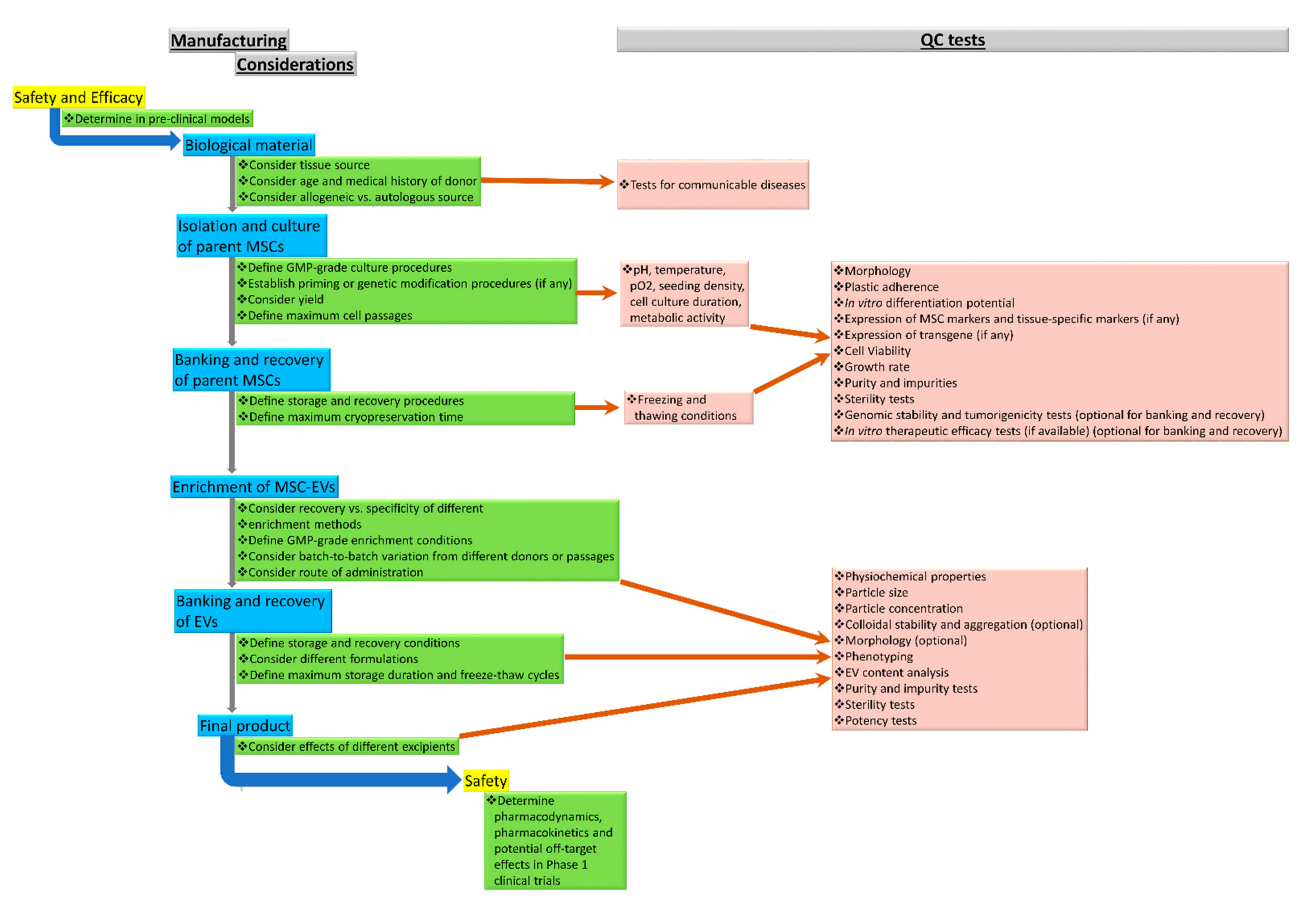
4. Considerations for the Manufacturing, Quality Control, Safety and Efficacy of MSC-EVs
4.1. MSC-EV Manufacturing
4.1.1. Parent Cell Source
- Tissue source of MSCs
- Age, medication, and medical history of donor
- Allogeneic versus autologous source
- Any priming of MSCs
- Any genetic modification of MSCs
- MSC culture conditions (MSc isolation procedures, seeding density, culture volume, culture vessel, oxygen level, culture medium, culture time, cell viability, passaging)
- MSC storage and recovery conditions
4.1.2. Culture Conditions
4.1.3. Enrichment Method
4.2. MSC-EV Quality Control
4.2.1. Quality and Identity
Physicochemical Properties
Particle Size and Concentration
Zeta Potential
Morphology
EV Phenotyping
MSC-EV Content
4.2.2. Purity and Impurities
Purity
Impurities
4.2.3. Sterility
4.2.4. Potency
4.2.5. Reproducibility
4.2.6. Storage and Formulation
4.3. Safety
4.4. Efficacy
5. Conclusions
Author Contributions
Funding
Institutional Review Board Statement
Informed Consent Statement
Data Availability Statement
Acknowledgments
Conflicts of Interest
References
- Andrzejewska, A.; Dabrowska, S.; Lukomska, B.; Janowski, M. Mesenchymal Stem Cells for Neurological Disorders. Adv. Sci. 2021, 8, 2002944. [Google Scholar] [CrossRef] [PubMed]
- Zhang, S.; Yang, Y.; Fan, L.; Zhang, F.; Li, L. The clinical application of mesenchymal stem cells in liver disease: The current situation and potential future. Ann. Transl. Med. 2020, 8, 565. [Google Scholar] [CrossRef] [PubMed]
- Guo, Y.; Yu, Y.; Hu, S.; Chen, Y.; Shen, Z. The therapeutic potential of mesenchymal stem cells for cardiovascular diseases. Cell Death Dis. 2020, 11, 349. [Google Scholar] [CrossRef] [PubMed]
- Markov, A.; Thangavelu, L.; Aravindhan, S.; Zekiy, A.O.; Jarahian, M.; Chartrand, M.S.; Pathak, Y.; Marofi, F.; Shamlou, S.; Hassanzadeh, A. Mesenchymal stem/stromal cells as a valuable source for the treatment of immune-mediated disorders. Stem Cell Res. Ther. 2021, 12, 192. [Google Scholar] [CrossRef] [PubMed]
- Lin, H.; Sohn, J.; Shen, H.; Langhans, M.T.; Tuan, R.S. Bone marrow mesenchymal stem cells: Aging and tissue engineering applications to enhance bone healing. Biomaterials 2019, 203, 96–110. [Google Scholar] [CrossRef]
- Arshi, A.; Petrigliano, F.A.; Williams, R.J.; Jones, K.J. Stem Cell Treatment for Knee Articular Cartilage Defects and Osteoarthritis. Curr. Rev. Musculoskelet. Med. 2020, 13, 20–27. [Google Scholar] [CrossRef] [PubMed]
- Spees, J.L.; Lee, R.H.; Gregory, C.A. Mechanisms of mesenchymal stem/stromal cell function. Stem Cell Res. Ther. 2016, 7, 125. [Google Scholar] [CrossRef] [PubMed]
- Marar, C.; Starich, B.; Wirtz, D. Extracellular vesicles in immunomodulation and tumor progression. Nat. Immunol. 2021, 22, 560–570. [Google Scholar] [CrossRef] [PubMed]
- Gomzikova, M.O.; James, V.; Rizvanov, A.A. Therapeutic Application of Mesenchymal Stem Cells Derived Extracellular Vesicles for Immunomodulation. Front. Immunol. 2019, 10, 2663. [Google Scholar] [CrossRef] [PubMed]
- Nagelkerke, A.; Ojansivu, M.; van der Koog, L.; Whittaker, T.E.; Cunnane, E.M.; Silva, A.M.; Dekker, N.; Stevens, M.M. Extracellular vesicles for tissue repair and regeneration: Evidence, challenges and opportunities. Adv. Drug Deliv. Rev. 2021, 175, 113775. [Google Scholar] [CrossRef] [PubMed]
- Wiklander, O.P.B.; Brennan, M.Á.; Lötvall, J.; Breakefield, X.O.; El Andaloussi, S. Advances in therapeutic applications of extracellular vesicles. Sci. Transl. Med. 2019, 11, eaav8521. [Google Scholar] [CrossRef] [PubMed]
- Bruno, S.; Grange, C.; Deregibus, M.C.; Calogero, R.A.; Saviozzi, S.; Collino, F.; Morando, L.; Busca, A.; Falda, M.; Bussolati, B.; et al. Mesenchymal stem cell-derived microvesicles protect against acute tubular injury. J. Am. Soc. Nephrol. 2009, 20, 1053–1067. [Google Scholar] [CrossRef] [PubMed]
- Doeppner, T.R.; Herz, J.; Görgens, A.; Schlechter, J.; Ludwig, A.K.; Radtke, S.; de Miroschedji, K.; Horn, P.A.; Giebel, B.; Hermann, D.M. Extracellular Vesicles Improve Post-Stroke Neuroregeneration and Prevent Postischemic Immunosuppression. Stem Cells Transl. Med. 2015, 4, 1131–1143. [Google Scholar] [CrossRef] [PubMed]
- He, J.; Wang, Y.; Sun, S.; Yu, M.; Wang, C.; Pei, X.; Zhu, B.; Wu, J.; Zhao, W. Bone marrow stem cells-derived microvesicles protect against renal injury in the mouse remnant kidney model. Nephrology 2012, 17, 493–500. [Google Scholar] [CrossRef]
- Szwedowicz, U.; Łapińska, Z.; Gajewska-Naryniecka, A.; Choromańska, A. Exosomes and Other Extracellular Vesicles with High Therapeutic Potential: Their Applications in Oncology, Neurology, and Dermatology. Molecules 2022, 27, 1303. [Google Scholar] [CrossRef] [PubMed]
- Tasso, R.; Augello, A.; Carida, M.; Postiglione, F.; Tibiletti, M.G.; Bernasconi, B.; Astigiano, S.; Fais, F.; Truini, M.; Cancedda, R.; et al. Development of sarcomas in mice implanted with mesenchymal stem cells seeded onto bioscaffolds. Carcinogenesis 2009, 30, 150–157. [Google Scholar] [CrossRef] [PubMed]
- Harris, M.T.; Butler, D.L.; Boivin, G.P.; Florer, J.B.; Schantz, E.J.; Wenstrup, R.J. Mesenchymal stem cells used for rabbit tendon repair can form ectopic bone and express alkaline phosphatase activity in constructs. J. Orthop. Res. 2004, 22, 998–1003. [Google Scholar] [CrossRef] [PubMed]
- Awad, H.A.; Boivin, G.P.; Dressler, M.R.; Smith, F.N.; Young, R.G.; Butler, D.L. Repair of patellar tendon injuries using a cell-collagen composite. J. Orthop. Res. 2003, 21, 420–431. [Google Scholar] [CrossRef]
- Karnoub, A.E.; Dash, A.B.; Vo, A.P.; Sullivan, A.; Brooks, M.W.; Bell, G.W.; Richardson, A.L.; Polyak, K.; Tubo, R.; Weinberg, R.A. Mesenchymal stem cells within tumour stroma promote breast cancer metastasis. Nature 2007, 449, 557–563. [Google Scholar] [CrossRef]
- Breitbach, M.; Bostani, T.; Roell, W.; Xia, Y.; Dewald, O.; Nygren, J.M.; Fries, J.W.; Tiemann, K.; Bohlen, H.; Hescheler, J.; et al. Potential risks of bone marrow cell transplantation into infarcted hearts. Blood 2007, 110, 1362–1369. [Google Scholar] [CrossRef] [PubMed]
- Galiè, M.; Konstantinidou, G.; Peroni, D.; Scambi, I.; Marchini, C.; Lisi, V.; Krampera, M.; Magnani, P.; Merigo, F.; Montani, M.; et al. stem cells share molecular signature with mesenchymal tumor cells and favor early tumor growth in syngeneic mice. Oncogene 2008, 27, 2542–2551. [Google Scholar] [CrossRef] [PubMed]
- Machova Urdzikova, L.; Sedlacek, R.; Suchy, T.; Amemori, T.; Ruzicka, J.; Lesny, P.; Havlas, V.; Sykova, E.; Jendelova, P. Human multipotent mesenchymal stem cells improve healing after collagenase tendon injury in the rat. Biomed. Eng. Online 2014, 13, 42. [Google Scholar] [CrossRef] [PubMed]
- Wood, M.J.; O’Loughlin, A.J.; Samira, L. Exosomes and the blood-brain barrier: Implications for neurological diseases. Ther. Deliv. 2011, 2, 1095–1099. [Google Scholar] [CrossRef] [PubMed]
- Skog, J.; Wurdinger, T.; van Rijn, S.; Meijer, D.H.; Gainche, L.; Sena-Esteves, M.; Curry, W.T., Jr.; Carter, B.S.; Krichevsky, A.M.; Breakefield, X.O. Glioblastoma microvesicles transport RNA and proteins that promote tumour growth and provide diagnostic biomarkers. Nat. Cell Biol. 2008, 10, 1470–1476. [Google Scholar] [CrossRef]
- Alvarez-Erviti, L.; Seow, Y.; Yin, H.; Betts, C.; Lakhal, S.; Wood, M.J. Delivery of siRNA to the mouse brain by systemic injection of targeted exosomes. Nat. Biotechnol. 2011, 29, 341–345. [Google Scholar] [CrossRef]
- Saint-Pol, J.; Gosselet, F.; Duban-Deweer, S.; Pottiez, G.; Karamanos, Y. Targeting and Crossing the Blood-Brain Barrier with Extracellular Vesicles. Cells 2020, 9, 851. [Google Scholar] [CrossRef]
- Shi, M.M.; Yang, Q.Y.; Monsel, A.; Yan, J.Y.; Dai, C.X.; Zhao, J.Y.; Shi, G.C.; Zhou, M.; Zhu, X.M.; Li, S.K.; et al. Preclinical efficacy and clinical safety of clinical-grade nebulized allogeneic adipose mesenchymal stromal cells-derived extracellular vesicles. J. Extracell Vesicles 2021, 10, e12134. [Google Scholar] [CrossRef]
- Zhu, Y.G.; Shi, M.M.; Monsel, A.; Dai, C.X.; Dong, X.; Shen, H.; Li, S.K.; Chang, J.; Xu, C.L.; Li, P.; et al. Nebulized exosomes derived from allogeneic adipose tissue mesenchymal stromal cells in patients with severe COVID-19: A pilot study. Stem Cell Res. Ther. 2022, 13, 220. [Google Scholar] [CrossRef]
- Sengupta, V.; Sengupta, S.; Lazo, A.; Woods, P.; Nolan, A.; Bremer, N. Exosomes Derived from Bone Marrow Mesenchymal Stem Cells as Treatment for Severe COVID-19. Stem Cells Dev. 2020, 29, 747–754. [Google Scholar] [CrossRef]
- Kaux, J.F.; Forthomme, B.; Goff, C.L.; Crielaard, J.M.; Croisier, J.L. Current opinions on tendinopathy. J. Sports Sci. Med. 2011, 10, 238–253. [Google Scholar]
- Engebretson, B.; Mussett, Z.; Williams, C.; Simmons, A.; Sikavitsas, V. Chapter 12—Tendon Tissue Engineering: Combined Tissue Engineering Approach for the Regeneration of Tendons. In Tendon Regeneration; Gomes, M.E., Reis, R.L., Rodrigues, M.T., Eds.; Academic Press: London, UK, 2015; pp. 321–347. [Google Scholar]
- Hevesi, M.; LaPrade, M.; Saris, D.B.F.; Krych, A.J. Stem cell treatment for ligament repair and reconstruction. Curr. Rev. Musculoskelet. Med. 2019, 12, 446–450. [Google Scholar] [CrossRef] [PubMed]
- Lui, P.P. Stem cell technology for tendon regeneration: Current status, challenges, and future research directions. Stem Cells Cloning 2015, 8, 163–174. [Google Scholar] [CrossRef] [PubMed]
- Lui, P.P.; Wong, O.T. Tendon stem cells: Experimental and clinical perspectives in tendon and tendon-bone junction repair. Muscles Ligaments Tendons J. 2012, 2, 163–168. [Google Scholar] [PubMed]
- Ni, M.; Lui, P.P.; Rui, Y.F.; Lee, Y.W.; Lee, Y.W.; Tan, Q.; Wong, Y.M.; Kong, S.K.; Lau, P.M.; Li, G.; et al. Tendon-derived stem cells (TDSCs) promote tendon repair in a rat patellar tendon window defect model. J. Orthop. Res. 2012, 30, 613–619. [Google Scholar] [CrossRef]
- Lui, P.P.; Wong, O.T.; Lee, Y.W. Application of tendon-derived stem cell sheet for the promotion of graft healing in anterior cruciate ligament reconstruction. Am. J. Sports Med. 2014, 42, 681–689. [Google Scholar] [CrossRef]
- Lui, P.P.; Wong, O.T.; Lee, Y.W. Transplantation of tendon-derived stem cells pre-treated with connective tissue growth factor and ascorbic acid in vitro promoted better tendon repair in a patellar tendon window injury rat model. Cytotherapy 2016, 18, 99–112. [Google Scholar] [CrossRef]
- Chamberlain, C.S.; Clements, A.; Kink, J.A.; Choi, U.; Baer, G.S.; Halanski, M.A.; Hematti, P.; Vanderby, R. Extracellular Vesicle-Educated Macrophages Promote Early Achilles Tendon Healing. Stem Cells 2019, 37, 652–662. [Google Scholar] [CrossRef]
- Shen, H.; Yoneda, S.; Abu-Amer, Y.; Guilak, F.; Gelberman, R.H. Stem cell-derived extracellular vesicles attenuate the early inflammatory response after tendon injury and repair. J. Orthop. Res. 2020, 38, 117–127. [Google Scholar] [CrossRef]
- Yao, Z.; Li, J.; Wang, X.; Peng, S.; Ning, J.; Qian, Y.; Fan, C. MicroRNA-21-3p Engineered Umbilical Cord Stem Cell-Derived Exosomes Inhibit Tendon Adhesion. J. Inflamm. Res. 2020, 13, 303–316. [Google Scholar] [CrossRef]
- Li, J.; Yao, Z.; Xiong, H.; Cui, H.; Wang, X.; Zheng, W.; Qian, Y.; Fan, C. Extracellular vesicles from hydroxycamptothecin primed umbilical cord stem cells enhance anti-adhesion potential for treatment of tendon injury. Stem Cell Res. Ther. 2020, 11, 500. [Google Scholar] [CrossRef]
- Han, Q.; Wang, S.; Chen, D.; Gan, D.; Wang, T. Exosomes derived from human umbilical cord mesenchymal stem cells reduce tendon injuries via the miR-27b-3p/ARHGAP5/RhoA signaling pathway. Acta Biochim. Biophys. Sin. 2022, 54, 232–242. [Google Scholar] [CrossRef] [PubMed]
- Gissi, C.; Radeghieri, A.; Antonetti Lamorgese Passeri, C.; Gallorini, M.; Calciano, L.; Oliva, F.; Veronesi, F.; Zendrini, A.; Cataldi, A.; Bergese, P.; et al. Extracellular vesicles from rat-bone-marrow mesenchymal stromal/stem cells improve tendon repair in rat Achilles tendon injury model in dose-dependent manner: A pilot study. PLoS ONE 2020, 15, e0229914. [Google Scholar] [CrossRef] [PubMed]
- Zhang, M.; Liu, H.; Cui, Q.; Han, P.; Yang, S.; Shi, M.; Zhang, T.; Zhang, Z.; Li, Z. Tendon stem cell-derived exosomes regulate inflammation and promote the high-quality healing of injured tendon. Stem Cell Res. Ther. 2020, 11, 402. [Google Scholar] [CrossRef] [PubMed]
- Yao, Z.; Li, J.; Xiong, H.; Cui, H.; Ning, J.; Wang, S.; Ouyang, X.; Qian, Y.; Fan, C. MicroRNA engineered umbilical cord stem cell-derived exosomes direct tendon regeneration by mTOR signaling. J. Nanobiotechnol. 2021, 19, 169. [Google Scholar] [CrossRef] [PubMed]
- Shi, Z.; Wang, Q.; Jiang, D. Extracellular vesicles from bone marrow-derived multipotent mesenchymal stromal cells regulate inflammation and enhance tendon healing. J. Transl. Med. 2019, 17, 211. [Google Scholar] [CrossRef] [PubMed]
- Yu, H.; Cheng, J.; Shi, W.; Ren, B.; Zhao, F.; Shi, Y.; Yang, P.; Duan, X.; Zhang, J.; Fu, X.; et al. Bone marrow mesenchymal stem cell-derived exosomes promote tendon regeneration by facilitating the proliferation and migration of endogenous tendon stem/progenitor cells. Acta Biomater. 2020, 106, 328–341. [Google Scholar] [CrossRef] [PubMed]
- Liu, H.; Zhang, M.; Shi, M.; Zhang, T.; Lu, W.; Yang, S.; Cui, Q.; Li, Z. Adipose-derived mesenchymal stromal cell-derived exosomes promote tendon healing by activating both SMAD1/5/9 and SMAD2/3. Stem Cell Res. Ther. 2021, 12, 338. [Google Scholar] [CrossRef]
- Song, K.; Jiang, T.; Pan, P.; Yao, Y.; Jiang, Q. Exosomes from tendon derived stem cells promote tendon repair through miR-144-3p-regulated tenocyte proliferation and migration. Stem Cell Res. Ther. 2022, 13, 80. [Google Scholar] [CrossRef]
- Wang, C.; Hu, Q.; Song, W.; Yu, W.; He, Y. Adipose Stem Cell-Derived Exosomes Decrease Fatty Infiltration and Enhance Rotator Cuff Healing in a Rabbit Model of Chronic Tears. Am. J. Sports Med. 2020, 48, 1456–1464. [Google Scholar] [CrossRef]
- Han, L.; Liu, H.; Fu, H.; Hu, Y.; Fang, W.; Liu, J. Exosome-delivered BMP-2 and polyaspartic acid promotes tendon bone healing in rotator cuff tear via Smad/RUNX2 signaling pathway. Bioengineered 2022, 13, 1459–1475. [Google Scholar] [CrossRef]
- Huang, Y.; He, B.; Wang, L.; Yuan, B.; Shu, H.; Zhang, F.; Sun, L. Bone marrow mesenchymal stem cell-derived exosomes promote rotator cuff tendon-bone healing by promoting angiogenesis and regulating M1 macrophages in rats. Stem Cell Res. Ther. 2020, 11, 496. [Google Scholar] [CrossRef]
- Fu, G.; Lu, L.; Pan, Z.; Fan, A.; Yin, F. Adipose-derived stem cell exosomes facilitate rotator cuff repair by mediating tendon-derived stem cells. Regen. Med. 2021, 16, 359–372. [Google Scholar] [CrossRef] [PubMed]
- Ren, Y.; Zhang, S.; Wang, Y.; Jacobson, D.S.; Reisdorf, R.L.; Kuroiwa, T.; Behfar, A.; Moran, S.L.; Steinmann, S.P.; Zhao, C. Effects of purified exosome product on rotator cuff tendon-bone healing in vitro and in vivo. Biomaterials 2021, 276, 121019. [Google Scholar] [CrossRef] [PubMed]
- Shi, Y.; Kang, X.; Wang, Y.; Bian, X.; He, G.; Zhou, M.; Tang, K. Exosomes Derived from Bone Marrow Stromal Cells (BMSCs) Enhance Tendon-Bone Healing by Regulating Macrophage Polarization. Med. Sci. Monit. 2020, 26, e923328. [Google Scholar] [CrossRef] [PubMed]
- Feng, W.; Jin, Q.; Ming-Yu, Y.; Yang, H.; Xu, T.; You-Xing, S.; Xu-Ting, B.; Wan, C.; Yun-Jiao, W.; Huan, W.; et al. MiR-6924-5p-rich exosomes derived from genetically modified Scleraxis-overexpressing PDGFRα(+) BMMSCs as novel nanotherapeutics for treating osteolysis during tendon-bone healing and improving healing strength. Biomaterials 2021, 279, 121242. [Google Scholar] [CrossRef]
- Wang, Y.; He, G.; Guo, Y.; Tang, H.; Shi, Y.; Bian, X.; Zhu, M.; Kang, X.; Zhou, M.; Lyu, J.; et al. Exosomes from tendon stem cells promote injury tendon healing through balancing synthesis and degradation of the tendon extracellular matrix. J. Cell. Mol. Med. 2019, 23, 5475–5485. [Google Scholar] [CrossRef] [PubMed]
- Liu, A.; Wang, Q.; Zhao, Z.; Wu, R.; Wang, M.; Li, J.; Sun, K.; Sun, Z.; Lv, Z.; Xu, J.; et al. Nitric Oxide Nanomotor Driving Exosomes-Loaded Microneedles for Achilles Tendinopathy Healing. ACS Nano. 2021, 15, 13339–13350. [Google Scholar] [CrossRef]
- Zhu, Z.; Gao, R.; Ye, T.; Feng, K.; Zhang, J.; Chen, Y.; Xie, Z.; Wang, Y. The Therapeutic Effect of iMSC-Derived Small Extracellular Vesicles on Tendinopathy Related Pain Through Alleviating Inflammation: An in vivo and in vitro Study. J. Inflamm. Res. 2022, 15, 1421–1436. [Google Scholar] [CrossRef]
- Chamberlain, C.S.; Kink, J.A.; Wildenauer, L.A.; McCaughey, M.; Henry, K.; Spiker, A.M.; Halanski, M.A.; Hematti, P.; Vanderby, R. Exosome-educated macrophages and exosomes differentially improve ligament healing. Stem Cells 2021, 39, 55–61. [Google Scholar] [CrossRef]
- Kornicka-Garbowska, K.; Pędziwiatr, R.; Woźniak, P.; Kucharczyk, K.; Marycz, K. Microvesicles isolated from 5-azacytidine-and-resveratrol-treated mesenchymal stem cells for the treatment of suspensory ligament injury in horse-a case report. Stem Cell Res. Ther. 2019, 10, 394. [Google Scholar] [CrossRef]
- Costa, L.A.; Eiro, N.; Fraile, M.; Gonzalez, L.O.; Saá, J.; Garcia-Portabella, P.; Vega, B.; Schneider, J.; Vizoso, F.J. Functional heterogeneity of mesenchymal stem cells from natural niches to culture conditions: Implications for further clinical uses. Cell. Mol. Life Sci. 2021, 78, 447–467. [Google Scholar] [CrossRef] [PubMed]
- Kim, J.Y.; Rhim, W.K.; Seo, H.J.; Lee, J.Y.; Park, C.G.; Han, D.K. Comparative Analysis of MSC-Derived Exosomes Depending on Cell Culture Media for Regenerative Bioactivity. Tissue Eng. Regen. Med. 2021, 18, 355–367. [Google Scholar] [CrossRef]
- Venugopal, C.; Shamir, C.; Senthilkumar, S.; Babu, J.V.; Sonu, P.K.; Nishtha, K.J.; Rai, K.S.; Dhanushkodi, A. Dosage and Passage Dependent Neuroprotective Effects of Exosomes Derived from Rat Bone Marrow Mesenchymal Stem Cells: An In Vitro Analysis. Curr. Gene Ther. 2017, 17, 379–390. [Google Scholar] [CrossRef]
- Lai, R.C.; Tan, S.S.; Yeo, R.W.; Choo, A.B.; Reiner, A.T.; Su, Y.; Shen, Y.; Fu, Z.; Alexander, L.; Sze, S.K.; et al. MSC secretes at least 3 EV types each with a unique permutation of membrane lipid, protein and RNA. J. Extracell. Vesicles 2016, 5, 29828. [Google Scholar] [CrossRef] [PubMed]
- Collino, F.; Pomatto, M.; Bruno, S.; Lindoso, R.S.; Tapparo, M.; Sicheng, W.; Quesenberry, P.; Camussi, G. Exosome and Microvesicle-Enriched Fractions Isolated from Mesenchymal Stem Cells by Gradient Separation Showed Different Molecular Signatures and Functions on Renal Tubular Epithelial Cells. Stem Cell Rev. Rep. 2017, 13, 226–243. [Google Scholar] [CrossRef] [PubMed]
- Department of Health. Guidance for Cell and Tissue Products. August 2021. Available online: https://www.advancedtherapyinfo.gov.hk/cbb/en/doc/Guidance_for_Cell_and_Tissue_Products.pdf (accessed on 15 June 2022).
- Lener, T.; Gimona, M.; Aigner, L.; Börger, V.; Buzas, E.; Camussi, G.; Chaput, N.; Chatterjee, D.; Court, F.A.; Del Portillo, H.A.; et al. Applying extracellular vesicles based therapeutics in clinical trials—An ISEV position paper. J. Extracell. Vesicles 2015, 4, 30087. [Google Scholar] [CrossRef] [PubMed]
- Witwer, K.W.; Van Balkom, B.W.M.; Bruno, S.; Choo, A.; Dominici, M.; Gimona, M.; Hill, A.F.; De Kleijn, D.; Koh, M.; Lai, R.C.; et al. Defining mesenchymal stromal cell (MSC)-derived small extracellular vesicles for therapeutic applications. J. Extracell. Vesicles 2019, 8, 1609206. [Google Scholar] [CrossRef] [PubMed]
- Pomatto, M.; Gai, C.; Negro, F.; Cedrino, M.; Grange, C.; Ceccotti, E.; Togliatto, G.; Collino, F.; Tapparo, M.; Figliolini, F.; et al. Differential Therapeutic Effect of Extracellular Vesicles Derived by Bone Marrow and Adipose Mesenchymal Stem Cells on Wound Healing of Diabetic Ulcers and Correlation to Their Cargoes. Int. J. Mol. Sci. 2021, 22, 3851. [Google Scholar] [CrossRef] [PubMed]
- Zhu, Y.; Wang, Y.; Zhao, B.; Niu, X.; Hu, B.; Li, Q.; Zhang, J.; Ding, J.; Chen, Y.; Wang, Y. Comparison of exosomes secreted by induced pluripotent stem cell-derived mesenchymal stem cells and synovial membrane-derived mesenchymal stem cells for the treatment of osteoarthritis. Stem Cell Res. Ther. 2017, 8, 64. [Google Scholar] [CrossRef]
- Haraszti, R.A.; Miller, R.; Stoppato, M.; Sere, Y.Y.; Coles, A.; Didiot, M.C.; Wollacott, R.; Sapp, E.; Dubuke, M.L.; Li, X.; et al. Exosomes Produced from 3D Cultures of MSCs by Tangential Flow Filtration Show Higher Yield and Improved Activity. Mol. Ther. 2018, 26, 2838–2847. [Google Scholar] [CrossRef] [PubMed]
- Tan, Q.; Lui, P.P.; Rui, Y.F.; Wong, Y.M. Comparison of potentials of stem cells isolated from tendon and bone marrow for musculoskeletal tissue engineering. Tissue Eng. Part A 2012, 18, 840–851. [Google Scholar] [CrossRef] [PubMed]
- Lotfy, A.; Salama, M.; Zahran, F.; Jones, E.; Badawy, A.; Sobh, M. Characterization of mesenchymal stem cells derived from rat bone marrow and adipose tissue: A comparative study. Int. J. Stem. Cells 2014, 7, 135–142. [Google Scholar] [CrossRef] [PubMed]
- Bi, Y.; Ehirchiou, D.; Kilts, T.M.; Inkson, C.A.; Embree, M.C.; Sonoyama, W.; Li, L.; Leet, A.I.; Seo, B.M.; Zhang, L.; et al. Identification of tendon stem/progenitor cells and the role of the extracellular matrix in their niche. Nat. Med. 2007, 13, 1219–1227. [Google Scholar] [CrossRef] [PubMed]
- Hu, C.; Zhang, Y.; Tang, K.; Luo, Y.; Liu, Y.; Chen, W. Downregulation of CITED2 contributes to TGFβ-mediated senescence of tendon-derived stem cells. Cell Tissue Res. 2017, 368, 93–104. [Google Scholar] [CrossRef] [PubMed]
- Inoue, O.; Usui, S.; Takashima, S.I.; Nomura, A.; Yamaguchi, K.; Takeda, Y.; Goten, C.; Hamaoka, T.; Ootsuji, H.; Murai, H.; et al. Diabetes impairs the angiogenic capacity of human adipose-derived stem cells by reducing the CD271+ subpopulation in adipose tissue. Biochem. Biophys. Res. Commun. 2019, 517, 369–375. [Google Scholar] [CrossRef] [PubMed]
- Cianfarani, F.; Toietta, G.; Di Rocco, G.; Cesareo, E.; Zambruno, G.; Odorisio, T. Diabetes impairs adipose tissue-derived stem cell function and efficiency in promoting wound healing. Wound Repair Regen. 2013, 21, 545–553. [Google Scholar] [CrossRef]
- Kornicka, K.; Houston, J.; Marycz, K. Dysfunction of Mesenchymal Stem Cells Isolated from Metabolic Syndrome and Type 2 Diabetic Patients as Result of Oxidative Stress and Autophagy may Limit Their Potential Therapeutic Use. Stem Cell Rev. Rep. 2018, 14, 337–345. [Google Scholar] [CrossRef]
- Abu-Shahba, N.; Mahmoud, M.; El-Erian, A.M.; Husseiny, M.I.; Nour-Eldeen, G.; Helwa, I.; Amr, K.; ElHefnawi, M.; Othman, A.I.; Ibrahim, S.A.; et al. Impact of type 2 diabetes mellitus on the immunoregulatory characteristics of adipose tissue-derived mesenchymal stem cells. Int. J. Biochem. Cell. Biol. 2021, 140, 106072. [Google Scholar] [CrossRef] [PubMed]
- Fadini, G.P.; Spinetti, G.; Santopaolo, M.; Madeddu, P. Impaired Regeneration Contributes to Poor Outcomes in Diabetic Peripheral Artery Disease. Arterioscler. Thromb. Vasc. Biol. 2020, 40, 34–44. [Google Scholar] [CrossRef]
- Santopaolo, M.; Sambataro, M.; Spinetti, G.; Madeddu, P. Bone marrow as a target and accomplice of vascular complications in diabetes. Diabetes Metab. Res. Rev. 2020, 36 (Suppl. S1), e3240. [Google Scholar] [CrossRef]
- Wuchter, P.; Bieback, K.; Schrezenmeier, H.; Bornhäuser, M.; Müller, L.P.; Bönig, H.; Wagner, W.; Meisel, R.; Pavel, P.; Tonn, T.; et al. Standardization of Good Manufacturing Practice-compliant production of bone marrow-derived human mesenchymal stromal cells for immunotherapeutic applications. Cytotherapy 2015, 17, 128–139. [Google Scholar] [CrossRef] [PubMed]
- Tigges, J.; Bielec, K.; Brockerhoff, G.; Hildebrandt, B.; Hübenthal, U.; Kapr, J.; Koch, K.; Teichweyde, N.; Wieczorek, D.; Rossi, A.; et al. Academic application of Good Cell Culture Practice for induced pluripotent stem cells. ALTEX 2021, 38, 595–614. [Google Scholar] [CrossRef] [PubMed]
- Pakzad, M.; Hassani, S.N.; Abbasi, F.; Hajizadeh-Saffar, E.; Taghiyar, L.; Fallah, N.; Haghparast, N.; Samadian, A.; Ganjibakhsh, M.; Dominici, M.; et al. A Roadmap for the Production of a GMP-Compatible Cell Bank of Allogeneic Bone Marrow-Derived Clonal Mesenchymal Stromal Cells for Cell Therapy Applications. Stem Cell Rev. Rep. 2022, 1–17. [Google Scholar] [CrossRef] [PubMed]
- Théry, C.; Witwer, K.W.; Aikawa, E.; Alcaraz, M.J.; Anderson, J.D.; Andriantsitohaina, R.; Antoniou, A.; Arab, T.; Archer, F.; Atkin-Smith, G.K.; et al. Minimal information for studies of extracellular vesicles 2018 (MISEV2018): A position statement of the International Society for Extracellular Vesicles and update of the MISEV2014 guidelines. J. Extracell. Vesicles 2018, 7, 1535750. [Google Scholar] [CrossRef]
- Chen, Y.S.; Lin, E.Y.; Chiou, T.W.; Harn, H.J. Exosomes in clinical trial and their production in compliance with good manufacturing practice. Ci Ji Yi Xue Za Zhi 2019, 32, 113–120. [Google Scholar]
- Andriolo, G.; Provasi, E.; Lo Cicero, V.; Brambilla, A.; Soncin, S.; Torre, T.; Milano, G.; Biemmi, V.; Vassalli, G.; Turchetto, L.; et al. Exosomes from Human Cardiac Progenitor Cells for Therapeutic Applications: Development of a GMP-Grade Manufacturing Method. Front. Physiol. 2018, 9, 1169. [Google Scholar] [CrossRef]
- Pachler, K.; Lener, T.; Streif, D.; Dunai, Z.A.; Desgeorges, A.; Feichtner, M.; Öller, M.; Schallmoser, K.; Rohde, E.; Gimona, M. A Good Manufacturing Practice-grade standard protocol for exclusively human mesenchymal stromal cell-derived extracellular vesicles. Cytotherapy 2017, 19, 458–472. [Google Scholar] [CrossRef]
- Tan, Q.; Lui, P.P.; Rui, Y.F. Effect of in vitro passaging on the stem cell-related properties of tendon-derived stem cells—Implications in tissue engineering. Stem Cells Dev. 2012, 21, 790–800. [Google Scholar] [CrossRef]
- Council of Europe. Nucleated cell count and viability. In The European Pharmacopoeia, 9th ed.; 2.7.29; EDQM: Strasbourg, France, 2017. [Google Scholar]
- Seaver, S. A new United States Pharmacopeia (USP) Chapter 1046: Cell and gene therapy products. Cytotherapy 2000, 2, 45–49. [Google Scholar] [CrossRef]
- Dominici, M.; Le Blanc, K.; Mueller, I.; Slaper-Cortenbach, I.; Marini, F.C.; Krause, D.S.; Deans, R.J.; Keating, A.; Prockop, D.J.; Horwitz, E.M. Minimal criteria for defining multipotent mesenchymal stromal cells. The International Society for Cellular Therapy position statement. Cytotherapy 2006, 8, 315–317. [Google Scholar] [CrossRef]
- Lui, P.P. Markers for the identification of tendon-derived stem cells in vitro and tendon stem cells in situ—Update and future development. Stem Cell Res. Ther. 2015, 6, 106. [Google Scholar] [CrossRef] [PubMed]
- Galipeau, J.; Krampera, M. The challenge of defining mesenchymal stromal cell potency assays and their potential use as release criteria. Cytotherapy 2015, 17, 125–127. [Google Scholar] [CrossRef] [PubMed]
- Ketterl, N.; Brachtl, G.; Schuh, C.; Bieback, K.; Schallmoser, K.; Reinisch, A.; Strunk, D. A robust potency assay highlights significant donor variation of human mesenchymal stem/progenitor cell immune modulatory capacity and extended radio-resistance. Stem Cell Res. Ther. 2015, 6, 236. [Google Scholar] [CrossRef] [PubMed]
- Oliver-Vila, I.; Ramírez-Moncayo, C.; Grau-Vorster, M.; Marín-Gallén, S.; Caminal, M.; Vives, J. Optimisation of a potency assay for the assessment of immunomodulative potential of clinical grade multipotent mesenchymal stromal cells. Cytotechnology 2018, 70, 31–44. [Google Scholar] [CrossRef]
- Grau-Vorster, M.; Rodríguez, L.; Del Mazo-Barbara, A.; Mirabel, C.; Blanco, M.; Codinach, M.; Gómez, S.G.; Querol, S.; García-López, J.; Vives, J. Compliance with Good Manufacturing Practice in the Assessment of Immunomodulation Potential of Clinical Grade Multipotent Mesenchymal Stromal Cells Derived from Wharton’s Jelly. Cells 2019, 8, 484. [Google Scholar] [CrossRef]
- De Wolf, C.; van de Bovenkamp, M.; Hoefnagel, M. Regulatory perspective on in vitro potency assays for human mesenchymal stromal cells used in immunotherapy. Cytotherapy 2017, 19, 784–797. [Google Scholar] [CrossRef]
- Guan, Q.; Li, Y.; Shpiruk, T.; Bhagwat, S.; Wall, D.A. Inducible indoleamine 2,3-dioxygenase 1 and programmed death ligand 1 expression as the potency marker for mesenchymal stromal cells. Cytotherapy 2018, 20, 639–649. [Google Scholar] [CrossRef]
- Poupardin, R.; Wolf, M.; Strunk, D. Adherence to minimal experimental requirements for defining extracellular vesicles and their functions. Adv. Drug Deliv. Rev. 2021, 176, 113872. [Google Scholar] [CrossRef]
- Andriolo, G.; Provasi, E.; Brambilla, A.; Lo Cicero, V.; Soncin, S.; Barile, L.; Turchetto, L.; Radrizzani, M. GMP-Grade Methods for Cardiac Progenitor Cells: Cell Bank Production and Quality Control. Methods Mol. Biol. 2021, 2286, 131–166. [Google Scholar]
- Lechanteur, C.; Briquet, A.; Bettonville, V.; Baudoux, E.; Beguin, Y. MSC Manufacturing for Academic Clinical Trials: From a Clinical-Grade to a Full GMP-Compliant Process. Cells 2021, 10, 1320. [Google Scholar] [CrossRef]
- Guadix, J.A.; López-Beas, J.; Clares, B.; Soriano-Ruiz, J.L.; Zugaza, J.L.; Gálvez-Martín, P. Principal Criteria for Evaluating the Quality, Safety and Efficacy of hMSC-Based Products in Clinical Practice: Current Approaches and Challenges. Pharmaceutics 2019, 11, 552. [Google Scholar] [CrossRef] [PubMed]
- Robb, K.P.; Fitzgerald, J.C.; Barry, F.; Viswanathan, S. Mesenchymal stromal cell therapy: Progress in manufacturing and assessments of potency. Cytotherapy 2019, 21, 289–306. [Google Scholar] [CrossRef] [PubMed]
- Mol, E.A.; Goumans, M.J.; Doevendans, P.A.; Sluijter, J.P.G.; Vader, P. Higher functionality of extracellular vesicles isolated using size-exclusion chromatography compared to ultracentrifugation. Nanomedicine 2017, 13, 2061–2065. [Google Scholar] [CrossRef] [PubMed]
- Nordin, J.Z.; Lee, Y.; Vader, P.; Mäger, I.; Johansson, H.J.; Heusermann, W.; Wiklander, O.P.; Hällbrink, M.; Seow, Y.; Bultema, J.J.; et al. Ultrafiltration with size-exclusion liquid chromatography for high yield isolation of extracellular vesicles preserving intact biophysical and functional properties. Nanomedicine 2015, 11, 879–883. [Google Scholar] [CrossRef]
- Lee, J.H.; Ha, D.H.; Go, H.K.; Youn, J.; Kim, H.K.; Jin, R.C.; Miller, R.B.; Kim, D.H.; Cho, B.S.; Yi, Y.W. Reproducible Large-Scale Isolation of Exosomes from Adipose Tissue-Derived Mesenchymal Stem/Stromal Cells and Their Application in Acute Kidney Injury. Int. J. Mol. Sci. 2020, 21, 4774. [Google Scholar] [CrossRef]
- Bari, E.; Perteghella, S.; Catenacci, L.; Sorlini, M.; Croce, S.; Mantelli, M.; Avanzini, M.A.; Sorrenti, M.; Torre, M.L. Freeze-dried and GMP-compliant pharmaceuticals containing exosomes for acellular mesenchymal stromal cell immunomodulant therapy. Nanomedicine 2019, 14, 753–765. [Google Scholar] [CrossRef]
- Watson, D.C.; Yung, B.C.; Bergamaschi, C.; Chowdhury, B.; Bear, J.; Stellas, D.; Morales-Kastresana, A.; Jones, J.C.; Felber, B.K.; Chen, X.; et al. Scalable, cGMP-compatible purification of extracellular vesicles carrying bioactive human heterodimeric IL-15/lactadherin complexes. J. Extracell. Vesicles 2018, 7, 1442088. [Google Scholar] [CrossRef]
- Gámez-Valero, A.; Monguió-Tortajada, M.; Carreras-Planella, L.; Franquesa, M.L.; Beyer, K.; Borràs, F.E. Size-Exclusion Chromatography-based isolation minimally alters Extracellular Vesicles’ characteristics compared to precipitating agents. Sci. Rep. 2016, 6, 33641. [Google Scholar] [CrossRef]
- Paolini, L.; Zendrini, A.; Di Noto, G.; Busatto, S.; Lottini, E.; Radeghieri, A.; Dossi, A.; Caneschi, A.; Ricotta, D.; Bergese, P. Residual matrix from different separation techniques impacts exosome biological activity. Sci. Rep. 2016, 6, 23550. [Google Scholar] [CrossRef]
- Zhang, X.; Borg, E.G.F.; Liaci, A.M.; Vos, H.R.; Stoorvogel, W. A novel three step protocol to isolate extracellular vesicles from plasma or cell culture medium with both high yield and purity. J. Extracell. Vesicles 2020, 9, 1791450. [Google Scholar] [CrossRef]
- McNamara, R.P.; Caro-Vegas, C.P.; Costantini, L.M.; Landis, J.T.; Griffith, J.D.; Damania, B.A.; Dittmer, D.P. Large-scale, cross-flow based isolation of highly pure and endocytosis-competent extracellular vesicles. J. Extracell. Vesicles 2018, 7, 1541396. [Google Scholar] [CrossRef] [PubMed]
- Buschmann, D.; Mussack, V.; Byrd, J.B. Separation, characterization, and standardization of extracellular vesicles for drug delivery applications. Adv. Drug Deliv. Rev. 2021, 174, 348–368. [Google Scholar] [CrossRef] [PubMed]
- Midekessa, G.; Godakumara, K.; Ord, J.; Viil, J.; Lättekivi, F.; Dissanayake, K.; Kopanchuk, S.; Rinken, A.; Andronowsk, A.; Bhattacharjee, S.; et al. Zeta Potential of Extracellular Vesicles: Toward Understanding the Attributes that Determine Colloidal Stability. ACS Omega 2020, 5, 16701–16710. [Google Scholar] [CrossRef]
- Le Saux, S.; Aarrass, H.; Lai-Kee-Him, J.; Bron, P.; Armengaud, J.; Miotello, G.; Bertrand-Michel, J.; Dubois, E.; George, S.; Faklaris, O.; et al. Post-production modifications of murine mesenchymal stem cell (mMSC) derived extracellular vesicles (EVs) and impact on their cellular interaction. Biomaterials 2020, 231, 119675. [Google Scholar] [CrossRef] [PubMed]
- Feng, K.; Xie, X.; Yuan, J.; Gong, L.; Zhu, Z.; Zhang, J.; Li, H.; Yang, Y.; Wang, Y. Reversing the surface charge of MSC-derived small extracellular vesicles by εPL-PEG-DSPE for enhanced osteoarthritis treatment. J. Extracell. Vesicles 2021, 10, e12160. [Google Scholar] [CrossRef] [PubMed]
- De Almeida Fuzeta, M.; Bernardes, N.; Oliveira, F.D.; Costa, A.C.; Fernandes-Platzgummer, A.; Farinha, J.P.; Rodrigues, C.A.V.; Jung, S.; Tseng, R.J.; Milligan, W.; et al. Scalable Production of Human Mesenchymal Stromal Cell-Derived Extracellular Vesicles Under Serum-/Xeno-Free Conditions in a Microcarrier-Based Bioreactor Culture System. Front. Cell. Dev. Biol. 2020, 8, 553444. [Google Scholar] [CrossRef]
- Koliha, N.; Wiencek, Y.; Heider, U.; Jüngst, C.; Kladt, N.; Krauthäuser, S.; Johnston, I.C.; Bosio, A.; Schauss, A.; Wild, S. A novel multiplex bead-based platform highlights the diversity of extracellular vesicles. J. Extracell. Vesicles 2016, 5, 29975. [Google Scholar] [CrossRef]
- Tian, Y.; Ma, L.; Gong, M.; Su, G.; Zhu, S.; Zhang, W.; Wang, S.; Li, Z.; Chen, C.; Li, L.; et al. Protein Profiling and Sizing of Extracellular Vesicles from Colorectal Cancer Patients via Flow Cytometry. ACS Nano 2018, 12, 671–680. [Google Scholar] [CrossRef]
- Rocha, S.; Carvalho, J.; Oliveira, P.; Voglstaetter, M.; Schvartz, D.; Thomsen, A.R.; Walter, N.; Khanduri, R.; Sanchez, J.C.; Keller, A.; et al. 3D Cellular Architecture Affects MicroRNA and Protein Cargo of Extracellular Vesicles. Adv. Sci. 2018, 6, 1800948. [Google Scholar] [CrossRef]
- Gandham, S.; Su, X.; Wood, J.; Nocera, A.L.; Alli, S.C.; Milane, L.; Zimmerman, A.; Amiji, M.; Ivanov, A.R. Technologies and Standardization in Research on Extracellular Vesicles. Trends Biotechnol. 2020, 38, 1066–1098. [Google Scholar] [CrossRef]
- Osteikoetxea, X.; Balogh, A.; Szabó-Taylor, K.; Németh, A.; Szabó, T.G.; Pálóczi, K.; Sódar, B.; Kittel, Á.; György, B.; Pállinger, É.; et al. Improved characterization of EV preparations based on protein to lipid ratio and lipid properties. PLoS ONE 2015, 10, e0121184. [Google Scholar] [CrossRef] [PubMed]
- Webber, J.; Clayton, A. How pure are your vesicles? J. Extracell. Vesicles 2013, 10, 2. [Google Scholar] [CrossRef] [PubMed]
- Maiolo, D.; Paolini, L.; Di Noto, G.; Zendrini, A.; Berti, D.; Bergese, P.; Ricotta, D. Colorimetric nanoplasmonic assay to determine purity and titrate extracellular vesicles. Anal. Chem. 2015, 87, 4168–4176. [Google Scholar] [CrossRef] [PubMed]
- Mihály, J.; Deák, R.; Szigyártó, I.C.; Bóta, A.; Beke-Somfai, T.; Varga, Z. Characterization of extracellular vesicles by IR spectroscopy: Fast and simple classification based on amide and CH stretching vibrations. Biochim. Biophys. Acta Biomembr. 2017, 1859, 459–466. [Google Scholar] [CrossRef]
- Veerman, R.E.; Teeuwen, L.; Czarnewski, P.; Güclüler Akpinar, G.; Sandberg, A.; Cao, X.; Pernemalm, M.; Orre, L.M.; Gabrielsson, S.; Eldh, M. Molecular evaluation of five different isolation methods for extracellular vesicles reveals different clinical applicability and subcellular origin. J. Extracell. Vesicles 2021, 10, e12128. [Google Scholar] [CrossRef] [PubMed]
- Whittaker, T.E.; Nagelkerke, A.; Nele, V.; Kauscher, U.; Stevens, M.M. Experimental artefacts can lead to misattribution of bioactivity from soluble mesenchymal stem cell paracrine factors to extracellular vesicles. J. Extracell. Vesicles 2020, 9, 1807674. [Google Scholar] [CrossRef] [PubMed]
- Gimona, M.; Brizzi, M.F.; Choo, A.B.H.; Dominici, M.; Davidson, S.M.; Grillari, J.; Hermann, D.M.; Hill, A.F.; de Kleijn, D.; Lai, R.C.; et al. Critical considerations for the development of potency tests for therapeutic applications of mesenchymal stromal cell-derived small extracellular vesicles. Cytotherapy 2021, 23, 373–380. [Google Scholar] [CrossRef]
- Lui, P.P.Y. Mesenchymal Stem Cell-Derived Extracellular Vesicles for the Promotion of Tendon Repair—An Update of Literature. Stem Cell Rev. Rep. 2021, 17, 379–389. [Google Scholar] [CrossRef]
- Mocchi, M.; Bari, E.; Marrubini, G.; Bonda, A.F.; Perteghella, S.; Tartara, F.; Cofano, F.; Perna, G.D.; Giovannelli, L.; Mandracchia, D.; et al. Freeze-Dried Mesenchymal Stem Cell-Secretome Pharmaceuticalization: Optimization of Formulation and Manufacturing Process Robustness. Pharmaceutics 2021, 13, 1129. [Google Scholar] [CrossRef]
- Laggner, M.; Gugerell, A.; Bachmann, C.; Hofbauer, H.; Vorstandlechner, V.; Seibold, M.; Gouya Lechner, G.; Peterbauer, A.; Madlener, S.; Demyanets, S.; et al. Reproducibility of GMP-compliant production of therapeutic stressed peripheral blood mononuclear cell-derived secretomes.; a novel class of biological medicinal products. Stem Cell Res. Ther. 2020, 11, 9. [Google Scholar] [CrossRef]
- Bahr, M.M.; Amer, M.S.; Abo-El-Sooud, K.; Abdallah, A.N.; El-Tookhy, O.S. Preservation techniques of stem cells extracellular vesicles: A gate for manufacturing of clinical grade therapeutic extracellular vesicles and long-term clinical trials. Int. J. Vet. Sci. Med. 2020, 8, 1–8. [Google Scholar] [CrossRef] [PubMed]
- Charoenviriyakul, C.; Takahashi, Y.; Nishikawa, M.; Takakura, Y. Preservation of exosomes at room temperature using lyophilization. Int. J. Pharm. 2018, 553, 1–7. [Google Scholar] [CrossRef] [PubMed]
- Bari, E.; Perteghella, S.; Di Silvestre, D.; Sorlini, M.; Catenacci, L.; Sorrenti, M.; Marrubini, G.; Rossi, R.; Tripodo, G.; Mauri, P.; et al. Production of Mesenchymal Stem/Stromal Freeze-Dried Secretome for Cell-Free Regenerative Nanomedicine: A Validated GMP-Compliant Process. Cells 2018, 7, 190. [Google Scholar] [CrossRef] [PubMed]
- Silva, A.; Morille, M.; Piffoux, M.; Arumugam, S.; Mauduit, P.; Larghero, J.; Bianchi, A.; Aubertin, K.; Blanc-Brude, O.; Noël, D.; et al. Development of extracellular vesicle-based medicinal products: A position paper of the group “Extracellular Vesicle translatiOn to clinicaL perspectiVEs—EVOLVE France”. Adv. Drug Deliv. Rev. 2021, 179, 114001. [Google Scholar] [CrossRef]
- Rohde, E.; Pachler, K.; Gimona, M. Manufacturing and characterization of extracellular vesicles from umbilical cord-derived mesenchymal stromal cells for clinical testing. Cytotherapy 2019, 21, 581–592. [Google Scholar] [CrossRef] [PubMed]
- Gimona, M.; Pachler, K.; Laner-Plamberger, S.; Schallmoser, K.; Rohde, E. Manufacturing of Human Extracellular Vesicle-Based Therapeutics for Clinical Use. Int. J. Mol. Sci. 2017, 18, 1190. [Google Scholar] [CrossRef]
- Lui, P.P.; Kong, S.K.; Lau, P.M.; Wong, Y.M.; Lee, Y.W.; Tan, C.; Wong, O.T. Allogeneic tendon-derived stem cells promote tendon healing and suppress immunoreactions in hosts: In vivo model. Tissue Eng. Part A 2014, 20, 2998–3009. [Google Scholar] [CrossRef]
- Lui, P.P.; Kong, S.K.; Lau, P.M.; Wong, Y.M.; Lee, Y.W.; Tan, C.; Wong, O.T. Immunogenicity and escape mechanisms of allogeneic tendon-derived stem cells. Tissue Eng. Part A 2014, 20, 3010–3020. [Google Scholar] [CrossRef]
- Lee, S.Y.; Kim, W.; Lim, C.; Chung, S.G. Treatment of Lateral Epicondylosis by Using Allogeneic Adipose-Derived Mesenchymal Stem Cells: A Pilot Study. Stem Cells 2015, 33, 2995–3005. [Google Scholar] [CrossRef]
- Wang, Y.; Shimmin, A.; Ghosh, P.; Marks, P.; Linklater, J.; Connell, D.; Hall, S.; Skerrett, D.; Itescu, S.; Cicuttini, F.M. Safety, tolerability, clinical, and joint structural outcomes of a single intra-articular injection of allogeneic mesenchymal precursor cells in patients following anterior cruciate ligament reconstruction: A controlled double-blind randomised trial. Arthritis Res. Ther. 2017, 19, 180. [Google Scholar] [CrossRef]
- Mendt, M.; Kamerkar, S.; Sugimoto, H.; McAndrews, K.M.; Wu, C.C.; Gagea, M.; Yang, S.; Blanko, E.V.R.; Peng, Q.; Ma, X.; et al. Generation and testing of clinical-grade exosomes for pancreatic cancer. JCI Insight 2018, 3, e99263. [Google Scholar] [CrossRef] [PubMed]
- Zhu, X.; Badawi, M.; Pomeroy, S.; Sutaria, D.S.; Xie, Z.; Baek, A.; Jiang, J.; Elgamal, O.A.; Mo, X.; Perle, K.; et al. Comprehensive toxicity and immunogenicity studies reveal minimal effects in mice following sustained dosing of extracellular vesicles derived from HEK293T cells. J. Extracell. Vesicles 2017, 6, 1324730. [Google Scholar] [CrossRef] [PubMed]
- Kordelas, L.; Rebmann, V.; Ludwig, A.K.; Radtke, S.; Ruesing, J.; Doeppner, T.R.; Epple, M.; Horn, P.A.; Beelen, D.W.; Giebel, B. MSC-derived exosomes: A novel tool to treat therapy-refractory graft-versus-host disease. Leukemia 2014, 28, 970–973. [Google Scholar] [CrossRef] [PubMed]
- Nassar, W.; El-Ansary, M.; Sabry, D.; Mostafa, M.A.; Fayad, T.; Kotb, E.; Temraz, M.; Saad, A.N.; Essa, W.; Adel, H. Umbilical cord mesenchymal stem cells derived extracellular vesicles can safely ameliorate the progression of chronic kidney diseases. Biomater. Res. 2016, 20, 21. [Google Scholar] [CrossRef] [PubMed]

| Number | Study Title | Target Health Condition Estimated Enrollment of Patients Clinical Trial Phase | Type of EV Administrated Dosage Route of Administration | Status and Results (If Applicable) | Study Location | Reference/ Identifier |
|---|---|---|---|---|---|---|
| 1 | Bone Marrow Mesenchymal Stem Cell Derived Extracellular Vesicles Infusion Treatment for Acute Respiratory Distress Syndrome: A Phase I/II Clinical Trial |
|
|
| Not specified | NCT05127122 |
| 2 | Bone Marrow Mesenchymal Stem Cell Derived Extracellular Vesicles Infusion Treatment: A Global Expanded Access Protocol for Patients With COVID-19 Associated ARDS Who Do Not Qualify for Phase II Randomized Control Trial |
|
|
| Not specified | NCT04657458 |
| 3 | Bone Marrow Mesenchymal Stem Cell Derived Extracellular Vesicles Infusion Treatment for COVID-19 Associated Acute Respiratory Distress Syndrome (ARDS): A Phase II Clinical Trial |
|
|
| United States | NCT04493242 |
| 4 | Bone Marrow Mesenchymal Stem Cell Derived Extracellular Vesicles as Early Goal Directed Therapy for COVID-19 Moderate-to-Severe Acute Respiratory Distress Syndrome (ARDS) |
|
|
| Not specified | NCT05354141 |
| 5 | Bone Marrow Mesenchymal Stem Cell Derived Extracellular Vesicles Infusion Treatment for Mild-to-Moderate COVID-19: A Phase II Clinical Trial |
|
|
| Not specified | NCT05125562 |
| 6 | ExoFlo™ Infusion for Post-Acute COVID-19 and Chronic Post-COVID-19 Syndrome |
|
|
| Not specified | NCT05116761 |
| 7 | Intermediate Size Expanded Access for the Use of ExoFlo in the Treatment of Abdominal Solid Organ Transplant Patients Who Are at Risk of Worsening Allograft Function with Conventional Immunosuppressive Therapy Alone |
|
|
| Not specified | NCT05215288 |
| 8 | A Phase I Study of ExoFlo, an ex Vivo Culture-expanded Adult Allogeneic Bone Marrow Mesenchymal Stem Cell Derived Extracellular Vesicle Isolate Product, for the Treatment of Medically Refractory Crohn’s Disease |
|
|
| Not specified | NCT05130983 |
| 9 | A Phase I Study of ExoFlo, an ex Vivo Culture-expanded Adult Allogeneic Bone Marrow Mesenchymal Stem Cell Derived Extracellular Vesicle Isolate Product, for the Treatment of Medically Refractory Ulcerative Colitis |
|
|
| Not specified | NCT05176366 |
| 10 | A Pilot Safety Study of the Administration of Mesenchymal Stem Cell Extracellular Vesicles in the Treatment of Burn Wounds |
|
|
| Not specified | NCT05078385 |
| 11 | A Safety Study of the Administration of Mesenchymal Stem Cell Extracellular Vesicles in the Treatment of Dystrophic Epidermolysis Bullosa Wounds |
|
|
| Not specified | NCT04173650 |
| 12 | A Safety Study of Intravenous Infusion of Bone Marrow Mesenchymal Stem Cell-derived Extracellular Vesicles (UNEX-42) in Preterm Neonates at High Risk for Bronchopulmonary Dysplasia |
|
|
| United States | NCT03857841 |
| 13 | Effect of Adipose Derived Stem Cells Exosomes as an Adjunctive Therapy to Scaling and Root Planning in the Treatment of Periodontitis: A Human Clinical Trial |
|
|
| Egypt | NCT04270006 |
| 14 | Study of Exosomes Derived from Mesenchymal Stem Cells on the Therapy for Children with Severe Infection |
|
|
| China | NCT04850469 |
| 15 | A Pilot Clinical Study on Inhalation of Mesenchymal Stem Cells Exosomes Treating Severe Novel Coronavirus Pneumonia |
|
|
| China | NCT04276987 |
| 16 | Exosome of Mesenchymal Stem Cells for Multiple Organ Dysfunction Syndrome After Surgical Repair of Acute Type A Aortic Dissection: A Pilot Study |
|
|
| Not specified | NCT04356300 |
| 17 | Effect of Umbilical Mesenchymal Stem Cells Derived Exosomes on Dry Eye in Patients with Chronic Graft Versus Host Diseases |
|
|
| China | NCT04213248 |
| 18 | A Tolerance Clinical Study on Aerosol Inhalation of Mesenchymal Stem Cells Exosomes in Healthy Volunteers |
|
|
| China | NCT04313647 |
| 19 | Mesenchymal Stem Cells Derived Exosomes Promote Healing of Large and Refractory Macular Holes |
|
|
| China | NCT03437759 |
| 20 | Phase 1 Study of The Effect of Cell-Free Cord Blood Derived Microvesicles On β-cell Mass in Type 1 Diabetes Mellitus (T1DM) Patients |
|
|
| Egypt | NCT02138331 |
| 21 | Mesenchymal Stem Cell Exosomes for the Treatment of COVID-19 Positive Patients with Acute Respiratory Distress Syndrome and/or Novel Coronavirus Pneumonia |
|
|
| United States | NCT04798716 |
| 22 | A Phase I Study Aiming to Assess Safety and Efficacy of a Single Intra-articular Injection of MSC-derived Exosomes (CelliStem®OA-sEV) in Patients with Moderate Knee Osteoarthritis (ExoOA-1) |
|
|
| Not specified | NCT05060107 |
| 23 | A Phase II Trial to Investigate Clinical Efficacy of Autologous Synovial Fluid Mesenchymal Stem Cell-Derived Exosome Application in Patients with Degenerative Meniscal Injury |
|
|
| Turkey | NCT05261360 |
| 24 | The Protocol of Evaluation of Safety and Efficiency of Method of Exosome Inhalation in SARS-CoV-2 Associated Two-Sided Pneumonia |
|
|
| Russia | NCT04491240 |
| 25 | The Extended Protocol of Evaluation of Safety and Efficiency of Method of Exosome Inhalation in COVID-19 Associated Two-Sided Pneumonia |
|
|
| Russia | NCT04602442 |
| 26 | A Multiple, Randomized, Double-blinded, Controlled Clinical Study of Allogeneic Human Mesenchymal Stem Cell Exosomes (hMSC-Exos) Nebulized Inhalation in the Treatment of Acute Respiratory Distress Syndrome |
|
|
| China | NCT04602104 |
| 27 | A Clinical Study of Allogeneic Human Adipose-derived Mesenchymal Progenitor Cell Exosomes (haMPC-Exos) Nebulizer for the Treatment of Carbapenem-resistant Gram-negative Bacilli-induced Pulmonary Infection |
|
|
| China | NCT04544215 |
| 28 | Open-Label, Single-Center, Phase I/II Clinical Trial to Evaluate the Safety and the Efficacy of Exosomes Derived from Allogeneic Adipose Mesenchymal Stem Cells in Patients with Mild to Moderate Dementia Due to Alzheimer’s Disease |
|
|
| China | NCT04388982 |
| 29 | The Use of Exosomes in Craniofacial Neuralgia |
|
|
| United States | NCT04202783 |
| 30 | Phase I Study of Mesenchymal Stromal Cells-Derived Exosomes with KrasG12D siRNA for Metastatic Pancreas Cancer Patients Harboring KrasG12D Mutation |
|
|
| United States | NCT03608631 |
| 31 | Safety and Efficacy of Allogeneic Mesenchymal Stem Cells Derived Exosome on Disability of Patients with Acute Ischemic Stroke: A Randomized, Single-blind, Placebo-controlled, Phase 1, 2 Trial |
|
|
| Iran | NCT03384433 |
| 32 | Safety and Efficacy of Injection of Human Placenta Mesenchymal Stem Cells Derived Exosomes for Treatment of Complex Anal Fistula |
|
|
| Iran | NCT05402748 |
| 33 | Efficacy and Safety of EXOSOME-MSC (Mesenchymal Stem Cell-Derived Exosomes) Therapy to Reduce Hyper-inflammation In Moderate COVID-19 (2019- New Corona Virus Disease) Patients |
|
|
| Indonesia | NCT05216562 |
| 34 | Exosome-based Nanoplatform for Ldlr mRNA Delivery in Familial Hypercholesterolemia |
|
|
| China | NCT05043181 |
| 35 | The Effect of Wharton Jelly-derived Mesenchymal Stem Cells and Stem Cell Exosomes on Visual Functions in Patients with Retinitis Pigmentosa |
|
|
| Not specified | NCT05413148 |
| Animal Model | EV Source Dosage Frequency Route of Administration | Follow Up Time Point | Results | References |
|---|---|---|---|---|
| Achilles tendon transection and repair in nude mouse |
| 7 and 14 days post-injury | EEM treatment substantially improved the biomechanical properties of the healing tendon but showed no improvement in collagen fiber organization. The EV or BMSC treatment showed biological responses but no effects on the biomechanical properties of tendon or collagen fiber organization. Both EEM and EV treatment reduced the MI/M2 ratio. EV, but not EEM, further increased the number of endothelial cells compared to injury only and EEM treatments. Although both EEM and EV treatments reduced the protein expression of collagen type I, no treatment effects were observed with the expression of type III collagen, type I/type III collagen ratio or collagen organization. | [38] |
| Mouse Achilles tendon two-third partial transection and repair |
| 1, 3 and 7 days post-injury | Compared with the untreated control group, primed ADSC-EVs, but not their unprimed counterparts, further reduced the rate of post-repair tendon gap formation and rupture and promoted collagen formation at the injury site. Primed ADSC-EVs, but not unprimed EVs, attenuated the early tendon inflammatory response after injury via modulation of the macrophage inflammatory response. | [39] |
| Rat mid-Achilles tendon transection and suture repair |
| 3 weeks post injury | HUMSC-Exos relieved tendon adhesion in rats when compared to PBS. The degree of inflammatory infiltration was lower in the HUMSC-Exos group compared to the PBS and sham groups. HUMSC-Exos significantly decreased COL III, α-SMA, p-p65 and COX2 expression. | [40] |
| Rat Achilles tendon transection and repair |
| 3 weeks post-injury | Both HCPT-EVs and unprimed EVs reduced tendon adhesion. However, only HCPT-EVs significantly improved the histological healing score. There was no significant improvement in the maximal tensile strengths of the healing tendon after treatment with HCPT-EVs or unprimed EVs. HCPT-EVs contained more endoplasmic reticulum stress (ERS)-associated protein compared to unprimed EVs and activated the ERS pathway in fibroblast to counteract myofibroblast differentiation. | [41] |
| Rat mid-Achilles tendon transection and suture repair |
| 7 days post-injury | HUMSC-Exos promoted tendon repair via exosomal miR-27b-3p, which increased cell proliferation, invasion and RhoA activity of primary injured tenocytes. | [42] |
| Rat Achilles tendon window injury |
| 30 days post-injury | BMSC-EVs accelerated tendon repair in a dose-dependent manner. Higher doses of BMSC-EVs resulted in better restoration of tendon architecture, improved tendon-fiber alignment and lower vascularity. Higher concentrations of EVs induced higher expression of collagen type I and lower expression of collagen type III compared to the PBS control group and BMSC group. | [43] |
| Rat Achilles tendon central one-third window injury |
| 1, 2 and 8 weeks post-injury | TDSC-Exos promoted tendon repair by improving collagen fiber alignment and diameter, as well as inhibiting inflammation, accumulation of apoptotic cells and scar formation. | [44] |
| Rat Achilles rectangular full-thickness defect |
| 2 and 4 weeks post-injury | HUMSC-Exos accelerated tendon healing via exosomal miR-29a-3p-mediated activation of PTEN/mTOR/TGF-β1 signaling pathway. | [45] |
| Rat patellar tendon window injury |
| 2 and 4 weeks post-injury | BMSC-EVs promoted tendon healing with improvement in collagen fiber alignment, expression of tendon matrix genes and tenogenic differentiation markers compared to the fibrin glue-only group and untreated group. Inflammation and accumulation of apoptotic cells were suppressed, while the numbers of tendon progenitor cells increased at the healing site. | [46] |
| Rat patellar tendon window injury |
| 3 days and 1, 2, 4 weeks post-injury | BMSC-Exos improved the histological scores, promoted the proliferation of resident tendon stem cells and enhanced the expression of tendomodulin and type I collagen, as well as the biomechanical properties of neotendon. | [47] |
| Rat patellar tendon central one-third window injury |
| 7, 14 and 28 days post-injury | Rat ADSC-Exos promoted tendon repair by improving the alignment of collagen fibers. The gene expression of TNMD, collagen I and SCXA, as well as the CD146+ TSCs at the injury site, increased significantly in the ADSC-derived exosome group. | [48] |
| Rat patellar tendon central one-third window injury |
| 2, 4 and 8 weeks post injury | pHA-TDSC-Exos promoted tendon healing with improvement in histology and biomechanical properties compared to the control group. TDSC-Exos enhanced tendon repair through miR-144-3p-regulated tenocyte proliferation and migration. | [49] |
| Rabbit chronic rotator cuff transection and tendon–bone repair |
| 18 weeks post-injury | Exos reduced fatty infiltration, increased the histological score with more fibrocartilage and improved biomechanical properties of the tendon–bone junction compared to the saline group. | [50] |
| Rabbit supraspinatus tendon injury |
| 6-, 12-, and 18-weeks post-injury | Exos wrapped with “BMP-2 supplemented PASP-PLA microcapsules” promoted tendon and bone interface healing after rotator cuff injury via the Smad/RUNX2 signaling pathway. The expressions of tendon regeneration- and cartilage differentiation-related proteins were significantly upregulated. | [51] |
| Rat rotator cuff repair |
| 4- and 8-weeks post-injury | BMSC-Exos promoted tendon–bone healing. It reduced the serum level of pro-inflammatory cytokines and inhibited the expression and distribution of M1 macrophages. The biomechanical properties and histology of the tendon–bone interface were also improved. | [52] |
| Rat supraspinatus tendon injury and repair |
| 4- and 8-weeks post-injury | The ADSC-Exos-hydrogel group promoted rotator cuff repair compared to the control group, with improved histology and biomechanical properties. | [53] |
| Rat supraspinatus tendon transection and repair |
| 6 weeks post-injury | PEP in TISSEEL increased the mRNA expression of Col1, Col3, Scx, Tnmd, Tnc, Dcn and IGF compared to the control group. It also promoted remodeling of collagen fibers and new cartilage-like tissue formation at the tendon–bone interface after 6 weeks. | [54] |
| Mouse Achilles tendon–bone reconstruction model |
| 7 and 14 days and 1 month post-injury | Mouse BMSC-Exos enhanced cell proliferation and reduced apoptosis at the injury site. It also increased the formation of fibrocartilage and improved M2 macrophage polarization and the biomechanical properties of the tendon–bone interface. | [55] |
| Mouse extra-articular Achilles tendon–bone tunnel model |
| 1, 2 and 3 weeks post-injury | Exosomes derived from Scx overexpressing PDGFRα(+) BMSCs reduced osteoclastogenesis and improved tendon–bone healing strength via exosomal miR-6924-5p. | [56] |
| Rat collagenase induced Achilles tendinopathy |
| 5 weeks post-injury | TDSC-Exos promoted tendon repair both histologically and biomechanically compared to the injury group; the effects were comparable to TDSC treatment. | [57] |
| Rat collagenase-induced Achilles tendinopathy |
| 14 days post-injury | TDSC-Exos promoted healing of collagenase-induced Achilles tendinopathy via enhancing tendon cell proliferation, increasing the expression of Col1a, suppressing inflammation and preventing extracellular matrix degradation. | [58] |
| Rat carrageenan-induced quadriceps tendon tendinopathy |
| 4 weeks after model establishment | iMSC-EVs alleviated pain and histologically improved tendinopathy characteristics by increasing cell proliferation and downregulating genes involved in inflammation and collagen degeneration. | [59] |
| Rat medial collateral ligament transection and repair |
| 14 days post-injury | Both BMSC-EVs and EEMs. BMSC-EVs promoted ligament repair with improvement in collagen (type I and III) production and collagen organization, as well as reduction of scar formation. EMMs improved the mechanical properties of healing ligament and reduced the M1/M2 macrophage ratio. | [60] |
| A case study of a horse suffering from suspensory ligament injury |
| 10 months and 12 months after first injection | MVs improved the lesion filling, angiogenesis and elasticity of injured tissue. | [61] |
| Phase (1) | ||||||
|---|---|---|---|---|---|---|
| Developmental Stage | Clinical Batch Production | |||||
| Assessment Category | Assessment Items | Method | Release Criteria | Cell Production (2) | Thawing and Recovery after Cryopreservation | |
| Cell culture conditions | pH, temperature, pO2, seeding density, cell culture duration, metabolic activity | Recording from cell incubator or bioreactor | Defined in-house | Mandatory | Mandatory | Not applicable |
| Maximum cell passages | Not applicable | Defined in-house | Mandatory | Mandatory | Not applicable | |
| Cryopreservation and thawing procedures | Cryopreservation and thawing methods, maximum cryopreservation time | Developed in-house | Defined in-house | Mandatory | Not applicable | Mandatory |
| Identity | Morphology | Microscopic observation | Defined in-house | Mandatory | Mandatory | Mandatory |
| Plastic adherence | Microscopic observation | Adherent to plastic in standard culture conditions [93] | Mandatory | Mandatory | Mandatory | |
| In vitro differentiation potential | Tri-lineage differentiation kit | Differentiate into osteoblasts, adipocytes and chondrocytes as shown by staining of in vitro cell culture [93] | Mandatory | Mandatory | Mandatory | |
| Expression of MSC markers | Flow cytometry | According to ISCT criteria, ≥95% of cells positive for CD90, CD105, CD73 ≤2% of stained cells positive for CD45, CD34, CD14 or CD11b, CD79a or CD19, HLA-DR [93] Vary with MSC type | Mandatory | Mandatory | Mandatory | |
| Expression of tissue-specific markers (if any) | Defined in-house | Defined in-house | Recommended | Recommended | Recommended | |
| Expression of transgene (if any) | Defined in-house | Defined in-house | Mandatory | Mandatory | Mandatory | |
| Viability and cell proliferation | Live/dead cell population | Cell counting/trypan blue dye exclusion | ≥80% cell viability for routine culture, ≥70% cell viability after cell recovery [91,92] | Mandatory | Mandatory | Mandatory |
| Growth rate | Calculation of population of doubling time and population doubling level (3) | Defined in-house | Mandatory | Mandatory | Mandatory | |
| Purity and impurities (4) | Residual chemicals and biologicals (e.g., cell priming molecules, vector of transgene, FBS, serum proteins such as albumin, fibrinogen, cryoprotectants) | Specific for a given biofluid or tissue source for MSC isolation, MSC isolation method or preservation method | Defined in-house | Mandatory | Mandatory | Mandatory |
| Sterility | Endotoxin | Limulus amebocyte lysate (LAL) test | <0.5 EU/mL | Mandatory | Mandatory | Mandatory |
| Bacteria | Direct inoculation | No growth of microorganisms | Mandatory | Mandatory | Mandatory | |
| Fungi | Direct inoculation | No growth of microorganisms | Mandatory | Mandatory | Mandatory | |
| Mycoplasma | PCR | Negative | Mandatory | Mandatory | Mandatory | |
| Adventitious virus | In vitro adventitious viral agent test | Negative | Mandatory | Mandatory | Mandatory | |
| Stability and safety | Genomic stability | Giemsa-banded karyotyping Comparative genomic hybridization Fluorescence in situ hybridization Genome sequencing | Absence of chromosomal and genomic abnormalities | Mandatory | Mandatory | Optional |
| Tumorigenicity | In vitro soft agar colony formation assay | Absence of cell colonies | Mandatory | Mandatory | Optional | |
| Potency (5) | In vitro test of therapeutic efficacy of MSCs (if available) | Developed in-house | Defined in-house | Optional | Optional | Optional |
| Phase (1) | |||||||
|---|---|---|---|---|---|---|---|
| Development Stage | Clinical Batch Production | ||||||
| Assessment Category | Assessment Items | Method | Release Criteria | In-Process Manufacturing Stage | Recovery Stage | Final Product | |
| Quantity and identity | |||||||
| Physiochemical properties | For EV in solution | ||||||
| pH | pH meter | Defined in-house | Mandatory | Mandatory | Mandatory | Mandatory | |
| Osmolality | Osmometer | Defined in-house | Mandatory | Mandatory | Mandatory | Mandatory | |
| Color | Physical appearance examination | Defined in-house | Mandatory | Mandatory | Mandatory | Mandatory | |
| Mass uniformity | Balance | Defined in-house | Not applicable | Not applicable | Not applicable | Mandatory | |
| Presence of visible particles | Physical appearance examination | Defined in-house | Mandatory | Mandatory | Mandatory | Mandatory | |
| For freeze dried EV | |||||||
| Appearance of lypoilisate | Physical appearance examination | Defined in-house | Mandatory | Mandatory | Mandatory | Mandatory | |
| Solubility | Dissolution time | Defined in-house | Mandatory | Mandatory | Mandatory | Mandatory | |
| Color | Physical appearance examination | Defined in-house | Mandatory | Mandatory | Mandatory | Mandatory | |
| Moisture content | Weight difference after drying | Defined in-house | Mandatory | Mandatory | Mandatory | Mandatory | |
| Clarity of reconstituted solution | Physical appearance examination | Defined in-house | Mandatory | Mandatory | Mandatory | Mandatory | |
| Particle size and concentration | Particle size range | TRPS/MRPS/NTA/DLS | Defined in-house | Mandatory | Mandatory | Mandatory | Mandatory |
| Particle concentration | TRPS/MRPS/NTA | Defined in-house | Mandatory | Mandatory | Mandatory | Mandatory | |
| Colloidal stability and aggregation | Zeta potential (surface charge) | TRPS/MRPS/NTA/DLS | Defined in-house | Optional | Optional | Optional | Optional |
| Morphology | Structure | Electron Microscopy | Cup-shaped structure | Mandatory | Recommended | Optional | Optional |
| Phenotyping | Positive EV marker | WB, ELISA, MASCPlex Exosome Kit, nano/small particle flow cytometry | Defined in-house; at least one positive GPI-anchored protein (e.g., CD9, CD63, CD80) and one cytosolic protein (e.g., TSG101, ALIX, HSC70, HSP84) according to MISEV2018 [86] | Mandatory | Mandatory | Mandatory | Mandatory |
| Negative non-EV marker | WB, ELISA, MACSPlex Exosome Kit, nano/small particle flow cytometry | Defined in-house; at least one negative marker depending on tissue source according to MISEV 2018. Examples are apolipoprotein A1/2, apolipoprotein B, albumin for plasma, endoplasmic reticulum markers, Golgi markers [86] | Mandatory | Mandatory | Mandatory | Mandatory | |
| EV content | Multi-omics study | Proteomics, metabolomics, lipidomics, transcriptomics | For exploratory purpose, no release criteria | Recommended | Not applicable | Not applicable | Not applicable |
| Protein concentration (2) | BCA protein assay | Defined in-house | Mandatory | Mandatory | Mandatory | Mandatory | |
| Lipid concentration (2) | Sulfovanilin assay/ Nile red assay | Defined in-house | Mandatory | Mandatory | Mandatory | Mandatory | |
| DNA/RNA concentration (2) | UV-Vis spectrophotometry (with or without Rnase/Dnase treatment) | Defined in-house | Mandatory | Mandatory | Mandatory | Mandatory | |
| Purity and impurities (3) | |||||||
| Particle-to-protein ratio [125,126] (4) | Refer to “Particle concentration” and “Protein concentration” | Defined in-house; Highly pure: >3 × 10¹⁰ P/μg, Less pure: 2 × 10⁹–2 × 10¹⁰ P/μg, Impure: <1.5 × 10⁹ P/μg [125] | Mandatory | Mandatory | Mandatory | Mandatory | |
| Protein-to-lipid ratio [124,127] (4) | Refer to “Protein concentration” and “Lipid concentration” | Defined in-house | Mandatory | Mandatory | Mandatory | Mandatory | |
| Particle-to-RNA ratio [128] (4) | Refer to “Particle concentration” and “DNA/RNA concentration” | Defined in-house | Mandatory | Mandatory | Mandatory | Mandatory | |
| Residual chemicals and biologicals (e.g., cryoprotectants, lysoprotectants, residual priming molecules, serum proteins if EV collection in complete culture medium, residual chemicals of EV enrichment) | Specific for a given biofluid or type of EV-producing cell, EV enrichment method or preservation method | Defined in-house | Mandatory | Mandatory | Mandatory | Mandatory | |
| Excipients | Specific to a given storage method or route of administration | Defined in-house | Mandatory | Mandatory | Mandatory | Mandatory | |
| Sterility | |||||||
| Endotoxin | Limulus amebocyte lysate (LAL) test | <0.5 EU/mL | Mandatory | Mandatory | Mandatory | Mandatory | |
| Bacteria | Direct inoculation | No growth of microorganisms | Mandatory | Mandatory | Mandatory | Mandatory | |
| Fungi | Direct inoculation | No growth of microorganisms | Mandatory | Mandatory | Mandatory | Mandatory | |
| Mycoplasma | PCR | Negative | Mandatory | Mandatory | Mandatory | Mandatory | |
| Adventitious virus | In vitro adventitious viral agent test | Negative | Mandatory | Mandatory | Mandatory | Mandatory | |
| Potency | |||||||
| Presence of specific RNA, proteins or lipids or in vitro activity assay(s) important for therapeutic functions (if available) | Developed in-house | Defined in-house | Mandatory | Mandatory | Mandatory | Mandatory | |
Publisher’s Note: MDPI stays neutral with regard to jurisdictional claims in published maps and institutional affiliations. |
© 2022 by the authors. Licensee MDPI, Basel, Switzerland. This article is an open access article distributed under the terms and conditions of the Creative Commons Attribution (CC BY) license (https://creativecommons.org/licenses/by/4.0/).
Share and Cite
Lui, P.P.Y.; Leung, Y.T. Practical Considerations for Translating Mesenchymal Stromal Cell-Derived Extracellular Vesicles from Bench to Bed. Pharmaceutics 2022, 14, 1684. https://doi.org/10.3390/pharmaceutics14081684
Lui PPY, Leung YT. Practical Considerations for Translating Mesenchymal Stromal Cell-Derived Extracellular Vesicles from Bench to Bed. Pharmaceutics. 2022; 14(8):1684. https://doi.org/10.3390/pharmaceutics14081684
Chicago/Turabian StyleLui, Pauline Po Yee, and Yung Tim Leung. 2022. "Practical Considerations for Translating Mesenchymal Stromal Cell-Derived Extracellular Vesicles from Bench to Bed" Pharmaceutics 14, no. 8: 1684. https://doi.org/10.3390/pharmaceutics14081684
APA StyleLui, P. P. Y., & Leung, Y. T. (2022). Practical Considerations for Translating Mesenchymal Stromal Cell-Derived Extracellular Vesicles from Bench to Bed. Pharmaceutics, 14(8), 1684. https://doi.org/10.3390/pharmaceutics14081684