Abstract
Human adenoviruses (HAdVs) are globally distributed pathogens capable of causing a wide range of clinical manifestations, particularly acute respiratory infections. However, their genomic diversity remains insufficiently characterized, with substantial geographic gaps in available sequence data, including for Russia, where only a few complete genomes have been deposited prior to this work. In this study, we analyzed more than 1200 PCR-positive respiratory specimens collected from hospitalized patients within routine surveillance projects and the Global Influenza Hospital Surveillance Network (GIHSN) across plenty of Russian regions during 2023–2024. Virus isolation followed by next-generation sequencing yielded 128 complete HAdV genomes representing species B, C, and D. The dataset included 27 B3, 9 B7, 44 B55, 12 C1, 16 C2, 4 C5, 7 C89, 5 C108, and one D109 genome, as well as three unassigned recombinant viruses with p89h5f5, p5h6f6 and p5h57f6 genomic structures (p, penton base; h, hexon; f, fiber). Phylogenetic analyses of whole genomes and capsid genes revealed extensive variability in immunogenic regions, particularly in species C, and identified clusters within B3 viruses. Notably, HAdV-D109 was identified in Russia, marking only the second reported detection of this genotype worldwide. Together, these findings substantially expand the currently available genomic landscape of HAdVs, highlighting the circulation of diverse and recombinant strains in Russia.
1. Introduction
Adenoviruses (AdVs) belong to the genus Mastadenovirus and are 90–100 nm, non-enveloped, double-stranded DNA viruses (26–45) kb [1] enclosed within an icosahedral capsid [2]. The name adenovirus originates from the fact that the first isolates were obtained from adenoid tissue in 1953 by Rowe et al. [3]. Since then, 116 human AdV types (Table 1) have been characterized and classified by the Human Adenovirus Working Group [4] based on viral nucleic acid content and the properties of their penton, hexon and fiber proteins; however, a substantial number of recombinant adenovirus strains remain unassigned or await formal classification. These viruses are divided into seven species, designated A–G [5], or named using a Latinized binomial format, which was implemented by the ICTV in 2023 (https://ictv.global/report/chapter/adenoviridae/adenoviridae (accessed on 18 January 2026) (Table 1).

Table 1.
Distribution of Human Adenovirus Types Across Species With Corresponding ICTV Species Names.
The coxsackievirus and adenovirus receptor [6] is utilized by adenovirus types exhibiting respiratory, ocular, and gastrointestinal tropism, whereas desmoglein-2 [7] serves as a receptor mediating adenoviral infection of renal and respiratory tissues [8]. Members of the same species exhibit a high degree of similarity in nucleotide sequence and in tissue tropism [9]. Species B, C, and E primarily target the respiratory tract, causing pneumonia and acute respiratory infections [10,11]. Species B and D infect the eye [12,13], leading to keratoconjunctivitis, and species B has additionally been associated with renal and urinary tract infections. Species A, F, and G preferentially infect the gastrointestinal tract, causing gastroenteritis and diarrhea. Because case reports frequently involve immunocompromised hosts, the clinical manifestations they describe may differ from those in the general population, and discussions of tissue tropism across adenovirus subtypes can therefore appear speculative [9].
Among these, HAdVs can cause a range of diseases in humans, including self-limiting respiratory infections that may become life-threatening in immunosuppressed patients [14]. Although they are typically self-limiting, they can, for reasons that remain unclear, give rise to localized epidemics [15,16].
Until now, the study by Kurskaya et al. [17] has been the sole work to examine adenoviral genetic diversity in Russia, reporting 12 sequences of various types, exclusively from the Novosibirsk region. Their analysis revealed the circulation of genotypes C1, C2, C5, C89, and C108 among hospitalized children between 2019 and 2022. Together with the sequences presented here, those analyzed in the study by Kurskaya et al. represent the only complete genomic data from Russia deposited in NCBI that characterize the genetic diversity of adenoviruses across the country and its regions. Additionally, a case report describing adenovirus B55 detected in Moscow, including the complete genome sequence, has been published [18].
Given the limited availability of adenovirus genomic data from Russia and the considerable regional heterogeneity in viral circulation, a systematic characterization of adenoviral diversity across the country is clearly warranted. Accordingly, the aim of this study is to generate a comprehensive dataset of complete adenovirus genomes representing the breadth of viral subtypes circulating in Russia and to conduct detailed genomic analyses of these viruses. The investigation focuses on human adenoviruses detected in nasopharyngeal swab specimens, thereby primarily addressing respiratory forms of infection.
2. Materials and Methods
2.1. Sample Collection
The study utilized nasopharyngeal and oropharyngeal swab specimens obtained from both adult and pediatric patients hospitalized with acute respiratory infections (ARIs). Samples were collected within the framework of the Global Influenza Hospital Surveillance Network (GIHSN) [19] during two consecutive years, 2023 and 2024. Immediately after collection, swabs were placed into 2 mL of transport medium (UTM-RT, COPAN, Murrieta, CA, USA) and transported to the Smorodintsev Research Institute of Influenza (St. Petersburg, Russia). Upon receipt, specimens were pulse-vortexed for 15 s, aliquoted, and stored at −80 °C until further analysis. A list of participating healthcare facilities contributing respiratory samples is provided in Appendix A Table A1.
2.2. Genomic DNA Extraction, Virus Detection and Sequencing
Nucleic acids were extracted from 100 μL of the sample using the column-based DNA extraction kit “Genomic DNA Extraction Kit from Cells, Tissues, and Blood” (Biolabmix DU-250) (Novosibirsk, Russia), in accordance with the manufacturer’s instructions. Adenovirus was initially detected by PCR in hospital laboratories and was not retested prior to isolation procedures. Upon delivery to the Smorodintsev Research Institute of Influenza, samples with a reported Cq value below 30 were selected for isolation. Library preparation for the MGI sequencing platform was performed using the MGIEasy Fast PCR-FREE FS DNA Library Prep Set (MGITech, Shenzhen, China) according to the manufacturer’s instructions. Sequencing was carried out on an MGI DNBSEQ G-400RS (MGI Tech, Shenzhen, China) instrument with the high-throughput sequencing kit FCL SE100 (single-end read mode with a length of 100 base pairs).
2.3. Virus Isolation
The human lung carcinoma cell line A-549 was obtained from the collection of the Smorodintsev Research Institute of Influenza, Ministry of Health of the Russian Federation. For adenovirus isolation, A-549 cells were seeded into 96-well culture plates at a concentration of 2 × 10⁵ cells/mL and incubated at 37 °C in 5% CO2 for 24 h until reaching 75–90% monolayer confluence. The monolayer was then washed twice with serum-free α-MEM, after which 135 µL of maintenance medium (α-MEM supplemented with 2 mM glutamine, 100 µg/mL penicillin, and 100 µg/mL streptomycin) and 15 µL of clinical samples were added to the experimental wells. Control wells received 150 µL of maintenance medium. Plates were incubated at 37 °C in 5% CO2 for 72 h. The condition of the monolayer was monitored daily for cytopathic effect (CPE) characteristic of adenovirus infection using an Axiovert 40C inverted light microscope (Carl Zeiss, Jena, Germany). To increase the efficiency of adenovirus isolation, up to three consecutive passages were performed in A-549 cells. Culture supernatants from wells showing characteristic adenovirus CPE were collected and used for library preparation and sequencing.
DNA extracted from several oropharyngeal swab samples with low Cq values for adenovirus PCR was used for library preparation without prior virus isolation or amplification.
2.4. Genome Assembly and Consensus Correction
FastQC [20] v.0.12.1 software was used for sequence data quality assessment. Raw reads were quality-filtered and trimmed using FastP [21] v.0.23.4 was applied for quality data trimming and adapter deletion with the following parameters: removal of the first 10 bases, a minimum read length of 50 bp, a quality cutoff of Q30, and a maximum of 20% low-quality bases per read. Adapter trimming and correction of MGI read IDs were also enabled. Reads were mapped onto reference sequences using BWA-MEM2 [22] v.2.2.1 against the multi-FASTA reference panel consisting of adenovirus reference sequences obtained from NCBI and recombinant reference sequences curated by the Human Adenovirus Working Group. Alignment statistics were computed with SAMtools [23] idxstats, and the reference sequence with the highest number of mapped reads was selected as the best reference for each sample. The corresponding FASTA entry was extracted with Biopython v.1.85 and used for downstream steps. SAMtools-mpileup v.1.20 was used to produce draft consensus sequences, which were then corrected with IVAR [24] v.1.4.3. The Python Matplotlib 3.9.1 library was used to generate coverage plots.
2.5. Genome Annotation, Alignments and Phylogenetics
Only consensus sequences that met specific quality criteria were included in the analysis and submitted to GenBank. These criteria required 100% coverage of the hexon, penton, and fiber genes; a minimum of 95% whole-genome coverage; and a median sequencing depth greater than 10×. Sequences that did not meet these thresholds were excluded from downstream analyses.
Multiple sequence alignments of adenoviral genomes were prepared as input using MAFFT [25] v7.526 in FASTA format. Maximum likelihood phylogenetic trees were inferred using RAxML-NG [26] v1.2.2. under the General Time Reversible (GTR) model of nucleotide substitution with gamma-distributed rate heterogeneity (GTR + G) and empirical base frequency estimations (GTR + G + FO). Bootstrap analysis with 1000 replicates was performed to assess the robustness and statistical support of the inferred tree topology. Genome annotation was performed using VAPiD [27] v1.2.
2.6. Recombination Analysis
The open-source IDplot [28] workflow, which integrates the GARD recombination detection algorithm [29] and FastTree2 [30] into an interactive tool for recombination analysis, was used. IDplot v1.1.3 was downloaded from https://github.com/brwnj/idplot (accessed on 18 January 2026) and executed using the following command: nextflow run brwnj/idplot -latest -with-docker -alignment/path/alignment.fasta -gard -cpus 64 -gff /path/AC_000008.gff3.
Genome-wide homology was assessed using sliding window analysis (window size, 1000 bp; step size, 200 bp; substitution model, Kimura 2-parameter) implemented in the SimPlot++ tool [31].
2.7. Sequence Data Deposition
All nucleotide sequence data generated in this study are available in the DDBJ/EMBL/GenBank databases under accession numbers PX637903, PX121473–PX121480, PX146717–PX146760, PV692083, and PV980131–PV980201, with detailed descriptions provided in Appendix A Table A2.
2.8. Ethics Statement
The study was approved by the Local Ethics Committee at the Smorodintsev Research Institute of Influenza, protocol dated 24 March 2023.
3. Results
3.1. Epidemiological Characteristics of Adenovirus Circulation in the 2023/24 and 2024/25 Seasons
According to routine weekly epidemiological surveillance conducted at the Influenza Research Institute, data obtained from 37 sentinel cities showed that approximately 596,973 samples were tested during the 2023/24 and 2024/25 seasons (203,159 in 2023/24 and 393,814 in 2024/25). Of these, 6016 samples tested positive for adenovirus (2144 and 3872 for the two seasons, respectively). The proportion of adenovirus-positive cases among all tested samples was about 1% overall and within each season.
Analysis of the etiology of diagnosed viruses showed that the proportion of adenovirus among respiratory virus–positive samples (excluding influenza and SARS-CoV-2) was 6% in the 2023/24 season and 7% in the 2024/25 season (Figure 1).
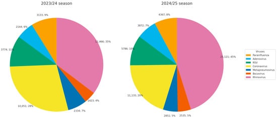
Figure 1.
Etiological structure of diagnosed acute respiratory infections in Russia during the 2023/24 and 2024/25 seasons.
The estimated annual incidence of adenoviral infection was 31.6 (30.2–32.9) and 26.7 (25.5–27.1) cases per 10,000 population in the 2023/24 and 2024/25 seasons, respectively. In absolute numbers, extrapolated to the national population, this corresponds to 461,426 (441,604–480,958) cases in 2023/24 and 389,429 (372,316–396,078) cases in 2024/25.
Analysis of the age distribution demonstrated that the highest detection rates (1.8% to 3.0%) occurred in children aged 0–2 and 3–6 years (Figure 2). In the 7–14-year age group, detection rates were lower (1.0% to 1.7%). The lowest rates (0.3% to 0.4%) were observed in the oldest age group (15 years and above).

Figure 2.
Age distribution of adenovirus-positive samples in Russia during the 2023/24 and 2024/25 seasons.
3.2. Overview of Sample Cohort and Genomic Dataset
In total, more than 1200 human adenovirus–positive samples, representing a subset of adenovirus-positive cases identified through national epidemiological surveillance, were received from participating hospitals in multiple shipment batches and processed during the study period. Over 200 viruses were successfully isolated and sequenced; however, complete genome assemblies were obtained only for a subset of these samples. Ultimately, 128 samples yielded high-quality genome sequences and were included in the present analysis.
In this study, we present 128 complete HAdV genomes from 16 Russian regions along with their genetic analysis. Among them are 27 B3 genomes, 9 B7, 44 B55, 12 C1, 16 C2, 4 C5, 7 C89, 5 C108, and one D109. Three more samples were found to contain recombinant viruses p89h5f5, p5h6f6 and p5h57f6, where p/h/f denote the penton base, hexon, and fiber genes, respectively. The distribution of successfully sequenced HAdV genomes, stratified by type and year of sample collection, is presented in Figure 3.
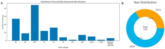
Figure 3.
(A) Distribution of successfully sequenced HAdV genomes. (B) Distribution of genome sequences by year of sample collection.
The successfully sequenced samples originated from 16 regions: Astrakhan Region, Khabarovsk Region, Komi Republic, Krasnodar Region, Lipetsk Region, Novosibirsk Region, Perm Region, Pskov Region, Republic of Bashkortostan, Republic of Buryatia, Republic of Tatarstan, Rostov Region, Saint Petersburg, Tambov Region, Vologda Region, and Yamalo-Nenets Autonomous Region.
However, sampling was geographically skewed, with the majority of samples originating from Saint Petersburg. Consequently, the dataset cannot be considered fully representative for assessing adenoviral genetic diversity across the entire country or for drawing definitive conclusions about the relative proportions of circulating viruses. Nevertheless, the data provide valuable insights into a problem that has, until now, remained largely unexplored. The geographic distribution of genetically characterized sequences is summarized in Figure 4.
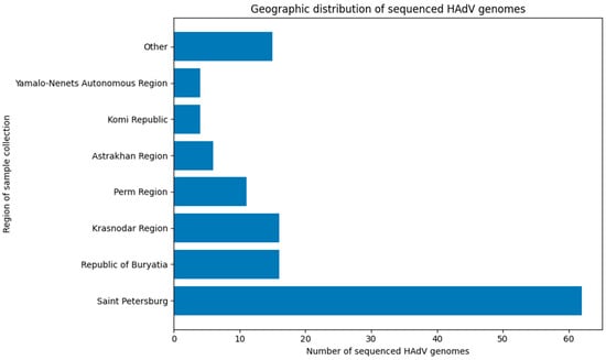
Figure 4.
Distribution of successfully sequenced adenovirus samples by region (regions with 1 or 2 samples combined as ‘Other’).
3.3. Virus Isolation and Virological Characteristics
Only samples with Cq values below 30 were used for isolation, and the median Cq among those that yielded successful whole-genome assemblies was 19.3 (Figure 5).
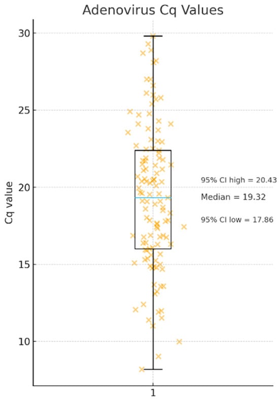
Figure 5.
Distribution of adenovirus Cq values obtained from clinical respiratory specimens that yielded successful whole-genome assemblies. Orange “×” symbols represent Cq values for single clinical specimens, while the blue horizontal line within the box indicates the median Cq value (19.3) with a 95% confidence interval ranging from 17.8 to 20.4, n = 128.
To increase the efficiency of adenovirus isolation, up to three successive passages were performed in A-549 cell culture. The virus-containing culture fluid from the wells that exhibited a cytopathic effect characteristic of an adenovirus was subsequently used for sequencing (Figure 6).
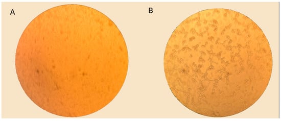
Figure 6.
Light microscopy images of A-549 cells: infected with clinical material and control, magnification ×400. (A) Control A-549 cell culture. (B) Cell culture A-549, 72 h after infection with clinical material. Cytopathic effect (CPE) characteristic of adenovirus is observed.
3.4. Sequencing
Only genomes that fulfilled predefined quality thresholds were retained for analysis and deposition in GenBank. Specifically, sequences were included only when the hexon, penton, and fiber genes were fully covered, whole-genome coverage exceeded 95%, and the median sequencing depth was greater than 10×. Genomes not meeting these criteria were excluded from further analysis.
The median sequencing coverage for the assembled adenovirus consensus genomes varied widely across samples, reflecting substantial differences in viral load, sequencing depth, and library quality. Coverage values ranged from 11× to 4865× (several high-quality samples) with the median coverage approximately 170×. Mapped read counts ranged from below 4000 to over 900,000 reads per sample, also reflecting substantial variability in sequencing depth.
Samples #1092, #1100 and #1124 (PV980131–PV980133) with low Cq values were additionally subjected to direct sequencing to test the hypothesis that successful genome recovery is possible without prior isolation. DNA extracted from several oropharyngeal swab samples was used for library preparation without virus isolation or amplification, and genomes with at least 10× coverage were successfully assembled, using the same estimated number of sequencing reads as for isolate-derived genomes (approximately one million reads per sample).
3.5. Phylogenetic and Genomic Analyses
The complete genomes of 128 HAdV isolates were successfully sequenced and deposited in the NCBI GenBank (accession numbers indicated in Appendix A Table A2). We then conducted phylogenetic analyses of these isolates together with representative HAdV sequences available in the GenBank database.
3.5.1. Adenovirus B
To contextualize the sequences generated in our laboratory within the broader evolutionary landscape of Adenovirus B, we constructed a maximum-likelihood phylogeny that includes both our newly obtained isolates and representative sequences retrieved from GenBank (Figure 7). The resulting tree resolves four major clades, each characterized by distinct temporal and geographic patterns. Most of our newly generated sequences cluster within three of these clades (27 B3 genomes, 9 B7, 44 B55), exhibiting minimal divergence and indicating close genetic relatedness.
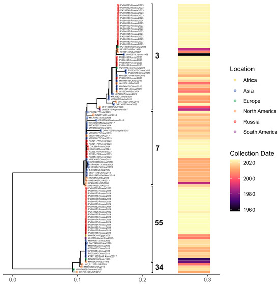
Figure 7.
Maximum-likelihood phylogenetic tree of Adenovirus B whole-genome sequences representing four major subtypes. Tips are colored by geographic region (with samples from Russia shown in red), brackets denote major clades, and the accompanying heatmap indicates the year of sample collection.
HAdV-B3 sequences produced in this study display multiple amino acid substitutions and segregate into two well-supported clusters according to their variation profiles. Additionally, a single-amino-acid S insertion in position 21 was identified in the B3 penton base for PV980200.1. By comparison, the immunogenic capsid protein sequences of the nine B7 and 44 B55 genomes generated here showed no detectable variation.
3.5.2. Adenovirus C
To characterize the genetic structure of our Adenovirus C isolates and assess the presence of potential recombinants, we constructed maximum-likelihood phylogenies for the three major capsid genes: penton base (Figure 8), hexon (Figure 9), and fiber (Figure 10). These gene-specific trees allowed us to compare our sequences with representative global strains and to evaluate whether individual genomic regions grouped consistently within established HAdV-C genotypes.
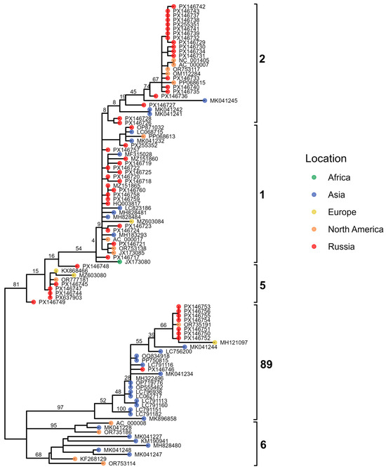
Figure 8.
Maximum-likelihood phylogenetic tree of Adenovirus C penton gene sequences. Tips are colored by geographic region, with Russian isolates shown in red. Major genotypes are indicated by brackets.
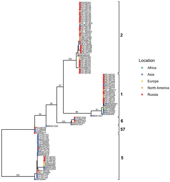
Figure 9.
Maximum-likelihood phylogenetic tree of Adenovirus C hexon gene sequences. Tips are colored by geographic region, with Russian isolates shown in red; brackets highlight major genotypes.
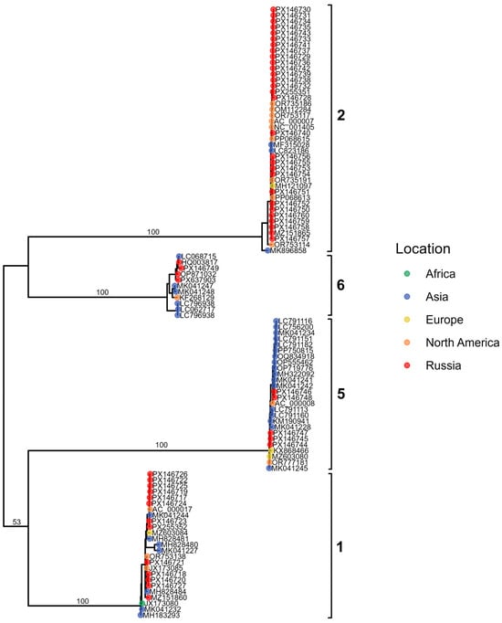
Figure 10.
Maximum-likelihood phylogenetic tree of Adenovirus C fiber gene sequences. Tips are colored by geographic region, with Russian isolates shown in red. Major genotypes are indicated by brackets.
In addition to the three gene-specific phylogenies, we also reconstructed a whole-genome maximum-likelihood tree to provide an integrated evolutionary framework for the Adenovirus C sequences (Figure 11). The whole-genome phylogeny was complemented by a visualization of genome structure, in which the capsid gene assignments inferred from the penton, hexon, and fiber trees were mapped onto each sequence. This approach enabled us to compare overall genomic relatedness with capsid–gene-specific clustering patterns and to detect signatures of recombination across the genome.
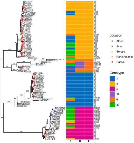
Figure 11.
Maximum-likelihood phylogenetic tree of Adenovirus C whole-genome sequences. Tips are colored by geographic region, with Russian samples shown in red. The heatmap indicates genotype assignment across the three genomic regions (P, H, and F). Brackets denote major clades.
The resulting phylogenies revealed clear clustering into the known genotypes (12 C1, 16 C2, 4 C5, 7 C89, 5 C108), enabling us to assign capsid gene types for each isolate. Notably, three recombinant viruses lacking assignment to any established genotype—p89h5f5, p5h6f6 and p5h57f6—were detected, each defined by unique combinations of capsid gene types.
Within the C5 genotype, the four genomes segregated into two distinct groups according to their amino-acid substitution profiles. Additional substitutions were observed in the fiber and penton proteins of the C1, C2, and C108 genomes, whereas the hexon proteins of these types showed no detectable variation. Notably, the C2 penton base exhibited both a single amino-acid deletion G10del (PX146727.1).
Based on phylogenetic analysis of the penton gene, HAdV-C/Russia/BU-RII-MH145796V/2023 (GenBank Acc. No. PX146746) was assigned to genotype 89. In contrast, phylogenetic trees inferred from the hexon and fiber genes show that this virus clusters with strains belonging to genotype 5. IDPlot analysis (see Figure 12) revealed extended genomic regions that distinguish HAdV-C/Russia/BU-RII-MH145796V/2023 from typical representatives of genotype 89. In particular, the hexon and fiber regions show the highest similarity to HAdV-C5. Notably, the putative recombinant segment at the fiber locus is substantially broader, spanning nt 28,275–33,020, and encompasses not only the fiber gene but also the E3 region, which encodes E3 12.5K, E3 CR1-α, E3 gp19K, 10.5 kDa, E3 RID-α, E3 RID-β, E3 14.7K, and the U exon. The IDPlot findings were corroborated by SimPlot analysis (Figure 12B).
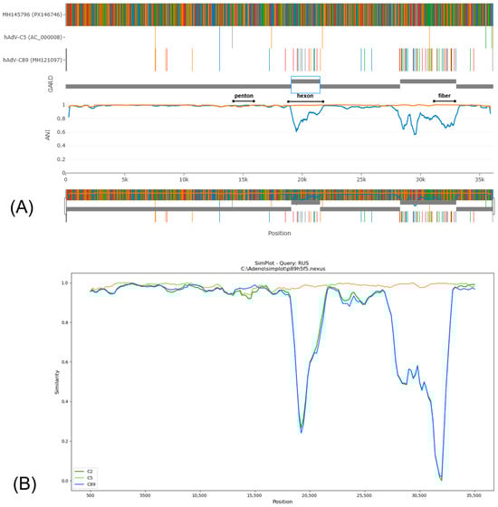
Figure 12.
(A) IDPlot analysis of HAdV-C/Russia/BU-RII-MH145796V/2023 in comparison with HAdV-C5 (orange line) and HAdV-C89 (blue line). (B) SimPlot analysis of HAdV-C/Russia/BU-RII-MH145796V/2023 in comparison with HAdV-C5 (orange line), HAdV-C2 (green line), and HAdV-C89 (blue line).
The penton sequences of genotypes 89 and 5 differ only slightly, and these differences are not attributable to recombination. Accordingly, in Figure 12, the penton similarity plots for genotype 89, genotype 5, and HAdV-C/Russia/BU-RII-MH145796V/2023 almost completely overlap. To confirm that the penton of HAdV-C/Russia/BU-RII-MH145796V/2023 belongs to genotype 89, we analyzed the amino acid sequence to identify a characteristic substitution profile. Amino acid sequence analysis of the penton revealed a substitution in HVR1 at position 153: P153Q in the genotype 89 reference strain and P153H in HAdV-C/Russia/BU-RII-MH145796V/2023. Of note, the HAdV-C/Russia/BU-RII-MH145796V/2023 strain also carries an additional HVR1 substitution, D156N. In the penton loop, the RGD region contains the A363E substitution and a deletion at position 364 (P364del), both of which are characteristic of genotype 89 (Figure 13).

Figure 13.
Amino acid substitutions in the penton protein of p89h5f5 strains, including HAdV-C/Russia/BU-RII-MH145796V/2023 (indicated by a red dot).
Whole-genome phylogenetic analysis identified adenovirus genomes from China and Japan that are closely related to HAdV-C/Russia/BU-RII-MH145796V/2023. Separate phylogenetic analyses of the capsid genes confirmed that these viruses belong to the p89h5f5 recombinant. Key metadata for these viruses are summarized in Table 2.
Table 2.
Recombinant p89h5f5 adenoviruses available in GenBank.
Phylogenetic analysis of complete genomes indicates that these viruses form a single cluster (with the exception of Kobe180113 and Kobe190208), which likely originated from genotype 5 adenoviruses. Exceptions are represented by two genomes from Kobe Prefecture, Japan—Kobe180113 (LC791113) and Kobe190208 (LC791160) [35]. These viruses conform to the p89h5f5 formula based on the penton, hexon, and fiber genes; however, analysis of genomic regions outside these loci indicates the presence of additional recombination events. Specifically, increased similarity to genotype 1 sequences was observed in the polymerase and E4 regions (see Supplementary Figures S1 and S2).
3.5.3. Adenovirus D109
To our knowledge, this represents the second reported detection of human adenovirus type D109 (HAdV-D109) worldwide. In the previously described case involving a one-year-old infant, clinical manifestations were typical of an acute respiratory infection, including fever, chills, cough, diarrhea, nasal congestion, and sore throat. In contrast, the present case involved a 43-year-old woman who required hospitalization and exhibited a severe clinical presentation characterized by fever, general malaise, sore throat, prominent ocular involvement (eye pain and photophobia), multiple painful erosions of the oral mucosa, and rapidly progressive diffuse cutaneous lesions with blistering, indicating a severe disease course. Rhinovirus was also detected concomitantly with adenovirus.
To contextualize the single Adenovirus D genome (D-109) identified in our study, we reconstructed a whole-genome maximum-likelihood phylogeny that includes representative HAdV-D sequences spanning multiple decades and geographic regions (Figure 14). The resulting tree shows extensive global heterogeneity within the HAdV-D group, where our D-109 sample clusters within one of the major HAdV-D lineages, closely grouping with contemporary isolates from North America and Europe.
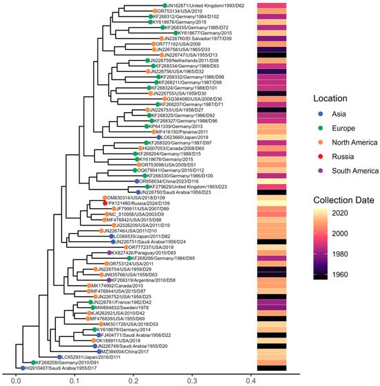
Figure 14.
Maximum-likelihood phylogenetic tree of Adenovirus D whole-genome sequences. Tips are colored by geographic region, and the accompanying heatmap shows collection dates from the 1950s to 2024.
Comparative genomic analysis revealed a very high level of similarity to the previously reported isolate 592275 (GenBank accession no. OM830314.1), with 99.94% nucleotide sequence identity, indicating minimal genomic divergence; consistent with this, both isolates clustered together in the phylogenetic tree.
4. Discussion
Adenoviruses undergo recombination and mutation, generating new variants with altered pathogenicity or transmissibility [38]. Genomic data provide valuable insights into these evolutionary processes. Monitoring adenoviral genetic diversity is essential for identifying emerging strains and adapting diagnostic assays, and it is also important given that adenoviruses are widely used in biotechnology as vectors for gene delivery, vaccine development, oncolytic cancer therapy, etc.
Information on genetic diversity remains highly uneven across pathogens. While millions of SARS-CoV-2 sequences are available, fewer than 2500 complete human adenovirus genomes have been deposited in GenBank, with only a few dozen represented for certain subtypes. Geographic representation is equally limited: for Russia, fewer than ten complete adenovirus genomes were available prior to our contribution. In fact, the global dataset on adenoviral genetic diversity remains insufficient overall, not merely at the regional level. The sequences generated in this study therefore provide a meaningful contribution to the global knowledge base on adenoviruses. For example, only two sequences of adenovirus D109 are currently available in NCBI: the reference genome and the sequence reported here.
The present study reports whole-genome sequences of HAdV isolates obtained from hospitalized patients with respiratory disease across different regions of Russia. The analysis revealed insights into the genetic diversity of circulating adenoviruses and identified several novel recombinant strains. The findings predominantly reflect adenoviruses causing respiratory disease, given that the analysis was limited to oropharyngeal swab specimens.
Over the past decade, advances in sequencing technologies that enable analysis of the entire viral genome have led to the identification of numerous recombinant types. Nevertheless, although the number of recognized HAdV types has increased rapidly, information regarding the clinical significance of these recombinant forms remains limited [39]. Genomic regions of HAdV that are most commonly involved in homologous recombination include the penton base, hexon, and fiber coding regions, which also represent the loci most frequently affected by mutations [40]. Recombinant adenoviruses do not necessarily possess entirely new genome sequences; rather, they may represent novel combinations of existing coding regions. For example, C89 and C57 contain genuinely new capsid protein sequences (p89 and h57, respectively), whereas C104 and C108 are not defined by unique sequence innovations but instead represent new genomic configurations—p1h1f2 for C104 and p1h2f2 for C108.
In our study, excluding the well-known recombinants B55, C89, C108, and D109, we identified three additional viruses that are likely unassigned recombinants. The first, PX146746.1, exhibits a p89h5f5 genome structure. In GenBank, 11 genomes share the same p89h5f5 profile, all of which are broadly classified as HAdV-C. The hAdV-C/Russia/BU-RII-MH145796V/2023 p89h5f5 recombinant virus identified in the Republic of Buryatia in 2023 shows high similarity to viruses detected in multiple provinces of China during 2002–2021 [32,33,36,37], as well as to viruses from Japan (Aichi and Kobe Prefectures) collected in 2017–2019 [34,35]. Notably, genotype 89 was first identified in fecal samples, and a substantial proportion of specimens containing the p89h5f5 recombinant form also originate from the gastrointestinal tract (Table 2). This observation suggests that these viruses may exhibit dual tropism for both the respiratory and gastrointestinal tracts. Of additional concern, the p89h5f5 group includes strain human/CHN/LN2017022/2017, which was recovered from a stool specimen collected from a patient with acute flaccid paralysis [33]. There is strong evidence that p89h5f5 recombinant viruses form a distinct, monophyletic clade in the whole-genome phylogenetic tree, with two exceptions: Kobe180113 and Kobe190208 strains. These two genomes fall outside the main clade, plausibly due to additional recombination events affecting the polymerase and E4 regions. The oldest representative of this lineage dates to 2002, suggesting that the p89h5f5 recombinant represents an East Asia–origin lineage that has been circulating cryptically for more than 24 years, causing sporadic infections in China and Japan over time. More broadly, recombination events outside the canonical “penton–hexon–fiber” gene set may substantially contribute to adenovirus genetic diversity, but this is likely underestimated under the current PHF-focused nomenclature.
A second virus without an established genotype, PX637903, displays a p5h6f6 structure. In GenBank, only two sequences (MH121112 and MH121113) show >99% similarity to this genome. These sequences were generated in the study by Dhingra et al. [41], which investigated adenovirus evolution and formally designated the C89 recombinant.
The third sequence generated in this study (PX146749.1) exhibited a p5h57f6 genomic structure. This genome shares very high similarity (>99%) with the Adenovirus Working Group reference strain HQ003817, which is classified as HAdV-C57 [42] (p1h57f6). However, in our phylogenetic analyses, HQ003817 consistently grouped as p1h57f6, whereas PX146749.1 clustered as p5h57f6, indicating that despite their overall genomic similarity, the penton gene of PX146749.1 corresponds to a different lineage.
It has long been assumed that, unlike HAdV-D, the evolution of HAdV-C is not characterized by recombination affecting the penton, hexon, and fiber genes. Accordingly, the emergence of genotype 89 was proposed to have resulted primarily from mutational changes in the HAdV-C2 penton and recombination in the E4 region [41]. However, the identification of HAdV-57 (p1h57f6) [42], HAdV-104 (p1h1f2) [43], HAdV-108 (p1h2f2) [44], and p89h5f5 suggests that the contribution of recombination to HAdV-C evolution warrants reassessment. It is clear that the current adenovirus classification system, which is based on the penton, hexon, and fiber genes, fails to capture substantial diversity generated by recombination in non-capsid genomic regions.
In addition to recombination events, we also identified a series of mutations in the immunogenic capsid proteins. The 27 B3 sequences generated in this study differed by multiple amino-acid substitutions and could be grouped into two distinct clusters based on these variation patterns. In contrast, the immunogenic capsid protein sequences of the 9 B7 and 44 B55 genomes obtained in our study showed no detectable variation. Among penton base, hexon, and fiber, the greatest diversity was observed in the fiber protein.
The greatest type-level diversity was observed among adenoviruses of species C (12 C1, 16 C2, 4 C5, 7 C89, and 5 C108). The four C5 genomes clustered into two groups based on their amino-acid substitution patterns. Additional amino-acid substitutions were identified in the fiber and penton proteins of C1, C2, and C108 genomes, whereas no substitutions were detected in the hexon protein for these types.
Several indels in the immunogenic capsid proteins were localized to the penton base, with no insertions or deletions detected in either the hexon or fiber genes. Specifically, a single–amino-acid insertion was observed in the B3 penton base in one sample, and a single–amino-acid deletion was identified in the C1 penton base.
Adenovirus D109 was first described in January 2018 [45] during surveillance at the California/Mexico border and was associated with acute respiratory disease. To the best of our knowledge, this subtype (p22h19f9) has not been identified elsewhere since its initial description. Comparative genomic analysis demonstrated 99.94% nucleotide sequence identity with the previously reported isolate 592275 (GenBank accession no. OM830314.1), indicating minimal genomic divergence. The limited clinical information provided by this case may nevertheless be valuable for future case identification and for improving the understanding of the potential pathogenic spectrum of HAdV-D109.
Although all of our results were obtained from sequencing adenoviral isolates, our preliminary experiments demonstrate that adenoviral samples with low Cq values can be successfully subjected to direct sequencing. DNA extracted directly from oropharyngeal swab samples was used for library preparation without prior virus isolation or amplification. Under these conditions, genomes achieving at least 10× coverage were successfully assembled against the reference. However, our experiment with direct sequencing was limited in scope, and additional work is required to more thoroughly evaluate sample quality parameters that enable efficient genome recovery without enrichment steps. Further studies are also needed to determine whether direct sequencing is a cost-effective and practical approach for routine adenovirus genomic surveillance. At the present stage, we point to the possibility of direct sequencing of adenoviruses from clinical swab samples using the same estimated number of sequencing reads as for isolate-derived genomes, with a relatively low read allocation per sample (approximately one million reads per sample).
Subtyping of human adenoviruses by PCR is currently restricted mainly to reference laboratories, research studies, or outbreak investigations. Given the large and continuously expanding number of recognized HAdV types, NGS represents a more comprehensive approach for adenovirus subtyping in clinical settings, particularly if whole-genome sequencing can be performed directly from clinical material without prior virus isolation. With respect to the development of other molecular techniques for HAdV subtyping, detailed knowledge of mutations and recombination events in immunogenic capsid proteins is highly relevant for diagnostic assay design, as sequence variation may compromise the performance of PCR primer/probe sets or antigen-based detection methods.
Limitations of the Study
This study was conducted using samples collected from different regions of Russia; however, their distribution was uneven and therefore may not fully represent the nationwide epidemiology of adenoviruses. In addition, the absence of clinical data limits the ability to assess potential associations between viral genotypes and disease severity or clinical outcomes. Another limitation is that only respiratory specimens (swabs) were analyzed, which restricts conclusions about adenovirus diversity in other types of infections.
Supplementary Materials
The following supporting information can be downloaded at: https://www.mdpi.com/article/10.3390/v18010136/s1, Figure S1: SimPlot analysis of Kobe180113 (LC791113) and Kobe190208 (LC791160) strains (brown line) in comparison with HAdV-C5 and p89h5f5 (pink line). Figure S2: Maximum-likelihood phylogenetic tree of Adenovirus C E4 region sequences. Tips are colored by geographic region, with Russian samples shown in red. Kobe180113 (LC791113) and Kobe190208 (LC791160) strains are indicated with red arrows. The heatmap indicates genotype assignment across the three genomic regions (P, H, and F).
Author Contributions
Bioinformatics, visualization, data curation, conceptualization, writing—original draft preparation, writing—review and editing, N.D.Y.; investigation, methodology, formal analysis, data curation A.A.I.; visualization, A.A.P.; investigation, methodology, A.A.P.; investigation, methodology, K.A.S. and I.V.A.; investigation, methodology, T.A.T.; writing—review and editing, supervision, project administration, A.B.K.; project administration, D.M.D.; funding acquisition, D.A.L. All authors have read and agreed to the published version of the manuscript.
Funding
This research was funded by the Ministry of Health of the Russian Federation (Project #TVKQ-2024-0003 “Complex approach to genetic characterization and early identifications of pathogens with epidemic and pandemic potential using metagenomic sequencing”).
Institutional Review Board Statement
The study was conducted in accordance with the Declaration of Helsinki and approved by the Local Ethics Committee at the Smorodintsev Research Institute of Influenza, protocol dated 24 March 2023.
Informed Consent Statement
Informed consent was obtained from all subjects involved in the study.
Data Availability Statement
All nucleotide sequence data generated in this study are available in the DDBJ/EMBL/GenBank databases under accession numbers PX637903, PX121473–PX121480, PX146717–PX146760, PV692083, and PV980131–PV980201, with detailed descriptions provided in Table A2, Appendix A.
Acknowledgments
We gratefully acknowledge the regional coordinators, clinicians, and laboratory specialists across 16 regions of Russia—Evgenia Lupyreva (Republic of Buryatia), Svetlana Netaliyeva (Astrakhan region), Elena Spirina (Lipetsk region), Larisa Rakova (Vologda region), Tatyana Fomina (Perm territory), Elena Yakovchuk (Krasnodar Territory), Gulbiyke Dzhammalova (Yamalo-Nenets Autonomous Okrug), Anastasia Kuzmenko (Rostov Region), Tatyana Gromova (Khabarovsk Territory), Ildar Magasumov (Republic of Bashkortostan), Olga Rakhmanova (Republic of Tatarstan), Tatyana Bezuglaya (Komi Republic), Olga Kurskaya (Novosibirsk region), Alexander Romanov (Pskov Region), Elena Pankova (Tambov region). We also thank Natalia Kolokolnikova for coordinating the shipment and logistics of biological specimens and Olga Shneider for administrative support, including coordination of paperwork and documentation. We also acknowledge the hospitals of Saint Petersburg for their collaboration and for delivering clinical samples from patients with ARVI symptoms: Clinical Infectious Diseases Hospital named after S.P. Botkin (Evgeniy Venev, Denis Gusev), City Children’s Hospital of St. Olga (Olga Afanasieva).
Conflicts of Interest
The authors declare no conflicts of interest.
Abbreviations
The following abbreviations are used in this manuscript:
| HAdV | Human adenovirus |
| ARI | Acute respiratory infection |
| CPE | Cytopathic effect |
| Cq | Quantification cycle |
| GenBank | International nucleotide sequence database |
| GIHSN | Global Influenza Hospital Surveillance Network |
| GTR | General Time Reversible |
| ML | Maximum likelihood |
| NCBI | National Center for Biotechnology Information |
| NGS | Next-generation sequencing |
| PCR | Polymerase chain reaction |
| UTM | Universal Transport Medium |
Appendix A
Table A1.
List of Healthcare Facilities Providing Respiratory Disease Patient Samples.
Table A1.
List of Healthcare Facilities Providing Respiratory Disease Patient Samples.
| Institution Name | Region | City |
|---|---|---|
| State Budgetary Healthcare Institution “Republican Clinical Infectious Diseases Hospital” | Republic of Buryatia | Ulan-Ude |
| St. Petersburg State Budgetary Healthcare Institution “Children’s City Polyclinic No. 35” | Saint Petersburg | Saint Petersburg |
| Astrakhan Regional Clinical Infectious Diseases Hospital named after A.M. Nichogi | Astrakhan Region | Astrakhan |
| Perm Regional Clinical Infectious Diseases Hospital | Perm Territory | Perm |
| Perm State Budgetary Healthcare Institution “City Children’s Polyclinic No. 1” | Perm Territory | Perm |
| Lipetsk Regional Clinical Infectious Diseases Hospital | Lipetsk Region | Lipetsk |
| Sheksninskaya Central District Hospital | Vologda Region | Sheksna |
| Perm Regional Infectious Diseases Hospital | Perm Territory | Perm |
| Chaykovsky Central City Hospital | Perm Territory | Chaykovsky |
| Specialized Clinical Children’s Infectious Diseases Hospital | Krasnodar Territory | Krasnodar |
| St. Petersburg Clinical Infectious Diseases Hospital named after Botkin | Saint Petersburg | Saint Petersburg |
| Children’s City Polyclinic No. 4 of Krasnodar | Krasnodar Territory | Krasnodar |
| Children’s City Polyclinic No. 5 of Krasnodar | Krasnodar Territory | Krasnodar |
| Infectious Diseases Hospital No. 3 of Novorossiysk | Krasnodar Territory | Novorossiysk |
| Infectious Diseases Hospital No. 2 | Krasnodar Territory | Sochi |
| Krymsk Central District Hospital | Krasnodar Territory | Krymsk |
| Specialized Clinical Children’s Infectious Diseases Hospital | Krasnodar Territory | Krasnodar |
| Noyabrsk Central City Hospital | Yamalo-Nenets Autonomous Okrug | Noyabrsk |
| St. Petersburg Children Infectious Diseases Hospital No. 3 | Saint Petersburg | Saint Petersburg |
| Children’s Polyclinic of the Railway District, Rostov-on-Don | Rostov Region | Rostov-on-Don |
| Children’s City Hospital (Komsomolsk-on-Amur) | Khabarovsk Territory | Komsomolsk-on-Amur |
| Republican Children’s Clinical Hospital | Republic of Bashkortostan | Ufa |
| Smorodintsev Research Institute of Influenza | Saint Petersburg | Saint Petersburg |
| St. Olga Children’s City Hospital | Saint Petersburg | Saint Petersburg |
| Republican Clinical Infectious Diseases Hospital | Republic of Tatarstan | Kazan |
| Children’s Polyclinic No. 2 of Krasnodar (Vostochnokruglikovskaya) | Krasnodar Territory | Krasnodar |
| Republican Infectious Diseases Hospital | Komi Republic | Syktyvkar |
| Novosibirsk Children’s City Clinical Hospital No. 6 | Novosibirsk Region | Novosibirsk |
| Infectious Diseases Hospital No. 3 of Novorossiysk | Krasnodar Territory | Novorossiysk |
| Children’s Infectious Diseases Department | Pskov Region | Pskov |
| Rasskazovo Central District Hospital | Tambov Region | Rasskazovo |
Table A2.
Sample Metadata and GenBank Accession Numbers for Sequenced Adenovirus Genomes.
Table A2.
Sample Metadata and GenBank Accession Numbers for Sequenced Adenovirus Genomes.
| Region | City | Subtype | Lab # | NCBI # |
|---|---|---|---|---|
| Republic of Buryatia | Ulan-Ude | B7 | MH145774 | PX121473.1 |
| Republic of Buryatia | Ulan-Ude | B7 | MH145795 | PX121474.1 |
| Republic of Buryatia | Ulan-Ude | p89h5f5 | MH145796 | PX146746.1 |
| Saint Petersburg | Saint Petersburg | B3 | MH147312 | PV980183.1 |
| Astrakhan Oblast | Astrakhan | B3 | MH147400 | PV980184.1 |
| Astrakhan Oblast | Astrakhan | C108 | MH147419 | PX146760.1 |
| Astrakhan Oblast | Astrakhan | B3 | MH147421 | PV980185.1 |
| Astrakhan Oblast | Astrakhan | B3 | MH147431 | PV980186.1 |
| Astrakhan Oblast | Astrakhan | C1 | MH147435 | PX146725.1 |
| Perm Krai | Perm | B3 | MH148372 | PV980199.1 |
| Perm Krai | Perm | B3 | MH148376 | PV980200.1 |
| Perm Krai | Perm | B7 | MH148387 | PX121475.1 |
| Perm Krai | Perm | C2 | MH148396 | PX146729.1 |
| Perm Krai | Perm | B3 | MH148410 | PV980201.1 |
| Saint Petersburg | Saint Petersburg | B3 | MH149423 | PV980187.1 |
| Saint Petersburg | Saint Petersburg | B3 | MH149429 | PV980188.1 |
| Lipetsk Oblast | Lipetsk | C1 | MH149917 | PX146720.1 |
| Republic of Buryatia | Ulan-Ude | B3 | MH166704 | PV980189.1 |
| Republic of Buryatia | Ulan-Ude | B3 | MH166710 | PV980190.1 |
| Republic of Buryatia | Ulan-Ude | B7 | MH166714 | PX121476.1 |
| Republic of Buryatia | Ulan-Ude | B3 | MH166738 | PV980194.1 |
| Republic of Buryatia | Ulan-Ude | C89 | MH167312 | PX146753.1 |
| Republic of Buryatia | Ulan-Ude | B7 | MH166717 | PX121477.1 |
| Republic of Buryatia | Ulan-Ude | B7 | MH166721 | PX121478.1 |
| Vologda Oblast | Sheksna | B3 | MH166724 | PV980191.1 |
| Perm Krai | Perm | C2 | MH166725 | PX146742.1 |
| Perm Krai | Chaikovsky | B55 | MH166726 | PV692083.1 |
| Perm Krai | Perm | B3 | MH166727 | PV980197.1 |
| Perm Krai | Perm | B3 | MH166728 | PV980192.1 |
| Perm Krai | Perm | B3 | MH166729 | PV980193.1 |
| Perm Krai | Perm | B3 | MH166730 | PV980198.1 |
| Krasnodar Krai | Krasnodar | C108 | MH166735 | PX146759.1 |
| Lipetsk Oblast | Lipetsk | C1 | MH166736 | PX146722.1 |
| Saint Petersburg | Saint Petersburg | B55 | MH209685 | PV980162.1 |
| Krasnodar Krai | Krasnodar | C2 | MH209686 | PX146735.1 |
| Krasnodar Krai | Krasnodar | C1 | MH209687 | PX146723.1 |
| Krasnodar Krai | Novorossiysk | C2 | MH209688 | PX146736.1 |
| Krasnodar Krai | Novorossiysk | C89 | MH209689 | PX146754.1 |
| Krasnodar Krai | Novorossiysk | C5 | MH209690 | PX146747.1 |
| Krasnodar Krai | Sochi | C2 | MH209691 | PX146732.1 |
| Krasnodar Krai | Krymsk | B55 | MH209692 | PV980163.1 |
| Krasnodar Krai | Krasnodar | C89 | MH209693 | PX146755.1 |
| Krasnodar Krai | Krasnodar | B55 | MH209694 | PV980164.1 |
| Krasnodar Krai | Krasnodar | C2 | MH209696 | PX146740.1 |
| Krasnodar Krai | Krasnodar | C89 | MH209697 | PX146756.1 |
| Krasnodar Krai | Krasnodar | C89 | MH209698 | PX146752.1 |
| Saint Petersburg | Saint Petersburg | B55 | MH209699 | PV980175.1 |
| Saint Petersburg | Saint Petersburg | D109 | MH209700 | PX121480.1 |
| Saint Petersburg | Saint Petersburg | C1 | MH209701 | PX146721.1 |
| Saint Petersburg | Saint Petersburg | B55 | MH209702 | PV980177.1 |
| Saint Petersburg | Saint Petersburg | B55 | MH209703 | PV980165.1 |
| Yamalo-Nenets Autonomous Okrug | Noyabrsk | B3 | MH209704 | PV980195.1 |
| Yamalo-Nenets Autonomous Okrug | Noyabrsk | C2 | MH209705 | PX146734.1 |
| Yamalo-Nenets Autonomous Okrug | Noyabrsk | C1 | MH209706 | PX146724.1 |
| Saint Petersburg | Saint Petersburg | B55 | MH209707 | PV980166.1 |
| Saint Petersburg | Saint Petersburg | B55 | MH209708 | PV980167.1 |
| Saint Petersburg | Saint Petersburg | C2 | MH209709 | PX146731.1 |
| Saint Petersburg | Saint Petersburg | C1 | MH209711 | PX146727.1 |
| Saint Petersburg | Saint Petersburg | B55 | MH209713 | PV980168.1 |
| Saint Petersburg | Saint Petersburg | B55 | MH209714 | PV980169.1 |
| Rostov Oblast | Rostov-on-Don | C5 | MH209715 | PX146748.1 |
| Saint Petersburg | Saint Petersburg | C1 | MH209716 | PX146726.1 |
| Khabarovsk Krai | Komsomolsk-on-Amur | C2 | MH209717 | PX146741.1 |
| Saint Petersburg | Saint Petersburg | B55 | MH209718 | PV980170.1 |
| Saint Petersburg | Saint Petersburg | C2 | MH209719 | PX146743.1 |
| Saint Petersburg | Saint Petersburg | B3 | MH209721 | PV980196.1 |
| Saint Petersburg | Saint Petersburg | B55 | MH209722 | PV980171.1 |
| Saint Petersburg | Saint Petersburg | B55 | MH209724 | PV980176.1 |
| Saint Petersburg | Saint Petersburg | B55 | MH209725 | PV980172.1 |
| Saint Petersburg | Saint Petersburg | B55 | MH209726 | PV980173.1 |
| Saint Petersburg | Saint Petersburg | B55 | MH209727 | PV980174.1 |
| Republic of Bashkortostan | Ufa | B55 | MH209728 | PV980178.1 |
| Saint Petersburg | Saint Petersburg | B55 | 1092 | PV980131.1 |
| Saint Petersburg | Saint Petersburg | B55 | 1110 | PV980132.1 |
| Saint Petersburg | Saint Petersburg | B55 | 1124 | PV980133.1 |
| Saint Petersburg | Saint Petersburg | B55 | MH204018 | PV980134.1 |
| Saint Petersburg | Saint Petersburg | C2 | MH204019 | PX146737.1 |
| Saint Petersburg | Saint Petersburg | B3 | MH204020 | PV980179.1 |
| Saint Petersburg | Saint Petersburg | B55 | MH204021 | PV980135.1 |
| Republic of Tatarstan | Kazan | B3 | MH204023 | PV980180.1 |
| Saint Petersburg | Saint Petersburg | B3 | MH204024 | PV980181.1 |
| Saint Petersburg | Saint Petersburg | C2 | MH204025 | PX146733.1 |
| Krasnodar Krai | Krasnodar | C2 | MH204026 | PX146738.1 |
| Saint Petersburg | Saint Petersburg | B55 | MH204027 | PV980136.1 |
| Saint Petersburg | Saint Petersburg | B55 | MH204028 | PV980137.1 |
| Saint Petersburg | Saint Petersburg | B55 | MH204029 | PV980138.1 |
| Astrakhan Oblast | Astrakhan | p5h6f6 | MH204031 | PX637903.1 |
| Saint Petersburg | Saint Petersburg | B55 | MH204032 | PV980139.1 |
| Saint Petersburg | Saint Petersburg | B55 | MH204033 | PV980140.1 |
| Saint Petersburg | Saint Petersburg | B55 | MH204034 | PV980141.1 |
| Saint Petersburg | Saint Petersburg | B55 | MH204035 | PV980142.1 |
| Saint Petersburg | Saint Petersburg | p5h57f6 | MH204036 | PX146749.1 |
| Saint Petersburg | Saint Petersburg | B55 | MH204037 | PV980143.1 |
| Saint Petersburg | Saint Petersburg | C5 | MH204038 | PX146744.1 |
| Saint Petersburg | Saint Petersburg | C108 | MH204039 | PX146728.1 |
| Komi Republic | Syktyvkar | B55 | MH204040 | PV980144.1 |
| Komi Republic | Syktyvkar | B55 | MH204041 | PV980145.1 |
| Komi Republic | Syktyvkar | B55 | MH204042 | PV980146.1 |
| Komi Republic | Syktyvkar | B55 | MH204043 | PV980147.1 |
| Saint Petersburg | Saint Petersburg | C89 | MH204046 | PX146750.1 |
| Novosibirsk Oblast | Novosibirsk | C1 | MH204047 | PX146717.1 |
| Krasnodar Krai | Novorossiysk | B7 | MH204049 | PX121479.1 |
| Krasnodar Krai | Krasnodar | C2 | MH204052 | PX146730.1 |
| Saint Petersburg | Saint Petersburg | B55 | MH204056 | PV980148.1 |
| Saint Petersburg | Saint Petersburg | B3 | MH204057 | PV980182.1 |
| Saint Petersburg | Saint Petersburg | B55 | MH204058 | PV980149.1 |
| Saint Petersburg | Saint Petersburg | B55 | MH204059 | PV980150.1 |
| Saint Petersburg | Saint Petersburg | B55 | MH204060 | PV980151.1 |
| Saint Petersburg | Saint Petersburg | C108 | MH204062 | PX146757.1 |
| Saint Petersburg | Saint Petersburg | B55 | MH204063 | PV980152.1 |
| Saint Petersburg | Saint Petersburg | B55 | MH204064 | PV980153.1 |
| Saint Petersburg | Saint Petersburg | C1 | MH204065 | PX146718.1 |
| Saint Petersburg | Saint Petersburg | B55 | MH204068 | PV980154.1 |
| Saint Petersburg | Saint Petersburg | B55 | MH204069 | PV980155.1 |
| Saint Petersburg | Saint Petersburg | C108 | MH204070 | PX146758.1 |
| Saint Petersburg | Saint Petersburg | C1 | MH204071 | PX146719.1 |
| Saint Petersburg | Saint Petersburg | C2 | MH204072 | PX146739.1 |
| Pskov Oblast | Pskov | C89 | MH204073 | PX146751.1 |
| Saint Petersburg | Saint Petersburg | C5 | MH204074 | PX146745.1 |
| Tambov Oblast | Rasskazovo | B55 | MH204075 | PV980156.1 |
| Yamalo-Nenets Autonomous Okrug | Noyabrsk | B3 | MH167300 | PX255345.1 |
| Republic of Buryatia | Ulan-Ude | B7 | MH167308 | PX255349.1 |
| Republic of Buryatia | Ulan-Ude | B3 | MH167309 | PX255346.1 |
| Republic of Buryatia | Ulan-Ude | B3 | MH167310 | PX255347.1 |
| Republic of Buryatia | Ulan-Ude | B7 | MH167311 | PX255350.1 |
| Republic of Buryatia | Ulan-Ude | C2 | MH167315 | PX255351.1 |
| Saint Petersburg | Saint Petersburg | B3 | MH167320 | PX255348.1 |
| Republic of Buryatia | Ulan-Ude | C1 | MH166713 | PX255352.1 |
References
- Harrach, B. Adenoviruses: General Features, Reference Module in Biomedical Sciences; Elsevier: Amsterdam, The Netherlands, 2014; ISBN 9780128012383. [Google Scholar] [CrossRef]
- Hearing, P. Adenoviridae: The viruses and their replication. In Fields Virology: DNA Viruses; Howley, P., Knipe, D., Cohen, J., Damania, B., Eds.; Walters Kluwer: Alphen aan den Rijn, The Netherlands, 2021; pp. 98–128. [Google Scholar]
- Rowe, W.P.; Huebner, R.J.; Gilmore, L.K.; Parrott, R.H.; Ward, T.G. Isolation of a cytopathogenic agent from human adenoids undergoing spontaneous degeneration in tissue culture. Proc. Soc. Exp. Biol. Med. 1953, 84, 570–573. [Google Scholar] [CrossRef]
- Human Adenovirus Working Group. 2025. Available online: http://hadvwg.gmu.edu (accessed on 1 May 2025).
- Jones, M.S., 2nd; Harrach, B.; Ganac, R.D.; Gozum, M.M.; Dela Cruz, W.P.; Riedel, B.; Pan, C.; Delwart, E.L.; Schnurr, D.P. New adeno-virus species found in a patient presenting with gastroenteritis. J. Virol. 2007, 81, 5978–5984. [Google Scholar] [CrossRef] [PubMed] [PubMed Central]
- Bergelson, J.M.; Cunningham, J.A.; Droguett, G.; Kurt-Jones, E.A.; Krithivas, A.; Hong, J.S.; Horwitz, M.S.; Crowell, R.L.; Finberg, R.W. Isolation of a common receptor for Coxsackie B viruses and adenoviruses 2 and 5. Science 1997, 275, 1320–1323. [Google Scholar] [CrossRef] [PubMed]
- Greber, U.F.; Flatt, J.W. Adenovirus Entry: From Infection to Immunity. Annu. Rev. Virol. 2019, 6, 177–197. [Google Scholar] [CrossRef] [PubMed]
- Wang, H.; Li, Z.Y.; Liu, Y.; Persson, J.; Beyer, I.; Möller, T.; Koyuncu, D.; Drescher, M.R.; Strauss, R.; Zhang, X.B.; et al. Desmoglein 2 is a receptor for adenovirus serotypes 3, 7, 11 and 14. Nat. Med. 2011, 17, 96–104. [Google Scholar] [CrossRef] [PubMed] [PubMed Central]
- Lion, T. Adenovirus infections in immunocompetent and immunocompromised patients. Clin. Microbiol. Rev. 2014, 27, 441–462. [Google Scholar] [CrossRef] [PubMed] [PubMed Central]
- Mandell, G.L.; Bennett, J.E.; Dolin, R. Mandell, Douglas, and Bennett’s Principles and Practice of Infectious Diseases, 7th ed; Elsevier: Philadelphia, PA, USA, 2009. [Google Scholar]
- Brandt, C.D.; Kim, H.W.; Vargosko, A.J.; Jeffries, B.C.; Arrobio, J.O.; Rindge, B.; Parrott, R.H.; Chanock, R.M. Infections in 18,000 infants and children in a controlled study of respiratory tract disease. I. Adenovirus pathogenicity in relation to serologic type and illness syndrome. Am. J. Epidemiol. 1969, 90, 484–500. [Google Scholar] [CrossRef] [PubMed]
- Ganzenmueller, T.; Wolf, M.; Wolfram, L.; Gkioule, C.; Steinbrück, L.; Heim, A. An Epidemic of Respiratory and Ocular Infections Caused by the Reemergence of a Recombinant Human Adenovirus, the Novel Type HAdV-B114 (P7H3F3). J. Med. Virol. 2025, 97, e70464. [Google Scholar] [CrossRef] [PubMed] [PubMed Central]
- Belser, J.A.; Rota, P.A.; Tumpey, T.M. Ocular tropism of respiratory viruses. Microbiol. Mol. Biol. Rev. 2013, 77, 144–156. [Google Scholar] [CrossRef] [PubMed] [PubMed Central]
- Yu, B.; Saravanan, L.; Tran, D.H.; Davies, A.J.; Kaur, H.; Naraynanan, S.; Verceles, A.C.; Kim, H.J. A case of fatal disseminated adenovirus and drug-resistant Pneumocystis pneumonia in a patient who received chemotherapy for mantle cell lymphoma. Clin. Case Rep. 2023, 11, e7220. [Google Scholar] [CrossRef] [PubMed]
- Nedelman, M. 11th Child Dies in Adenovirus Outbreak in New Jersey. CNN, 19 November 2018. Available online: https://edition.cnn.com/2018/11/16/health/wanaque-adenovirus-deaths-new-jersey-bn (accessed on 18 January 2026).
- Heinonen, S.; Erra, E.; Lundell, R.; Nyqvist, A.; Hepo-Oja, P.; Mannonen, L.; Jarva, H.; Loginov, R.; Lindh, E.; Lakoma, L.; et al. Adenovirus type 7d outbreak associated with severe clinical presentation, Finland, February to June 2024. Eurosurveillance 2025, 30, 2500061. [Google Scholar] [CrossRef] [PubMed] [PubMed Central]
- Kurskaya, O.G.; Prokopyeva, E.A.; Dubovitskiy, N.A.; Solomatina, M.V.; Sobolev, I.A.; Derko, A.A.; Nokhova, A.R.; Anoshina, A.V.; Leonova, N.V.; Simkina, O.A.; et al. Genetic Diversity of the Human Adenovirus C Isolated from Hospitalized Children in Russia (2019–2022). Viruses 2024, 16, 386. [Google Scholar] [CrossRef] [PubMed] [PubMed Central]
- Shein, D.A.; Ryzhova, N.N.; Kunda, M.S.; Ermolova, E.I.; Ozharovskaia, T.A.; Popova, O.; Nikitenko, N.A.; Krasnoslobodtsev, K.G.; Burtseva, E.I.; Zubkova, O.V.; et al. Genetic characteristics of the isolate of human adenovirus type 55 (Adenoviridae: Mastadenovirus) isolated in Moscow in 2022. Vopr Virusol. 2025, 70, 431–443. [Google Scholar] [CrossRef] [PubMed]
- Puig-Barberà, J.; Tormos, A.; Trushakova, S.; Sominina, A.; Pisareva, M.; Ciblak, M.A.; Badur, S.; Yu, H.; Cowling, B.J.; Burtseva, E.; et al. The Global Influenza Hospital Surveillance Network (GIHSN): A new platform to describe the epidemiology of severe influenza. Influenza Other Respir. Viruses 2015, 9, 277–286. [Google Scholar] [CrossRef] [PubMed] [PubMed Central]
- Andrews, S. FastQC: A Quality Control Tool for High Throughput Sequence Data. 2010. Available online: http://www.bioinformatics.babraham.ac.uk/projects/fastqc/ (accessed on 18 January 2026).
- Chen, S. Ultrafast one-pass FASTQ data preprocessing, quality control, and deduplication using fastp. iMeta 2023, 2, e107. [Google Scholar] [CrossRef]
- Li, H.; Durbin, R. Fast and accurate short read alignment with Burrows-Wheeler transform. Bioinformatics 2009, 25, 1754–1760. [Google Scholar] [CrossRef] [PubMed]
- Li, H.; Handsaker, B.; Wysoker, A.; Fennell, T.; Ruan, J.; Homer, N.; Marth, G.; Abecasis, G.; Durbin, R. 1000 Genome Project Data Processing Subgroup The Sequence Alignment/Map format and SAMtools. Bioinformatics 2009, 25, 2078–2079. [Google Scholar] [CrossRef]
- Grubaugh, N.D.; Gangavarapu, K.; Quick, J.; Matteson, N.L.; De Jesus, J.G.; Main, B.J.; Tan, A.L.; Paul, L.M.; Brackney, D.E.; Grewal, S.; et al. An amplicon-based sequencing framework for accurately measuring intrahost virus diversity using PrimalSeq and iVar. Genome Biol. 2019, 20, 8. [Google Scholar] [CrossRef]
- Katoh, K.; Standley, D.M. MAFFT multiple sequence alignment software version 7: Improvements in performance and usability. Mol. Biol. Evol. 2013, 30, 772–780. [Google Scholar] [CrossRef] [PubMed] [PubMed Central]
- Stamatakis, A. RAxML version 8: A tool for phylogenetic analysis and post-analysis of large phylogenies. Bioinformatics 2014, 30, 1312–1313. [Google Scholar] [CrossRef] [PubMed] [PubMed Central]
- Shean, R.C.; Makhsous, N.; Stoddard, G.D.; Lin, M.J.; Greninger, A.L. VAPiD: A lightweight cross-platform viral annotation pipeline and identification tool to facilitate virus genome submissions to NCBI GenBank. BMC Bioinform. 2019, 20, 48. [Google Scholar] [CrossRef] [PubMed Central]
- Goldstein, S.A.; Brown, J.; Pedersen, B.S.; Quinlan, A.R.; Elde, N.C. Extensive Recombination-driven Coronavirus Diversifi-cation Expands the Pool of Potential Pandemic Pathogens. Genome Biol. Evol. 2022, 14, evac161. [Google Scholar] [CrossRef] [PubMed] [PubMed Central]
- Kosakovsky Pond, S.L.; Posada, D.; Gravenor, M.B.; Woelk, C.H.; Frost, S.D.W. GARD: A genetic algorithm for recombination detection. Bioinformatics 2006, 22, 3096–3098. [Google Scholar] [CrossRef] [PubMed]
- Price, M.N.; Dehal, P.S.; Arkin, A.P. Fasttree 2—Approximately Maximum-likelihood trees for large alignments. PLoS ONE 2010, 5, e9490. [Google Scholar] [CrossRef] [PubMed]
- Samson, S.; Lord, É.; Makarenkov, V. SimPlot++: A Python application for representing sequence similarity and detecting recombination. Bioinformatics 2022, 38, 3118–3120. [Google Scholar] [CrossRef] [PubMed]
- Mao, N.; Zhu, Z.; Rivailler, P.; Yang, J.; Li, Q.; Han, G.; Yin, J.; Yu, D.; Sun, L.; Jiang, H.; et al. Multiple divergent Human mastadenovirus C co-circulating in mainland of China. Infect. Genet. Evol. 2019, 76, 104035. [Google Scholar] [CrossRef] [PubMed]
- Shao, Y.; Wang, Z.; Ren, L.; Chen, T.; Fang, X.; Wang, Y. Genetic characteristics of recombinant human adenovirus C5 and 89 isolated from an acute flaccid paralysis case in Liaoning, a study based on whole genome sequencing. Dis. Surveill. 2025, 41, 1–9. [Google Scholar] [CrossRef]
- Hirose, E.; Minagawa, H.; Adachi, H.; Suzuki, M.; Nakamura, N.; Saito, N.; Ito, M.; Mitsuoka, C.; Sato, K.; Yasui, Y. Detection of Recombinant Type of Human Adenovirus C Composed of Types 89 and 5 in Aichi Prefecture, Japan (2016–2019). Jpn. J. Infect. Dis. 2025, 78, 139–142. [Google Scholar] [CrossRef] [PubMed]
- Tanimoto, Y.; Ohyama, M.; Ito, E.; Akiyoshi, K.; Onishi, Y.; Mori, A.; Nomoto, R. Whole genome-based surveillance for human adenovirus-related diseases in Kobe City, Japan, 2018–2022. BMC Res. Notes 2025, 18, 170. [Google Scholar] [CrossRef] [PubMed Central]
- Cao, Y.; Yang, J.; Li, N.; Zhang, R.; Jiang, L.; Zhou, X.; Xiang, Y.; Cun, J.; Qiao, E. Detection and complete genome sequence analysis of human adenovirus in children with acute diarrhea in Yunnan, China, 2015–2021. Arch. Virol. 2024, 169, 34. [Google Scholar] [CrossRef] [PubMed] [PubMed Central]
- Lei, Y.; Zhuang, Z.; Liu, Y.; Tan, Z.; Gao, X.; Li, X.; Yang, D. Whole Genomic Sequence Analysis of Human Adenovirus Species C Shows Frequent Recombination in Tianjin, China. Viruses 2023, 15, 1004. [Google Scholar] [CrossRef] [PubMed Central]
- Robinson, C.M.; Singh, G.; Lee, J.Y.; Dehghan, S.; Rajaiya, J.; Liu, E.B.; Yousuf, M.A.; Betensky, R.A.; Jones, M.S.; Dyer, D.W.; et al. Molecular evolution of human adenoviruses. Sci. Rep. 2013, 3, 1812. [Google Scholar] [CrossRef] [PubMed] [PubMed Central]
- Kosulin, K. Intestinal HAdV Infection: Tissue Specificity, Persistence, and Implications for Antiviral Therapy. Viruses 2019, 11, 804. [Google Scholar] [CrossRef] [PubMed Central]
- Lukashev, A.N.; Ivanova, O.E.; Eremeeva, T.P.; Iggo, R.D. Evidence of frequent recombination among human adenoviruses. J. Gen. Virol. 2008, 89, 380–388. [Google Scholar] [CrossRef] [PubMed]
- Dhingra, A.; Hage, E.; Ganzenmueller, T.; Böttcher, S.; Hofmann, J.; Hamprecht, K.; Obermeier, P.; Rath, B.; Hausmann, F.; Dobner, T.; et al. Molecular Evolution of Human Adenovirus (HAdV) Species C. Sci. Rep. 2019, 9, 1039. [Google Scholar] [CrossRef] [PubMed] [PubMed Central]
- Walsh, M.P.; Seto, J.; Liu, E.B.; Dehghan, S.; Hudson, N.R.; Lukashev, A.N.; Ivanova, O.; Chodosh, J.; Dyer, D.W.; Jones, M.S.; et al. Computational analysis of two species C human adenoviruses provides evidence of a novel virus. J. Clin. Microbiol. 2011, 49, 3482–3490. [Google Scholar] [CrossRef] [PubMed] [PubMed Central]
- Ji, T.; Li, L.; Li, W.; Zheng, X.; Ye, X.; Chen, H.; Zhou, Q.; Jia, H.; Chen, B.; Lin, Z.; et al. Emergence and characterization of a putative novel human adenovirus recombinant HAdV-C104 causing pneumonia in Southern China. Virus Evol. 2021, 7, veab018. [Google Scholar] [CrossRef] [PubMed] [PubMed Central]
- Chen, M.; Zhu, Z.; Huang, F.; Liu, D.; Zhang, T.; Ying, D.; Wu, J.; Xu, W. Adenoviruses associated with acute respiratory diseases reported in Beijing from 2011 to 2013. PLoS ONE 2015, 10, e0121375. [Google Scholar] [CrossRef] [PubMed] [PubMed Central]
- Gonzalez, G.; Hayes, M.; Balansay, M.; Underwood, R.; Balagot, C.; Pan, R.; Pineda, K.; Iniguez-Stevens, E.; Kriner, P.; Estrada, I.; et al. Two novel recombinant human mastadenovirus D genotypes associated with acute respiratory illness. J. Med. Virol. 2023, 95, e28653. [Google Scholar] [CrossRef] [PubMed]
Disclaimer/Publisher’s Note: The statements, opinions and data contained in all publications are solely those of the individual author(s) and contributor(s) and not of MDPI and/or the editor(s). MDPI and/or the editor(s) disclaim responsibility for any injury to people or property resulting from any ideas, methods, instructions or products referred to in the content. |
© 2026 by the authors. Licensee MDPI, Basel, Switzerland. This article is an open access article distributed under the terms and conditions of the Creative Commons Attribution (CC BY) license.