Article

Use of Capsid Integrity-qPCR for Detecting Viral Capsid Integrity in Wastewater

1
School of Environmental and Natural Sciences, Bangor University, Bangor, Gwynedd LL57 2UW, UK
2
School of Ocean Sciences, Bangor University, Menai Bridge, Anglesey LL59 5AB, UK
3
Centre for Sustainable Farming Systems, Food Futures Institute, Murdoch University, Murdoch, WA 6150, Australia
*
Author to whom correspondence should be addressed.
Viruses 2024, 16(1), 40; https://doi.org/10.3390/v16010040
Submission received: 20 November 2023 / Revised: 18 December 2023 / Accepted: 21 December 2023 / Published: 26 December 2023
(This article belongs to the Special Issue Wastewater-Based Epidemiology (WBE) in COVID-19 Pandemics)

Abstract

:
Quantifying viruses in wastewater via RT-qPCR provides total genomic data but does not indicate the virus capsid integrity or the potential risk for human infection. Assessing virus capsid integrity in sewage is important for wastewater-based surveillance, since discharged effluent may pose a public health hazard. While integrity assays using cell cultures can provide this information, they require specialised laboratories and expertise. One solution to overcome this limitation is the use of photo-reactive monoazide dyes (e.g., propidium monoazide [PMAxx]) in a capsid integrity-RT-qPCR assay (ci-RT-qPCR). In this study, we tested the efficiency of PMAxx dye at 50 μM and 100 μM concentrations on live and heat-inactivated model viruses commonly detected in wastewater, including adenovirus (AdV), hepatitis A (HAV), influenza A virus (IAV), and norovirus GI (NoV GI). The 100 μM PMAxx dye concentration effectively differentiated live from heat-inactivated viruses for all targets in buffer solution. This method was then applied to wastewater samples (n = 19) for the detection of encapsulated AdV, enterovirus (EV), HAV, IAV, influenza B virus (IBV), NoV GI, NoV GII, and SARS-CoV-2. Samples were negative for AdV, HAV, IAV, and IBV but positive for EV, NoV GI, NoV GII, and SARS-CoV-2. In the PMAxx-treated samples, EV, NoV GI, and NoV GII showed −0.52–1.15, 0.9–1.51, and 0.31–1.69 log reductions in capsid integrity, indicating a high degree of potentially infectious virus in wastewater. In contrast, SARS-CoV-2 was only detected using RT-qPCR but not after PMAxx treatment, indicating the absence of encapsulated and potentially infectious virus. In conclusion, this study demonstrates the utility of PMAxx dyes to evaluate capsid integrity across a diverse range of viruses commonly monitored in wastewater.

1. Introduction

The use of wastewater-based surveillance (WBS) to monitor community-level health via the detection of harmful disease-causing pathogens, excreted in faeces and urine, has greatly increased in recent years [1]. For example, many studies now exist showing the utility of WBS to detect viruses, such as norovirus [2], polio and non-polio enterovirus [3,4,5], adenovirus [6,7,8], hepatitis A [9,10], hepatitis E [11,12], and respiratory viruses such as coronaviruses, influenza viruses, and respiratory syncytial virus [13,14,15]. During the COVID-19 pandemic, WBS was widely implemented for population-level surveillance of SARS-CoV-2 by research groups and laboratories globally [16,17,18,19,20,21,22], further showcasing the usefulness of WBS for disease monitoring and public health policy implementation.
Viruses in wastewater are typically detected by first concentrating and precipitating the viruses, extracting their nucleic acids, and then performing molecular techniques on the samples such as RT-qPCR (real-time quantitative polymerase chain reaction), dd-PCR (digital droplet PCR), or next generation sequencing. However, these methods cannot distinguish between intact potentially infectious viruses and degraded non-infectious viral particles; rather, they detect genetic markers from viruses regardless of viability. Thus, while the data gathered by these techniques provide an overview of infection levels in the community, it does not indicate the viral stability or the potential for infectivity if an individual comes into contact with sewage-contaminated water. This aspect of WBS is becoming more important, given the increased discharge of untreated sewage into rivers and seas and the associated risks to human and environmental health (i.e., One Health) [23,24].
Cell culture is the gold standard for virus viability testing. However, it requires specialised equipment like incubators, inverted microscopes, centrifuges, plate readers, hemacytometers, cryogenic storage, CO2, media, and other consumables [25]. In addition, it can take 4–7 days to generate infectivity data, causing delays in the implementation of public health interventions. It also requires staff with expertise to maintain the cell lines. Furthermore, many human respiratory viruses can only be cultured in biosecurity level (BLS) 3 laboratories, which are usually not available in environmental monitoring facilities. These requirements can be limiting factors, especially in lower- or middle-income countries (LMICs), where, in addition, frequent power outages may be common. Furthermore, isolating viruses from wastewater for cell culture assays is extremely challenging. Viruses are often present at low viral loads; so, large volumes (>10 l) of wastewater must be concentrated prior to culturing. Wastewater also contains debris and microbes that can contaminate and destroy cell cultures used for virus viability assays. Therefore, methods like filtration are needed to remove these contaminants without damaging viable viruses [26]. Antibiotics also may need to be added to the culture media to reduce microbial activity, albeit at levels that do not compromise the cell line [27]. Therefore, simpler methods like capsid integrity-PCR/qPCR (ci-PCR or ci-qPCR) (also referred to as viability qPCR/RT-qPCR) are needed to assess the potential infectivity of viruses in wastewater samples.
Capsid integrity-PCR/qPCR has become an important technique in virology for selectively detecting potentially infectious viral particles. Photo-reactive monoazide dyes such as ethidium monoazide (EMA), propidium monoazide (PMA), and PMAxx® penetrate damaged viral capsids with exposed genomes, which are incapable of cell attachment and entry but are excluded from viruses with intact capsids that maybe capable of host cell infection. After addition of the dye to a wastewater viral concentrate, the sample is exposed to light at which point the dye modifies any exposed nucleic acids, blocking their subsequent amplification by PCR [28]. In contrast, the genomes of viruses with intact capsids that are potentially infectious can be successfully amplified. This allows selective quantification of the encapsulated viral genomes versus the non-infectious nucleic acids present in the sample. Capsid integrity-PCR using monoazide-based dyes has been applied to differentiate infectious and non-infectious viruses including influenza-A virus [29], norovirus [30], and SARS-CoV-2 [31]. Furthermore, the quantification of encapsulated viral genomes has provided insights into viral persistence in the environment [32,33] and the disinfection efficiency [34,35,36]. It is worth noting that the capsids of viruses inactivated using UV are still intact, and as a result, ci-qPCR is not suitable for assessing UV disinfection [36]. However, the sensitivity and specificity of this approach continues to be improved through optimising dye types, the concentration of dye used, incubation times, and light exposure length [37].
Many studies have utilised ci-qPCR to assess the capsid integrity of viruses in contaminated drinking water and food, as reviewed [38]; however, the use of ci-qPCR in wastewater monitoring has received much less attention. Of the wastewater studies that do exist, ci-qPCR has been used to assess the capsid integrity of adenovirus [39], enterovirus [39,40], norovirus GI and GII [30,32,40,41], rotavirus [32,39], hepatitis A virus [10,42], and SARS-CoV-2 [31,32]. Human enteric virus genomes (adenovirus, enterovirus, and norovirus) in wet and dry wastewater-derived struvite have also been shown to remain encapsulated using intercalating dyes [43]. We hypothesize that wastewater-based ci-qPCR can provide improved estimates of public health exposure risks compared to conventional PCR, especially regarding the potential infection risks posed by sewage discharges into the environment. This study therefore aims to optimise and assess the usefulness of ci-RT-qPCR using PMAxx dyes alongside conventional wastewater monitoring via RT-qPCR, for important viruses of public health concern including adenovirus (AdV), enterovirus (EV), hepatitis A (HAV), influenza A virus (IAV), influenza B virus (IBV), norovirus GI (NoV GI), norovirus GII (NoV GII), and SARS-CoV-2. The optimisation of PMAxx dye for a range of viral targets is also presented.

2. Materials and Methods

2.1. Viral Stocks

The viral stocks used for PMAxx dye optimisation were AdV 40 cultured in-house, as described previously [44], HAV (Culture Collections of the UK Health Security Agency), IAV vaccine strain (H1N1) (kindly provided by Dr Eleanor Gaunt, University of Edinburgh), and NoV GI (Culture Collections of the UK Health Security Agency). To optimise the PMAxx dye assays on live and heat-inactivated viruses, samples were made containing AdV at concentration of ~108–109 gene copies/ml (gc/ml), HAV at ~105–106 gc/ml, IAV at ~107–108 gc/ml, and NoV GI at ~105–106 gc/ml. Viruses were suspended in phosphate buffer saline (PBS), pH 7.4, to a final volume of 5 ml. An aliquot of the virus mixture was then heat inactivated at 70 °C for 30 min in a benchtop heating block, to allow for a comparison of viruses with damaged capsids, alongside samples containing live viruses. All samples were made in triplicate.

2.2. PMAxx Optimisation

PMAxx dye 20 mM in H2O (Biotium, Cat no: 40069; Biotium Inc., Freemont, CA, USA) was diluted with molecular grade H2O to a final volume of 10 mM to make a working stock. PMA enhancer at 1 X concentration was added to each sample and mixed by pipetting. Working in the dark, PMAxx dye working solution was added to the samples at a final concentration of 50 µM or 100 µM. Once the PMAxx dye was added, samples were incubated in the dark, at 20 °C on a bench top rocker (PMR-30 2D Rocker, Cat No: PMR-30-UK) at 30 oscillations/min. The samples were then exposed to light (465–475 nm) for 20 min at room temperature, using the PMA-Lite™ 2.0 Photolysis Device (Biotium, Cat No: BTE90006). Nucleic acids were extracted directly after light exposure.

2.3. Wastewater Sample Collection

On 13 June 2023, 1 litre of influent wastewater samples was collected from behind the primary screen at 17 wastewater treatment plants (WWTP) and the main sewer line leaving 2 hospital sites, located in North Wales, resulting in a total of 19 samples. These sites formed part of the national wastewater-based public health surveillance programme funded by the Welsh Government. All wastewater samples were 24 h composites collected using refrigerated autosamplers, with samples taken every 15 min. The samples were collected and transported chilled to the laboratory, where on their arrival they were stored at 4 °C overnight and processed the following day. Sample storage at 4 °C is deemed appropriate for wastewater samples, as minimal impact on the viral decay and recovery of SARS-CoV-2 and surrogates from wastewater has been evidenced [45,46].

Wastewater Processing

Samples were processed via the polyethylene glycol (PEG) precipitation method, as previously described [47,48] but with modifications [49]. Briefly, a PEG-NaCl solution at 40% PEG (PEG8000, Sigma-Aldrich, St. Louis, MO, USA, Cat. No. P5413) with 8% NaCl (Sigma-Aldrich, St. Louis, MO, USA, Cat. No. S7653) was prepared. Wastewater (200 ml, n = 19) and dH2O negative controls were clarified by centrifugation at 10,000× g, 4 °C, for 10 min. The supernatant (150 ml) was transferred to a sterile 250 ml PCCO bottle, and the pH was adjusted to 7–7.5 using 1 M NaOH. PEG-NaCI (50 ml) was then added to each sample, and the sample was mixed and incubated overnight at 4 °C. The following day, the samples in PEG-NaCI were centrifuged (10,000× g, 4 °C, 30 min). The resulting pellet was resuspended in 200 μl of PBS. All samples were processed in duplicate, one for RT-qPCR and one for ci-RT-qPCR. Concentrates were stored at 4 °C until nucleic acid extraction. All sample preparation and processing were conducted in a Biosafety Level 2 (BSL2) laboratory, adhering to WHO and national biosafety guidelines [50].

2.4. Nucleic Acid Extraction

Nucleic acids were extracted from concentrates using NucliSens lysis buffer (BioMerieux, Marcy-lÉtoile, France, Cat No. 280134 or 200292), NucliSens extraction reagent kit (BioMerieux, Cat. No. 200293), and a Kingfisher 96 Flex system (Thermo Scientific, Waltham, MA, USA), following a previously published protocol [49]. Extracts were stored at −80 °C until analysis.

2.5. Real-Time Quantitative PCR (RT-qPCR) and Capsid Integrity-RT-qPCR (ci-RT-qPCR) Analysis

All qPCRs (RT-qPCR and ci-RT-qPCR) were carried out on a Quant Studio Flex 6 Real-time PCR machine (Applied Biosystems Inc., Waltham, MA, USA). To quantify AdV, the qPCR reactions contained 2 X PowerUP™ SYBR™ Green Master Mix (Applied Biosystems™, ThermoFisher, Cat No: A25742), 10 pmol forward primer, 10 pmol reverse primer (Supplementary Table S1), molecular grade water, and a 4 µl sample, to make a 20 µl final reaction volume. The cycling conditions were to hold at 95 °C for 5 min, followed by 40 cycles of 95 °C for 0.15 s, 56 °C for 30 s annealing, and melt curve at 95 °C 15 s, then 60 °C for 1 min and 95 °C for 15 s at increments of 1.6° C/s. The qPCRs for EV, HAV, IAV, IBV, NoV GI, NoV GII, and SARS-CoV-2 contained 5 µl 4 × TaqMan™ Fast Virus 1-Step Master Mix for qPCR (Applied Biosystems™, ThermoFisher, Cat No: 4444432), 10 pmol forward primer, 20 pmol reverse primer, 5 pmol probe (Supplementary Table S1), 6 nmol MgSO4, 1 μg bovine serum albumin (BSA), molecular grade water, and a 4 µl sample to make a final reaction volume of 20 µl. RT-qPCR was conducted as follows: hold at 55 °C for 60 min for reverse transcription, 95 °C for 5 min for reverse transcriptase inactivation, followed by 45 amplification cycles of 95 °C for 15 s, 60 °C for 1 min, and 65 °C for 1 min at increments of 1.6 °C/s. All viral targets were run alongside a dsDNA (AdV), ssDNA (HAV), or ssRNA standard curve ranging from 1 to 105 copies/µl The DNA standards were purchased from IDT (Integrated DNA Technologies Inc., Coralville, IA, USA), whilst the RNA standards were prepared in-house using a protocol previously described by Kevill et al. (2021) [49]. All samples were run in duplicate with each plate containing four non-template controls with molecular grade water.

2.6. Data Analysis

The qPCR data were analysed and quality checked as per MIQE guidelines for RT-PCR [51]. The limit of detection (LOD) and limit of quantification (LOQ) for viral targets in wastewater have been descried previously [52]. The gene copies per PCR reaction were transformed into gc/ml for viruses in PBS and gc/l for viruses detected in wastewater, for statistical analysis. The data followed a non-normal distribution; therefore, nonparametric statistical analysis was performed using a Kruskal–Wallis test, followed by post hoc multi-comparisons using a Dunn’s test [53], with a Bonferroni corrected alpha of p < 0.003. Statistical analysis was performed using R studio, utilising the “MultNonParam” package, “Kruskal.test” [54], and “dunn.test” [55]. Log reductions were calculated as:
L o g   r e d u c t i o n = l o g 10   ( V i r a l   c o p i e s   o f   u n t r e a t e d   s a m p l e s V i r a l   c o p i e s   o f   t r e a t e d   s a m p l e s )

3. Results

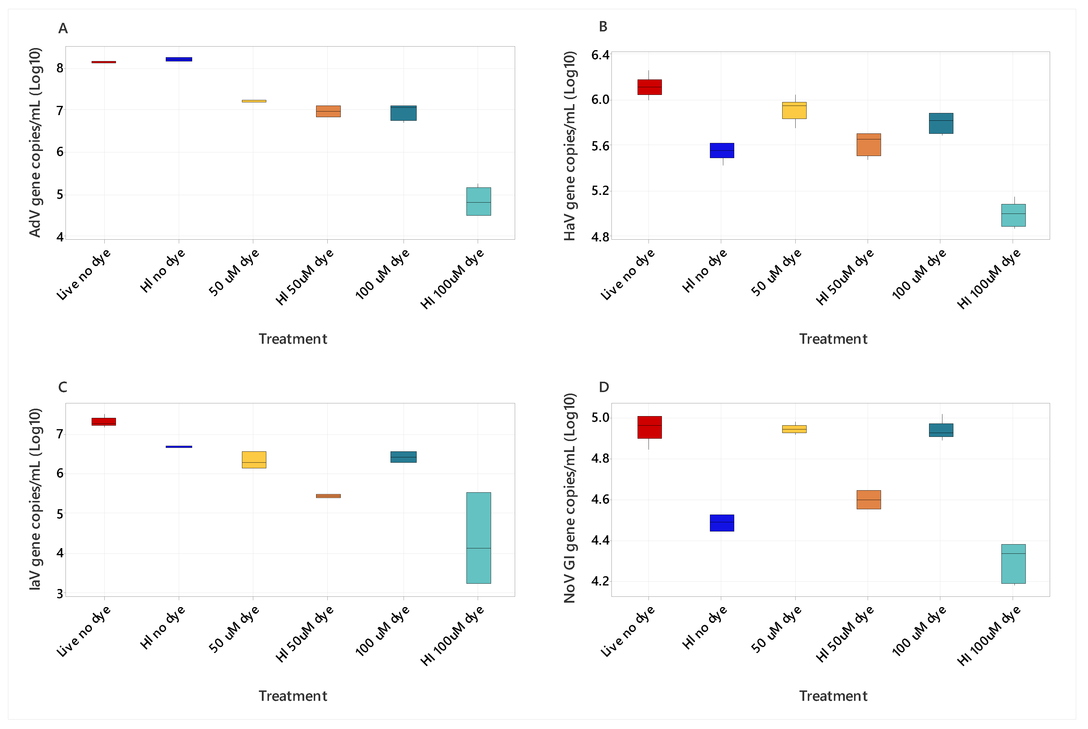
3.1. PMAXX Dye Concentration on Live and Heat-Inactivated Viruses (AdV, HAV, IAV, NoV GI)

Comparisons of live and heat-inactivated viruses (HI) (AdV, HAV, IAV, and NoV GI) pretreated with PMAxx dyes at concentrations of 50 μM and 100 μM and untreated samples (no PMAxx dye) (Figure 1) were made using a Kruskal–Wallis test. The analysis revealed significant differences between all viral targets, across treatment groups (p < 0.001). The post hoc Dunn’s test with Bonferroni-corrected alpha of p < 0.003 identified significant differences between mean ranks of paired comparisons (Table 1). Pretreatment with the 100 μM PMAxx dye significantly reduced the AdV copy number compared to the untreated samples (no dye) (p 0.0024, Table 1). The HI AdV untreated had 4 × 10⁷ gc/ml versus 2 × 10⁶ gc/ml for live AdV, pretreated with 100 μM PMAxx dye (Figure 1). This result indicated the AdV sample contained degraded/semi-assembled virus. The HI 100 μM dye significantly reduced the viral copy number >100-fold compared to HI no dye for AdV and IAV (Table 1, Figure 1). A significant difference between the 100 μM pretreated live and 100 μM pretreated HI samples for NoV GI (p <0.0003, Table 1) also occurred, as the gc/ml were higher for live NoV GI (2 × 10⁴ gc/ml) than for the HI samples (5 × 10³ gc/ml) (Figure 1). The gc/ml was significantly lower for all viruses when comparing HI 100 μM to the live no dye treatment (Figure 1, Table 1), indicating the dyes at this concentration reduce the gene copies detected by RT-qPCR and are suitable for ci-RT-qPCR assays for the target viruses.

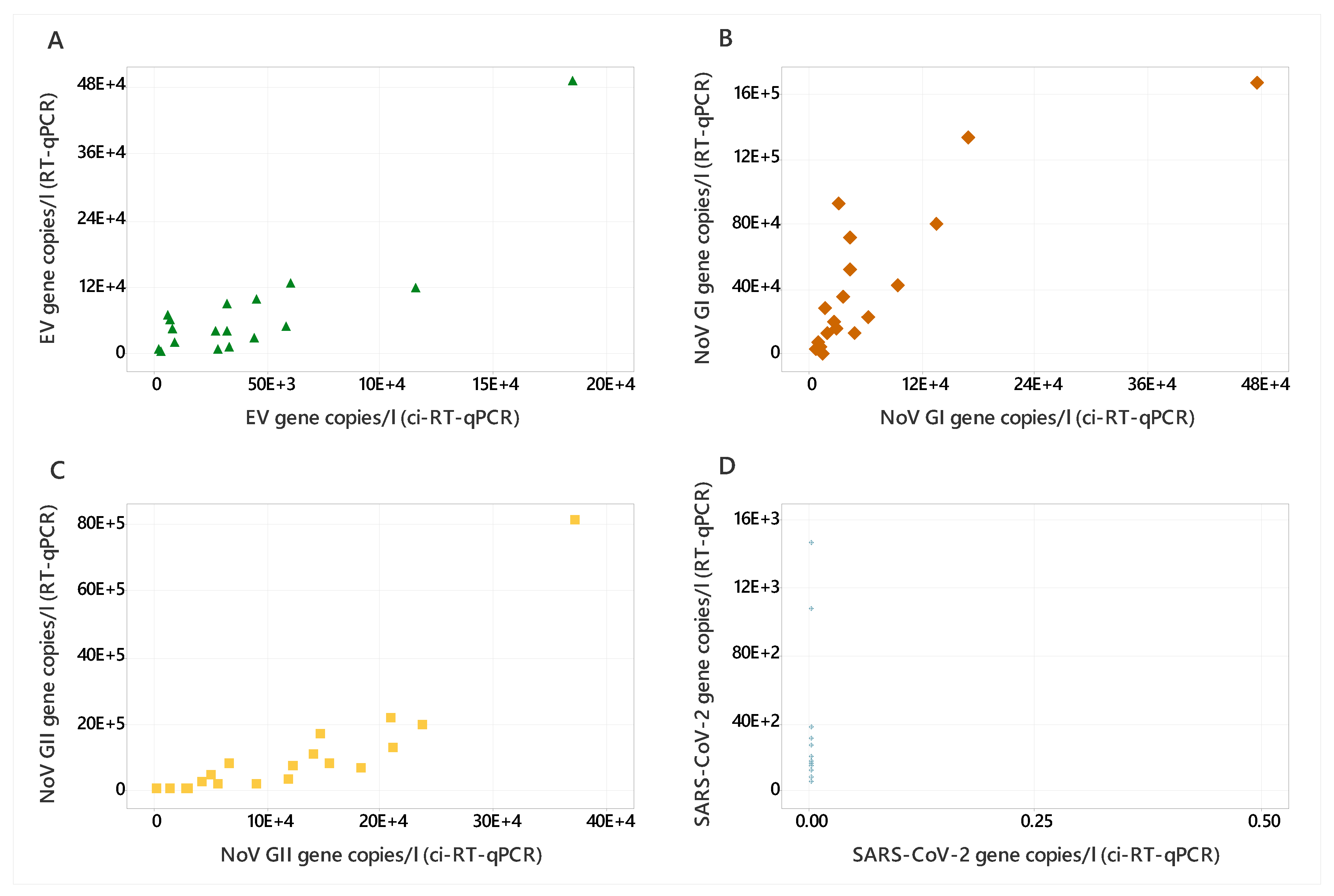
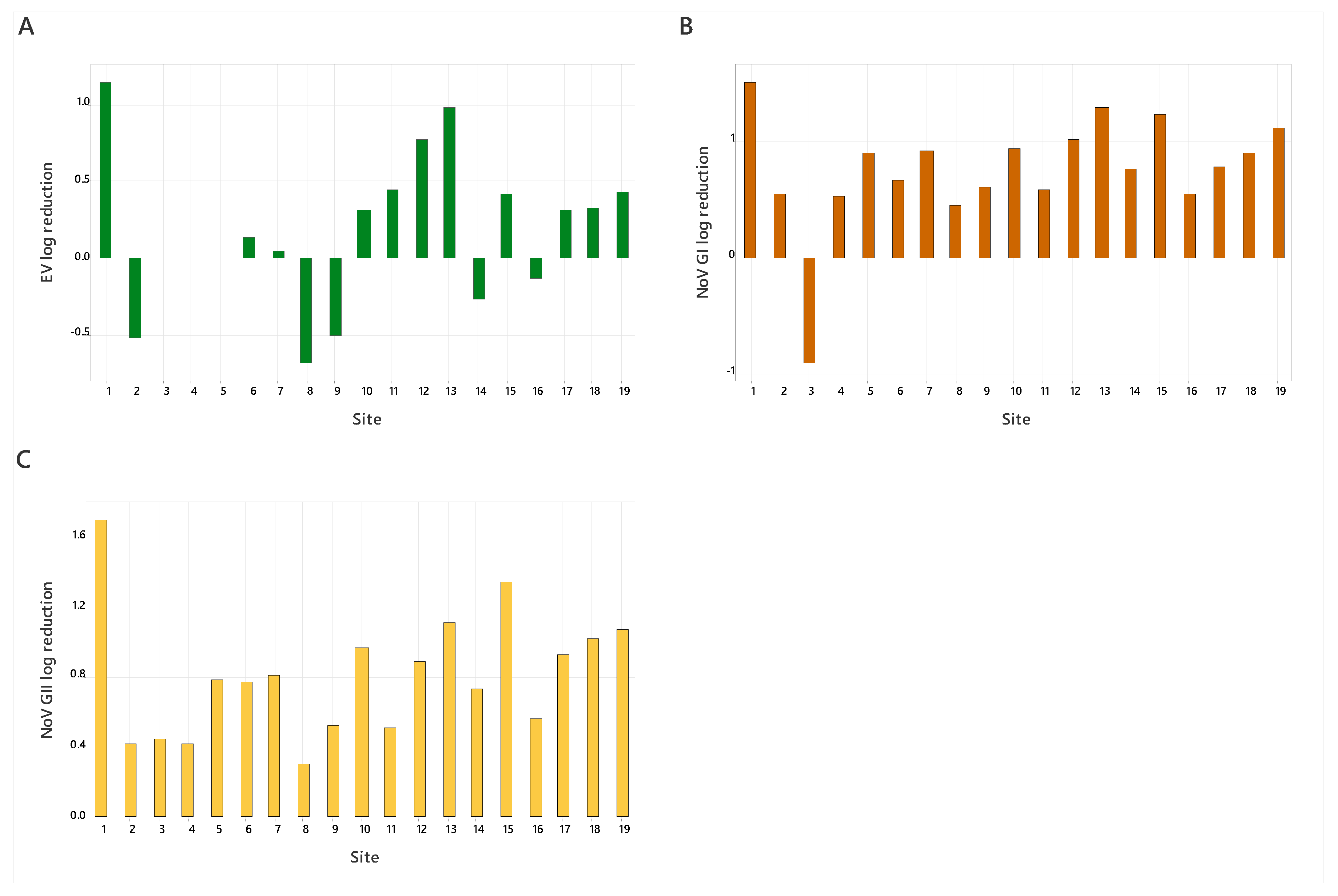
3.2. Wastewater Samples

Wastewater samples were negative for AdV, HAV, IAV, and IBV; so, a comparison of RT-qPCR and ci-RT-qPCR was conducted for EV, NoV GI, NoV GII, and SARS-CoV-2, as these viruses were detected in the samples. SARS-CoV-2 was detected in all wastewater samples via RT-qPCR only, while NoV GI, NoV GII, and EV were detected by both RT-qPCR and ci-RT-qPCR (Figure 2). EV was detected in 17 out of 19 samples, ranging from 7.82 × 10² to 1.84 × 10⁵ and 6.14 × 10² to 4.87 × 10⁵ gc/l for ci-RT-qPCR and RT-qPCR, respectively (Figure 2). Additionally, EV showed the lowest log reduction across all target viruses, at -0.52–1.15 (Figure 3), indicating that the EV capsid remains intact in wastewater. NoV GI and NoV GII were detected in all wastewater samples. NoV GI ranged from 5.89 × 10³ to 4.72 × 10⁵ and 1.43 × 10³ to 1.69 × 10⁶ gc/l for ci-RT-qPCR and RT-qPCR, respectively, and the log reductions were between 0.9 and 1.51 (Figure 3). NoV GII was detected at higher copy numbers than NoV GI, ranging from 9.20 × 10² to 3.70 × 10⁵ and 3.72 × 10⁴ to 8.12 × 10⁶ gc/l, respectively (Figure 2). The log reductions for NoV GII were between 0.31 and 1.69 (Figure 3).

4. Discussion

Molecular methods used to assess the capsid integrity of viruses present in wastewater are needed to provide more in-depth risk analysis of human exposure to wastewater [38], especially where sewage is discharged into the environment, as it may contain intact potentially infectious viruses [56]. It may also be relevant for undertaking quantitative microbial risk assessments (QMRA) to estimate the potential viral exposure of operators at wastewater treatment plants or those working within the sewage network [57]. As evidenced, EV, NoV GI, and NoV GII capsids remain intact, and as a result are potentially infectious.
In this study, we further advance the use of intercalating dyes for viral capsid integrity-RT-qPCR assays used in WBS applications. PMAxx dye at 50 μM and 100 μM concentrations was selected based on a review by Leifels et al. 2021 [38], where over half of the research papers reported dye concentrations of 50 μM and 100 μM to be sufficient for detecting damaged viral capsids. Due to the increased cost associated with using 200 μM of PMAxx dye, and the fact that 100 μM was deemed sufficient, we decided to assess lower concentrations of the dye. The heat-inactivated viruses exposed to PMAxx dye had a lower amplification resulting in lower gc/ml values than that of the live and no-dye-treated heat-inactivated viruses, a result consistent with other studies that assessed the effectiveness of PMAxx on heat treated Norovirus GI and GII and HAV [42,58]. In this study, we used heat inactivation to damage the viral capsid and compared the results to corresponding samples that did not undergo heat inactivation. Exposing viruses to high temperatures (>50 °C) causes them to become non-infectious and is a well-established method, as evidenced in cell culture assays [59]. However, using lower temperatures (<50 °C), while potentially inactivating viruses, may not sufficiently destabilise the viral capsid enough to expose the viral genome. In such cases ci-qPCR can overestimate the remaining infectious potential by detecting encapsulated viral genomes of viruses that can no longer replicate. Overall, the 100 μM dye concentration effectively differentiated live viruses from heat-inactivated viruses and reduced the detection of inactivated viruses for the targets tested. The PMAxx dyes also allowed us to identify viruses that were degraded or semi-assembled, as was the case for the AdV culture stock used in this study. These findings demonstrate that the PMAxx dye at 100 μM reduces the detection of heat-inactivated and degraded viruses compared to live viruses, allowing differentiation of capsid integrity by qPCR. Other studies have demonstrated that 50 µM PMAxx is sufficient for detecting human AdV in aquatic matrices [60] and similarly for NoV on vegetables [58]. However, the concentration of PMAxx dye treatment required varies depending on the sample type and what compounds maybe present in the sample [30]. Therefore, initial optimisation is required before the use of PMAxx in capsid integrity assays, across the sample type and target viruses.
Once the concentration of PMAxx dye, effective on several viruses, was determined, we tested our assay on wastewater samples for more targets (AdV, EV, HAV, IAV, IBV, NoV GI, NoV GII, and SARS-CoV-2). Wastewater samples were negative for AdV, HAV, IAV, and IBV. Therefore, we compared the RT-qPCR and ci-RT-qPCR data for EV, NoV GI, NoV GII, and SARS-CoV-2, which were detected in the wastewater samples. SARS-CoV-2 was detected in all samples by RT-qPCR only, showing that SARS-CoV-2 did not remain viable in the wastewater samples tested; as a result, the log reductions were not calculated, as no intact SARS-CoV-2 viruses were detected. The integrity of SARS-CoV-2 in wastewater samples has previously been investigated using PMAxx dyes and cell culture [31]. SARS-CoV-2 that had been spiked into wastewater had strongly (>3 log) or moderately (>1 log) reduced infectivity, and encapsulated SARS-CoV-2 RNA was always detected at a lower titre than total RNA in wastewater [31]. The reduced integrity of SARS-CoV-2 in wastewater is likely due to the enveloped capsid, which is less stable than that of non-enveloped viruses (e.g., NoVs, EV) [61], a result which mirrors the known modes of SARS-CoV-2 transmission. EV, NoV GI, and NoV GII were detected by RT-qPCR and ci-RT-qPCR. EV showed the lowest log reductions between RT-qPCR and ci-RT-qPCR, suggesting the viral particles remain intact and potentially infectious in wastewater. The EV RT-qPCR assay used in this study is a non-specific assay that detects a highly conserved region of the 62 nonpolio enteroviruses and three poliovirus types characterised at the time the assay was developed [62]. Therefore, the exact enterovirus strains detected are unknown. Norovirus GI and GII were detected in all samples, with NoV GII at higher gene copies. The log reductions for NoV GI and GII were < 1 log for most samples, indicating the presence of viable virus particles in the wastewater samples. PMAxx dyes have previously been used to detect viable NoV GI and GII in wastewater samples [30], while PMA dyes have been used to detect NoV GI and GII in contaminated drinking water [63], freshwater [29], and sewage samples [64]; again, these viruses remain intact and pose a human health risk.
Reductions in viral gene copies for inactivated viruses were observed in our assays, and our results show that PMAxx dyes are suitable for use in WBS; however, there are limitations to the approach, and where possible, steps should be taken to avoid these. Intercalating dyes such as PMAxx rely on a photolysis step; therefore, the sample matrix needs to be considered, as shown when detecting viruses in shellfish [30], as when a sample has a high volume of suspended solids, light penetration maybe insufficient to induce the reaction. The floccing of viruses in the sample may also need to be considered, as damaged viruses in the middle of viral aggregations maybe protected from the dye, and samples may need to be agitated to limit this. In addition, the dyes cannot differentiate between non-viable viruses that have an intact capsid and live infectious viruses, as seen with UV treatment [36,39]. Furthermore, the target virus, concentration of dye, length of dye incubation period, and length of light exposure all need to be considered [38]. Therefore, while ci-RT-qPCR gives a better estimation of the number of viable virus particles than RT-qPCR alone, an overestimation of viable viruses within a sample may still occur.

5. Conclusions

This study demonstrates the utility of PMAxx dye for quantifying viable viruses, even when cell culture-based assays are unavailable. With careful optimisation, ci-RT-qPCR can build upon knowledge from public health surveillance to provide estimates of viral infectivity. Assessing infectious viruses will enable better evaluation of potential public health risks associated with wastewater discharge and environmental transmission routes. Overall, capsid integrity-PCR shows promise for enhancing the specificity of wastewater-based epidemiology by revealing insights into community-level transmission dynamics of infectious pathogens.

Supplementary Materials

The following supporting information can be downloaded at: https://www.mdpi.com/article/10.3390/v16010040/s1, Table S1: Primers, probes, and standard sequences used in this study.

Author Contributions

Conceptualisation; J.L.K. and K.F. Laboratory work; J.L.K., N.R. and N.W. Data analysis and curation; J.L.K. Authorship; J.L.K. Final suggested edits; K.F., S.K.M. and D.L.J. Funding acquisition; D.L.J., K.F. and S.K.M. All authors have read and agreed to the published version of the manuscript.

Funding

This research has been co-funded through the BlueAdapt Project (www.blueadapt.eu, accessed on 26/12/2023). BlueAdapt has received funding from the European Union’s Horizon Europe research and innovation programme under grant agreement No 101057764 and by the UKRI/HM Government.

Institutional Review Board Statement

Not applicable.

Informed Consent Statement

Not applicable.

Data Availability Statement

Data are contained within the article or Supplementary Material.

Acknowledgments

Thank to Eleanor Gaunt, University of Edinburgh, who kindly provided influenza A vaccine strain virus (H1N1).

Conflicts of Interest

The authors declare no conflicts of interest.

References

  1. Barcellos, D.S.; Barquilha, C.E.R.; Oliveira, P.E.; Prokopiuk, M.; Etchepare, R.G. How Has the COVID-19 Pandemic Impacted Wastewater-Based Epidemiology? Sci. Total Environ. 2023, 892, 164561. [Google Scholar] [CrossRef] [PubMed]
  2. Huang, Y.; Zhou, N.; Zhang, S.; Yi, Y.; Han, Y.; Liu, M.; Han, Y.; Shi, N.; Yang, L.; Wang, Q.; et al. Norovirus Detection in Wastewater and Its Correlation with Human Gastroenteritis: A Systematic Review and Meta-Analysis. Environ. Sci. Pollut. Res. 2022, 29, 3. [Google Scholar] [CrossRef] [PubMed]
  3. Tedcastle, A.; Wilton, T.; Pegg, E.; Klapsa, D.; Bujaki, E.; Mate, R.; Fritzsche, M.; Majumdar, M.; Martin, J. Detection of Enterovirus D68 in Wastewater Samples from the UK between July and November 2021. Viruses 2022, 14, 143. [Google Scholar] [CrossRef] [PubMed]
  4. Kitakawa, K.; Kitamura, K.; Yoshida, H. Monitoring Enteroviruses and SARS-CoV-2 in Wastewater Using the Polio Environmental Surveillance System in Japan. Appl. Environ. Microbiol. 2023, 89, e01853-22. [Google Scholar] [CrossRef]
  5. Bisseux, M.; Debroas, D.; Mirand, A.; Archimbaud, C.; El Ene Peigue-Lafeuille, H.; Bailly, J.-L.; Henquell, E. Monitoring of Enterovirus Diversity in Wastewater by Ultra-Deep Sequencing: An Effective Complementary Tool for Clinical Enterovirus Surveillance. Water Res. 2019, 169, 115246. [Google Scholar] [CrossRef] [PubMed]
  6. Reyne, M.I.; Allen, D.M.; Levickas, A.; Allingham, P.; Lock, J.; Fitzgerald, A.; Mcsparron, C.; Nejad, B.F.; Mckinley, J.; Lee, A.; et al. Detection of Human Adenovirus F41 in Wastewater and Its Relationship to Clinical Cases of Acute Hepatitis of Unknown Aetiology. Sci. Total Environ. 2022, 857, 159579. [Google Scholar] [CrossRef] [PubMed]
  7. Takuissu, G.R.; Kenmoe, S.; Ebogo-Belobo, J.T.; Kengne-Ndé, C.; Mbaga, D.S.; Bowo-Ngandji, A.; Ondigui Ndzie, J.L.; Kenfack-Momo, R.; Tchatchouang, S.; Kenfack-Zanguim, J.; et al. Exploring Adenovirus in Water Environments: A Systematic Review and Meta-Analysis. Int. J. Environ. Health Res. 2023, 33. [Google Scholar] [CrossRef]
  8. Fong, T.T.; Phanikumar, M.S.; Xagoraraki, I.; Rose, J.B. Quantitative Detection of Human Adenoviruses in Wastewater and Combined Sewer Overflows Influencing a Michigan River. Appl. Environ. Microbiol. 2010, 76, 715–723. [Google Scholar] [CrossRef]
  9. Fantilli, A.; Cola, G.D.; Castro, G.; Sicilia, P.; Cachi, A.M.; de los Ángeles Marinzalda, M.; Ibarra, G.; López, L.; Valduvino, C.; Barbás, G.; et al. Hepatitis A Virus Monitoring in Wastewater: A Complementary Tool to Clinical Surveillance. Water Res. 2023, 241, 120102. [Google Scholar] [CrossRef]
  10. Rachida, S.; Taylor, M.B. Potentially Infectious Novel Hepatitis A Virus Strains Detected in Selected Treated Wastewater Discharge Sources, South Africa. Viruses 2020, 12, 1468. [Google Scholar] [CrossRef]
  11. Beyer, S.; Szewzyk, R.; Gnirss, R.; Johne, R.; Selinka, H.C. Detection and Characterization of Hepatitis E Virus Genotype 3 in Wastewater and Urban Surface Waters in Germany. Food Environ. Virol. 2020, 12, 137–147. [Google Scholar] [CrossRef] [PubMed]
  12. Masclaux, F.G.; Hotz, P.; Friedli, D.; Savova-Bianchi, D.; Oppliger, A. High Occurrence of Hepatitis E Virus in Samples from Wastewater Treatment Plants in Switzerland and Comparison with Other Enteric Viruses. Water Res. 2013, 47, 5101–5109. [Google Scholar] [CrossRef] [PubMed]
  13. Boehm, A.B.; Hughes, B.; Duong, D.; Chan-Herur, V.; Buchman, A.; Wolfe, M.K.; White, B.J. Wastewater Concentrations of Human Influenza, Metapneumovirus, Parainfluenza, Respiratory Syncytial Virus, Rhinovirus, and Seasonal Coronavirus Nucleic-Acids during the COVID-19 Pandemic: A Surveillance Study. Lancet Microbe 2023, 4, e340–e348. [Google Scholar] [CrossRef]
  14. Wolfe, M.K.; Duong, D.; Bakker, K.M.; Ammerman, M.; Mortenson, L.; Hughes, B.; Arts, P.; Lauring, A.S.; Fitzsimmons, W.J.; Bendall, E.; et al. Wastewater-Based Detection of Two Influenza Outbreaks. Environ. Sci. Technol. Lett. 2022, 9, 687–692. [Google Scholar] [CrossRef]
  15. Toribio-Avedillo, D.; Gómez-Gómez, C.; Sala-Comorera, L.; Rodríguez-Rubio, L.; Carcereny, A.; García-Pedemonte, D.; Pintó, R.M.; Guix, S.; Galofré, B.; Bosch, A.; et al. Monitoring Influenza and Respiratory Syncytial Virus in Wastewater. Beyond COVID-19. Sci. Total Environ. 2023, 892, 164495. [Google Scholar] [CrossRef]
  16. Dzinamarira, T.; Murewanhema, G.; Iradukunda, P.G.; Madziva, R.; Herrera, H.; Cuadros, D.F.; Tungwarara, N.; Chitungo, I.; Musuka, G. Utilization of SARS-CoV-2 Wastewater Surveillance in Africa—A Rapid Review. Int. J. Environ. Res. Public. Health 2022, 19, 969. [Google Scholar] [CrossRef]
  17. Kumar, M.; Kumar Patel, A.; Shah, A.V.; Raval, J.; Rajpara, N.; Joshi, M.; Joshi, C.G. First Proof of the Capability of Wastewater Surveillance for COVID-19 in India through Detection of Genetic Material of SARS-CoV-2. Sci. Total Environ. 2020, 746, 141326. [Google Scholar] [CrossRef]
  18. Yu, A.T.; Hughes, B.; Wolfe, M.K.; Leon, T.; Duong, D.; Rabe, A.; Kennedy, L.C.; Ravuri, S.; White, B.J.; Wigginton, K.R.; et al. Estimating Relative Abundance of 2 SARS-CoV-2 Variants through Wastewater Surveillance at 2 Large Metropolitan Sites, United States. Emerg. Infect. Dis. 2022, 28, 940. [Google Scholar] [CrossRef]
  19. Agrawal, S.; Orschler, L.; Schubert, S.; Zachmann, K.; Heijnen, L.; Tavazzi, S.; Gawlik, B.M.; de Graaf, M.; Medema, G.; Lackner, S. Prevalence and Circulation Patterns of SARS-CoV-2 Variants in European Sewage Mirror Clinical Data of 54 European Cities. Water Res. 2022, 214, 118162. [Google Scholar] [CrossRef]
  20. Brunner, F.S.; Brown, M.R.; Bassano, I.; Denise, H.; Khalifa, M.S.; Wade, M.J.; van Aerle, R.; Kevill, J.L.; Jones, D.L.; Farkas, K.; et al. City-Wide Wastewater Genomic Surveillance through the Successive Emergence of SARS-CoV-2 Alpha and Delta Variants. Water Res. 2022, 226, 119306. [Google Scholar] [CrossRef]
  21. Hillary, L.S.; Farkas, K.; Maher, K.H.; Lucaci, A.; Thorpe, J.; Distaso, M.A.; Gaze, W.H.; Paterson, S.; Burke, T.; Connor, T.R.; et al. Monitoring SARS-CoV-2 in Municipal Wastewater to Evaluate the Success of Lockdown Measures for Controlling COVID-19 in the UK. Water Res. 2021, 200, 117214. [Google Scholar] [CrossRef] [PubMed]
  22. Ahmed, W.; Angel, N.; Edson, J.; Bibby, K.; Bivins, A.; O’brien, J.W.; Choi, P.M.; Kitajima, M.; Simpson, S.L.; Li, J.; et al. First Confirmed Detection of SARS-CoV-2 in Untreated Wastewater in Australia: A Proof of Concept for the Wastewater Surveillance of COVID-19 in the Community. Sci. Total Environ. 2020, 728, 138764. [Google Scholar] [CrossRef] [PubMed]
  23. Hassard, F.; Sharp, J.H.; Taft, H.; Levay, L.; Harris, J.P.; Mcdonald, J.E.; Tuson, K.; Wilson, J.; Jones, L.D.; Malham, S.K. Critical Review on the Public Health Impact of Norovirus Contamination in Shellfish and the Environment: A UK Perspective. Food Environ. Virol. 2017, 9, 123–141. [Google Scholar] [CrossRef] [PubMed]
  24. Botturi, A.; Ozbayram, E.G.; Tondera, K.; Gilbert, N.I.; Rouault, P.; Caradot, N.; Gutierrez, O.; Daneshgar, S.; Frison, N.; Akyol, Ç.; et al. Combined Sewer Overflows: A Critical Review on Best Practice and Innovative Solutions to Mitigate Impacts on Environment and Human Health. Crit. Rev. Environ. Sci. Technol. 2021, 51, 1585–1618. [Google Scholar] [CrossRef]
  25. Lee, J.H.; Lee, G.-C.; Kim, J.I.; Yi, H.A.; Lee, C.H. Development of a New Cell Culture-Based Method and Optimized Protocol for the Detection of Enteric Viruses. J. Virol. Methods 2013, 191, 16–23. [Google Scholar] [CrossRef] [PubMed]
  26. Iaconelli, M.; Muscillo, M.; Libera, S.D.; Fratini, M.; Meucci, L.; De Ceglia, M.; Giacosa, D.; Rosa, G. La One-Year Surveillance of Human Enteric Viruses in Raw and Treated Wastewaters, Downstream River Waters, and Drinking Waters. Food Environ. Virol. 2017, 9, 79–88. [Google Scholar] [CrossRef] [PubMed]
  27. Schmidt, N.J.; Ho, H.H.; Riggs, J.L.; Lennette, E.H. Comparative Sensitivity of Various Cell Culture Systems for Isolation of Viruses from Wastewater and Fecal Samples. Appl. Environ. Microbiol. 1978, 36, 480–486. [Google Scholar] [CrossRef]
  28. Fittipaldi, M.; Nocker, A.; Codony, F. Progress in Understanding Preferential Detection of Live Cells Using Viability Dyes in Combination with DNA Amplification. J. Microbiol. Methods 2012, 91, 276–289. [Google Scholar] [CrossRef]
  29. Prevost, B.; Goulet, M.; Lucas, F.S.; Joyeux, M.; Moulin, L.; Wurtzer, S. Viral Persistence in Surface and Drinking Water: Suitability of PCR Pre-Treatment with Intercalating Dyes. Water Res. 2016, 91, 68–76. [Google Scholar] [CrossRef]
  30. Randazzo, W.; Khezri, M.; Ollivier, J.; Le Guyader, F.S.; Rodríguez-Díaz, J.; Aznar, R.; Sánchez, G. Optimization of PMAxx Pretreatment to Distinguish between Human Norovirus with Intact and Altered Capsids in Shellfish and Sewage Samples. Int. J. Food Microbiol. 2018, 266, 1–7. [Google Scholar] [CrossRef]
  31. Wurtzer, S.; Waldman, P.; Ferrier-Rembert, A.; Frenois-Veyrat, G.; Mouchel, J.M.; Boni, M.; Maday, Y.; Consortium, O.; Marechal, V.; Moulin, L. Several Forms of SARS-CoV-2 RNA Can Be Detected in Wastewaters: Implication for Wastewater-Based Epidemiology and Risk Assessment. Water Res. 2021, 198, 117183. [Google Scholar] [CrossRef] [PubMed]
  32. Stobnicka-Kupiec, A.; Gołofit-Szymczak, M.; Cyprowski, M.; Górny, R.L. Detection and Identification of Potentially Infectious Gastrointestinal and Respiratory Viruses at Workplaces of Wastewater Treatment Plants with Viability QPCR/RT-QPCR. Sci. Rep. 2022, 12, 4517. [Google Scholar] [CrossRef] [PubMed]
  33. Palowski, A.; Balestreri, C.; Urriola, P.E.; van de Ligt, J.L.G.; Sampedro, F.; Dee, S.; Shah, A.; Yancy, H.F.; Shurson, G.C.; Schroeder, D.C. Survival of a Surrogate African Swine Fever Virus-like Algal Virus in Feed Matrices Using a 23-Day Commercial United States Truck Transport Model. Front. Microbiol. 2022, 13, 1059118. [Google Scholar] [CrossRef] [PubMed]
  34. Anfruns-Estrada, E.; Bottaro, M.; Pintó, R.M.; Guix, S.; Bosch, A. Effectiveness of Consumers Washing with Sanitizers to Reduce Human Norovirus on Mixed Salad. Foods 2019, 8, 637. [Google Scholar] [CrossRef] [PubMed]
  35. Shirasaki, N.; Matsushita, T.; Matsui, Y.; Koriki, S. Suitability of Pepper Mild Mottle Virus as a Human Enteric Virus Surrogate for Assessing the Efficacy of Thermal or Free-Chlorine Disinfection Processes by Using Infectivity Assays and Enhanced Viability PCR. Water Res. 2020, 186, 116409. [Google Scholar] [CrossRef] [PubMed]
  36. Leifels, M.; Jurzik, L.; Wilhelm, M.; Hamza, I.A. Use of Ethidium Monoazide and Propidium Monoazide to Determine Viral Infectivity upon Inactivation by Heat, UV- Exposure and Chlorine. Int. J. Hyg. Environ. Health 2015, 218, 686–693. [Google Scholar] [CrossRef]
  37. Canh, V.D.; Liu, M.; Sangsanont, J.; Katayama, H. Capsid Integrity Detection of Pathogenic Viruses in Waters: Recent Progress and Potential Future Applications. Sci. Total Environ. 2022, 827, 154258. [Google Scholar] [CrossRef]
  38. Leifels, M.; Cheng, D.; Sozzi, E.; Shoults, D.C.; Wuertz, S.; Mongkolsuk, S.; Sirikanchana, K. Capsid Integrity Quantitative PCR to Determine Virus Infectivity in Environmental and Food Applications—A Systematic Review. Water Res. X 2021, 11, 100080. [Google Scholar] [CrossRef]
  39. Leifels, M.; Hamza, I.A.; Krieger, M.; Wilhelm, M.; Mackowiak, M.; Jurzik, L. From Lab to Lake—Evaluation of Current Molecular Methods for the Detection of Infectious Enteric Viruses in Complex Water Matrices in an Urban Area. PLoS ONE 2016, 11, e0167105. [Google Scholar] [CrossRef]
  40. Parshionikar, S.; Laseke, I.; Fout, G.S. Use of propidium monoazide in reverse transcriptase PCR to distinguish between infectious and noninfectious enteric viruses in water samples. Appl. Environ. Microbiol. 2010, 76, 4318–4326. [Google Scholar] [CrossRef]
  41. Gyawali, P.; Hewitt, J. Detection of Infectious Noroviruses from Wastewater and Seawater Using PEMAXTM Treatment Combined with RT-QPCR. Water 2018, 10, 841. [Google Scholar] [CrossRef]
  42. Randazzo, W.; Piqueras, J.; Rodriguez, D.; Iaz, J.; Aznar, R.; Anchez, G.S. Improving Efficiency of Viability-QPCR for Selective Detection of Infectious HAV in Food and Water Samples. J. Appl. Microbiol. 2017, 124, 958–964. [Google Scholar] [CrossRef] [PubMed]
  43. Yee, R.A.; Leifels, M.; Scott, C.; Ashbolt, N.J.; Liu, Y. Evaluating Microbial and Chemical Hazards in Commercial Struvite Recovered from Wastewater. Environ. Sci. Technol. 2019, 53, 5378–5386. [Google Scholar] [CrossRef] [PubMed]
  44. Ogorzaly, L.; Cauchie, H.M.; Penny, C.; Perrin, A.; Gantzer, C.; Bertrand, I. Two-Day Detection of Infectious Enteric and Non-Enteric Adenoviruses by Improved ICC-QPCR. Appl. Microbiol. Biotechnol. 2013, 97, 4159–4166. [Google Scholar] [CrossRef] [PubMed]
  45. Ahmed, W.; Bertsch, P.M.; Bibby, K.; Haramoto, E.; Hewitt, J.; Huygens, F.; Gyawali, P.; Korajkic, A.; Riddell, S.; Sherchan, S.P.; et al. Decay of SARS-CoV-2 and Surrogate Murine Hepatitis Virus RNA in Untreated Wastewater to Inform Application in Wastewater-Based Epidemiology. Environ. Res. 2020, 191, 110092. [Google Scholar] [CrossRef]
  46. Beattie, R.E.; Denene Blackwood, A.; Clerkin, T.; Dinga, C.; Noble, R.T. Evaluating the Impact of Sample Storage, Handling, and Technical Ability on the Decay and Recovery of SARS-CoV-2 in Wastewater. PLoS ONE 2022, 17, e0270659. [Google Scholar] [CrossRef]
  47. Farkas, K.; Hillary, L.S.; Thorpe, J.; Walker, D.I.; Lowther, J.A.; McDonald, J.E.; Malham, S.K.; Jones, D.L. Concentration and Quantification of SARS-CoV-2 RNA in Wastewater Using Polyethylene Glycol-Based Concentration and QRT-PCR. Methods Protoc. 2021, 4, 17. [Google Scholar] [CrossRef]
  48. Lewis, G.D.; Metcalf, T.G. Polyethylene Glycol Precipitation for Recovery of Pathogenic Viruses, Including Hepatitis A Virus and Human Rotavirus, from Oyster, Water, and Sediment Samples. Appl. Environ. Microbiol. 1988, 54, 1983–1988. [Google Scholar] [CrossRef]
  49. Kevill, J.L.; Pellett, C.; Farkas, K.; Brown, M.R.; Bassano, I.; Denise, H.; Mcdonald, J.E.; Malham, S.K.; Porter, J.; Warren, J.; et al. A Comparison of Precipitation and Filtration-Based SARS-CoV-2 Recovery Methods and the Influence of Temperature, Turbidity, and Surfactant Load in Urban Wastewater. Sci. Total Environ. 2021, 808, 151916. [Google Scholar] [CrossRef]
  50. WHO. Laboratory Biosafety Manual, 3rd ed.; World Health Organization: Geneva, Switzerland, 2004. [Google Scholar] [CrossRef]
  51. Bustin, S.A.; Benes, V.; Garson, J.A.; Hellemans, J.; Huggett, J.; Kubista, M.; Mueller, R.; Nolan, T.; Pfaffl, M.W.; Shipley, G.L.; et al. The MIQE Guidelines: Minimum Information for Publication of Quantitative Real-Time PCR Experiments. Clin. Chem. 2009, 55, 611–622. [Google Scholar] [CrossRef]
  52. Farkas, K.; Pellett, C.; Alex-Sanders, N.; Bridgman, M.T.P.; Corbishley, A.; Grimsley, J.M.S.; Kasprzyk-Hordern, B.; Kevill, J.L.; Pântea, I.; Richardson-O’Neill, I.S.; et al. Comparative Assessment of Filtration- and Precipitation-Based Methods for the Concentration of SARS-CoV-2 and Other Viruses from Wastewater. Microbiol. Spectr. 2022, 10, e01102-22. [Google Scholar] [CrossRef] [PubMed]
  53. Dunn, O.J. Multiple Comparisons Using Rank Sums. Technometrics 1964, 6, 241–252. [Google Scholar] [CrossRef]
  54. Kolassa, J.E.; Jankowski, S. R: Multivariate Nonparametric Methods. Available online: https://search.r-project.org/CRAN/refmans/MultNonParam/html/00Index.html (accessed on 1 November 2023).
  55. Dinno, A. Dunn’s Test of Multiple Comparisons Using Rank Sums. Available online: https://cran.r-project.org/web/packages/dunn.test/dunn.test.pdf (accessed on 1 November 2023).
  56. Cuevas-Ferrando, E.; Pérez-Cataluña, A.; Falcó, I.; Randazzo, W.; Sánchez, G. Monitoring Human Viral Pathogens Reveals Potential Hazard for Treated Wastewater Discharge or Reuse. Front. Microbiol. 2022, 13, 836193. [Google Scholar] [CrossRef] [PubMed]
  57. Morales, E.; Badilla, A.; Rivera, P.; Alfaro, E.; Symonds, E.M.; Barrantes, K.; Ruepert, C.; Crowe, J.; Chacón, L. A Novel Approach to Biological Risk Surveillance in Occupational Hygiene: A Case Study on the Use of Quantitative Microbial Risk Assessment to Determine Occupational Risks for Wastewater Treatment Plants Workers in Costa Rica. Microb. Risk Anal. 2023, 24, 100262. [Google Scholar] [CrossRef]
  58. Randazzo, W.; López-Gálvez, F.; Allende, A.; Aznar, R.; Sánchez, G. Evaluation of Viability PCR Performance for Assessing Norovirus Infectivity in Fresh-Cut Vegetables and Irrigation Water. Int. J. Food Microbiol. 2016, 229, 1–6. [Google Scholar] [CrossRef] [PubMed]
  59. Bozkurt, H.; D’souza, D.H.; Davidson, P.M. Thermal inactivation of foodborne enteric viruses and their viral surrogates in foods. J. Food Prot. 2015, 78, 1597–1617. [Google Scholar] [CrossRef] [PubMed]
  60. Da Graça Pedrosa De Macena, L.; Simone De Oliveira Pereira, J.; Couto Da Silva, J.; Ferreira, F.C.; Gonçalves Maranhão, A.; Lanzarini, N.M.; Pereira Miagostovich, M. Environmental Microbiology-Quantification of Infectious Human Mastadenovirus in Environmental Matrices Using PMAxx-QPCR. Braz. J. Microbiol. 2022, 53, 1456–1471. [Google Scholar] [CrossRef]
  61. Roos, Y.H. Water and Pathogenic Viruses Inactivation-Food Engineering Perspectives. Food Eng. Rev. 2020, 12, 251–267. [Google Scholar] [CrossRef]
  62. Gregory, J.B.; Litaker, R.W.; Noble, R.T. Rapid One-Step Quantitative Reverse Transcriptase PCR Assay with Competitive Internal Positive Control for Detection of Enteroviruses in Environmental Samples. Appl. Environ. Microbiol. 2006, 72, 3960–3967. [Google Scholar] [CrossRef]
  63. Blanco, A.; Guix, S.; Fuster, N.; Fuentes, C.; Bartolomé, R.; Cornejo, T.; Pintó, R.M.; Bosch, A. Norovirus in Bottled Water Associated with Gastroenteritis Outbreak, Spain, 2016. Emerg. Infect. Dis. 2016, 23, 1531–1534. [Google Scholar] [CrossRef]
  64. Fuster, N.; Pint, R.M.; Fuentes, C.; Beguiristain, N.; Bosch, A.; Guix, S. Propidium Monoazide RTqPCR Assays for the Assessment of Hepatitis A Inactivation and for a Better Estimation of the Health Risk of Contaminated Waters. Water Res. 2016, 101, 226–232. [Google Scholar] [CrossRef] [PubMed]
Figure 1. Effect of PMAxx addition (0, 50, or 100 µM) on the RT-qPCR-based quantification of live and heat-inactivated (HI) viruses (AdV (A), HAV (B), IAV (C), and NoV GI (D)).
Viruses 16 00040 g001
Figure 2. Scatter diagrams comparing RT-qPCR and ci-RT-qPCR gene copies/l for EV (A), NoV GI (B), NoV GII (C), and SARS-CoV-2 (D) detected in wastewater samples (n 19).
Viruses 16 00040 g002
Figure 3. Log reductions of viruses (EV (A), NoV GI (B), and NoV GII (C)) detected in wastewater for samples 1–19.
Viruses 16 00040 g003
Table 1. The p values from Dunn’s multi-comparison post hoc test, showing the significance between ranked pairs for each target virus. Comparisons were made between the gene copies/ml obtained from live and heat-inactivated (HI) viruses (AdV, HAV, IAV, or NoV GI) that were exposed to 50 µM or 100 µM PMAxx dye or no dye. Significant (Bonferroni corrected) p values are highlighted in bold.
ComparisonAdVHAVIAVNov GI
Live 100 µM—Live 50 µM0.27000.43000.68000.8813
Live 100 µM—HI 100 µM0.25000.00370.02600.0003
Live 100 µM—HI 50 µM0.88000.32000.08200.1419
Live 100 µM—HI no dye0.00240.08700.17000.0112
Live 100 µM—Live no dye0.02500.05200.01600.5462
Live 50 µM—HI 100 µM0.01400.00020.06800.0002
Live 50 µM—HI 50 µM0.20000.08900.17000.1091
Live 50 µM—HI no dye0.03100.01300.07700.0072
Live 50 µM—Live no dye0.20000.25000.00490.6447
HI 100 µM—HI 50 µM0.33000.11000.80000.0780
HI 100 µM—HI no dye<0.00010.23000.00030.2825
HI 100 µM—Live no dye0.00030.0000<0.00010.0001
HI 50 µM—HI no dye0.00130.60000.00310.4231
HI 50 µM—Live no dye0.01600.00620.00010.0502
HI no dye—Live no dye0.37000.00030.30000.0025
Disclaimer/Publisher’s Note: The statements, opinions and data contained in all publications are solely those of the individual author(s) and contributor(s) and not of MDPI and/or the editor(s). MDPI and/or the editor(s) disclaim responsibility for any injury to people or property resulting from any ideas, methods, instructions or products referred to in the content.

