
Effects of Clinical Use on the Mechanical Properties of Bio-Active® (BA) and TriTanium® (TR) Multiforce Nickel-Titanium Orthodontic Archwires

 , ,
, ,
Abstract
1. Introduction
2. Materials and Methods
2.1. Ethics Statement
2.2. Materials
2.2.1. Selection of the Investigated Archwires
2.2.2. Marking Code of the Samples
2.3. Methods
2.3.1. Disinfection Protocol
2.3.2. Segmentation of the Archwires
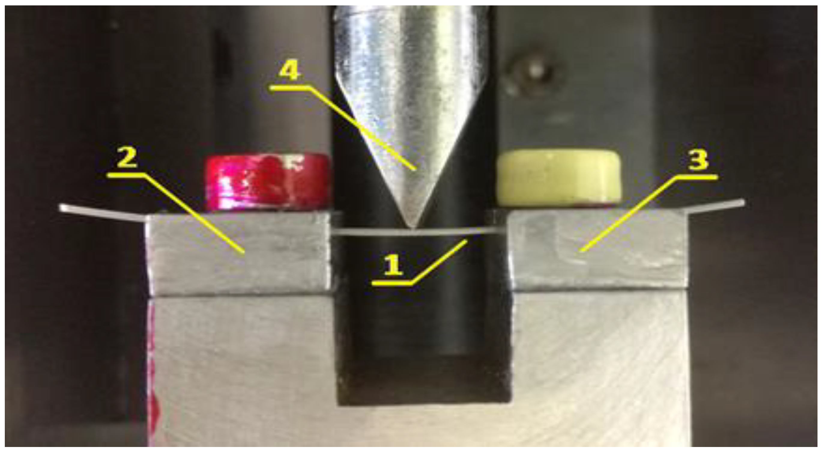
2.3.3. Three-Point Bending Test
2.3.4. Statistical Methods
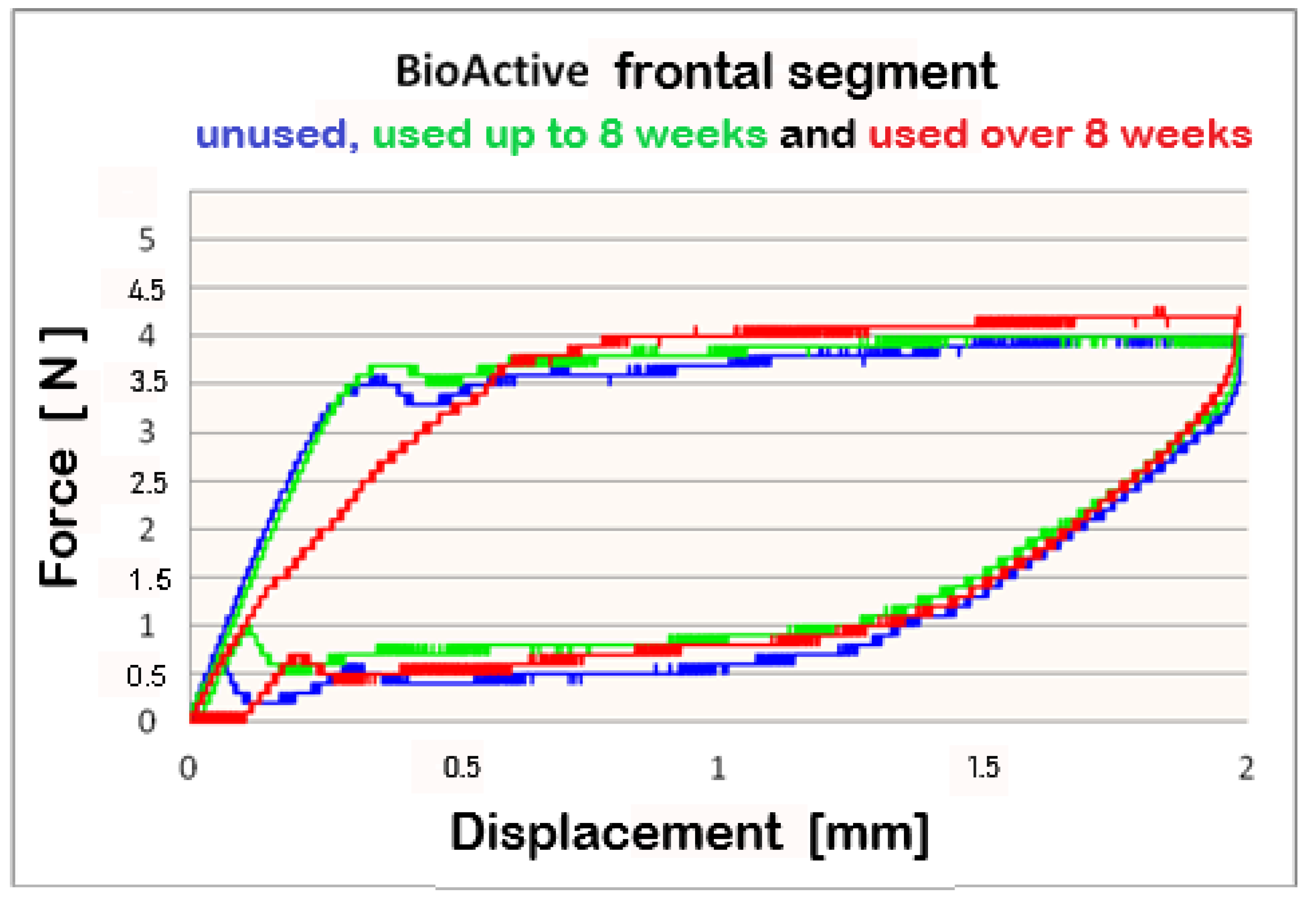
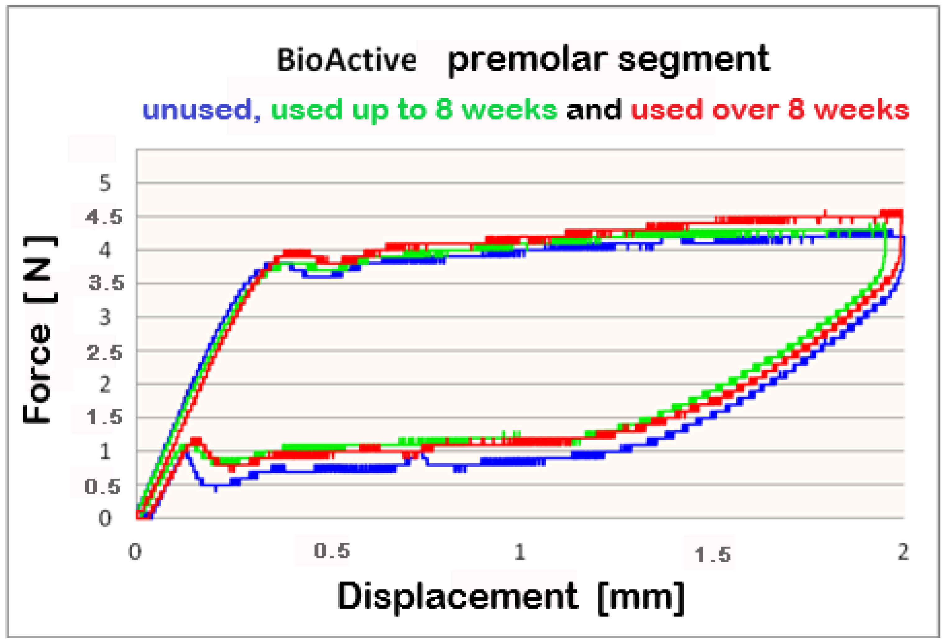
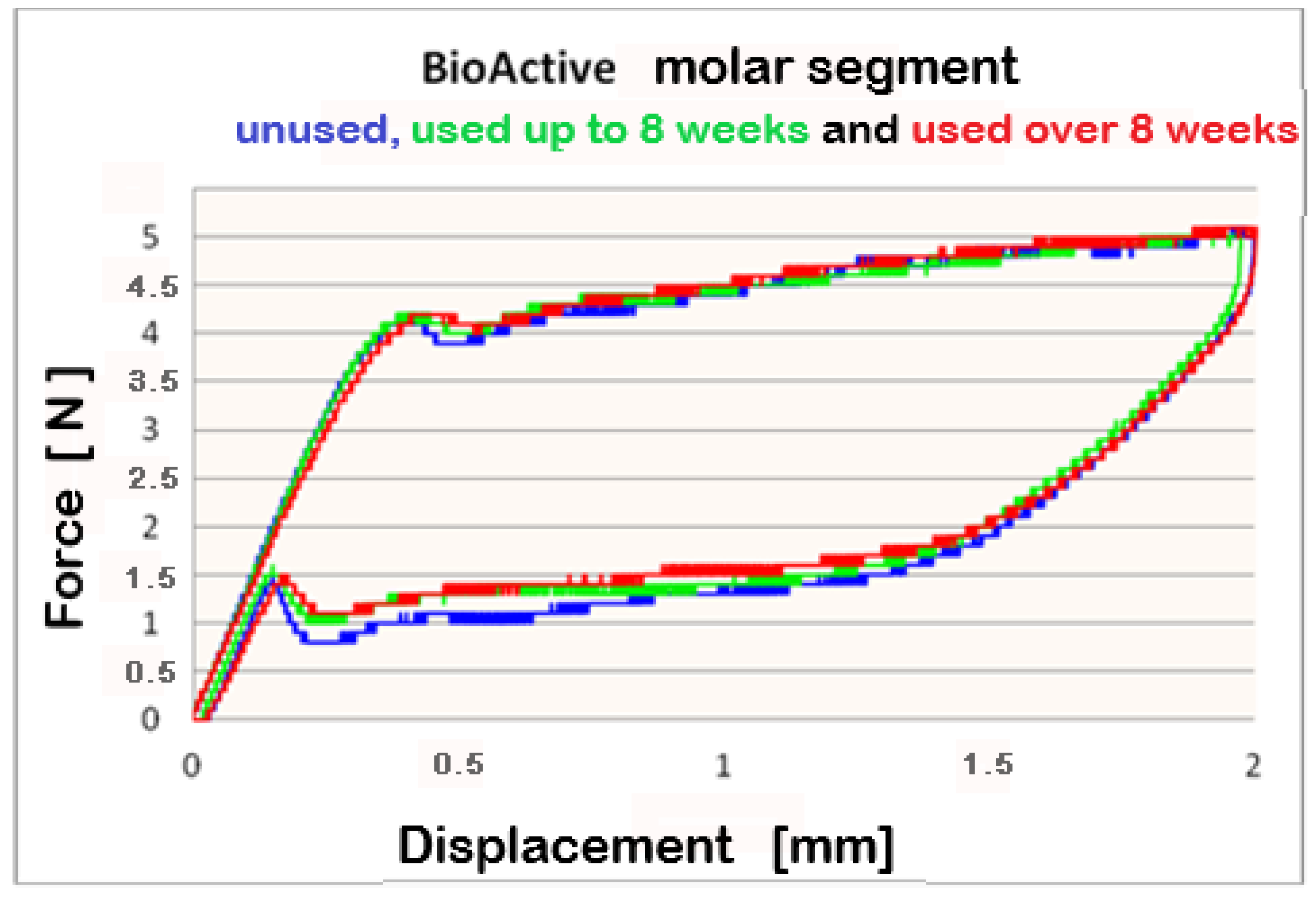
3. Results
4. Discussion
5. Conclusions
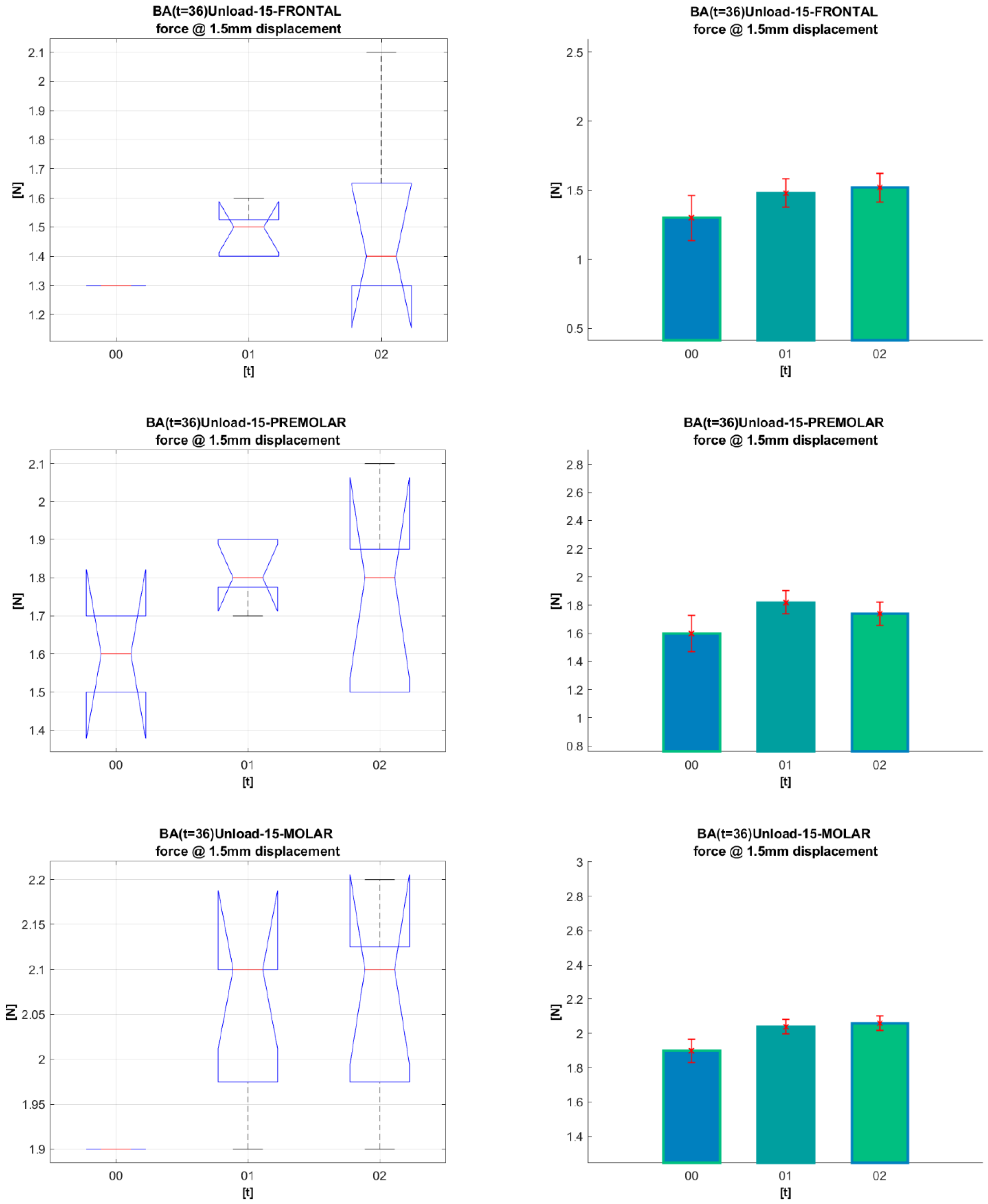
- The results show that the released forces and the mechanical properties of the archwires (Bio-Active® and TriTanium®) are preserved for a period of over 8 weeks.
- Comparing the results for groups I, II, and III, no statistically significant differences were found.
- This makes their use possible even when regular monthly meetings with patients are impossible.
- With their graduated biologically tolerable forces multi-force archwires are particularly suitable for patients with periodontal problems and little crowding.
Author Contributions
Funding
Institutional Review Board Statement
Informed Consent Statement
Data Availability Statement
Acknowledgments
Conflicts of Interest
References
- Taneja, P.; Duncanson, M.G.; Khajotia, S.S.; Nanda, R.S. Deactivation Force-Deflection Behavior of Multistranded Stainless Steel Wires. Am. J. Orthod. Dentofac. Orthop. 2003, 124, 61–68. [Google Scholar] [CrossRef] [PubMed]
- Ballard, D.J.; Jones, A.S.; Petocz, P.; Darendeliler, M.A. Physical Properties of Root Cementum: Part 11. Continuous vs Intermittent Controlled Orthodontic Forces on Root Resorption. A Microcomputed-Tomography Study. Am. J. Orthod. Dentofac. Orthop. 2009, 136, e1–e8. [Google Scholar] [CrossRef] [PubMed]
- Burstone, C.J. Variable-Modulus Orthodontics. Am. J. Orthod. 1981, 80, 1–16. [Google Scholar] [CrossRef]
- Burstone, C.J.; Qin, B.; Morton, J.Y. Chinese NiTi Wire-d New Orthodontic Alloy. Am. J. Orthod. 1985, 87, 445–452. [Google Scholar] [CrossRef] [PubMed]
- Linge, L.; Linge, B.O. Patient Characteristics and Treatment Variables Associated with Apical Root Resorption during Orthodontic Treatment. Am. J. Orthod. Dentofac. Orthop. 1991, 99, 35–43. [Google Scholar] [CrossRef] [PubMed]
- Wang, Y.; Liu, C.; Jian, F.; Mcintyre, G.T.; Millett, D.T.; Hickman, J.; Lai, W. Initial Arch Wires Used in Orthodontic Treatment with Fixed Appliances. Cochrane Database Syst. Rev. 2018, 2018. [Google Scholar] [CrossRef] [PubMed]
- Kauffman, G.; Mayo, I. The Story of Nitinol: The Serendipitous Discovery of the Memory Metal and Its Applications. Chem. Educ. 1997, 2, 1–21. [Google Scholar] [CrossRef]
- Buehler, W.J.; Gilfrich, J.V.; Wiley, R.C. Effect of Low-Temperature Phase Changes on the Mechanical Properties of Alloys near Composition TiNi. J. Appl. Phys. 1963, 34, s1475–s1477. [Google Scholar] [CrossRef]
- Wadood, A. Brief Overview on Nitinol as Biomaterial. Adv. Mater. Sci. Eng. 2016, 4173138. [Google Scholar] [CrossRef]
- Yoneyama, T.; Miyazaki, S. Shape Memory Alloys for Biomedical Applications, 1st ed.; Woodhead Publishing: Sawston, Cambridge, UK, 2008; pp. 261–278. [Google Scholar]
- Evans, T.J.; Durning, P. Aligning Archwires, the Shape of Things to Come?—A Fourth and Fifth Phase of Force Delivery. Br. J. Orthod. 1996, 23, 269–275. [Google Scholar] [CrossRef]
- Ibe, D.M.; Segner, D. Superelastic Materials Displaying Different Force Levels within One Archwire. J. Orofac. Orthop. 1998, 59, 29–38. [Google Scholar] [CrossRef] [PubMed]
- Sanders, E.; Johannessen, L.; Nadal, J.; Jäger, A.; Bourauel, C. Comparison of Multiforce Nickel–Titanium Wires to Multistrand Wires without Force Zones in Bending and Torque Measurements. J. Orofac. Orthop. 2022, 83, 382–394. [Google Scholar] [CrossRef] [PubMed]
- Lombardo, L.; Ceci, M.; Mollica, F.; Mazzanti, V.; Palone, M.; Siciliani, G. Mechanical Properties of Multi-Force vs. Conventional NiTi Archwires. J. Orofac. Orthop. 2019, 80, 57–67. [Google Scholar] [CrossRef]
- Sarul, M.; Kawala, B.; Kawala, M.; Antoszewska-Smith, J. Do the NiTi Low and Constant Force Levels Remain Stable in Vivo? Eur. J. Orthod. 2015, 37, 656–664. [Google Scholar] [CrossRef]
- Cherneva, S.; Stoyanova-Ivanova, A.; Gueorguieva, M.; Andreeva, L.; Petkov, A.; Petrov, V.; Petrova, V.; Mikli, V. Nanoindentation and Surface Characterization of Clinically Retrieved Multi-Force NiTi Orthodontic Archwires. Russ. J. Biomech. 2020, 24, 282–299. [Google Scholar]
- Georgieva, M.; Stoyanova-Ivanova, A.; Cherneva, S.; Petrov, V.; Petrova, V.; Andreeva, L.; Mihailov, V.; Petkov, A.; Mikli, V. Characterization and Comparison of as Received and Clinically Retrieved Bio-ActiveTM Orthodontic Archwires. Biotechnol. Biotechnol. Equip. 2021, 35, 1301–1311. [Google Scholar] [CrossRef]
- Alcock, J.P.; Barbour, M.E.; Sandy, J.R.; Ireland, A.J. Nanoindentation of Orthodontic Archwires: The Effect of Decontamination and Clinical Use on Hardness, Elastic Modulus and Surface Roughness. Dent. Mater. 2009, 25, 1039–1043. [Google Scholar] [CrossRef]
- Iijima, M.; Muguruma, T.; Brantley, W.A.; Mizoguchi, I. Comparisons of Nanoindentation, 3-Point Bending, and Tension Tests for Orthodontic Wires. Am. J. Orthod. Dentofac. Orthop. 2011, 140, 65–71. [Google Scholar] [CrossRef]
- Khatri, J.M.; Mehta, V.P. Evaluation of Force Deflection Properties of Various Types of Initial Orthodontic Archwires. J. Indian Orthod. Soc. 2014, 48, 309–312. [Google Scholar] [CrossRef]
- Santoro, M.; Nicolay, O.F.; Cangialosi, T.J. Pseudoelasticity and Thermoelasticity of Nickel-Titanium Alloys: A Clinically Oriented Review. Part I: Temperature Transitional Ranges. Am. J. Orthod. Dentofac. Orthop. 2001, 119, 587–593. [Google Scholar] [CrossRef]
- Proffit, W.; Fields, H., Jr.; Sarver, D. Contemporary Orthodontics, 4th ed.; Mosby: St. Louis, MI, USA, 2007; pp. 331–358. [Google Scholar]
- Thilander, B. Introduction to Orthodontics, 5th ed.; Tandlakarforlaget: Stockholm, Sweden, 1985; pp. 205–224. [Google Scholar]
- Wayman, C.M. Shape Memory Alloys. MRS Bull. 1993, 18, 49–56. [Google Scholar] [CrossRef]
- Liu, X.D.; Nagumo, M.; Umemoto, M. The hall-patch relationship in nanocrystalline materials. Mater. Trans. JIM 1997, 38, 1033–1039. [Google Scholar] [CrossRef]
- Petrov, V.; Andreeva, L.; Petkov, G.; Gueorguieva, M.; Stoyanova-Ivanova, A.; Kalitzin, S. Modelling of nickel release dynamics for three types of nickel-titan orthodontic wires. In Proceedings of the 2nd International Conference on Applications of Intelligent Systems, Las Palmas de Gran Canaria, Spain, 7–9 January 2019. [Google Scholar] [CrossRef]
- Bhagyaraj, J.; Ramaiah, K.V.; Saikrishna, C.N.; Bhaumik, S.K. Behavior and effect of Ti2Ni phase during processing of NiTi shape memory alloy wire from cast ingot. J. Alloys Compd. 2013, 581, 344–351. [Google Scholar] [CrossRef]
- Sfondrini, M.F.; Cacciafesta, V.; Maffia, E.; Massironi, S.; Scribante, A.; Alberti, G.; Biesuz, R.; Klersy, C. Chromium release from new stainless steel, recycled and nickel-free orthodontic brackets. Angle Orthod. 2009, 79, 361–367. [Google Scholar] [CrossRef] [PubMed]
- Sfondrini, M.F.; Cacciafesta, V.; Maffia, E.; Scribante, A.; Alberti, G.; Biesuz, R.; Klersy, C. Nickel release from new conventional stainless steel, recycled, and nickel-free orthodontic brackets: An in vitro study. Am. J. Orthod. Dentofac. Orthop. 2010, 137, 809–815. [Google Scholar] [CrossRef]


| CODE | LOAD Displacement [mm] | UNLOAD Displacement [mm] | |||||||||
|---|---|---|---|---|---|---|---|---|---|---|---|
| 0.5 | 1.0 | 1.5 | 2.0 | 0.5 | 1.0 | 1.5 | 2.0 | ||||
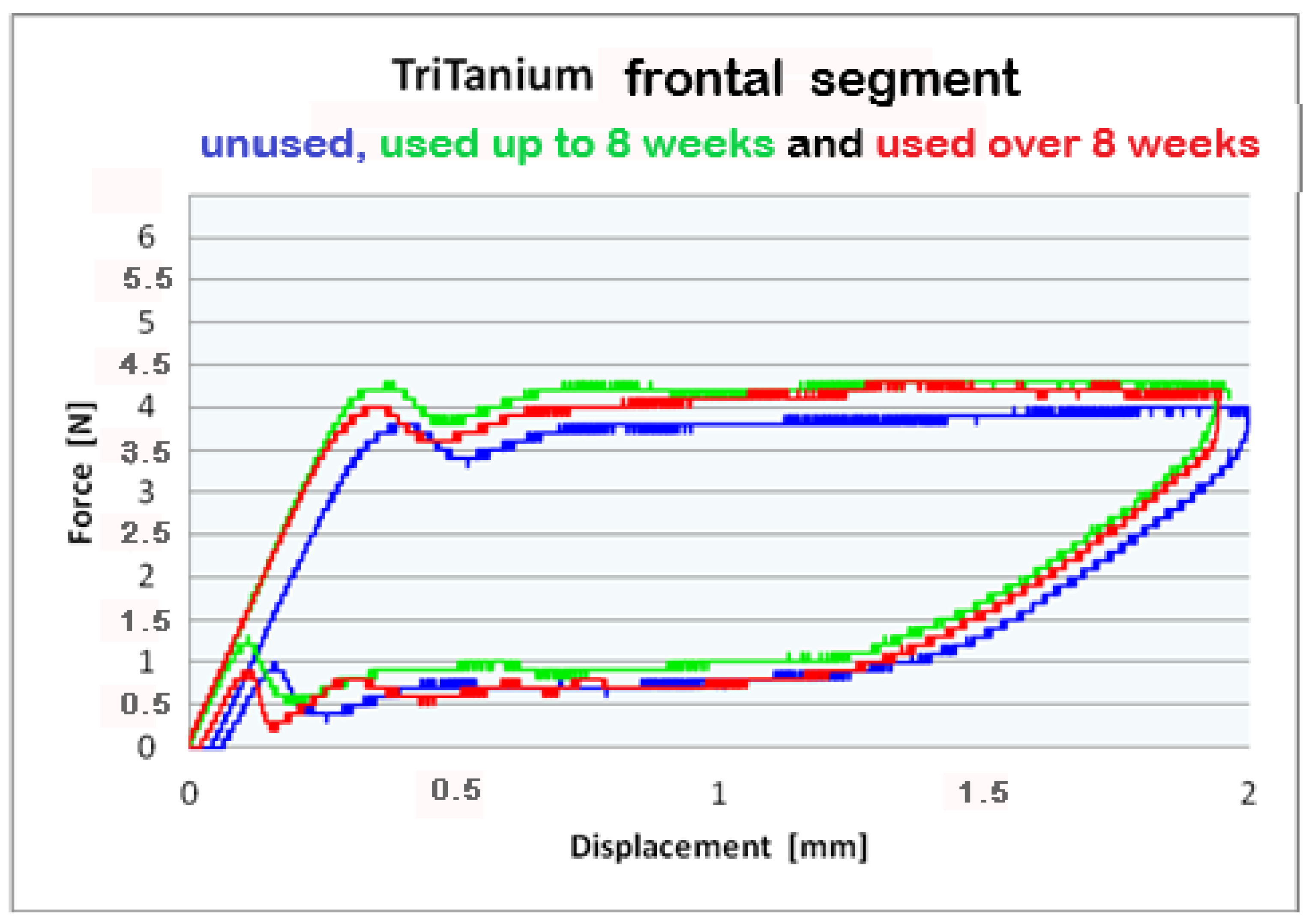
| FRONT | 201 | 3.3 | 3.6 | 3.9 | 3.9 | 0.3 | 0.5 | 1.3 | 3.9 | FORCE | |
| SD | 0.2 | 0.1 | 0.0 | 0.0 | |||||||
| 211 | 3.5 | 3.8 | 4.0 | 4.0 | 0.7 | 0.9 | 1.5 | 4.0 | |||
| SD | 0.1 | 0.1 | 0.0 | 0.2 | |||||||
| 221 | 3.6 | 4.1 | 4.3 | 4.2 | 0.8 | 1.0 | 1.7 | 4.2 | |||
| SD | 0.5 | 0.3 | 0.4 | 0.2 | |||||||
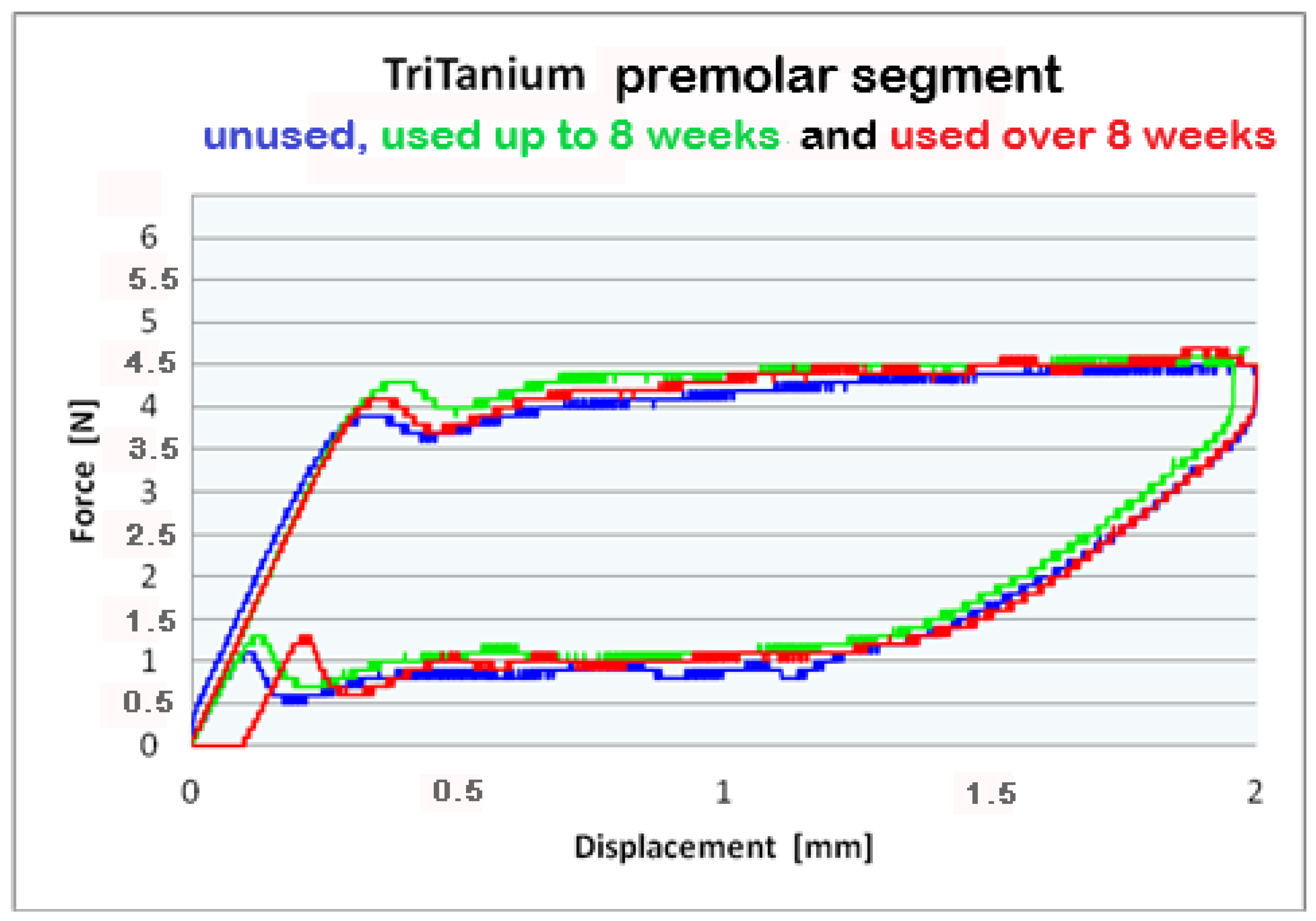
| PREMOLAR | 202 | 3.6 | 3.9 | 4.1 | 4.1 | 0.6 | 0.8 | 1.6 | 4.1 | ||
| SD | 0.2 | 0.1 | 0.1 | 0.1 | |||||||
| 212 | 3.8 | 4.2 | 4.3 | 4.3 | 1.1 | 1.2 | 1.9 | 4.3 | |||
| SD | 0.1 | 0.1 | 0.1 | 0.1 | |||||||
| 222 | 3.9 | 4.2 | 4.4 | 4.4 | 1.1 | 1.3 | 1.9 | 4.4 | |||
| SD | 0.2 | 0.2 | 0.2 | 0.2 | |||||||
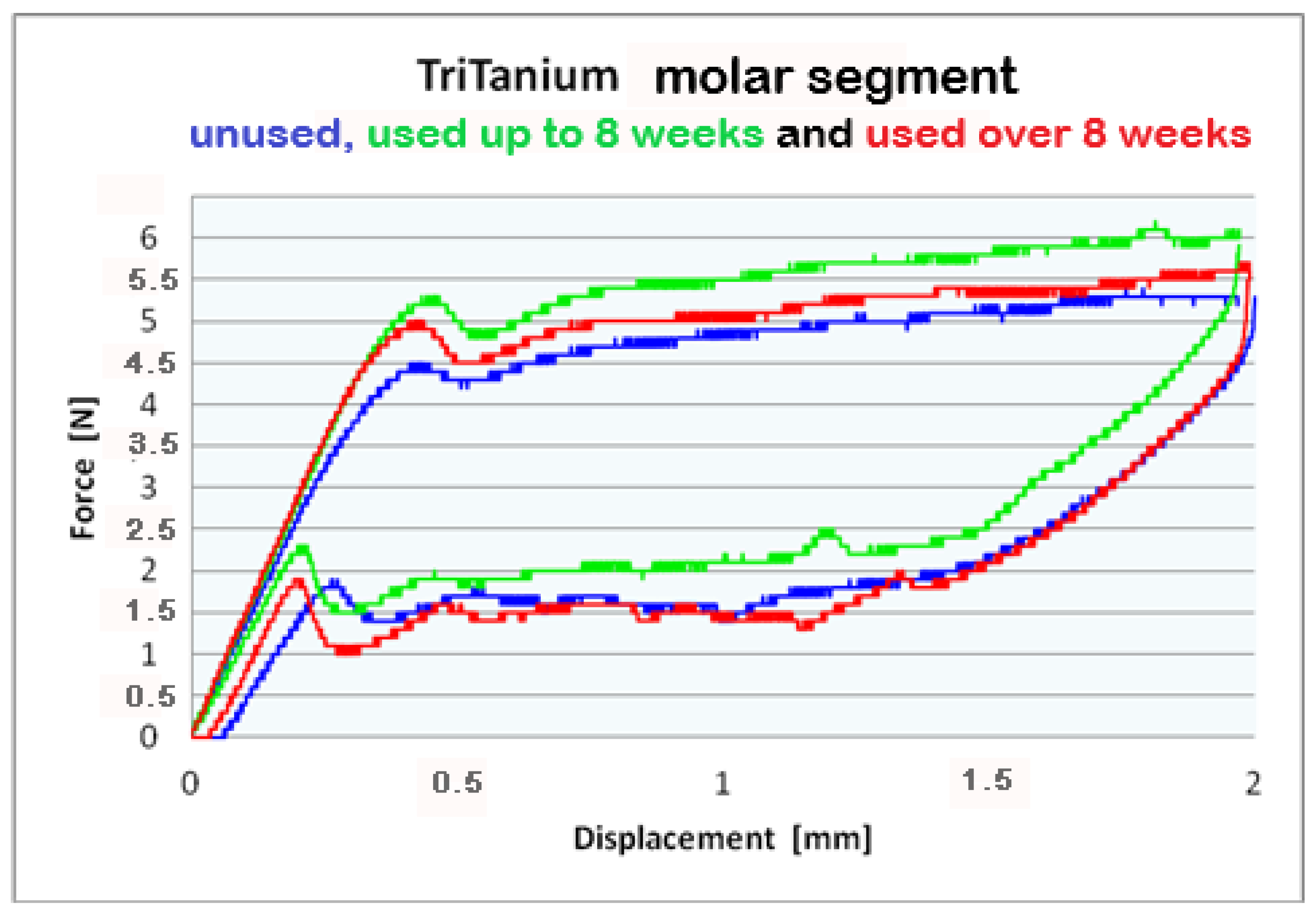
| MOLAR | 203 | 3.8 | 4.4 | 4.8 | 5.0 | 1.1 | 1.3 | 1.9 | 5.0 | ||
| SD | 0.1 | 0.1 | 0.0 | 0.0 | |||||||
| 213 | 4.0 | 4.5 | 4.8 | 5.0 | 1.3 | 1.5 | 2.1 | 5.0 | |||
| SD | 0.1 | 0.1 | 0.0 | 0.1 | |||||||
| 223 | 4.2 | 4.6 | 5.1 | 5.2 | 1.4 | 1.6 | 2.1 | 5.2 | |||
| SD | 0.1 | 0.1 | 0.1 | 0.3 | |||||||
| CODE | LOAD Displacement [mm] | UNLOAD Displacement [mm] | |||||||||
|---|---|---|---|---|---|---|---|---|---|---|---|
| 0.5 | 1.0 | 1.5 | 2.0 | 0.5 | 1.0 | 1.5 | 2.0 | ||||
| FRONT | 101 | 3.5 | 3.9 | 4.0 | 4.0 | 0.7 | 0.8 | 1.5 | 4.0 | FORCE | |
| SD | 0.0 | 0.0 | 0.1 | 0.1 | |||||||
| 111 | 3.7 | 4.1 | 4.2 | 4.1 | 0.8 | 0.8 | 1.6 | 4.1 | |||
| SD | 0.1 | 0.2 | 0.1 | 0.1 | |||||||
| 121 | 3.6 | 4.0 | 4.0 | 3.9 | 0.5 | 0.7 | 1.5 | 3.9 | |||
| SD | 0.3 | 0.3 | 0.3 | 0.3 | |||||||
| PREMOLAR | 102 | 3.8 | 4.2 | 4.4 | 4.5 | 0.8 | 0.9 | 1.6 | 4.5 | ||
| SD | 0.0 | 0.0 | 0.0 | 0.1 | |||||||
| 112 | 4.1 | 4.6 | 4.8 | 4.8 | 1.1 | 1.3 | 1.9 | 4.8 | |||
| SD | 0.3 | 0.4 | 0.4 | 0.5 | |||||||
| 122 | 3.6 | 4.1 | 4.3 | 4.3 | 0.7 | 0.9 | 1.6 | 4.3 | |||
| SD | 0.3 | 0.3 | 0.1 | 0.4 | |||||||
| MOLAR | 103 | 4.4 | 5.0 | 5.3 | 5.6 | 1.6 | 1.7 | 2.5 | 5.6 | ||
| SD | 0.1 | 0.1 | 0.2 | 0.4 | |||||||
| 113 | 4.5 | 5.0 | 5.2 | 5.3 | 1.5 | 1.6 | 2.3 | 5.3 | |||
| SD | 0.5 | 0.5 | 0.4 | 0.8 | |||||||
| 123 | 4.3 | 4.9 | 5.4 | 5.4 | 1.4 | 1.4 | 2.1 | 5.4 | |||
| SD | 0.3 | 0.3 | 0.2 | 0.6 | |||||||
| p-Values Bio-Active® | Displacements | ||||
|---|---|---|---|---|---|
| Segments | Groups | 0.5 | 1.0 | 1.5 | 2.0 |
| Frontal | G0–G1 | 0.99357 | 0.69191 | 1 | 1 |
| G0–G2 | 0.667518 | 0.69191 | 0.847142 | 0.967188 | |
| G1–G2 | 1 | 1 | 1 | 1 | |
| Pre-molar | G0–G1 | 0.214609 | 0.379306 | 0.550995 | 1 |
| G0–G2 | 0.489692 | 0.68973 | 1 | 1 | |
| G1–G2 | 1 | 1 | 1 | 1 | |
| Molar | G0–G1 | 1 | 0.919958 | 0.35192 | 1 |
| G0–G2 | 0.430823 | 0.341768 | 0.237375 | 1 | |
| G1–G2 | 0.687607 | 1 | 1 | 1 | |
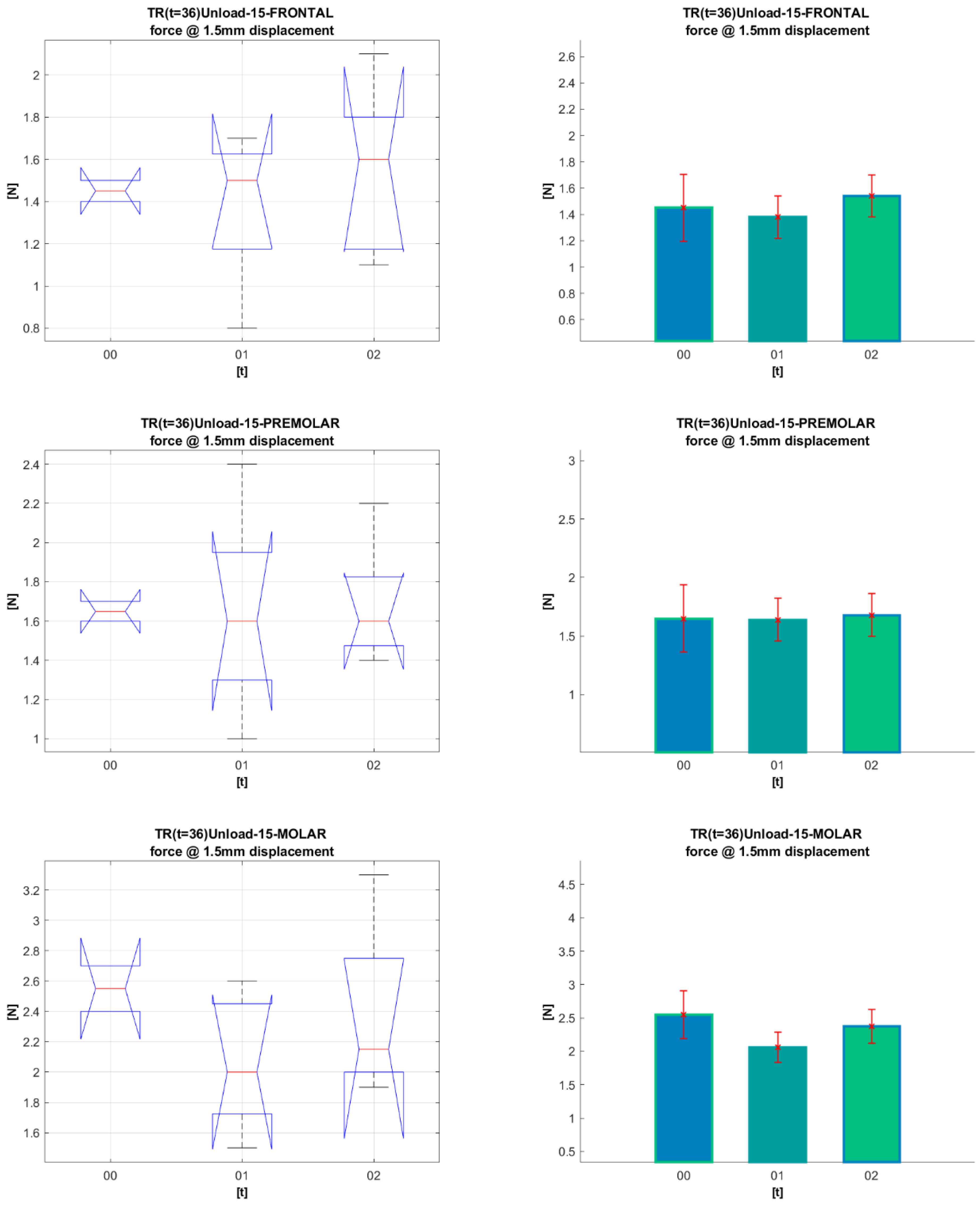
| p-Values TriTanium® | Displacements | ||||
|---|---|---|---|---|---|
| Segments | Groups | 0.5 | 1.0 | 1.5 | 2.0 |
| Frontal | G0–G1 | 1 | 1 | 1 | 1 |
| G0–G2 | 1 | 1 | 1 | 1 | |
| G1–G2 | 1 | 1 | 1 | 1 | |
| Pre-molar | G0–G1 | 1 | 1 | 1 | 1 |
| G0–G2 | 1 | 1 | 1 | 1 | |
| G1–G2 | 1 | 1 | 1 | 1 | |
| Molar | G0–G1 | 1 | 1 | 0.833637 | 1 |
| G0–G2 | 1 | 1 | 1 | 1 | |
| G1–G2 | 0.980193 | 1 | 1 | 1 | |
Disclaimer/Publisher’s Note: The statements, opinions and data contained in all publications are solely those of the individual author(s) and contributor(s) and not of MDPI and/or the editor(s). MDPI and/or the editor(s) disclaim responsibility for any injury to people or property resulting from any ideas, methods, instructions or products referred to in the content. |
© 2023 by the authors. Licensee MDPI, Basel, Switzerland. This article is an open access article distributed under the terms and conditions of the Creative Commons Attribution (CC BY) license (https://creativecommons.org/licenses/by/4.0/).
Share and Cite
Stoyanova-Ivanova, A.; Georgieva, M.; Petrov, V.; Andreeva, L.; Petkov, A.; Georgiev, V. Effects of Clinical Use on the Mechanical Properties of Bio-Active® (BA) and TriTanium® (TR) Multiforce Nickel-Titanium Orthodontic Archwires. Materials 2023, 16, 483. https://doi.org/10.3390/ma16020483
Stoyanova-Ivanova A, Georgieva M, Petrov V, Andreeva L, Petkov A, Georgiev V. Effects of Clinical Use on the Mechanical Properties of Bio-Active® (BA) and TriTanium® (TR) Multiforce Nickel-Titanium Orthodontic Archwires. Materials. 2023; 16(2):483. https://doi.org/10.3390/ma16020483
Chicago/Turabian StyleStoyanova-Ivanova, Angelina, Mirela Georgieva, Valeri Petrov, Laura Andreeva, Alexander Petkov, and Velizar Georgiev. 2023. "Effects of Clinical Use on the Mechanical Properties of Bio-Active® (BA) and TriTanium® (TR) Multiforce Nickel-Titanium Orthodontic Archwires" Materials 16, no. 2: 483. https://doi.org/10.3390/ma16020483
APA StyleStoyanova-Ivanova, A., Georgieva, M., Petrov, V., Andreeva, L., Petkov, A., & Georgiev, V. (2023). Effects of Clinical Use on the Mechanical Properties of Bio-Active® (BA) and TriTanium® (TR) Multiforce Nickel-Titanium Orthodontic Archwires. Materials, 16(2), 483. https://doi.org/10.3390/ma16020483