Article

Enhanced Photoluminescence and Photocatalytic Efficiency of La-Doped Bismuth Molybdate: Its Preparation and Characterization

1
Institute of Chemistry, Punjab University Lahore-Pakistan, Lahore 54590, Pakistan
2
Department of Chemistry, King Saud University, Riyadh 11451, Saudi Arabia
3
Department of Chemistry, University of Lahore-Pakistan, Lahore 54590, Pakistan
*
Authors to whom correspondence should be addressed.
Materials 2020, 13(1), 35; https://doi.org/10.3390/ma13010035
Submission received: 26 November 2019 / Revised: 17 December 2019 / Accepted: 18 December 2019 / Published: 20 December 2019
(This article belongs to the Section Materials Chemistry)

Abstract

:
Herein, a systematic study of the enhanced physicochemical properties of lanthanide doped (La-doped) bismuth molybdate (Bi2MoO6) is performed. For this purpose, Bi2MoO6 and La-doped Bi2MoO6 were prepared by the sol-gel method. BiCl3, Na2MoO4·2H2O, and LaCl3·7H2O were taken as the main precursors while sodium dodecyl sulfate was used as a surfactant. Both Bi2MoO6 and La-doped Bi2MoO6 were calcined at 650 °C for 2 h. These prepared materials were characterized by spectroscopic techniques such as UV–VIS, FT-IR, XRD, photoluminescence, XPS, along with other techniques such as SEM, TEM, TGA, etc. The investigation of luminescence behavior revealed that the La-doped Bi2MoO6 nanocomposite exhibited much greater luminescence compared to the undoped Bi2MoO6. The photocatalytic behavior of the prepared materials was explored by studying the degradation of methylene blue (MB) at room temperature. The degradation of MB with Bi2MoO6 and La-doped bismuth molybdate were observed to be 68% and 75% @ 45 s, respectively, indicating an enhancement of catalytic performance due to the La doping.

1. Introduction

Light emitting diodes with higher efficiency (>100,000 h) are a potential solution for the growing needs of urban civilization. Among various materials applied for the preparation of LEDs, indium gallium nitride (InGaN), aluminum gallium nitride (AlGaN) have been found to be more efficient as LEDs. Additionally, various other materials have also been applied for this purpose, such as, borates [1], phosphors [2], MOFs [3], different types of mixed metal oxides including CeO2, ZnO, ZnAl2O4 [4], Cr-doped ZnGa2O4 [5]. In recent times, scientists have paid more attention towards the preparation and characterization of trivalent lanthanide doped materials due to their applicability as solid-state laser media, fiber amplifiers, infrared to-visible up-converters, field emission displays [6,7] and, therefore, numerous luminescent materials were synthesized with suitable rare earth activators [Ln3+].
Nanomaterials have gained great importance due to remarkable physicochemical properties such as, catalytic, dielectric and luminescence. These properties can be further enhanced by altering the morphology of the nanomaterials. Among the plethora of nanomaterials, a series of Bi-ion based materials such as Bi2O3, BiOBr, Bi2S3, BiVO4, Bi2WO6, Bi2Ru2O7−d, Bi2Ti2O7, Bi12TiO20, and Bi2InNbO7 have attracted lot of attention as photocatalysts that operate under visible range [8,9,10,11,12] and have been reportedly employed for a variety of applications such as photocatalytic decomposition of water, and degradation of harmful dyes from water bodies [13,14,15], as almost 15% of the dyes used are released in water, which have adverse effects on the environment [16,17].
Apart from bismuth, molybdenum oxide (Mo)- and tungsten (W)-based compounds were also reported as efficient materials in the field of photo-catalysis and other important applications, such as photoluminescence, optical fibers, and microwave applications, etc. [18,19]. In this regard, the combination of Bi with Mo and W, such as, Bi2MO6 (M = Mo, W) types of materials have great potential as efficient photocatalysts in the visible solar spectrum for the degradation of organic compounds and water spitting [20,21]. Particularly, bismuth molybdate (Bi2MoO6)-based photocatalysts exhibit several special properties among other types of metal molybdates for the degradation of organic compounds, especially dyes. So far, the Bi2MoO6 nano-structure has been prepared through various conventional procedures, such as co-precipitation, hydrothermal, solid state reaction and sol-gel methods [20].
As it is well documented in literature that the incorporation of metals and non-metals with metal oxides enhances the catalytic efficiency [22,23]. In this research work, we employ a simple and facile sol-gel method to synthesize Bi2MoO6 and La-doped Bi2MoO6 nanocomposites using sodium dodecyl sulfate as a surfactant. The sol-gel method used in the present research work has a number of advantages, such as the control of the morphology and being a low-temperature process. The prepared materials were subjected to thorough characterization, such as SEM, TEM, BET, FT-IR, TGA, XPS, XRD, and photoluminescence spectra. The prepared materials were employed as photo-degradation catalysts for the degradation of methylene blue, an environmentally hazardous dye.

2. Materials and Methods

2.1. Materials

All chemicals used for the synthesis such as, sodium molybdate (Na2Mo2O4·2H2O), bismuth chloride (BiCl3), and lanthanum chloride (LaCl3·7H2O), were of analytical grade purchased from Sigma Aldrich and PubChem (Saint Louis, USA).
The Bi2MoO6 was synthesized by the following method. Sodium dodecyl sulfate (0.576 g, 2 mmol) was dispersed into deionized water with constant stirring and temperature was elevated up to 50 °C. At this temperature sodium molybdate (0.483 g, 2 mmol) was added drop wise followed by drop wise addition of bismuth chloride (0.63 g, 2 mmol) to from sol with continuous stirring. The resulting mixture was stirred for further 2 h, while the temperature was raised to 90 °C. After that the solution was filtered and washed with deionized water and ethanol. The obtained precipitate was dried in oven at 60 °C for 3 h. The dried precipitate was ground into powder by mortar and pestle followed by calcination at 650 °C in furnace for 2 h.
La-doped Bi2MoO6 catalyst was synthesized in a similar manner as described above with a slight modification. The sodium dodecyl sulfate (0.576 g, 2 mmol) was dispersed into deionized water at 50 °C, to this solution sodium molybdate (0.483 g, 2 mmol) was added drop-wise followed by the drop-wise addition of lanthanum chloride (0.742 g, 2 mmol) which yielded a white suspension. To this, bismuth chloride (0.63 g, 2 mmol) was added drop-wise and the resulting mixture was heated at 90 °C under continuous stirring for 2 h which resulted in the formation of yellow precipitate. The obtained precipitate was dried in oven at 60 °C for 3 h. The dried precipitate was ground into powder with a mortar and pestle followed by calcination at 650 °C in furnace for 2 h.

2.2. Characterization Techniques

The characterizations of synthesized Bi2MoO6 and La-doped Bi2MoO6 were carried out by using different analytical techniques to understand their morphology and other catalytic activities. The functional groups in Bi2MoO6 and La-doped Bi2MoO6 were determined by Agilent Technologies Cary 630 Fourier transform infrared (FT-IR) spectrometer (Agilent, Santa Clara, USA). Degradation of dye was recorded by T90+ UV–VIS spectrometer (Perkin Elmer lambda 35, Waltham, MA, USA). The photoluminescence (PL) properties of synthetic materials were analyzed by FLS 1000 photoluminescence spectrometer. BET surface area (NOVA 4200e surface area and pore size analyzer (Quantachrome Instruments, FL, USA)). The XRD analysis of the as-prepared nanocatalyst was carried out using a D2 Phaser X-ray diffractometer (Bruker, Bremen, Germany) with Cu Kα radiation (k = 1.5418 A°). XPS spectra were measured on a PHI 5600 Multi-Technique XPS (Physical Electronics, Lake Drive East, Chanhassen, MN, USA) using monochromatized Al Kα at 1486.6 eV. Peak fitting was performed using Origin software (2019a), USA. TGA was carried out using a TGA/DSC 1 (Mettler Toledo AG, Analytical, Schwerzenbach, Switzerland).

2.3. Photocatalytic Activity Measurements

The catalytic activities of prepared Bi2MoO6 and La-doped Bi2MoO6 catalysts were determined by the degradation of methylene blue (MB) as model reaction in the presence of reducing agent NaBH4. We designed three reactions for degradation of MB. The time dependent degradation of methylene blue was examined from decrease in the absorbance observed in the UV–VIS spectra, scanned in the range of 400–800 at room temperature. To evaluate the catalytic activity of the prepared material (a) 5 mL solution of methylene blue was mixed with NaBH4 (0.2 mL, 0.16 M); (b) same solution in the presence of 2 mg of Bi2MoO6 catalyst at room temperature; and (c) the same solution in the presence of 2 mg of La-doped Bi2MoO6 catalyst at room temperature. The blue color of methylene blue disappeared with time which shows the degradation of methylene blue. The reduction efficiency and degradation were determined by the following equation:
Reduction   efficiency ( % ) = ( C 0 C t ) C 0 × 100
where C0 and Ct represent the initial and residual concentration of methylene blue in solution with time. The reduction of methylene blue followed the reaction mechanism in which electrons passes from the reductant BH4 to methylene blue which contains double bond. The Bi2MoO6 and La-doped Bi2MoO6 help to decrease the potential difference between donor and acceptor and facilitate the transfer of electron from BH4 to MB for the hydrogenation of double bond present in methylene blue.

3. Results and Discussion

3.1. Microscopic Analysis

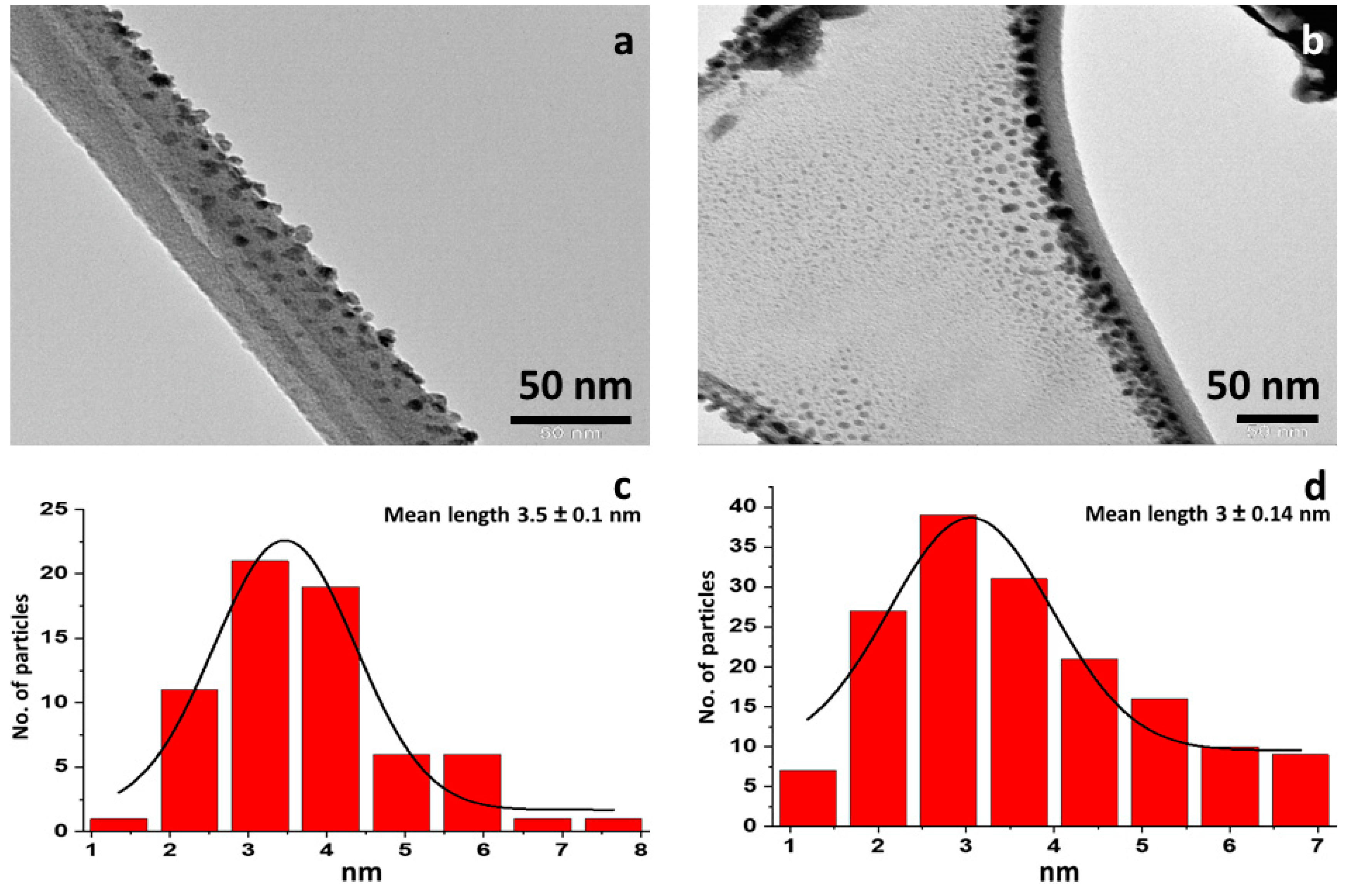
Figure 1 shows the SEM micrograms of the prepared Bi2MoO6 and La-doped Bi2MoO6 nanocomposite. The Bi2MoO6 surface morphology appears to be dense rod-like structures, however, the surface morphology of La-doped Bi2MoO6 is found to be globular and hemispherical particles are engaged coarse granular clustering with high porosity. The TEM images of the prepared Bi2MoO6 and La-doped Bi2MoO6 nanocomposite and their particle size distribution graph is given Figure 2. From the TEM images and the particle size distribution graph (Figure 2a,c) of Bi2MoO6, it is evident that the nanoparticles range from 1–8 nm with a mean size of 3.5 nm, while that of La-doped Bi2MoO6 nanocomposites have a size range of 1–7 nm with a mean size of 3 nm (Figure 2b,d), indicating the effect of doping on the particle size of the composite.

3.2. BET Analysis

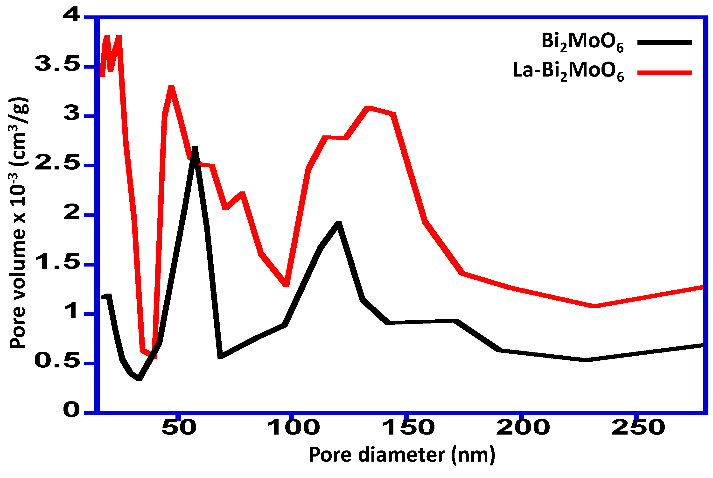
The synthesized Bi2MoO6 and La-doped Bi2MoO6 nanocomposites were subjected to BET analysis to understand the porosity and it was found to be mesoporous in nature. The surface area of the Bi2MoO6 and La-doped Bi2MoO6 nanocomposite was found to be 1.0 m²/g and 3.4 m²/g, respectively. The pore distribution plot revealed that the surface porosity for both the prepared material is different, the Bi2MoO6 possessed a pore volume in the range of 0.3–2.4 × 10⁻³ cm3/g, while the La-doped Bi2MoO6 nanocomposite was found to possess a pore volume in the range of 0.6–3.8 × 10⁻³ cm3/g, which is mostly represents granular materials [22]. The pore distribution plot is given in Figure 3.

3.3. FT-IR Spectral Analysis

FT-IR spectral data provided valuable information about functional groups present in synthesized Bi2MoO6 and La-doped Bi2MoO6 nanocomposites. The FT-IR spectra of Bi2MoO6 and La-doped Bi2MoO6 were recorded in the range 4000–400 cm−1. The spectrum of bismuth nanoparticle showed the peaks at 797 cm−1 is due to (Mo = O). The peak observed at 499 cm−1 was assigned to Bi-O bond [23]. The infrared spectrum of La-doped Bi2MoO6 was compared to that of un-doped bismuth molybdate and a shift was observed in the peaks of Mo-O and Bi-O from 797 cm−1 to 794 cm−1 and 499 cm−1 to 493 cm−1, respectively. This shift can be attributed to the influence of the dual lanthanum in the host matrix of Bi2MoO6 [24]. The IR spectra of Bi2MoO6 and La-doped Bi2MoO6 nanocomposite is given in Supplementary File (Figure S1).

3.4. X-Ray Diffraction Spectroscopy

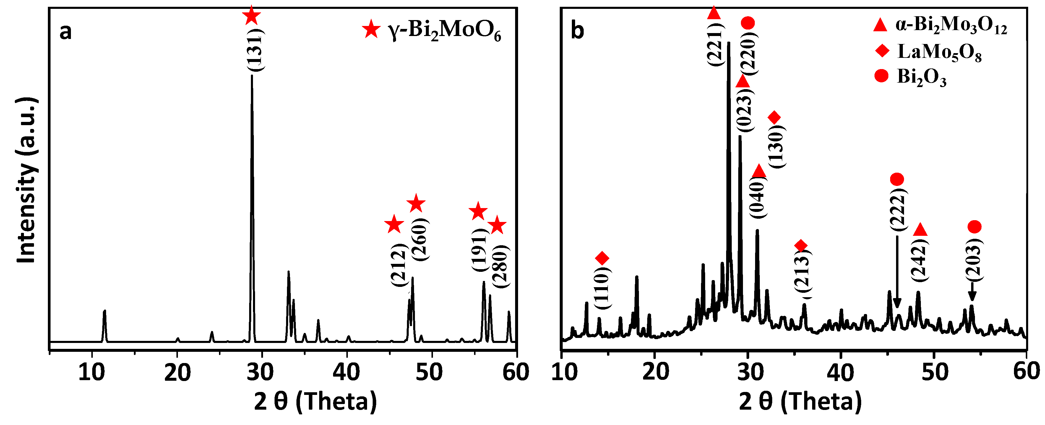
The XRD pattern of the synthesized sample of bismuth molybdate (BW) is given in Figure 4a. It shows pronounced diffraction peaks of (131), (212), (260), (191) and (280) planes at 2-Theta = 28.271, 47.094, 47.214, 56.283, and 56.452, which can be ascribed to the characteristic orthorhombic phase of Bi2MoO6 (JCPDS card no. 77-1246). However, the lanthanum doped bismuth molybdate (LW) exhibits more than one crystal phases as shown in Figure 4b. It shows the main diffraction peaks of α-Bi2Mo3O12 at 2θ = 27.947°, 29.170°, 30.995°, and 48.348° corresponding to diffraction planes of (221), (023), (040) and (242), respectively, and match with JCPDS card no. 21-0103. It was found that the addition of La to the Bi2MoO6 induced a phase transition of synthesized bismuth molybdenum from γ-Bi2MoO6 to α-Bi2Mo3O12. The diffraction peaks of planes (110), (130), and (213) at diffraction angles 13.55, 30.98 and 37.57, respectively shows the presence of monoclinic crystal phase of lanthanum molybdenum oxide (LaMo5O8). Along with these two phases, the XRD pattern also shows the presence of bismuth oxide (Bi2O3) as presented by the diffraction peaks at 27.94, 46.20, and 54.24 correspond to (201), (222), and (203) planes. Moreover, the crystallite size of the Bi2MoO6 and La-doped Bi2MoO6 was calculated using Debye–Scherrer equation and the crystallite size obtained was found to be 18 and 14 nm. Normally the size of crystallites as obtained by XRD are smaller than the particle size obtained by TEM, as the particles are made up of crystallites. However, in our case the observation is opposite, this may be because XRD measurements are usually on bulk of the sample, whereas TEM is usually on a small selected area, which in our case showed smaller particle size.

3.5. Luminescence

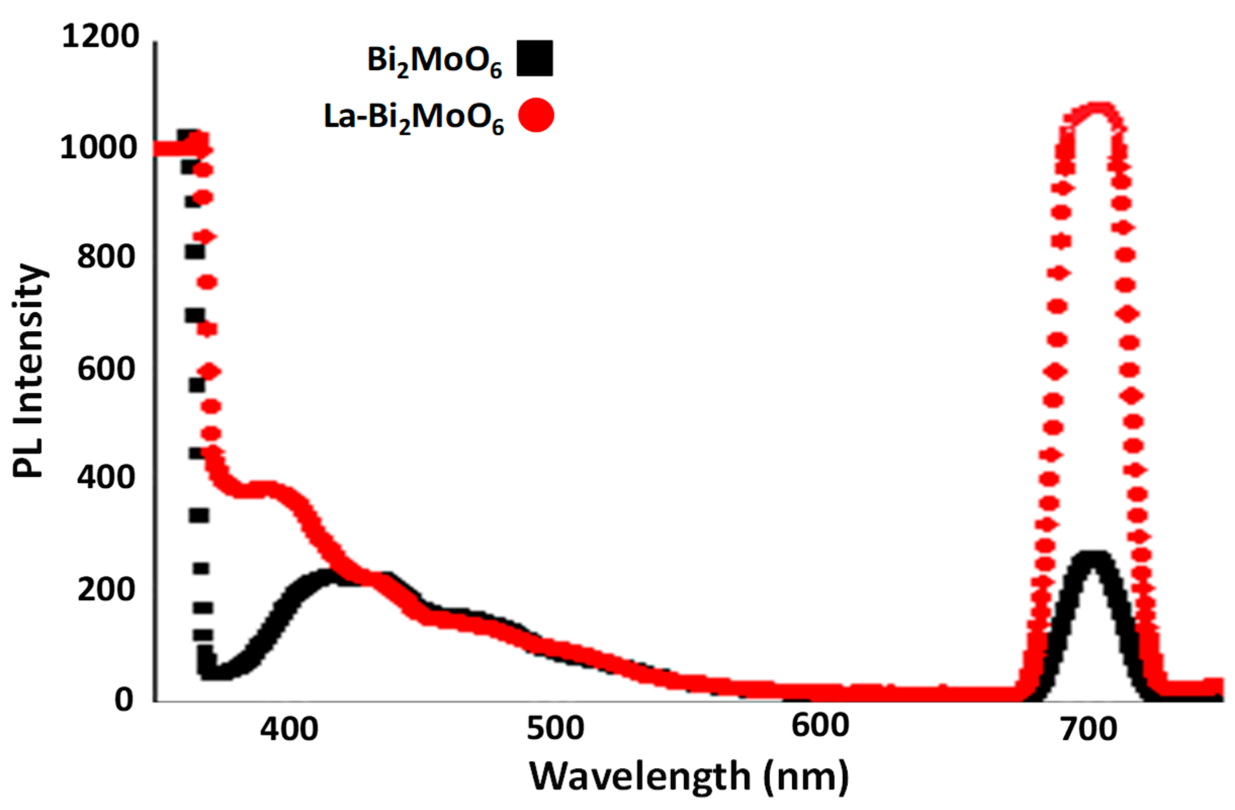
The optical property of Bi3+ ions have been explained in terms of Russell–Saunders type electronic energy terms with s2 configuration in the ground and sp configuration (first excited state). Among these, the energetically lowest possible 3P01S0 excitation is strongly forbidden but the 3P11S0 transition is possible because of spin-orbit coupling of the 3P1 and 1P1 states [25]. Figure 5 represented the PL spectra of un-doped Bi2MoO6 and La-Bi2MoO6 in the range 350 to 750 nm excited by 350 nm [26]. The relatively broad PL peaks appeared around 700 nm with enhanced emission intensity in La-doped Bi2MoO6 compared to un-doped Bi2MoO6. PL intensity normally depends on crystallinity, materials having more crystallinity exhibits enhanced PL intensity. [27]. The same possible reason might be attributed to the enhanced emission intensity of La-Bi2MoO6. A similar observation has been reported for CaMoO4:Eu3+ in the literature [27].

3.6. X-Ray Photoelectron Spectroscopy

Surface composition analyses of the prepared nanocomposites were analyzed using X-Ray photoelectron spectroscopy (XPS). The survey scan revealed the presence of Bi, Mo, O elements in the composites prepared, and an additional signal corresponding to the element La revealed the successful doping of La in the nanocomposite given in Figure 6.
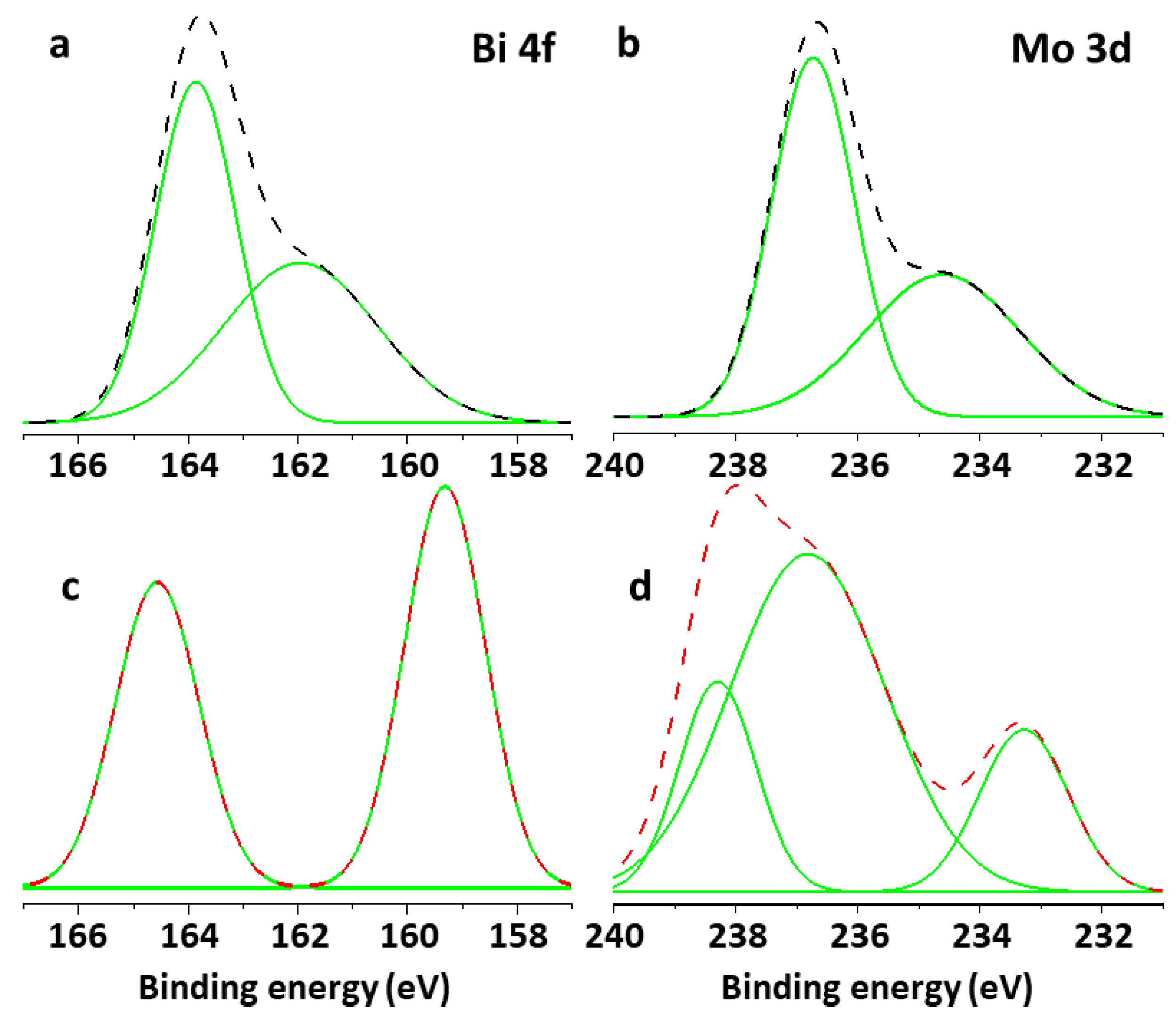
The deconvolution of the Bi 4f spectrum of Bi2MoO6 yielded peaks at 161.8 eV and 163.8 eV corresponding to Bi 4f5/2 and the difference in BE between the two Bi 4f5/2 peaks was 2.0 eV, which coincided with that between Bi0 and Bi3. While in the case of La-doped Bi2MoO6 nanocomposite the Bi 4f spectrum yielded peaks at 159.3 eV and 164.6 eV corresponding to Bi 4f7/2 and Bi 4f5/2, respectively. However, the Mo 3d deconvoluted spectrum for the Bi2MoO6 yielded peaks at 234.6 eV and 236.7 eV attributed to Mo 3d5/2 which correspond to the Mo6+, while that of La-doped Bi2MoO6 nanocomposite yielded peaks at 233.2 eV, 236.7 eV, and 238.3 eV [28]. The deconvolution of the Bi 4f and Mo 3d spectrum of Bi2MoO6 and La-doped Bi2MoO6 nanocomposites are given in Figure 7.

3.7. TGA Analysis

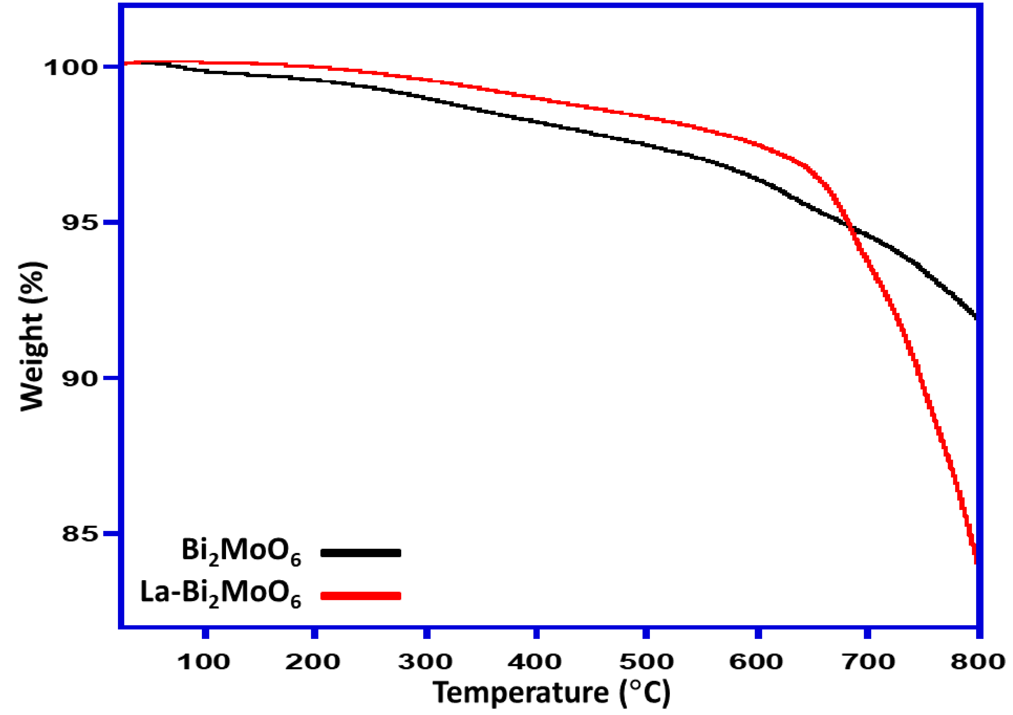
Thermogravimetric analysis (TGA) was conducted to identify the thermal behavior of the Bi2MoO6 and La-doped Bi2MoO6 nanocomposites and the results were compared as displayed in Figure 8. TGA of Bi2MoO6 reveals that there is an 8% weight loss when the sample is heated to 800 °C starting from 25 °C at a heating rate of 10 °C/minute under an inert atmosphere. However, when the La-doped Bi2MoO6 was subjected to similar study it is found that the weight loss is up to 16% indicating that the later nanocomposite is thermally less stable than un-doped Bi2MoO6.

3.8. Photocatalytic Activity

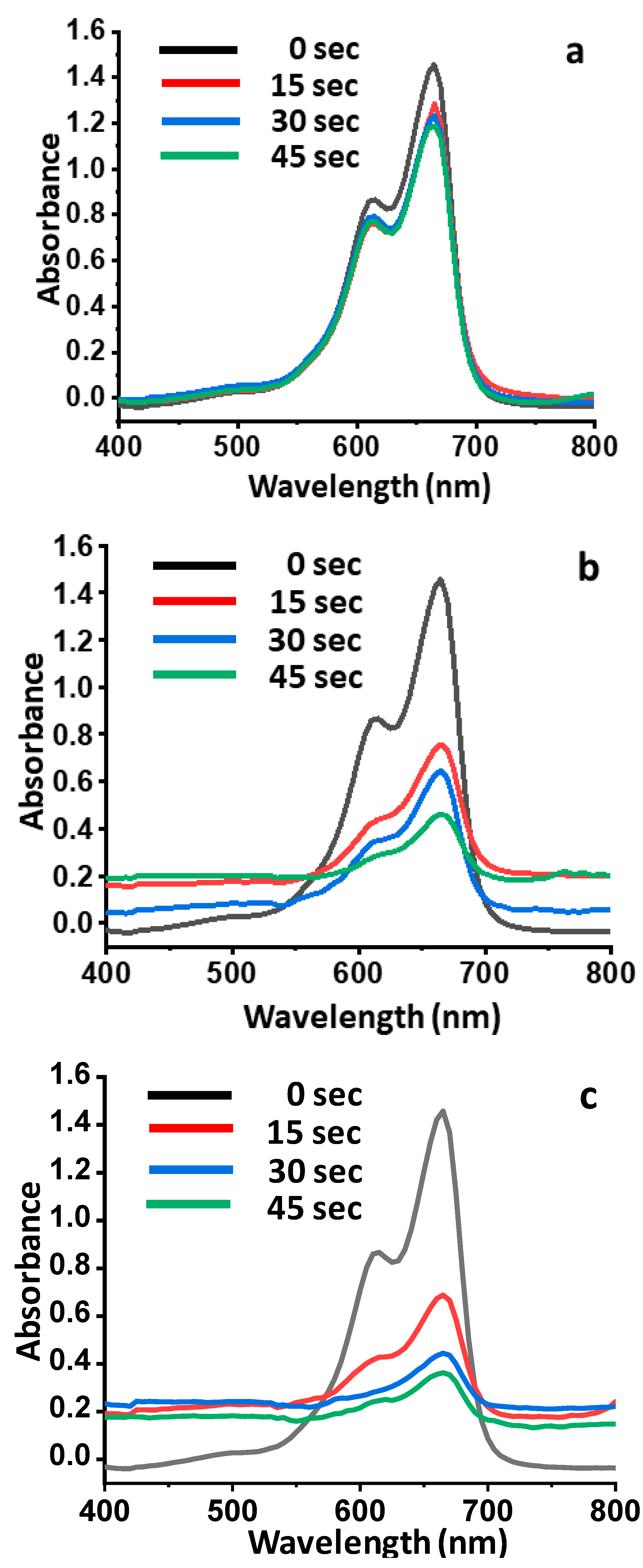
The catalytic activities of undoped Bi2MoO6 and La-doped Bi2MoO6 were evaluated by the degradation of methylene blue (MB) using UV-visible spectroscopy. λ-max for pure methylene blue was noted at 665 nm. The gradual change in color was observed at different time intervals which ensured the enhanced reduction. Maximum degradation was observed at a time interval of 45 s with complete decolorization. The presence of the reaction intermediates was eradicated as no new band appeared in the spectrum. The degradation of methylene blue using NaBH4 was recorded minimal 18% at 45 s, while in the presence of undoped BiMoO6 catalyst, the degradation of MB was enhanced up to 48% within first 15 s, as the reaction proceeds, the % age degradation was recorded as 55% and 68% at 30 and 45 s, respectively (Figure 9). However, when a similar reaction was carried out using NaBH4 in the presence of La-doped Bi2MoO6, a drastic increase in the degradation of methylene blue was observed, with a percentage of degradation of 53% in the first 15 s, which further proceeds to 69% and 75% at 30 and 45 s, respectively. The degradation kinetics of the reaction are illustrated graphically in Figure 10.
Relevant literature reveals that similar pattern of degradation has been observed with Pd-Nano-diamond-GO composite for the degradation of MB from 15 s to 60 s [23]. This enhancement of degradation efficiency can be attributed to the inclusion of La in the system. By correlating the characterization data obtained and the results of photocatalytic activity it can be concluded that the inclusion of La leads to the formation of nanocomposites with smaller dimension and larger surface area. These changes along with the phase transformation of γ-Bi2MoO6 to α-Bi2Mo3O12, and the presence of monoclinic crystal phase of lanthanum molybdenum oxide (LaMo5O8) are assumed to be responsible in the enhancement of photocatalytic activity towards the degradation of MB.

4. Conclusion

In conclusion the Bi2MoO6 and La-doped Bi2MoO6 materials were successfully synthesized through the environmental friendly sol-gel method using sodium dodecyl sulfate as a surfactant. The morphological and structural properties of the synthesized material were determined by various analytical techniques. The morphology of Bi2MoO6 was changed with lanthanum doping, i.e., from dense rod-like structures to globular and hemispherical particles engaged into clusters with high porosity. The photo-degradation efficiency of La-Bi2MoO6 for MB was enhanced due to lanthanum doping and can be attributed to the efficient separation and preventing of the recombination of electron-hole pairs. Hence, the environmentally friendly sol-gel method for synthesis of La-Bi2MoO6 is an efficient tool for possible applications in the degradation of dyes and sequestering of MB pollutants from environment, and this study can be further extended to other pollutants, which is in progress and shall be published later.

Supplementary Materials

The following are available online at https://www.mdpi.com/1996-1944/13/1/35/s1, Figure S1: FT-IR spectra of Bi2MoO6 and La-doped Bi2MoO6 nanocomposite.

Author Contributions

Methodology, M.W. and S.N.; Conceptualization, Supervision, Resources, Writing-Original Draft Preparation, M.I. and S.L.: acted as supervisor and co-supervisor respectively. Formal Analysis, Investigation, Writing-Review & Editing, S.F.A., M.R.H.S. and M.K.; Project Administration & Funding Acquisition, M.R.H.S. All the authors equally contributed in this manuscript. All authors have read and agreed to the published version of the manuscript.

Funding

This research was funded by Researchers Supporting Project number (RSP-2019/60), King Saud University, Riyadh, Saudi Arabia.

Acknowledgments

Researchers Supporting Project number (RSP-2019/60), King Saud University, Riyadh, Saudi Arabia.

Conflicts of Interest

The authors declare no conflict of interest.

References

  1. Fouassier, C.E.; Fu, W.T.; Hagenmuller, P. Luminescent Mixed Borates Based on Rare Earths. U.S. Patent 4,946,621, 7 August 1990. [Google Scholar]
  2. Jayachandiran, M.; Annadurai, G.; Kennedy, S.M.M. Photoluminescence properties of red emitting Ba3Bi2 (PO4) 4: Eu3+ phosphor for WLEDs applications. J. Lumin. 2018, 201, 196–202. [Google Scholar] [CrossRef]
  3. Guo, H.; Zhu, Y.; Qiu, S.; Lercher, J.A.; Zhang, H. Coordination modulation induced synthesis of nanoscale Eu1-xTbx-metal-organic frameworks for luminescent thin films. Adv. Mater. 2010, 22, 4190–4192. [Google Scholar] [CrossRef] [PubMed]
  4. Subhan, M.A.; Ahmed, T.; Sarker, P.; Pakkanen, T.T.; Suvanto, M.; Horimoto, M.; Nakata, H. Structural, luminescence and photophysical properties of novel trimetallic nanocomposite CeO2· ZnO ZnAl2O4. J. Lumin. 2014, 148, 98–102. [Google Scholar] [CrossRef]
  5. Srivastava, B.B.; Kuang, A.; Mao, Y. Persistent luminescent sub-10 nm Cr doped ZnGa 2 O 4 nanoparticles by a biphasic synthesis route. Chem. Commun. 2015, 51, 7372–7375. [Google Scholar] [CrossRef] [Green Version]
  6. Nayab Rasool, S.; Rama Moorthy, L.; Kulala Jayasankar, C. Optical and luminescence properties of Eu3+-doped phosphate based glasses. Mater. Express 2013, 3, 231–240. [Google Scholar] [CrossRef]
  7. Lin, H.; Liu, K.; Pun, E.; Ma, T.; Peng, X.; An, Q.; Yu, J.; Jiang, S. Infrared and visible fluorescence in Er3+-doped gallium tellurite glasses. Chem. Phys. Lett. 2004, 398, 146–150. [Google Scholar] [CrossRef]
  8. Niishiro, R.; Kato, H.; Kudo, A. Nickel and either tantalum or niobium-codoped TiO2 and SrTiO3 photocatalysts with visible-light response for H2 or O2 evolution from aqueous solutions. Phys. Chem. Chem. Phys. 2005, 7, 2241–2245. [Google Scholar] [CrossRef]
  9. Fu, H.; Zhang, S.; Xu, T.; Zhu, Y.; Chen, J. Photocatalytic degradation of RhB by fluorinated Bi2WO6 and distributions of the intermediate products. Environ. Sci. Technol. 2008, 42, 2085–2091. [Google Scholar] [CrossRef]
  10. Xie, L.; Liu, Z.; Zhang, J.; Ma, J. Preparation of a novel Bi3. 64Mo0. 36O6. 55 nanophotocatalyst by molten salt method and evaluation for photocatalytic decomposition of rhodamine B. J. Alloys Compd. 2010, 503, 159–162. [Google Scholar] [CrossRef]
  11. Labib, S. Preparation, characterization and photocatalytic properties of doped and undoped Bi2O3. J. Saudi Chem. Soc. 2017, 21, 664–672. [Google Scholar] [CrossRef]
  12. Singh, P.; Raizada, P.; Sudhaik, A.; Shandilya, P.; Thakur, P.; Agarwal, S.; Gupta, V.K. Enhanced photocatalytic activity and stability of AgBr/BiOBr/graphene heterojunction for phenol degradation under visible light. J. Saudi Chem. Soc. 2019, 23, 586–599. [Google Scholar] [CrossRef]
  13. Lu, L.; Kobayashi, A.; Tawa, K.; Ozaki, Y. Silver nanoplates with special shapes: Controlled synthesis and their surface plasmon resonance and surface-enhanced Raman scattering properties. Chem. Mater. 2006, 18, 4894–4901. [Google Scholar] [CrossRef]
  14. Yu, J.; Kudo, A. Effects of structural variation on the photocatalytic performance of hydrothermally synthesized BiVO4. Adv. Funct. Mater. 2006, 16, 2163–2169. [Google Scholar] [CrossRef]
  15. Zou, Z.; Ye, J.; Abe, R.; Arakawa, H. Photocatalytic decomposition of water with Bi2 InNbO7. Catal. Lett. 2000, 68, 235–239. [Google Scholar] [CrossRef]
  16. Yao, W.; Zhang, B.; Huang, C.; Ma, C.; Song, X.; Xu, Q. Synthesis and characterization of high efficiency and stable Ag3 PO4/TiO2 visible light photocatalyst for the degradation of methylene blue and rhodamine B solutions. J. Mater. Chem. 2012, 22, 4050–4055. [Google Scholar] [CrossRef]
  17. Zhao, D.; Sheng, G.; Chen, C.; Wang, X. Enhanced photocatalytic degradation of methylene blue under visible irradiation on graphene@ TiO2 dyade structure. Appl. Catal. B Environ. 2012, 111, 303–308. [Google Scholar] [CrossRef]
  18. Karaoglu, E.; Baykal, A. CoFe2O4–Pd (0) nanocomposite: Magnetically recyclable catalyst. J. Supercond. Nov. Magn. 2014, 27, 2041–2047. [Google Scholar] [CrossRef]
  19. Liyan, W.; Hongxia, W.; Aijie, W.; Min, L. Surface modification of a magnetic SiO2 support and immobilization of a nano-TiO2 photocatalyst on it. Chin. J. Catal. 2009, 30, 939–944. [Google Scholar]
  20. Baux, N.; Vannier, R.; Mairesse, G.; Nowogrocki, G. Oxide ion conductivity in Bi2W1− xMExO6− x/2 (ME = Nb, Ta). Solid State Ion. 1996, 91, 243–248. [Google Scholar] [CrossRef]
  21. Islam, M.S.; Lazure, S.; Vannier, R.-n.; Nowogrocki, G.; Mairesse, G. Structural and computational studies of Bi 2 WO 6 based oxygen ion conductors. J. Mater. Chem. 1998, 8, 655–660. [Google Scholar] [CrossRef]
  22. Karathanos, V.; Saravacos, G. Porosity and pore size distribution of starch materials. J. Food Eng. 1993, 18, 259–280. [Google Scholar] [CrossRef]
  23. Umapathy, V.; Manikandan, A.; Antony, S.A.; Ramu, P.; Neeraja, P. Structure, morphology and opto-magnetic properties of Bi2MoO6 nano-photocatalyst synthesized by sol–gel method. Trans. Nonferrous Met. Soc. China 2015, 25, 3271–3278. [Google Scholar] [CrossRef]
  24. Rambabu, U.; Han, S.D. Synthesis and luminescence properties of broad band greenish-yellow emitting LnVO4: Bi3+ and (Ln1, Ln2) VO4: Bi3+ (Ln= La, Gd and Y) as down conversion phosphors. Ceram. Int. 2013, 39, 701–708. [Google Scholar] [CrossRef]
  25. Adhikari, R.; Gyawali, G.; Cho, S.H.; Narro-García, R.; Sekino, T.; Lee, S.W. Er3+/Yb3+ co-doped bismuth molybdate nanosheets upconversion photocatalyst with enhanced photocatalytic activity. J. Solid State Chem. 2014, 209, 74–81. [Google Scholar] [CrossRef]
  26. Zhou, T.; Hu, J.; Li, J. Er3+ doped bismuth molybdate nanosheets with exposed {0 1 0} facets and enhanced photocatalytic performance. Appl.Catal. B Environ. 2011, 110, 221–230. [Google Scholar] [CrossRef]
  27. Wang, Y.; Jie, S.; Zhao, Y.; Xu, L.; He, D.; Jiao, H. Effects of organic additives on morphology and luminescent properties of Eu3+-doped calcium molybdate red phosphors. Powder Technol. 2015, 275, 1–11. [Google Scholar] [CrossRef]
  28. Haoran, W.; Keryn, L. The Development of Pseudocapacitive Molybdenum Oxynitride Electrodes for Super capacitors. ECS Trans. 2014, 58, 67–75. [Google Scholar]
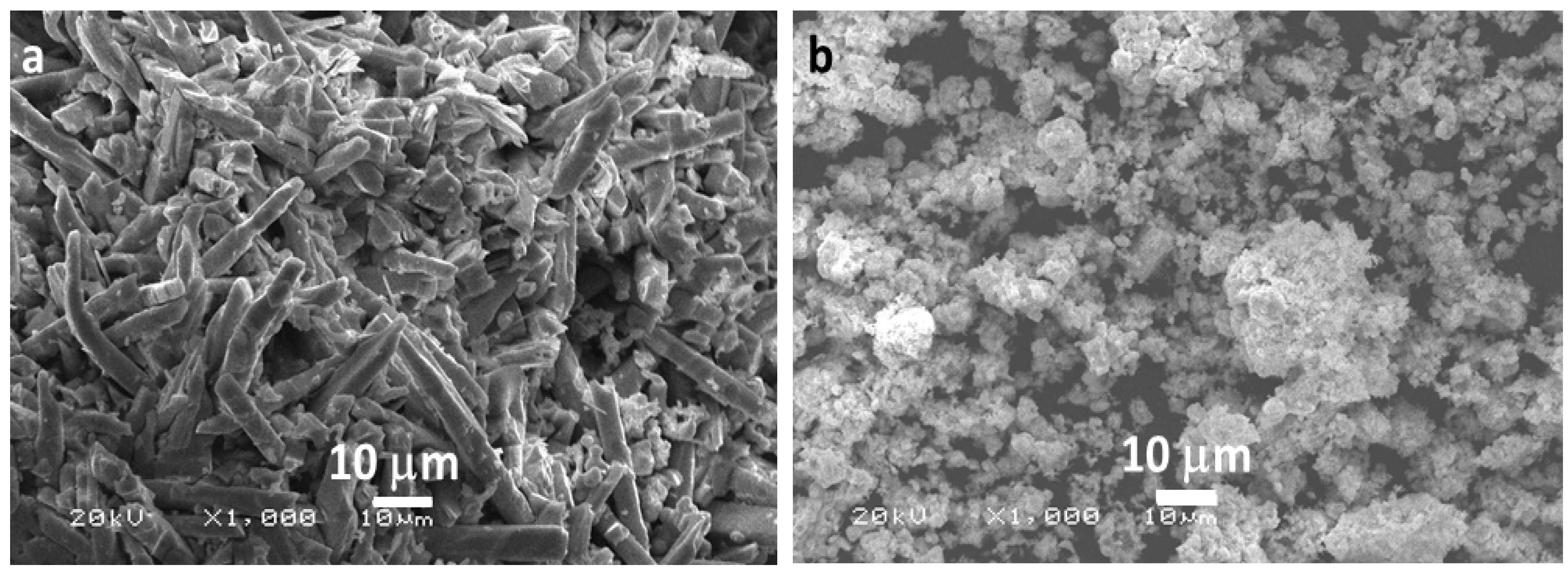
Figure 1. Scanning electron micrograms of (a) Bi2MoO6 and (b) La- Bi2MoO6 nanocomposite.
Materials 13 00035 g001
Figure 2. Transmission electron micrograms of (a) Bi2MoO6 and (b) La-doped Bi2MoO6 nanocomposite: Particle distribution graph of (c) Bi2MoO6, and (d) La-doped Bi2MoO6 nanocomposite.
Materials 13 00035 g002
Figure 3. Pore size distribution curve of Bi2MoO6 and La-doped Bi2MoO6 nanocomposite.
Materials 13 00035 g003
Figure 4. XRD pattern of (a) Bi2MoO6 and (b) La-doped Bi2MoO6 nanocomposite.
Materials 13 00035 g004
Figure 5. Photoluminescence (PL) spectra of Bi2MoO6 and La-doped Bi2MoO6.
Materials 13 00035 g005
Figure 6. XPS survey scan of Bi2MoO6 and La-doped Bi2MoO6 nanocomposites.
Materials 13 00035 g006
Figure 7. XPS results of deconvoluted Bi 4f region of (a) Bi2MoO6 and (c) La-doped Bi2MoO6 nanocomposites and of deconvoluted Mo 4f region of (b) Bi2MoO6 and (d) La-doped Bi2MoO6 nanocomposites.
Materials 13 00035 g007
Figure 8. Thermal degradation patterns of Bi2MoO6 and La-doped Bi2MoO6 nanocomposites.
Materials 13 00035 g008
Figure 9. (a) UV–VIS spectra showing the degradation of methylene blue using NaBH4. (b) UV–VIS spectra showing the degradation of methylene blue using NaBH4 in the presence of Bi2MoO6. (c) UV–VIS spectra showing the degradation of methylene blue using NaBH4 in the presence of La-doped Bi2MoO6.
Materials 13 00035 g009
Figure 10. Graphical illustration of the photocatalytic degradation kinetics of methylene blue using NaBH4, NaBH4 in the presence of Bi2MoO6, and NaBH4 in the presence of La-doped Bi2MoO6.
Materials 13 00035 g010