Le Fort I Osteotomy with Bone Grafts in Preprosthetic Surgery: Technical Note
Abstract
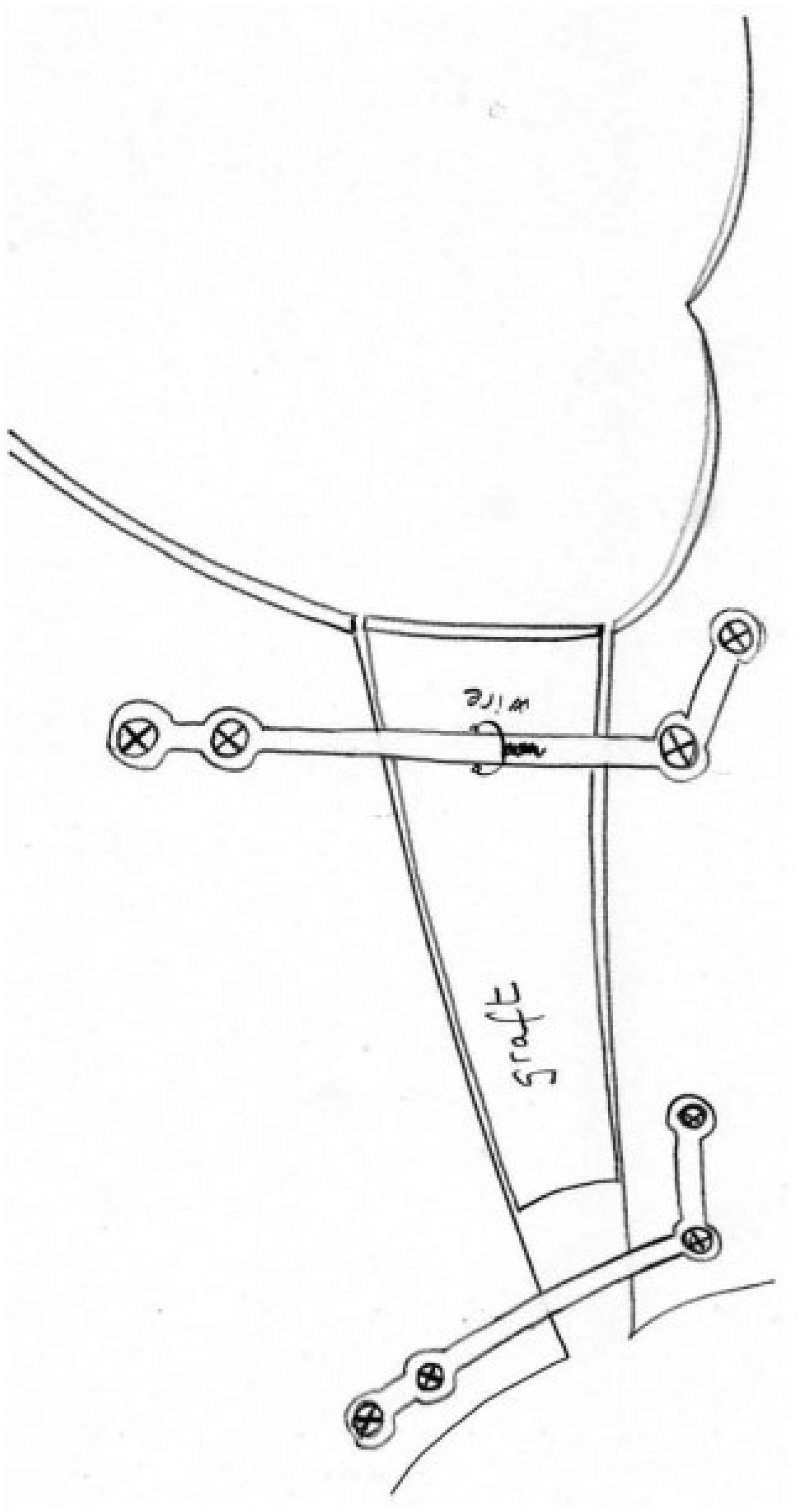
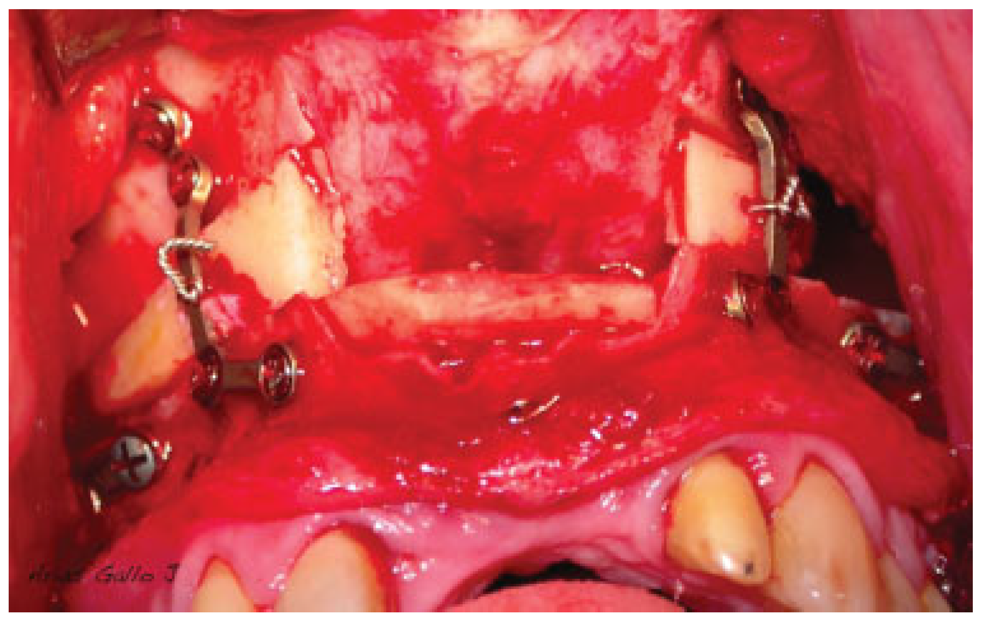
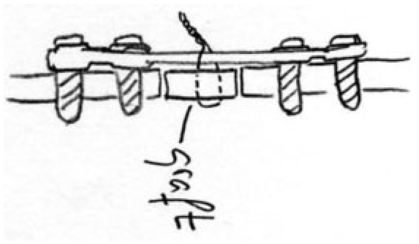
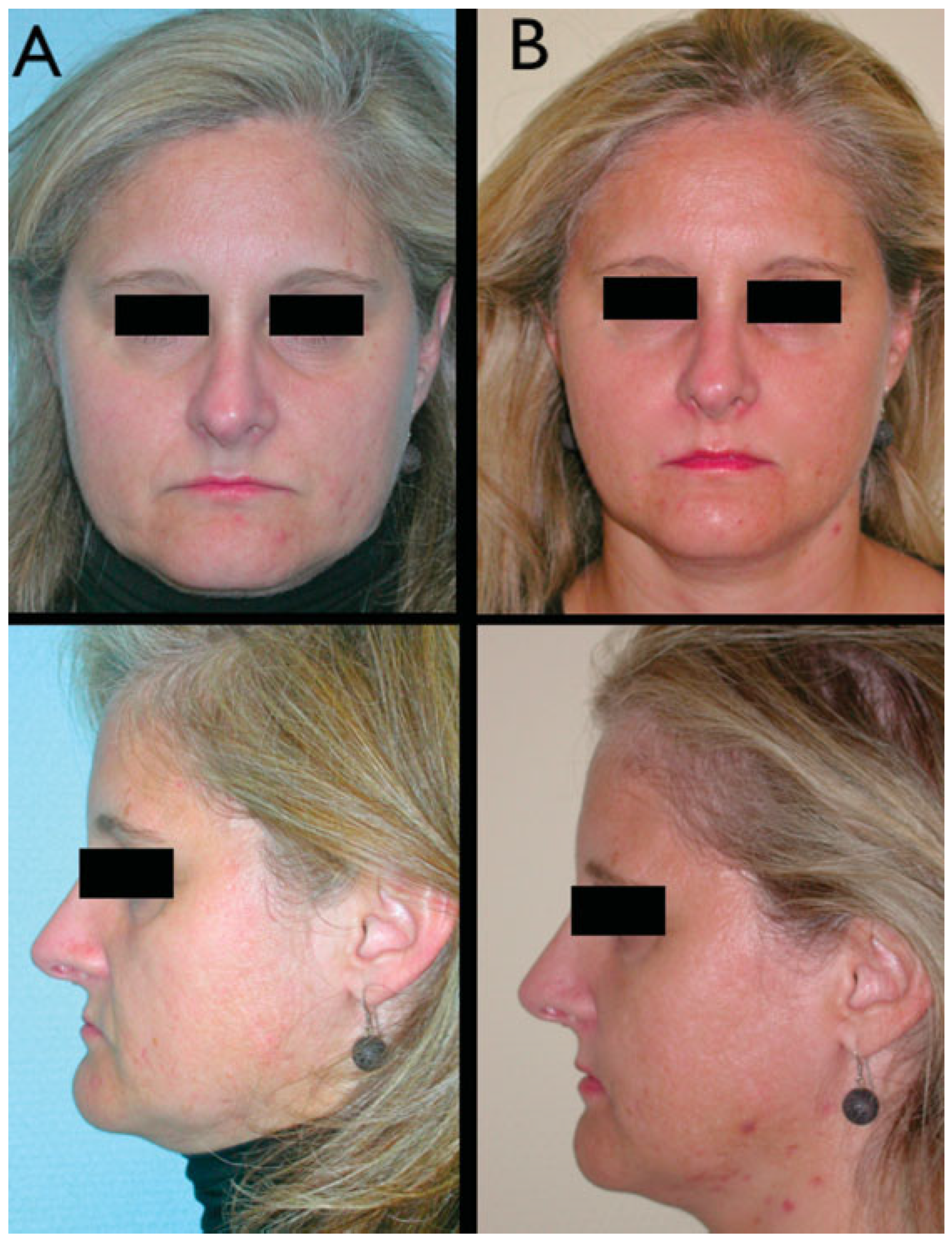
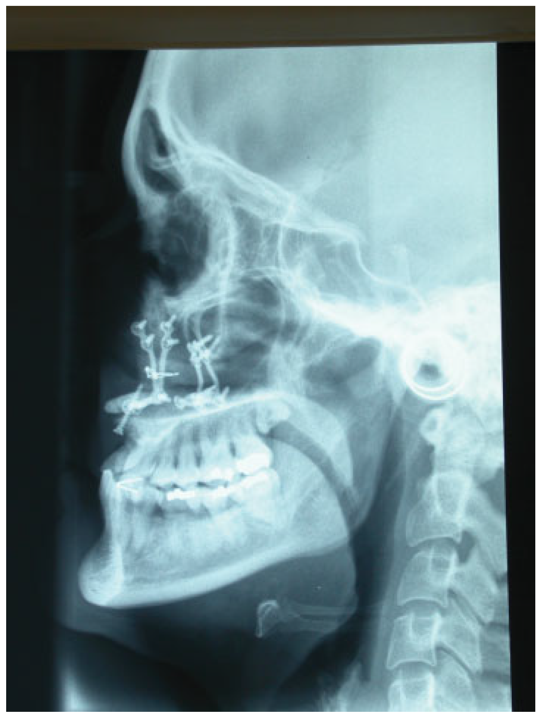
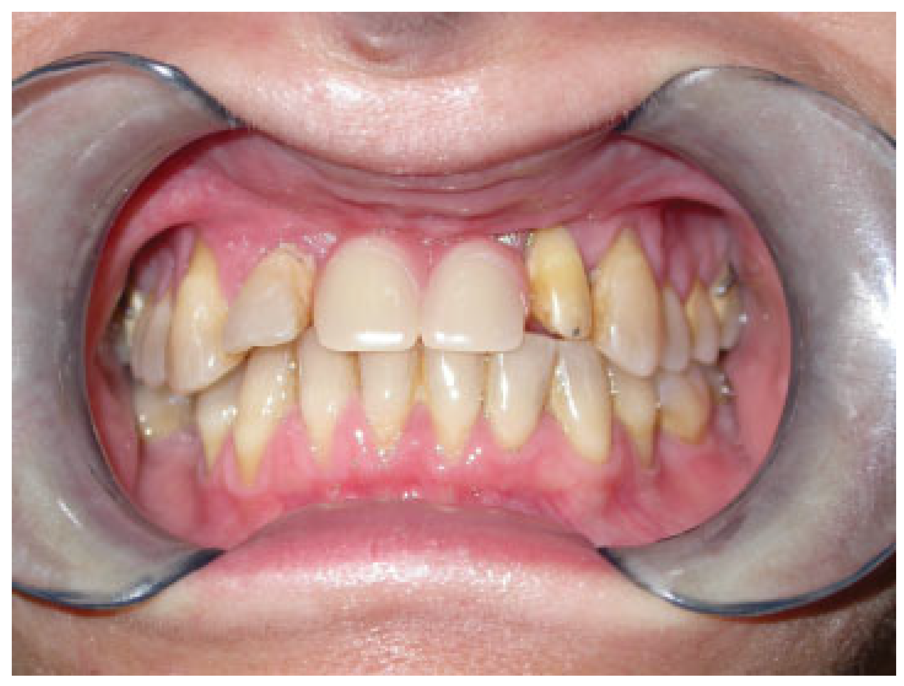
:Case Report
Discussion
Conclusion
References
- Jensen, J.; Sindet-Pedersen, S.; Oliver, A.J. Varying treatment strategies for reconstruction of maxillary atrophy with implants: results in 98 patients. J Oral Maxillofac Surg 1994, 52, 210–216; discussion 216–218. [Google Scholar] [PubMed]
- Ferri, J.; Lauwers, L.; Jeblaoui, Y.; Genay, A.; Raoul, G. Le Fort I osteotomy and calvarial bone grafting for dental implants. Rev Stomatol Chir Maxillofac 2010, 111, 63–67. [Google Scholar] [PubMed]
- Ferri, J.; Dujoncquoy, J.P.; Carneiro, J.M.; Raoul, G. Maxillary reconstruction to enable implant insertion: a retrospective study of 181 patients. Head Face Med 2008, 4, 31. [Google Scholar] [PubMed]
- Breine, U.; Brånemark, P.I. Reconstruction of alveolar jaw bone. An experimental and clinical study of immediate and preformed autologous bone grafts in combination with osseointegrated implants. Scand J Plast Reconstr Surg 1980, 14, 23–48. [Google Scholar] [PubMed]
- Yerit, K.C.; Posch, M.; Guserl, U.; et al. Rehabilitation of the severely atrophied maxilla by horseshoe Le Fort I osteotomy (HLFO). Oral Surg Oral Med Oral Pathol Oral Radiol Endod 2004, 97, 683–692. [Google Scholar] [PubMed]
- Sjöström, M.; Sennerby, L.; Nilson, H.; Lundgren, S. Reconstruction of the atrophic edentulous maxilla with free iliac crest grafts and implants: a 3-year report of a prospective clinical study. Clin Implant Dent Relat Res 2007, 9, 46–59. [Google Scholar] [PubMed]
- Serhan, H.; Slivka, M.; Albert, T.; Kwak, S.D. Is galvanic corrosion between titanium alloy and stainless steel spinal implants a clinical concern? Spine J 2004, 4, 379–387. [Google Scholar] [CrossRef] [PubMed]
- Høl, P.J.; Mølster, A.; Gjerdet, N.R. Should the galvanic combination of titanium and stainless steel surgical implants be avoided? Injury 2008, 39, 161–169. [Google Scholar] [CrossRef] [PubMed]

Disclaimer/Publisher’s Note: The statements, opinions and data contained in all publications are solely those of the individual author(s) and contributor(s) and not of MDPI and/or the editor(s). MDPI and/or the editor(s) disclaim responsibility for any injury to people or property resulting from any ideas, methods, instructions or products referred to in the content. |
© 2013 by the author. The Author(s) 2013.
Share and Cite
Pingarron-Martin, L.; Arias-Gallo, J.; Ong, H.S.; Pons, M.C. Le Fort I Osteotomy with Bone Grafts in Preprosthetic Surgery: Technical Note. Craniomaxillofac. Trauma Reconstr. 2013, 6, 143-146. https://doi.org/10.1055/s-0033-1333876
Pingarron-Martin L, Arias-Gallo J, Ong HS, Pons MC. Le Fort I Osteotomy with Bone Grafts in Preprosthetic Surgery: Technical Note. Craniomaxillofacial Trauma & Reconstruction. 2013; 6(2):143-146. https://doi.org/10.1055/s-0033-1333876
Chicago/Turabian StylePingarron-Martin, Lorena, Javier Arias-Gallo, Hui Shan Ong, and Manuel Chamorro Pons. 2013. "Le Fort I Osteotomy with Bone Grafts in Preprosthetic Surgery: Technical Note" Craniomaxillofacial Trauma & Reconstruction 6, no. 2: 143-146. https://doi.org/10.1055/s-0033-1333876
APA StylePingarron-Martin, L., Arias-Gallo, J., Ong, H. S., & Pons, M. C. (2013). Le Fort I Osteotomy with Bone Grafts in Preprosthetic Surgery: Technical Note. Craniomaxillofacial Trauma & Reconstruction, 6(2), 143-146. https://doi.org/10.1055/s-0033-1333876