Next Article in Journal
Le Fort I Osteotomy with Bone Grafts in Preprosthetic Surgery: Technical Note
Previous Article in Journal
Blowout Fracture in a 3-Year-Old
 
 
Craniomaxillofacial Trauma & Reconstruction is published by MDPI from Volume 18 Issue 1 (2025). Previous articles were published by another publisher in Open Access under a CC-BY (or CC-BY-NC-ND) licence, and they are hosted by MDPI on mdpi.com as a courtesy and upon agreement with Sage.
Case Report

Mandible and Zygomatic Fracture in a 2-Year-Old Patient Due to Dog Bite

by
Jesús R. Manzani Baldi
1,* and
Daniel A. Wolff de Freitas
2
1
Plastic Surgery, Clinicas Hospital, Montevideo, Uruguay
2
Plastic Surgery, Hospital Pasteur, Montevideo, Uruguay
*
Author to whom correspondence should be addressed.
Craniomaxillofac. Trauma Reconstr. 2013, 6(2), 137-141; https://doi.org/10.1055/s-0033-1333879
Submission received: 16 March 2012 / Revised: 6 May 2012 / Accepted: 6 May 2012 / Published: 5 March 2013

Abstract

:
Dog bite injury frequently occurs in children, and many of these bites involve the facial region. On the other hand, facial fractures due to dog attacks are a rare complication, with the orbital, nasal, and maxillary bones most often affected. We present a case report of a child who suffered a double facial fracture, mandible and left zygoma, due to a dog bite. The clinical diagnosis was supported by X-rays and computed tomography, which also provided information about the characteristics of the fracture. Internal fixation was done with titanium miniplates. Finally, the pathophysiological mechanism and the biomechanics of the fracture, as well as the use of resorbable versus nonresorbable material on infants, are discussed.

Frequently, cases of apparently quiet and friendly dogs that unexpectedly attack infants with anger are presented, and they usually result in severe soft tissue injuries [1,2]. The majority of injuries are to the head region, with the lips, cheeks, and nose often affected [3]. Most injuries experienced are confined to the soft tissues; nevertheless, maxillofacial fracture is a potential although rare complication. The incidence of facial fractures in the literature is discordant, and the majority of cases are not well investigated 1]. We reviewed the literature for pediatric facial fracture due to dog attack, and we found just four reports of mandible fracture and no reports for the combination of mandible and zygomatic fracture [4,5,6,7].
In the following case report we describe a dog bite injury in a child patient who, besides the soft tissue injuries (eyelid in this case), also presented two severe open fractures of the mandible and zygoma with an interesting biomechanical pattern analyzed later (Figure 1).

Case Report

We describe a 2-year-old girl without previous history, who was attacked by her pet (Rottweiler cross-breed, ~40 kg), suffering injuries mostly on the left side of her face. At the physical examination she presented in the middle of her left upper eyelid a three-layer vertical injury, without globe injury, and also skin lacerations at the lateral orbital rim and inferior mandible edge were found. Considerable swelling and hematoma of the inferior third of the face were also found. When we explored her oral occlusion, we found that she had partial opening of the mouth and right deviation (Figure 2).
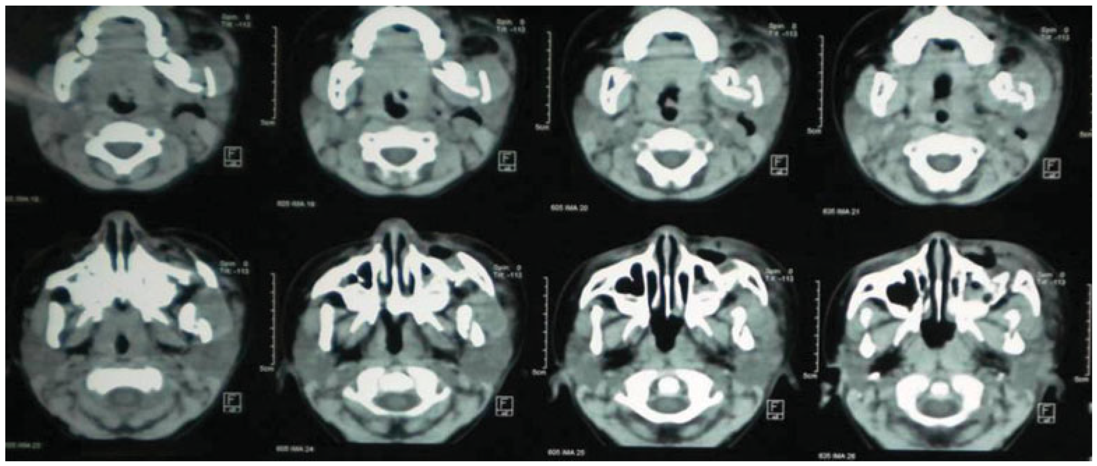
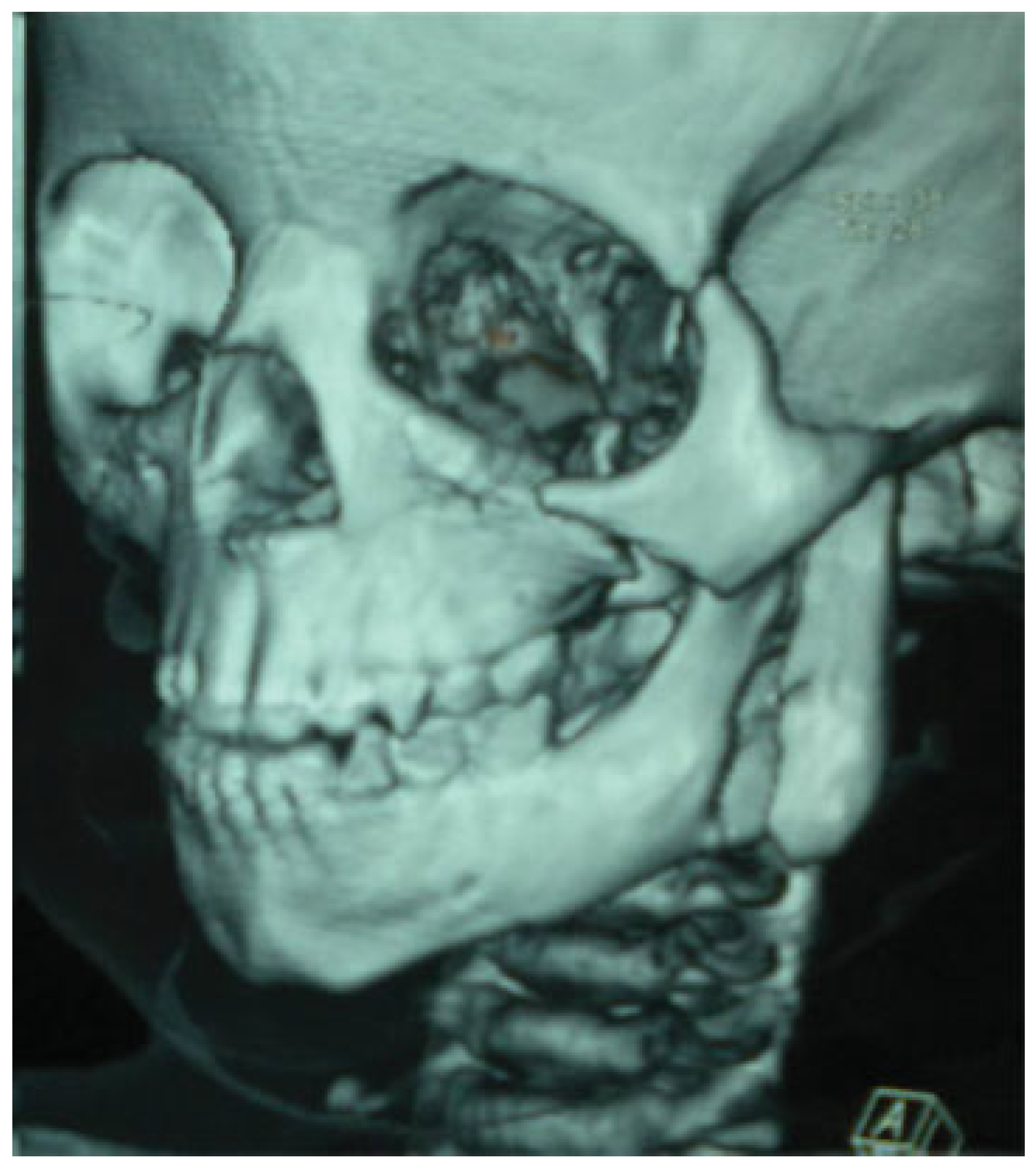
The imaging obtained showed a left zygoma fracture with inferior and lateral displacement of the body and a displaced left vertical ramus fracture extending from the sigmoid notch to the angle of the mandible in a sagittal direction (Figure 3, Figure 4 and Figure 5).

Procedure

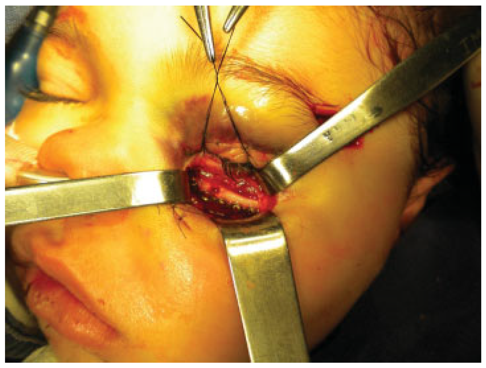
The procedure was done under general anesthesia with nasotracheal intubation. The first step was to repair the three layers of the upper eyelid laceration. A subciliary approach was then utilized (Figure 6) for reduction and stabilization of the zygomatic fracture. With this approach in children we have excellent exposure, and in our hands no ectropion was presented. Once the anatomic reduction was reached, internal fixation was performed at the inferior orbital rim with a single titanium six-hole miniplate that uses 1.3-mm monocortical screws. Then, we approached to the lateral orbital rim, through the ciliary wound. The fixation was done with a single titanium four-hole miniplate that uses 1.3-mm monocortical screws (Figure 7).
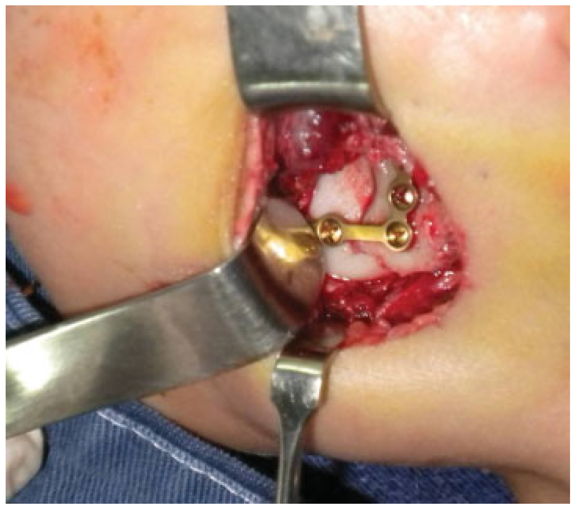
For the mandible approach (Figure 8), we extended the submandibular wound, with careful subplatysmal dissection to avoid the injury of the mandibular branch of the facial nerve. Because of the small size of the mandible and the impossibility of intermaxillary fixation (with cerclage or screws due to the immature mandible), the right dental occlusion (interincisive line) was maintained in position by the hands of the assistant. Only an L-shaped titanium six-hole miniplate that used 1.5-mm bicortical screws at the basal side of the mandible was used to avoid dental germs.
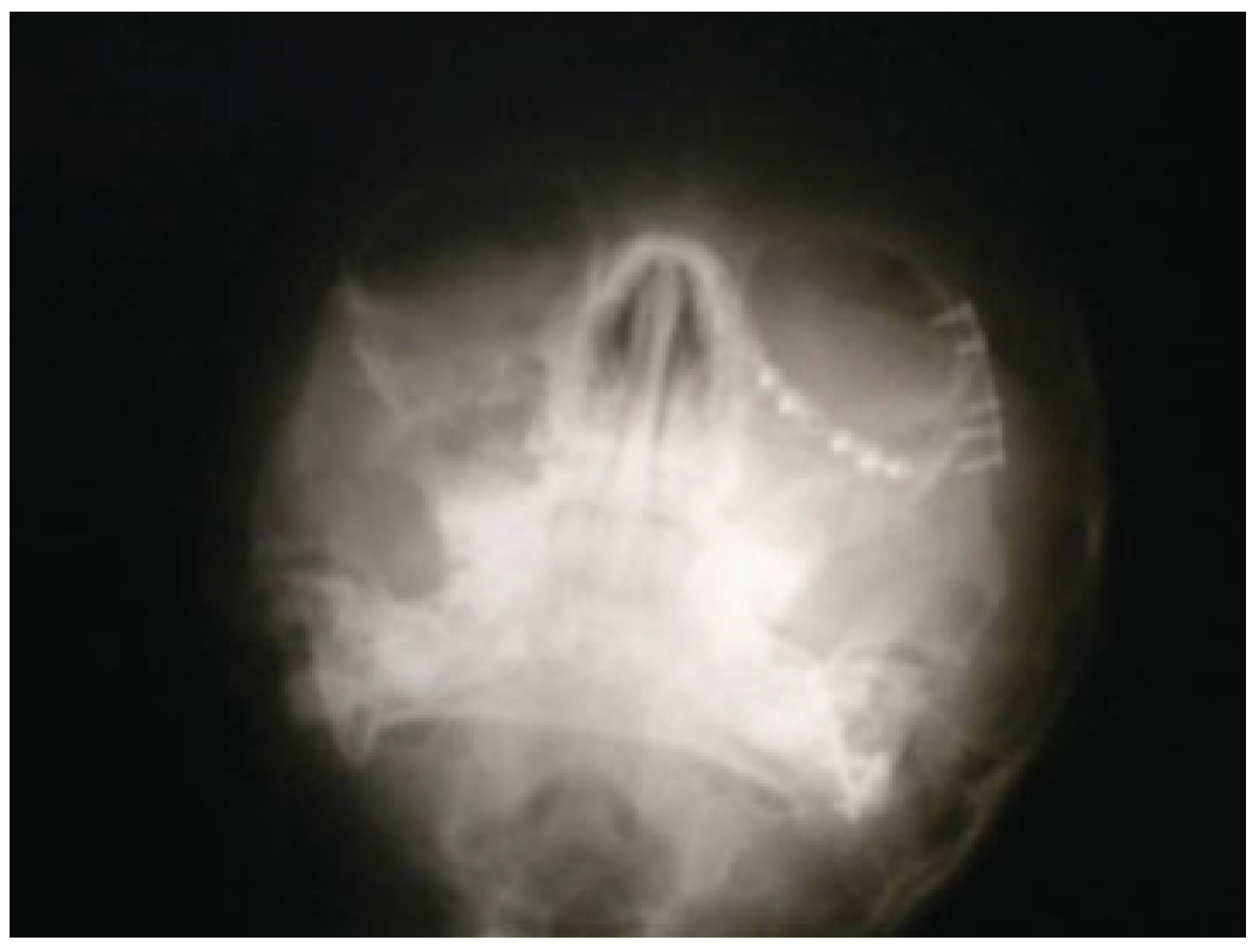
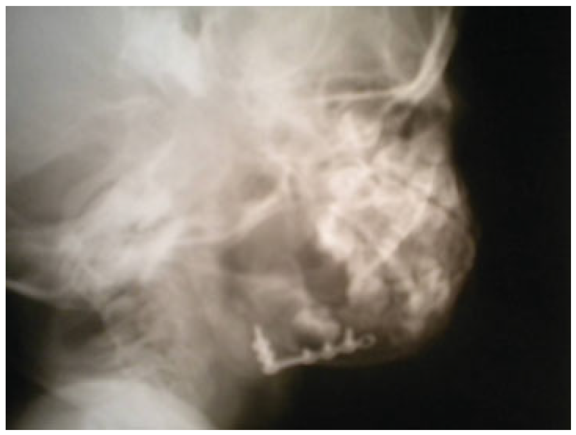
Postoperatively, antibiotics and corticoids were used without any kind of complication. One week later the patient was discharged. In the followings exams, the patient reached a good dental occlusion and aperture and normal zygomatic projection. X-rays showed the hardware in the correct position (Figure 9 and Figure 10). Our department does not use 3-D computed tomography with children unless it is strictly necessary (radioprotection policies). The soft tissue reconstruction healed correctly without functional impairment.

Discussion

The actual incidence of facial fractures related to dog attacks is currently unknown. The majority of the reviews documented no maxillofacial fractures in cases of facial dog-bite injuries [8,9,10]. Tu et al reported facial fractures in fewer than 5% of dog bites [1]. In our review, we found that in cases of facial fractures, the most frequent sites were the orbital, nasal, and maxillary bones. We found four reports of mandible fractures and no reports for the combination of zygomatic and mandible fractures like this case [4,5,6,7].
The biomechanics and the physiopathology of these kinds of mandible fractures are one of the main points of discussion. The compression force applied in the vertical vector rests on two points: zygoma and mandible angle. The applied force was ~100 kg calculated by the dog breed and age (Figure 1) [11]. This force destroyed the zygomatic toehold with its down displacement and at the same time produced a rare kind of mandible fracture line [12,13,14,15], characterized by a split of the inner from the external cortical to the sigmoid notch direction, resulting in a posterior fragment with the condyle and an anterior fragment attached to the dental arch [16].
Our opinion is that the dog tooth produced this fracture plane within the two corticals, and the elasticity of the young mandible contributed to this type of fracture, similar to the “greenstick” fracture that occurs in long bones [15].
Dog-bite wounds are often left open for some days because of their reputation for infection if primarily closed. Like other authors [16], our experience shows that open-wound dog bites could be safety closed primarily without increased incidence of infection if you clean wounds properly and use adequate antibiotic therapy.
On one hand, it is also interesting to discuss the use of titanium miniplates versus resorbable miniplates (polylactic acid, polyglycolic acid, and polydioxanone) often used in children [17,18,19,20]. Even though we have no wide experience with resorbable materials, we consider that mandible fractures always have to be treated with titanium because it gave us more biomechanical safety in this clinical case.
On the other hand, the zygomatic fracture has different biomechanics and could have been repaired with resorbable material. However, according to some authors the nonreabsorbable material would not interfere with bone growth unless it was fixated over ossification points.1,15 We decided to place miniplates at the orbital rim over some malar ossification points, knowing that our surgical plan is to remove the miniplates in 6 months. Finally, our decision to use nonreabsorbable plates is based on the economic costs and overcoming the initial learning curve for the placement of resorbable plates.

References

  1. Tu, A.H.; Girotto, J.A.; Singh, N.; et al. Facial fractures from dog bite injuries. Plast Reconstr Surg 2002, 109, 1259–1265. [Google Scholar] [PubMed]
  2. Hogg, N.J.; Stewart, T.C.; Armstrong, J.E.; Girotti, M.J. Epidemiology of maxillofacial injuries at trauma hospitals in Ontario, Canada, between 1992 and 1997. J Trauma 2000, 49, 425–432. [Google Scholar] [CrossRef] [PubMed]
  3. Wolff, K.D. Management of animal bite injuries of the face: experience with 94 patients. J Oral Maxillofac Surg 1998, 56, 838–843; discussion 843–844. [Google Scholar] [CrossRef] [PubMed]
  4. Rapuano, R.; Stratigos, G.T. Mandibular fracture resulting from dog bite: report of a case. J Oral Surg 1976, 34, 359–361. [Google Scholar] [PubMed]
  5. Case Report of Fractured Mandible in a Child Aged 1 Year. Available online: http://www.aofoundation.org/portal/wps/portal/!ut/p/_s.7_0_A/7_0_3K7?contentUrl=/aca/case_of_the/2006/08/ex_alumni_cases_0000024.jsp (accessed on 2 October 2012).
  6. Walker, T.; Modayil, P.; Cascarini, L.; Collyer, J. Dog bite-fracture of the mandible in a 9 month old infant: a case report. Cases Journal 2009, 2, 1–3. [Google Scholar] [CrossRef] [PubMed]
  7. Cottom, H.; Tuopar, D.; Ameerally, P. Mandibular fracture in a child resulting from a dog attack: a case report. Case Reports in Dentistry 2011, 1–4. [Google Scholar] [CrossRef] [PubMed]
  8. Palmer, J.; Rees, M. Dog bites of the face: a 15 year review. Br J Plast Surg 1983, 36, 315–318. [Google Scholar] [PubMed]
  9. Schalamon, J.; Ainoedhofer, H.; Singer, G.; et al. Analysis of dog bites in children who are younger than 17 years. Pediatrics 2006, 117, e374–e379. [Google Scholar] [CrossRef] [PubMed]
  10. Karlson, T.A. The incidence of facial injuries from dog bites. JAMA 1984, 251, 3265–3267. [Google Scholar] [PubMed]
  11. Lidner, D.; Marretta, H.; Pijanowski, G.; Johnson, A.; Smith, C. Measurament of bit force in dogs to pilot study. Veterinary Dentistry 1995, 12, 49–52. [Google Scholar] [CrossRef]
  12. Yaremchuk, M.J. Revisional surgery of nasoethmoidal orbital fractures. In Operative Techniques in Plastic Surgery; Manson, P.N., Ed.; W.B. Saunders: Philadelphia, PA, USA, 1998. [Google Scholar]
  13. Merville, L.; Vincent, J. Traumatismes de la face. Fractures du malaire. Fractures de la corniche zygomatique. Encycl Méd Chir 1992, 3, 23. [Google Scholar]
  14. Merville, L.; Vincent, J. Traumatismes de la face. Fractures mandibulaires. Encycl Méd Chir 1992, 3, 23–10. [Google Scholar]
  15. Dufresnese, C.; Manson, P. Cirugía Plástica McCarthy. Traumatismos faciales en niños. Ed. Med Panam 1991, 269–313. [Google Scholar]
  16. Maimaris, C.; Quinton, D.N. Dog-bite lacerations: a controlled trial of primary wound closure. Arch Emerg Med 1988, 5, 156–161. [Google Scholar] [CrossRef] [PubMed]
  17. Cheung, L.K.; Chow, L.K.; Chiu, W.K. A randomized controlled trial of resorbable versus titanium fixation for orthognathic surgery. Oral Surg Oral Med Oral Pathol Oral Radiol Endod 2004, 98, 386–397. [Google Scholar] [PubMed]
  18. Costa, F.; Robiony, M.; Zerman, N.; Zorzan, E.; Politi, M. Bone biological plate for stabilization of maxillary inferior repositioning. Minerva Stomatol 2005, 54, 227–236. [Google Scholar] [PubMed]
  19. Fedorowicz, Z.; Nasser, M.; Newton, J.T.; Oliver, R.J. Resorbable versus titanium plates for orthognathic surgery. Cochrane Database Syst Rev 2007, 2, CD006204. [Google Scholar]
  20. Simon, J.A.; Ricci, J.L.; Di Cesare, P.E. Bioresorbable fracture fixation in orthopedics: a comprehensive review. Part I. Basic science and preclinical studies. Am J Orthop 1997, 26, 665–671. [Google Scholar] [PubMed]
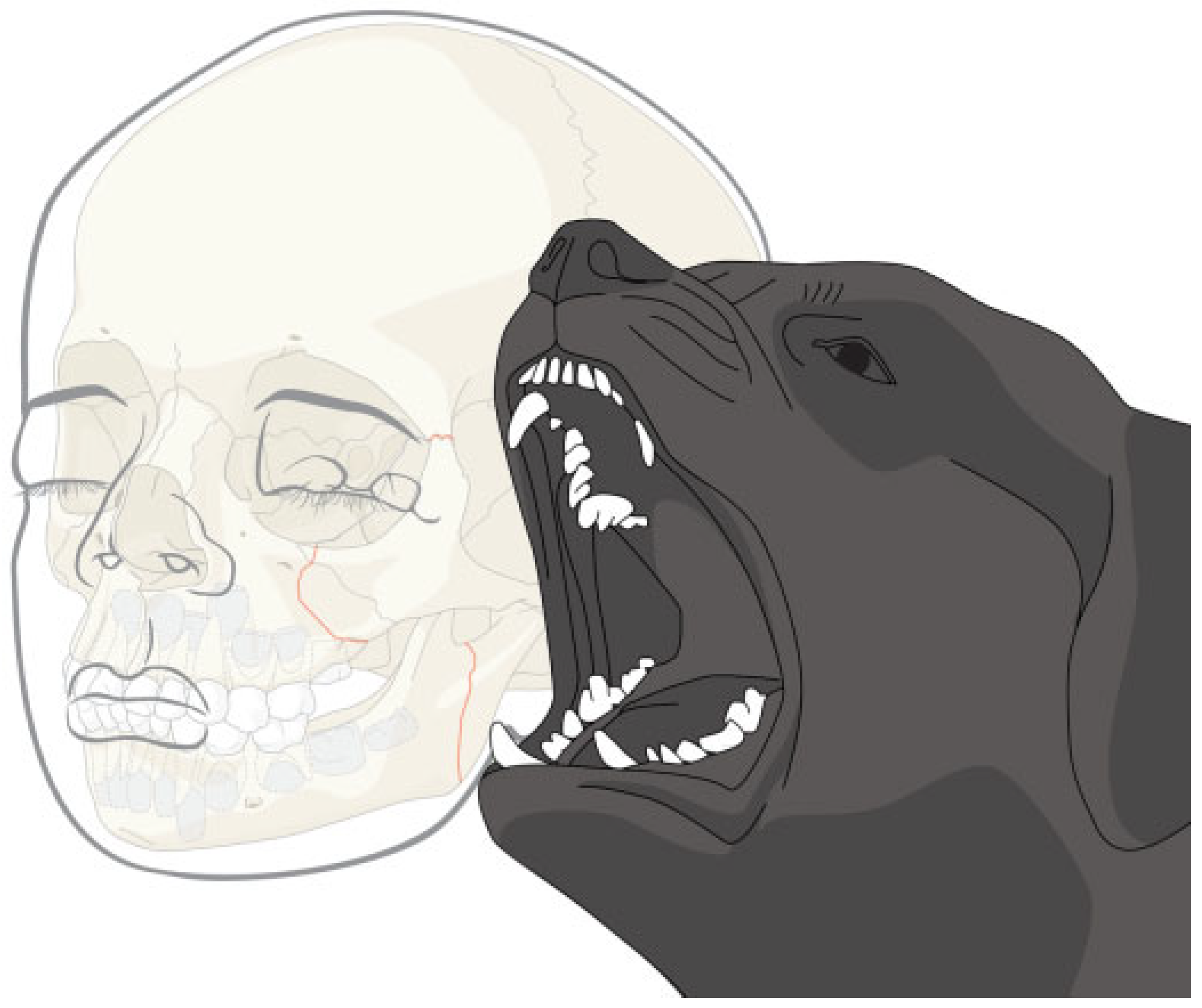
Figure 1. Biomechanics of the fracture.
Cmtr 06 00022 g001
Figure 2. Partial opening of the mouth and right deviation.
Cmtr 06 00022 g002
Figure 3. CT coronal view of the sagittal ramus fracture. Abbreviation: CT, computed tomography.
Cmtr 06 00022 g003
Figure 4. CT axial view of zygomatic and mandible fracture. Abbreviation: CT, computed tomography.
Cmtr 06 00022 g004
Figure 5. Three-dimensional view.
Cmtr 06 00022 g005
Figure 6. Subciliary approach.
Cmtr 06 00022 g006
Figure 7. Dingman’s approach.
Cmtr 06 00022 g007
Figure 8. Mandible approach.
Cmtr 06 00022 g008
Figure 9. Postoperative. Water’s view.
Cmtr 06 00022 g009
Figure 10. Postoperative. Left oblique lateral view.
Cmtr 06 00022 g010
Disclaimer/Publisher’s Note: The statements, opinions and data contained in all publications are solely those of the individual author(s) and contributor(s) and not of MDPI and/or the editor(s). MDPI and/or the editor(s) disclaim responsibility for any injury to people or property resulting from any ideas, methods, instructions or products referred to in the content.