A 59-year-old man with severe mitral regurgitation underwent successful mitral valve repair. Postoperative recovery was uneventful except for the development of a third-degree atrio-ventricular block (fig. 1A). The haemodynamic consequences of the atrio-ventricular dissociation were observed during echocardiography (fig. 1B and fig. 1C): at the beginning and the end of the re- cordings, right atrial depolarisation and contraction occurs during ventricular systole when the tricuspid valve is closed, leading to reversal of blood flow (short arrows). This echocardiographic finding represents the clinically observable jugular venous pulsation called “Cannon waves”. In contrast, when the atrio-ventricular sequence is more or less correct, the backflow into the IVC and the HV is minimal (long arrows).
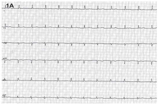
Figure 1A.
Third-degree atrio-ventricular block.
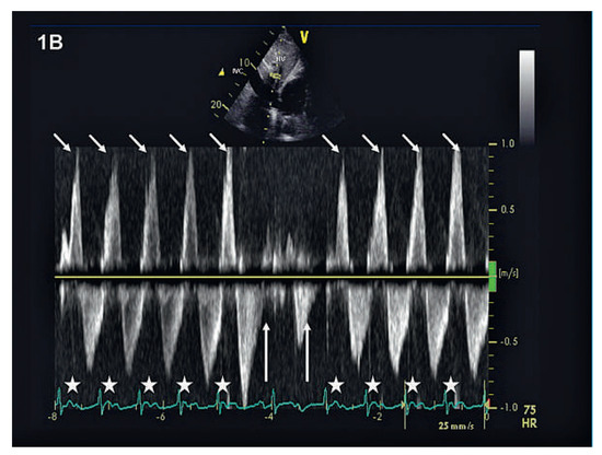
Figure 1B.
Pulse-wave Doppler through a hepatic vein demonstrating blood flow reversals during atrioventricular block. If atrioventricular sequence is correct (arrows), backflow into the hepatic vein is minimal.
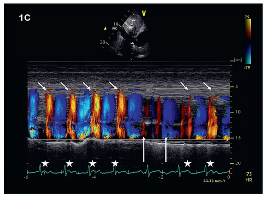
Figure 1C.
Colour-Doppler M-inode through a hepatic vein confirming the findings from Figure 1B.
The differential diagnosis of systolic flow reversal includes tricuspid regurgitation (excluded in fig. 1D, dashed circle) as well as rhythm disorders (atrial or ventricular premature beats, atrioventricular block of any degree and atrioventricular dissociation during ventricular tachycardia).
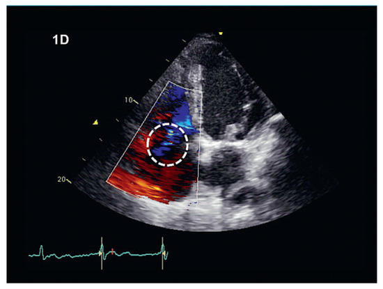
Figure 1D.
Colour-Doppler image. Relevant tricospid regurgitation can be excluded.
Funding/potential conflict of interest
No financial support and no other potential conflict of interest relevant to this article were reported.
© 2012 by the authors. Licensee MDPI, Basel, Switzerland. This article is an open access article distributed under the terms and conditions of the Creative Commons Attribution (CC BY) license (https://creativecommons.org/licenses/by/4.0/).