
Prevalence, Features and Degree of Association of Oral Lesions in COVID-19: A Systematic Review of Systematic Reviews

 ,
, 

 , ,
, , 
Abstract
:1. Introduction
2. Materials and Methods
2.1. Search Strategy
- COVID-19 OR SARS-CoV-2 OR Coronavirus disease 2019 OR novel coronavirus
- AND oral lesions OR oral manifestations OR oral signs
- AND Systematic review OR meta-analysis.
2.2. Study Selection and Eligibility Criteria
2.3. Data Extraction
2.4. Quality Assessment and Data Synthesis
2.5. Synthesis Methods
3. Results
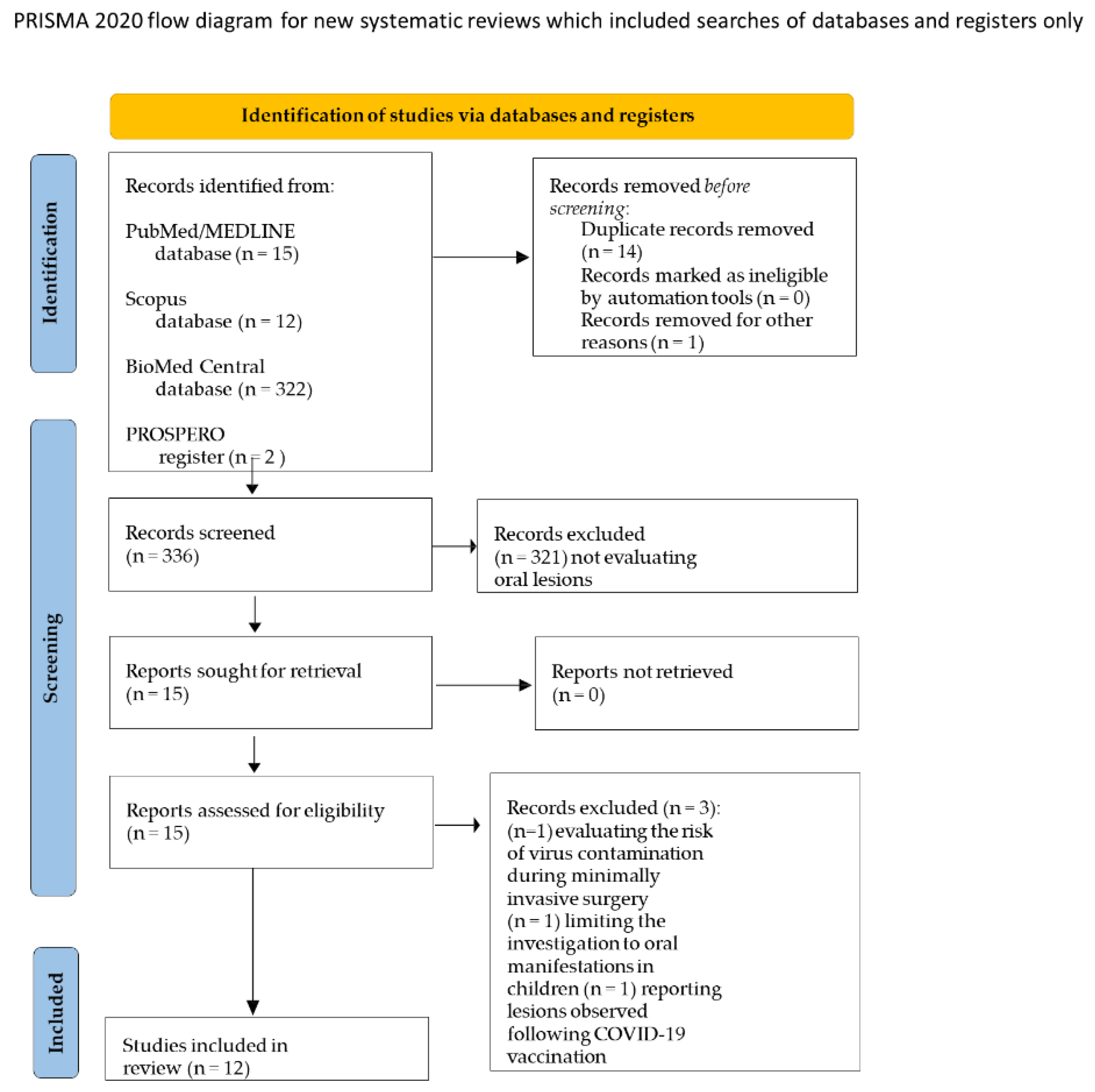
3.1. Study Selection
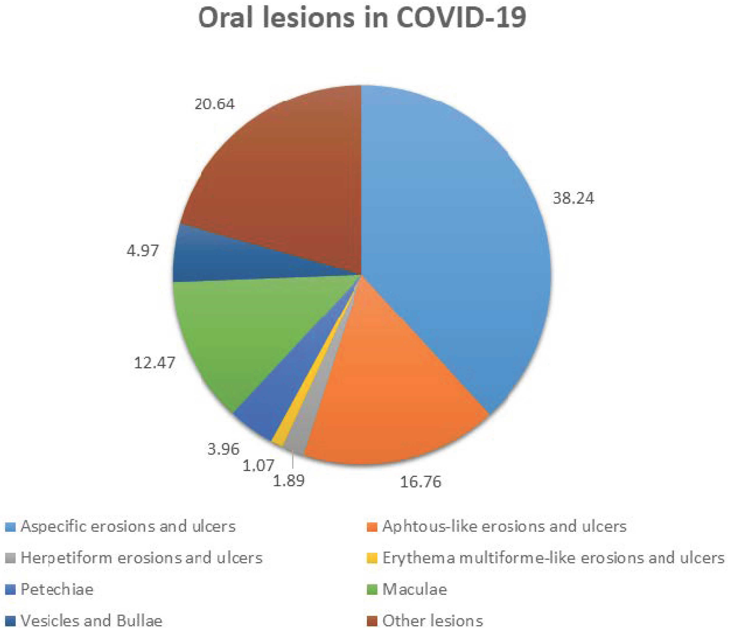
3.2. Studies’ Characteristics and Qualitative Synthesis
4. Discussion
5. Conclusions
Author Contributions
Funding
Institutional Review Board Statement
Informed Consent Statement
Data Availability Statement
Acknowledgments
Conflicts of Interest
References
- Orozco, M.S.; Niño-Martínez, N.; Martínez-Castañón, G.-A.; Marín, N.P.; Valencia, C.S.; Velázquez, F.D.; Munguía, P.S.; Santana, M.C. Presence of SARS-CoV-2 and Its Entry Factors in Oral Tissues and Cells: A Systematic Review. Medicina 2021, 57, 523. [Google Scholar] [CrossRef] [PubMed]
- Wan, S.; Li, M.; Ye, Z.; Yang, C.; Cai, Q.; Duan, S.; Song, B. CT Manifestations and Clinical Characteristics of 1115 Patients with Coronavirus Disease 2019 (COVID-19): A Systematic Review and Meta-analysis. Acad. Radiol. 2020, 27, 910–921. [Google Scholar] [CrossRef] [PubMed]
- Recalcati, S. Cutaneous manifestations in COVID-19: A first perspective. J. Eur. Acad. Dermatol. Venereol. 2020, 34, e212–e213. [Google Scholar] [CrossRef] [PubMed]
- Rocha, B.A.; Souto, G.R.; Grossmann, S.D.M.C.; de Aguiar, M.C.F.; de Andrade, B.A.B.; Romañach, M.J.; Horta, M.C.R. Viral enanthema in oral mucosa: A possible diagnostic challenge in the COVID-19 pandemic. Oral Dis. 2021, 27, 776–778. [Google Scholar] [CrossRef] [PubMed]
- Xu, J.; Li, Y.; Gan, F.; Du, Y.; Yao, Y. Salivary Glands: Potential Reservoirs for COVID-19 Asymptomatic Infection. J. Dent. Res. 2020, 99, 989. [Google Scholar] [CrossRef] [PubMed] [Green Version]
- Xu, H.; Zhong, L.; Deng, J.; Peng, J.; Dan, H.; Zeng, X.; Li, T.; Chen, Q. High expression of ACE2 receptor of 2019-nCoV on the epithelial cells of oral mucosa. Int. J. Oral Sci. 2020, 12, 8. [Google Scholar] [CrossRef] [PubMed]
- Mazur, M.; Duś-Ilnicka, I.; Jedliński, M.; Ndokaj, A.; Janiszewska-Olszowska, J.; Ardan, R.; Radwan-Oczko, M.; Guerra, F.; Luzzi, V.; Vozza, I.; et al. Facial and Oral Manifestations Following COVID-19 Vaccination: A Survey-Based Study and a First Perspective. Int. J. Environ. Res. Public Health 2021, 18, 4965. [Google Scholar] [CrossRef]
- Martín Carreras-Presas, C.; Amaro Sánchez, J.; López-Sánchez, A.F.; Jané-Salas, E.; Somacarrera Pérez, M.L. Oral vesiculobullous lesions associated with SARS-CoV-2 infection. Oral Dis. 2021, 27, 710–712. [Google Scholar] [CrossRef]
- Petrescu, N.; Lucaciu, O.; Roman, A. Oral mucosa lesions in COVID-19. Oral Dis. 2022, 28, 935–936. [Google Scholar] [CrossRef]
- Vieira, A.R. Oral manifestations in coronavirus disease 2019 (COVID-19). Oral Dis. 2020, 27, 770. [Google Scholar] [CrossRef]
- Di Spirito, F.; Pelella, S.; Argentino, S.; Sisalli, L.; Sbordone, L. Oral manifestations and the role of the oral healthcare workers in COVID-19. Oral Dis. 2022, 28, 1003–1004. [Google Scholar] [CrossRef] [PubMed]
- Classification and diagnostic criteria for oral lesions in HIV infection. EC-Clearinghouse on Oral Problems Related to HIV Infection and WHO Collaborating Centre on Oral Manifestations of the Immunodeficiency Virus. J. Oral Pathol. Med. Off. Publ. Int. Assoc. Oral Pathol. Am. Acad. Oral Pathol. 1993, 22, 289–291. [Google Scholar]
- World Health Organization (WHO). Living Guidance for Clinical Management of COVID-19: Living Guidance. Available online: WHO-2019-nCoV-clinical-2021.2-eng.pdf (accessed on 7 May 2022).
- Page, M.J.; McKenzie, J.E.; Bossuyt, P.M.; Boutron, I.; Hoffmann, T.C.; Mulrow, C.D.; Shamseer, L.; Tetzlaff, J.M.; Akl, E.A.; Brennan, S.E.; et al. The PRISMA 2020 statement: An updated guideline for reporting systematic reviews. BMJ 2021, 372, n71. [Google Scholar] [CrossRef] [PubMed]
- Higgins, J.; Thomas, J. Cochrane Handbook for Systematic Reviews of Interventions; John Wiley & Sons, Ltd.: Chichester, UK, 2008. [Google Scholar]
- Oivio, U.-M.; Pesonen, P.; Ylipalosaari, M.; Kullaa, A.; Salo, T. Prevalence of oral mucosal normal variations and lesions in a middle-aged population: A Northern Finland Birth Cohort 1966 study. BMC Oral Health 2020, 20, 357. [Google Scholar] [CrossRef]
- dos Santos, J.A.; Normando, A.G.; Da Silva, R.C.; Acevedo, A.; Canto, G.D.L.; Sugaya, N.; Santos-Silva, A.; Guerra, E. Oral Manifestations in Patients with COVID-19: A Living Systematic Review. J. Dent. Res. 2021, 100, 141–154. [Google Scholar] [CrossRef]
- Shea, B.J.; Reeves, B.C.; Wells, G.; Thuku, M.; Hamel, C.; Moran, J.; Moher, D.; Tugwell, P.; Welch, V.; Kristjansson, E.; et al. AMSTAR 2: A critical appraisal tool for systematic reviews that include randomised or non-randomised studies of healthcare interventions, or both. BMJ 2017, 358, j4008. [Google Scholar] [CrossRef] [Green Version]
- Pavan, N.; Crestani, A.; Abrate, A.; De Nunzio, C.; Esperto, F.; Giannarini, G.; Galfano, A.; Gregori, A.; Liguori, G.; Bartoletti, R.; et al. Risk of Virus Contamination Through Surgical Smoke During Minimally Invasive Surgery: A Systematic Review of the Literature on a Neglected Issue Revived in the COVID-19 Pandemic Era. Eur. Urol. Focus 2020, 6, 1058–1069. [Google Scholar] [CrossRef]
- Nascimento, R.B.; Araujo, N.S.; Silva, J.C.; Xavier, F.C.A. Oral manifestations of multisystemic inflammatory syndrome in children (MIS-C) and Kawasaki disease associated to COVID-19: A systematic review. Spec. Care Dent. Off. Publ. Am. Assoc. Hosp. Dent. Acad. Dent. Handicap. Am. Soc. Geriatr. Dent. 2021, 42, 266–280. [Google Scholar] [CrossRef]
- Triantafyllidis, K.K.; Giannos, P.; Mian, I.T.; Kyrtsonis, G.; Kechagias, K.S. Varicella Zoster Virus Reactivation Following COVID-19 Vaccination: A Systematic Review of Case Reports. Vaccines 2021, 9, 1013. [Google Scholar] [CrossRef]
- dos Santos, J.A.; Normando, A.; da Silva, R.C.; Acevedo, A.; Canto, G.D.L.; Sugaya, N.; Santos-Silva, A.; Guerra, E. Oral Manifestations in Patients with COVID-19: A 6-Month Update. J. Dent. Res. 2021, 100, 1321–1329. [Google Scholar] [CrossRef]
- Aragoneses, J.; Suárez, A.; Algar, J.; Rodríguez, C.; López-Valverde, N.; Aragoneses, J.M. Oral Manifestations of COVID-19: Updated Systematic Review with Meta-Analysis. Front. Med. 2021, 8, 726753. [Google Scholar] [CrossRef] [PubMed]
- Bhujel, N.; Zaheer, K.; Singh, R.P. Oral mucosal lesions in patients with COVID-19: A systematic review. Br. J. Oral Maxillofac. Surg. 2021, 59, 1024–1030. [Google Scholar] [CrossRef] [PubMed]
- Cuevas-Gonzalez, M.V.; Espinosa-Cristóbal, L.F.; Donohue-Cornejo, A.; Tovar-Carrillo, K.L.; Saucedo-Acuña, R.A.; García-Calderón, A.G.; Guzmán-Gastelum, D.A.; Cuevas-Gonzalez, J.C. COVID-19 and its manifestations in the oral cavity: A systematic review. Medicine 2021, 100, e28327. [Google Scholar] [CrossRef] [PubMed]
- Doceda, M.V.; Gavriiloglou, M.; Petit, C.; Huck, O. Oral Health Implications of SARS-CoV-2/COVID-19: A Systematic Review. Oral Health Prev. Dent. 2022, 20, 207–218. [Google Scholar] [CrossRef] [PubMed]
- Erbaş, G.S.; Botsali, A.; Erden, N.; Arı, C.; Taşkın, B.; Alper, S.; Vural, S. COVID-19-related oral mucosa lesions among confirmed SARS-CoV-2 patients: A systematic review. Int. J. Dermatol. 2022, 61, 20–32. [Google Scholar] [CrossRef] [PubMed]
- Nijakowski, K.; Wyzga, S.; Singh, N.; Podgórski, F.; Surdacka, A. Oral Manifestations in SARS-CoV-2 Positive Patients: A Systematic Review. J. Clin. Med. 2022, 11, 2202. [Google Scholar] [CrossRef]
- Orilisi, G.; Mascitti, M.; Togni, L.; Monterubbianesi, R.; Tosco, V.; Vitiello, F.; Santarelli, A.; Putignano, A.; Orsini, G. Oral Manifestations of COVID-19 in Hospitalized Patients: A Systematic Review. Int. J. Environ. Res. Public Health 2021, 18, 12511. [Google Scholar] [CrossRef]
- Qi, X.; Northridge, M.E.; Hu, M.; Wu, B. Oral health conditions and COVID-19: A systematic review and meta-analysis of the current evidence. Aging Health Res. 2022, 2, 100064. [Google Scholar] [CrossRef]
- Prevalence of Oral Manifestations in COVID-19: A Systematic Review. Available online: https://onlinelibrary.wiley.com/doi/10.1002/rmv.2345 (accessed on 7 May 2022).
- Silveira, F.M.; Mello, A.L.R.; Fonseca, L.D.S.; Ferreira, L.D.S.; Kirschnick, L.B.; Martins, M.D.; Schuch, L.F.; de Arruda, J.A.A.; Soares, C.D.; Sales, A.D.O.; et al. Morphological and tissue-based molecular characterization of oral lesions in patients with COVID-19: A living systematic review. Arch. Oral Biol. 2022, 136, 105374. [Google Scholar] [CrossRef]
- Uzêda-E-Silva, V.D.; de Sá, I.B.; Martins, J.; Pedreira, N.; Vieira, V.; Silva, B. Oral lesions associated with COVID-19: A systematic review. Stomatologija 2021, 23, 3–8. [Google Scholar]
- Nuno-Gonzalez, A.; Martin-Carrillo, P.; Magaletsky, K.; Rios, M.M.; Mañas, C.H.; Almazan, J.A.; Casasola, G.G.; Castro, E.P.; Arenas, A.G.; Ibarguren, A.M.; et al. Prevalence of mucocutaneous manifestations in 666 patients with COVID-19 in a field hospital in Spain: Oral and palmoplantar findings. Br. J. Dermatol. 2021, 184, 184–185. [Google Scholar] [CrossRef]
- Gebhard, C.; Regitz-Zagrosek, V.; Neuhauser, H.K.; Morgan, R.; Klein, S.L. Impact of sex and gender on COVID-19 outcomes in Europe. Biol. Sex Differ. 2020, 11, 29. [Google Scholar] [CrossRef]
- Tay, M.Z.; Poh, C.M.; Rénia, L.; Macary, P.A.; Ng, L.F.P. The trinity of COVID-19: Immunity, inflammation and intervention. Nat. Rev. Immunol. 2020, 20, 363–374. [Google Scholar] [CrossRef]
- Iranmanesh, B.; Khalili, M.; Amiri, R.; Zartab, H.; Aflatoonian, M. Oral manifestations of COVID-19 disease: A review article. Dermatol. Ther. 2021, 34, e14578. [Google Scholar] [CrossRef]
- Di Spirito, F.; Iacono, V.J.; Alfredo, I.; Alessandra, A.; Sbordone, L.; Lanza, A. Evidence-based Recommendations on Periodontal Practice and the Management of Periodontal Patients during and after the COVID-19 Era: Challenging Infectious Diseases Spread by Air-borne Transmission. Open Dent. 2021, 15, 325–336. [Google Scholar] [CrossRef]
- Fidan, V.; Koyuncu, H.; Akin, O. Oral lesions in COVID-19 positive patients. Am. J. Otolaryngol. 2021, 42, 102905. [Google Scholar] [CrossRef]
- Elamrousy, W.A.H.; Nassar, M.; Issa, D.R. Prevalence of Oral Lesions in COVID-19 Egyptian Patients. J. Int. Soc. Prev. Community Dent. 2021, 11, 712–720. [Google Scholar] [CrossRef]
- Favia, G.; Tempesta, A.; Barile, G.; Brienza, N.; Capodiferro, S.; Vestito, M.C.; Crudele, L.; Procacci, V.; Ingravallo, G.; Maiorano, E.; et al. COVID-19 Symptomatic Patients with Oral Lesions: Clinical and Histopathological Study on 123 Cases of the University Hospital Policlinic of Bari with a Purpose of a New Classification. J. Clin. Med. 2021, 10, 757. [Google Scholar] [CrossRef]
- Soares, C.; Carvalho, R.; Carvalho, M.; Almeida, O. Letter to Editor: Oral lesions in a patient with COVID-19. Med. Oral Patol. Oral Y Cir. Bucal 2020, 25, e563–e564. [Google Scholar] [CrossRef]
- Gupta, S.; Mohindra, R.; Singla, M.; Khera, S.; Sahni, V.; Kanta, P.; Soni, R.K.; Kumar, A.; Gauba, K.; Goyal, K.; et al. The clinical association between Periodontitis and COVID-19. Clin. Oral Investig. 2022, 26, 1361–1374. [Google Scholar] [CrossRef]
- Di Spirito, F.; Schiavo, L.; Pilone, V.; Lanza, A.; Sbordone, L.; D’Ambrosio, F. Periodontal and Peri-Implant Diseases and Systemically Administered Statins: A Systematic Review. Dent. J. 2021, 9, 100. [Google Scholar] [CrossRef]
- Pitones-Rubio, V.; Chávez-Cortez, E.G.; Hurtado-Camarena, A.; González-Rascón, A.; Serafín-Higuera, N. Is periodontal disease a risk factor for severe COVID-19 illness? Med. Hypotheses 2020, 144, 109969. [Google Scholar] [CrossRef]
- Sampson, V.; Kamona, N.; Sampson, A. Could there be a link between oral hygiene and the severity of SARS-CoV-2 infections? Br. Dent. J. 2020, 228, 971–975. [Google Scholar] [CrossRef]
- Vergara-Buenaventura, A.; Castro-Ruiz, C. Use of mouthwashes against COVID-19 in dentistry. Br. J. Oral Maxillofac. Surg. 2020, 58, 924–927. [Google Scholar] [CrossRef]
- Scully, C.; Samaranayake, L.P. Emerging and changing viral diseases in the new millennium. Oral Dis. 2016, 22, 171–179. [Google Scholar] [CrossRef] [Green Version]
- Lo Giudice, R.; Famà, F. Health care and health service digital revolution. Int. J. Environ. Res. Public Health 2020, 17, 4913. [Google Scholar] [CrossRef]
- Zannella, C.; Shinde, S.; Vitiello, M.; Falanga, A.; Galdiero, E.; Fahmi, A.; Santella, B.; Nucci, L.; Gasparro, R.; Galdiero, M.; et al. Antibacterial activity of indolicidin-coated silver nanoparticles in oral disease. Appl. Sci. 2020, 10, 1837. [Google Scholar] [CrossRef] [Green Version]
- Amato, A.; Ciacci, C.; Martina, S.; Caggiano, M.; Amato, M. COVID-19: The Dentists’ Perceived Impact on the Dental Practice. Eur. J. Dent. 2021, 15, 469–474. [Google Scholar] [CrossRef]
- Gasparro, R.; Scandurra, C.; Maldonato, N.M.; Dolce, P.; Bochicchio, V.; Valletta, A.; Sammartino, G.; Sammartino, P.; Mariniello, M.; Di Lauro, A.E.; et al. Perceived job insecurity and depressive symptoms among italian dentists: The moderating role of fear of COVID-19. Int. J. Environ. Res. Public Health 2020, 17, 5338. [Google Scholar] [CrossRef]
- Martina, S.; Amato, A.; Faccioni, P.; Iandolo, A.; Amato, M.; Rongo, R. The perception of COVID-19 among Italian dental patients: An orthodontic point of view. Prog. Orthod. 2021, 22, 11. [Google Scholar] [CrossRef]
- Lo Giudice, R. The Severe Acute Respiratory Syndrome Coronavirus-2 (SARS CoV-2) in Dentistry. Management of Biological Risk in Dental Practice. Int. J. Environ. Res. Public Health 2020, 17, 3067. [Google Scholar] [CrossRef]
- Schiavo, L.; Calabrese, P.; Aliberti, S.M.; Tramontano, S.; Iannelli, A.; Pilone, V. Impact of SARS-CoV-2 Lockdown on the Preoperative Care Program of Patients Scheduled for Bariatric Surgery. Nutrients 2022, 14, 1488. [Google Scholar] [CrossRef]
- Marinaci, T.; Carpinelli, L.; Venuleo, C.; Savarese, G.; Cavallo, P. Emotional distress, psychosomatic symptoms and their relationship with institutional responses: A survey of Italian frontline medical staff during the COVID-19 pandemic. Heliyon 2020, 6, e05766. [Google Scholar] [CrossRef]
- Sica, A.; Casale, D.; Rossi, G.; Casale, B.; Ciccozzi, M.; Fasano, M.; Ciotti, M.; Sagnelli, E.; Papa, A.; Sagnelli, C. The impact of the SARS-CoV-2 infection, with special reference to the hematological setting. J. Med. Virol. 2020, 93, 223–233. [Google Scholar] [CrossRef]

| Source | Analyzed Studies | Cases Diagnosed with Oral Lesions | Macroscopic and Microscopic Features of Reported Primary Oral Lesions |
|---|---|---|---|
| First author, year Journal Online date Funding Meta-Analysis Quality (AMSTAR2) | Number Design Investigated Population Sample size (n.) Gender ratio (M/F) Mean age (y.o.) | Number and/or Prevalence (n.—%) Gender ratio (M/F) Mean age (y.o.) COVID-19 Severity COVID-19 Treatment | Number or Prevalence (n.—%) Single/Multiple—Location Cyto/Histopathology Erosions and Ulcers Aphthous-like; Erythema Multiforme-like; Herpetiform Plaques White; Red Vesicles and Bullae Maculae and Petechiae |
| Amorim Dos Santos, 2021 J Dent Res 11 Sept 2020 No funding No meta-analysis Critically low quality | Studies, n.8 OBS (n.0) CS (n.0) CR (n.7) Sample size, n.8 Gender ratio, 5M/3F Mean age, 53.75 y.o. | Cases, n.8—100% Gender ratio, 5M/3F Mean age, 53.75 y.o. COVID-19 severity: Hospitalized, n.3; Non-hospitalized, n.3 MD, n.2 COVID-19 treatment: MD | Erosions and Ulcers, n.5—62.5% Apht., n.1—12.5%; EM, n.0—0%; Herp., n.1—12.5% 2Single/3Multiple—Location: tongue, n.3; hard palate, n.1 cheeks, n.1; N/D, n.1 Cyto/Histopathology (Soares et al., 2020): Epithelium—severe vacuolization and occasional exocytosis. Mesenchymal layer—diffuse chronic inflammatory infiltrate (mostly positive for CD3 and CD8), mainly adjacent to salivary glands, focal areas of necrosis and hemorrhage, thrombi in deep small vessels. Insitu hybridization negative for HHV-1, HHV-2, CMV, Treponema Pallidum and EBV; (Ansari et al., 2020): Edema, desquamation, granulation and ulceration with invasion of mononuclear and neutrophilic cells, suggesting a bacterial superinfection. Plaques, n.1—12.5% White, n.1—12.5%; Red, n.0—0% Single—Location: tongue Cyto/Histopathology: MD COVID-19 severity: hospitalized Vesicles and Bullae, n.1—12.5% Multiple—Location: lip Cyto/Histopathology: MD COVID-19 severity: hospitalized Maculae and Petechiae, n.3—37.5% Erythematous Maculae, n.2—25% Multiple—Location: palate, n.2; tongue, n.1; lip, n.1 Petechiae, n.1—12.5% Multiple—Location:palate Cyto/Histopathology: MD Other lesions described Nodule, n.1; Desquamative gingivitis |
| Aragoneses, 2021 Front Med (Lausanne) 25 Aug 2021 No funding Meta-analysis Critically low quality | Studies, n.6 OBS (n.4) CS (n.0) CR (n.2) Sample size n.2762 Gender ratio, MD Mean age, MD | Cases, 33% Gender ratio, MD Mean age, MD COVID-19 severity: MD COVID-19 treatment: MD | Erosions and Ulcers, n.163—17.9% Apht., n.35—3.8%; EM, n.1—0.1%; Herp., n.3—0.33% Single/Multiple: MD—Location: MD Cyto/Histopathology: MD Plaques, n.0—0% White, n.0—0%; Red, n.0—0% Vesicles and Bullae, n.0—0% Maculae and Petechiae, n.34—3.7% Erythematous maculae, n.20—2.2% Single/Multiple: MD—Location: MD Cyto/Histopathology: MD Petechiae, n.14 Single/Multiple: MD—Location: MD Cyto/Histopathology: MD |
| Bhujel, 2021 Br J Oral Maxillofac Surg Nov 2021 No funding No meta-analysis Critically low quality | Studies, n.7 OBS (n.3) CS (n.4) CS (n.0) Sample size, n.255 Gender ratio, 160M/95F Mean age, 56.6 y.o. | Cases, n.210—82.75% Gender ratio, MD Mean age, MD COVID-19 severity: MD COVID-19 treatment: MD | Erosions and Ulcers, n.5—2.4% Apht., n.1—0.5%; EM, n.0—0%; Herp., n.1—0.5% Single/Multiple: MD—Location: MD Cyto/Histopathology: MD Plaques, n.0—0% White, n.0—0%; Red, n.0—0% Vesicles and Bullae, n.19—9.0% Single/Multiple: MD—Location: MD Cyto/Histopathology: MD Maculae and Petechiae, n.22—10.5% Erythematous Maculae, n.21—10% Single/Multiple: MD—Location: MD Cyto/Histopathology: MD Petechiae, n.1—0.5% Single/Multiple: MD—Location: MD Cyto/Histopathology: MD Location: tongue, n.34; cheeks, n.33; gingiva, n.13; lip, n.4; palate, n.11; diffuse, n.1 Other lesions described Candidiasis, n.28—13.3%; Oral lichen Planus, n.28—13.3%; Hemorrhagic lesions, n.16—7.6%; Desquamative gingivitis, n.1—0.5%; NPD n.7—3.33% |
| Cuevas-Gonzalez, 2021 Medicine 23 Dec 2021 No funding No meta-analysis Critically low quality | Studies, n.18 OBS (n.10) CS (n.8) CR (n.0) Sample size, n.756 Gender ratio, MD Mean age, MD | Cases, n.10—1.32% Gender ratio, MD Mean age, MD COVID-19 severity: Hospitalization COVID-19 treatment: MD | Erosions and Ulcers, n.7—70.0% Apht., n.0—0%; EM, n.0—0%; Herp., n.1—10% Single/Multiple: MD—Location: MD Cyto/Histopathology: MD Plaques n.1—10% White, n.1—10%; Red, n.0—0% Single/Multiple: MD—Location: MD Cyto/Histopathology: MD Vesicles and Bullae, n.0—0% Single/Multiple: MD—Location: MD Cyto/Histopathology: MD Maculae and Petechiae, n.5—50% Erythematous Maculae, n.5—50% Single/Multiple: MD—Location: MD Cyto/Histopathology: MD Petechiae, n.0—0% Other lesions described Candidiasis, n.3—30%; Thrombi, n.1—10% |
| Doceda, 2022 Oral Health Prev Dent 27 Apr 2022 No funding No meta-analysis Low quality | Studies, n.6 OBS (n.3) CS (n.3) CR (n.0) Sample size, n.1606 Gender ratio, 535M/375F Mean age, MD | Cases, n.277—42.21% Gender ratio, MD Mean age, MD COVID-19 severity: 3Hospitalized/3MD COVID-19 treatment: MD | Erosions and Ulcers, n.117—42.2% Apht., n.48—17.3%; EM, n.0—0%; Herp., n.0—0% Single/Multiple: MD—Location: MD Cyto/Histopathology: MD Plaques, n.0—0% White, n.0—0%; Red, n.0—0% Vesicles and Bullae, n.5—1.8% Single/Multiple: MD—Location: MD Cyto/Histopathology: MD Maculae and Petechiae, n.45—16.2% Erythematous Maculae, n.30—10.8% Single/Multiple: MD—Location: MD Cyto/Histopathology: MD Petechiae, n.15—5.4% Single/Multiple: MD—Location: MD Cyto/Histopathology: MD Location: tongue, n.3; lip, n.3; palate, n.6 Other lesions described Candidiasis, n.32—11.5%; Oral lichen Planus, n.12—4.33%; Hemorrhagic lesions, n.3—1.1%; NPD, n.6—2.17% |
| Erbaş, 2021 Int J Dermatol 22 Sept 2021 No funding No meta-analysis Critically low quality | Studies, n.19 OBS (n.0) CS (n.4) CR (n.15) Sample size, n.23 Gender ratio, MD Mean age, MD | Cases, n.23—100% Gender ratio, MD Mean age, MD COVID-19 severity: MD COVID-19 treatment | Erosions and Ulcers, n.23—104.4% Apht., n.14—60.9%; EM, n.2—8.7%; Herp., n.0—0% Multiple—Location: MD Cyto/Histopathology: MD Plaques, n.0—0% White, n.0—0%; Red, n.0—0% Vesicles and Bullae, n.2—8.7% Multiple—Location: MD Cyto/Histopathology: MD Maculae and Petechiae, n.7—30.4% Erythematous Maculae n.6—26% Single/Multiple: MD—Location: MD Cyto/Histopathology: MD Petechiae, n.1—4.3% Multiple—Location: MD Cyto/Histopathology: MD Location:tongue, n.7; cheeks, n.3; gingiva, n.1; lip, n.6; palate, n.6; diffuse, n.5 Other lesions described Tongue depapillation, n.2—8.6%; Hemorrhagic lesions, n.3—12.9% Multiple |
| Nijakowski, 2022 J Clin Med 14 Apr 2022 No funding Meta-analysis Low quality | Studies, n.12 OBS (n.10) CS (n.2) CR (n.0) Sample size, n.3287 Gender ratio, 1997M/920F (6792MD) Mean age, 50.5 y.o. | Cases, n.1587—13.54% Gender ratio, MD Mean age, MD COVID-19 severity: MD COVID-19 treatment: MD | Erosions and Ulcers, n.347—49.6% Apht., n.266—16.8%; EM, n.30—1.9%; Herp., n.17—1.1%; Multiple Cyto/Histopathology: MD Plaques, n.4—0.25% White, n.4—0.25%; Red, n.0—0% Single/Multiple: MD Cyto/Histopathology: MD Vesicles and Bullae, n.79—4.97% Multiple Cyto/Histopathology: MD Maculae and Petechiae, n.261—16.44% Erythematous Maculae, n.198—12.47% Single/Multiple: MD Cyto/Histopathology: MD Petechiae, n.63—3.96% Single/Multiple: MD Cyto/Histopathology: MD Location:tongue, n.295; cheeks and lip, n.218; gingiva, n.45; palate, n.43; diffuse n.37 Other lesions described Candidiasis, n.161—10.1%; Oral lichen Planus, n.67—4.2%; Hemorrhagic lesions, n.36—2.3%; Desquamative gingivitis, n.10—0.6%; NPD n.20—1.3% |
| Orilisi, 2021 Int J Environm Res Public Health 27 Nov 2021 No funding No meta-analysis Low quality | Studies, n.36 OBS (n.8) CS (n.15) CR (n.13) Sample size, n.1219 Gender ratio, 456M/374F and 389 N/D Mean age, 50.8 y.o. | Cases, n.834—68.41% Gender ratio, 423M/380F Mean age, 55.7 y.o. COVID-19 severity. Hospitalized/Non-hospitalized COVID-19 treatment: MD | Erosions and Ulcers, n.76—9.11% Apht., n.10—1.2%; EM, n.0—0%; Herp., n.0—0% Single/Multiple: MD—Location: MD Cyto/Histopathology: MD Plaques, n.1—1.2% White, n.1—1.2%; Red, n.0—0% Single/Multiple: MD—Location: MD Cyto/Histopathology: MD Vesicles and Bullae, n.19—2.3% Single/Multiple: MD—Location: MD Cyto/Histopathology: MD Maculae and Petechiae, n.18—2.15% Erythematous Maculae n.2—0.2% Single/Multiple: MD—Location: MD Cyto/Histopathology: MD Petechiae n.16—1.9% Single/Multiple: MD—Location: MD Cyto/Histopathology: MD Other lesions described Candidiasis, n.5—0.6%; Swelling of the tongue, n.2—0.2%; NPD, n.7—0.8% |
| Qi, 2022 AHR 5 Mar 2022 Partial funding from national Institutes of Health (NIH)/National Institute of Dental and Craniofacial Research (NIDCR) U01 DE027512-01 and NIH/National Institute of Aging (NIA) R56 AG067619-01 Meta-analysis Critically low quality | Studies, n.6 OBS (n.6) CS (n.0) CR (n.0) Sample size, n.1562 Gender ratio, MD Mean age, MD | Cases, n.250–16.00% Gender ratio, MD Mean age, MD COVID-19 severity: Hospitalized/Non-hospitalized COVID-19 treatment: MD | Erosions and Ulcers, n.77—30.8% Apht.-like, n.61—24.4%; EM, n.26—10.4%; Herp., n.0—0%; Single/Multiple: MD—Location: MD Cyto/Histopathology: MD Plaques, n.0—0% White, n.0—0%; Red, n.0—0% Vesicles and Bullae, n.19—7.6% Single/Multiple: MD—Location: MD Cyto/Histopathology: MD Maculae and Petechiae n.36—14.4% Erythematous Maculae n.22—8.8% Single/Multiple: MD—Location: MD Cyto/Histopathology: MD Petechiae n.14—5.6% Single/Multiple: MD—Location: MD Cyto/Histopathology: MD |
| Sharma, 2022 Rev Med Virol 10 Mar 2022 No funding No meta-analysis Critically low quality | Studies, n.34 OBS (n.21) CS (n.3) CR (n.10) Sample size, n.764 Gender ratio, MD Mean age, MD | Cases, n.69–9.03% Gender ratio, MD Mean age, MD COVID-19 severity: MD COVID-19 treatment: MD | Erosions and Ulcers, n.48—69.6% Apht., n.46—67.7%; EM, n.0—0%; Herp., n.2—2.9% Single/Multiple: MD—Location: MD Cyto/Histopathology: MD Plaques, n.0—0% White, n.0—0%; Red, n.0—0% Vesicles and Bullae, n.1—1.4% Multiple—Location: MD Cyto/Histopathology: MD Maculae and Petechiae, n.2—2.9% Erythematous Maculae, n.2—2.9% Single/Multiple: MD—Location: MD Cyto/Histopathology: MD Petechiae, n.0—0% Other lesions described Candidiasis, n.7—10.1% |
| Silveira, 2022 Arch Oral Biol 12 Feb 2022 Partialfunding fromSectorial Commission for Scientific Research (CSIC) 2018,University of the Republic, Uruguay No meta-analysis Critically low quality | Studies, n.4 OBS (n.0) CS (NA) CR (NA) Sample size, n.5 Gender ratio, 3M/2F Mean age, 47.6 y.o. | Cases, n.5–100% Gender ratio, 3M/2F Mean age, 47.6 y.o. COVID-19 severity: MD COVID-19 treatment: Supportive, n.3 Antibiotics, n.2; Antivirals, n.1 | Erosions and Ulcers, n.5—100% Apht., n.0—0%; EM, n.0—0%; Herp., n.0—0% Single/Multiple: MD Cyto/Histopathology: Epithelial Layer—Vacuolization (100.0%); Exocytosis 5 (100.0%). Mesenchymal Layer—Inflammation (60.0%); Thrombi/microvascular thrombosis (60.0%); Hemorrhage and Necrosis (40.0) Plaques, n.0—0% White, n.0—0%; Red, n.0—0% Vesicles and Bullae, n.1—20% Multiple Cyto/Histopathology: MD Maculae and Petechiae, n.2—40% Erythematous Maculae, n.2—40% Single/Multiple: MD Cyto/Histopathology: MD Petechiae, n.0—0% Location: tongue, n.2; cheeks, n.1; lip, n.2; palate, n.3 |
| Uzêda-E-Silva, 2021 Stomatologija Mar 2022 No funding No meta-analysis Critically low quality | Studies, n.20 OBS (n.0) CS (n.0) CR (n.1) Letters (n.15) Comments (n.2) Sample size, n.16 Gender ratio, MD Mean age, 40.5 y.o. | Cases, n.16—100% Gender ratio, MD Mean age, 40.5 y.o. COVID-19 severity: MD COVID-19 treatment: MD | Erosions and Ulcers, n.5—31.2% Apht., n.2—12.5%; EM, n.1—6.2%; Herp., n.2—12.5% Single/Multiple: MD—Location: MD Cyto/Histopathology: MD Plaques, n.1 White, n.1; Red, n.0—0% Single/Multiple: MD—Location: MD Cyto/Histopathology: MD Vesicles and Bullae, n.2—12.5% Multiple—Location: MD Cyto/Histopathology: MD Maculae and Petechiae, n.3—18.7% Erythematous Maculae, n.2—12.5% Single/Multiple: MD Cyto/Histopathology: MD Petechiae, n.1—6.5% Single/Multiple: MD Cyto/Histopathology: MD Other lesions described NPD, n. 1—6.25% |
Publisher’s Note: MDPI stays neutral with regard to jurisdictional claims in published maps and institutional affiliations. |
© 2022 by the authors. Licensee MDPI, Basel, Switzerland. This article is an open access article distributed under the terms and conditions of the Creative Commons Attribution (CC BY) license (https://creativecommons.org/licenses/by/4.0/).
Share and Cite
Di Spirito, F.; Iandolo, A.; Amato, A.; Caggiano, M.; Raimondo, A.; Lembo, S.; Martina, S. Prevalence, Features and Degree of Association of Oral Lesions in COVID-19: A Systematic Review of Systematic Reviews. Int. J. Environ. Res. Public Health 2022, 19, 7486. https://doi.org/10.3390/ijerph19127486
Di Spirito F, Iandolo A, Amato A, Caggiano M, Raimondo A, Lembo S, Martina S. Prevalence, Features and Degree of Association of Oral Lesions in COVID-19: A Systematic Review of Systematic Reviews. International Journal of Environmental Research and Public Health. 2022; 19(12):7486. https://doi.org/10.3390/ijerph19127486
Chicago/Turabian StyleDi Spirito, Federica, Alfredo Iandolo, Alessandra Amato, Mario Caggiano, Annunziata Raimondo, Serena Lembo, and Stefano Martina. 2022. "Prevalence, Features and Degree of Association of Oral Lesions in COVID-19: A Systematic Review of Systematic Reviews" International Journal of Environmental Research and Public Health 19, no. 12: 7486. https://doi.org/10.3390/ijerph19127486
APA StyleDi Spirito, F., Iandolo, A., Amato, A., Caggiano, M., Raimondo, A., Lembo, S., & Martina, S. (2022). Prevalence, Features and Degree of Association of Oral Lesions in COVID-19: A Systematic Review of Systematic Reviews. International Journal of Environmental Research and Public Health, 19(12), 7486. https://doi.org/10.3390/ijerph19127486