Neutrophil-To-Lymphocyte and Platelet-To-Lymphocyte Ratios as Prognostic Markers of Survival in Patients with Head and Neck Tumours—Results of a Retrospective Multicentric Study

 , , ,
, , ,
Abstract
1. Introduction
2. Materials and Methods
Statistical Analysis
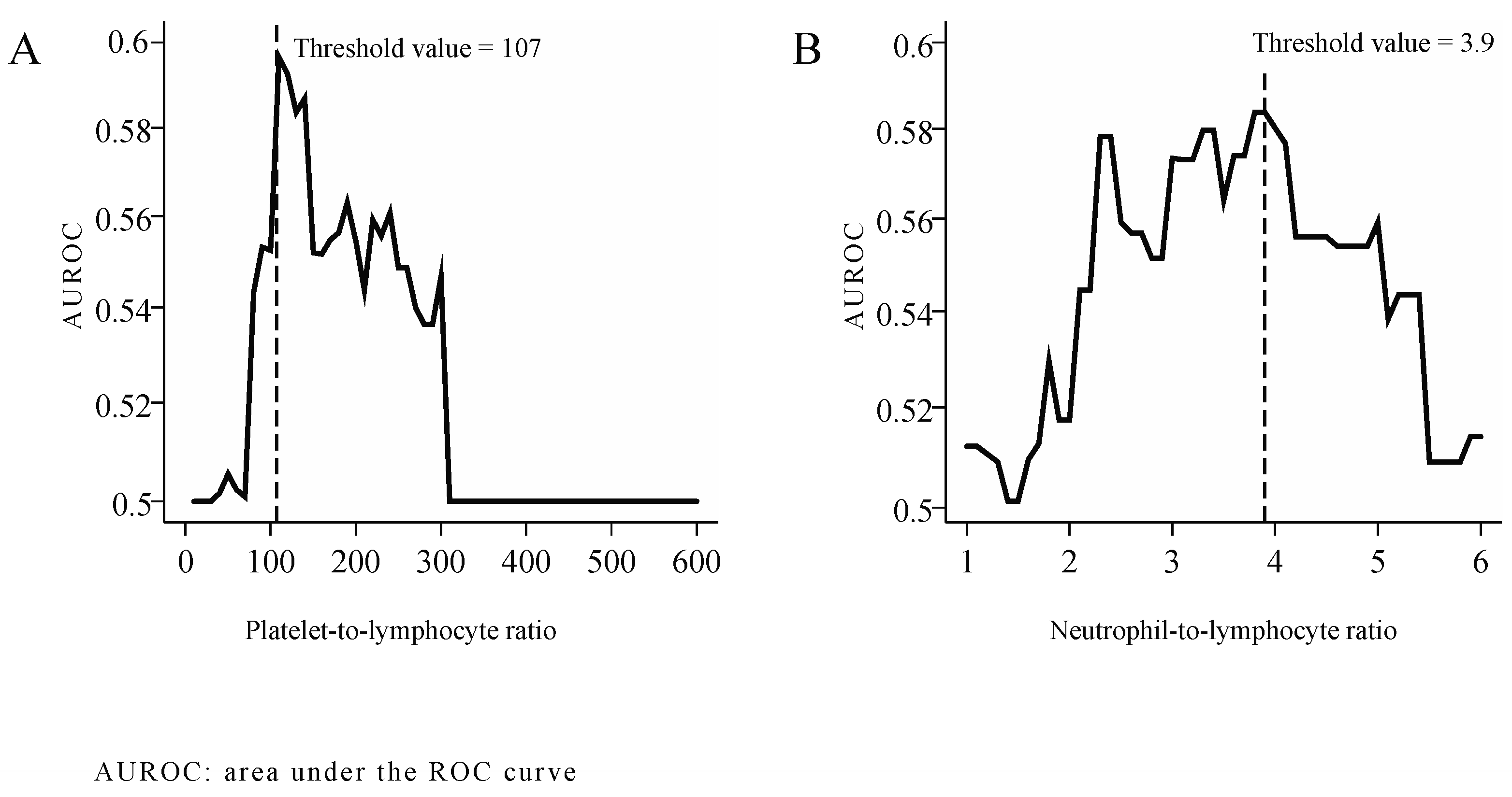
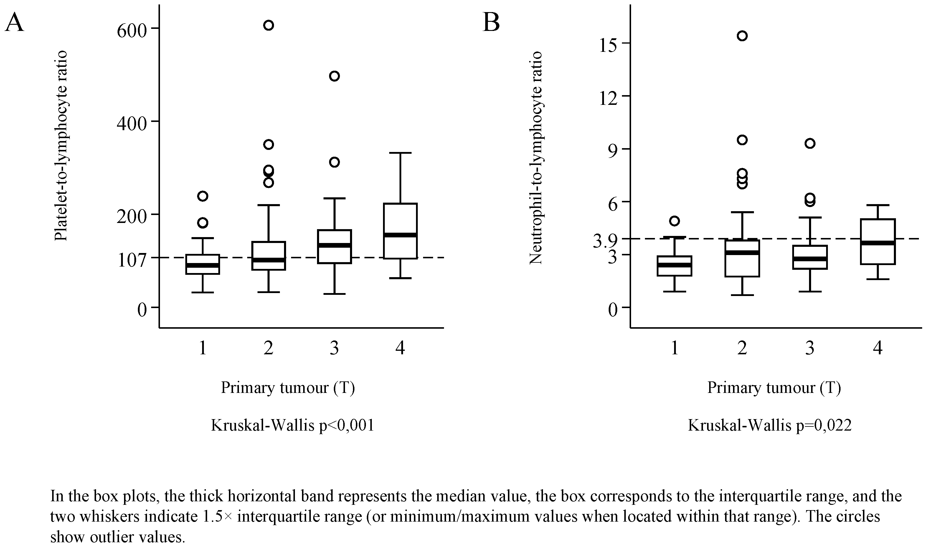
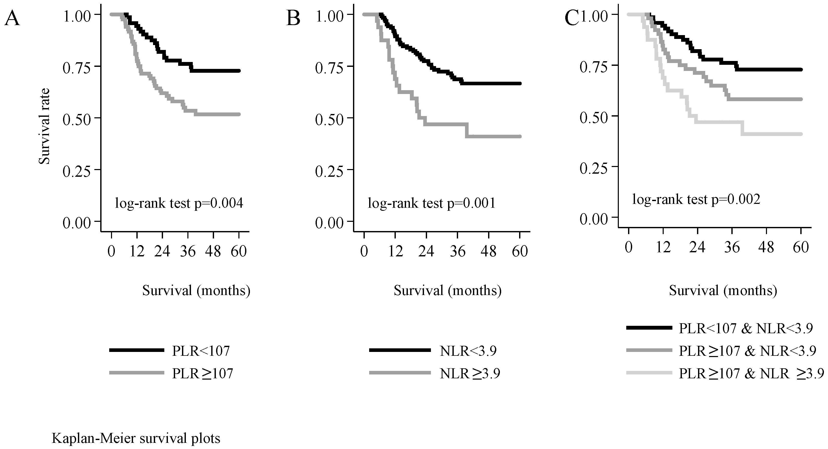
3. Results
4. Discussion
5. Conclusions
Author Contributions
Funding
Conflicts of Interest
Ethical Standards
Abbreviations
| PLR | platelet-to-lymphocyte ratio |
| NLR | neutrophil-to-lymphocyte ratio |
| HNSCC | head and neck squamous cell carcinoma |
References
- Singh, J.; Jayaraj, R.; Baxi, S.; Ramamoorthi, R.; Thomas, M. Incidence and mortality from mucosal head and neck cancers amongst Australian states and territories: What it means for the northern territory. Asian Pac. J. Cancer Prev. 2013, 14, 5621–5624. [Google Scholar] [CrossRef][Green Version]
- Buergy, D.; Wenz, F.; Groden, C.; Brockmann, M.A. Tumor—Platelet interaction in solid tumors. Int. J. Cancer 2012, 130, 2747–2760. [Google Scholar] [CrossRef]
- Ardies, C.M. Inflammation as cause for scar cancers of the lung. Integr. Cancer Ther. 2003, 2, 238–246. [Google Scholar] [CrossRef]
- Van der Auwera, I.; Van Laere, S.J.; Van den Eynden, G.G.; Benoy, I.; van Dam, P.; Colpaert, C.G.; Fox, S.B.; Turley, H.; Harris, A.L.; Van Marck, E.A.; et al. Increased angiogenesis and lymphangiogenesis in inflammatory versus noninflammatory breast cancer by real-time reverse transcriptase-PCR gene expression quantification. Clin. Cancer Res. Off. J. Am. Assoc. Cancer Res. 2004, 10, 7965–7971. [Google Scholar] [CrossRef]
- Karaman, H.; Karaman, A.; Erden, A.; Poyrazoglu, O.K.; Karakukcu, C.; Tasdemir, A. Relationship between colonic polyp type and the neutrophil/ lymphocyte ratio as a biomarker. Asian Pac. J. Cancer Prev. APJCP 2013, 14, 3159–3161. [Google Scholar] [CrossRef] [PubMed][Green Version]
- Wang, W.; Bergh, A.; Damber, J.E. Chronic inflammation in benign prostate hyperplasia is associated with focal upregulation of cyclooxygenase-2, Bcl-2, and cell proliferation in the glandular epithelium. Prostate 2004, 61, 60–72. [Google Scholar] [CrossRef] [PubMed]
- Altinoz, M.A.; Korkmaz, R. NF-kappaB, macrophage migration inhibitory factor and cyclooxygenase-inhibitions as likely mechanisms behind the acetaminophen- and NSAID-prevention of the ovarian cancer. Neoplasma 2004, 51, 239–247. [Google Scholar] [PubMed]
- Hanna, G.J.; Adkins, D.R.; Zolkind, P.; Uppaluri, R. Rationale for neoadjuvant immunotherapy in head and neck squamous cell carcinoma. Oral Oncol. 2017, 73, 65–69. [Google Scholar] [CrossRef]
- Ambatipudi, S.; Langdon, R.; Richmond, R.C.; Suderman, M.; Koestler, D.C.; Kelsey, K.T.; Kazmi, N.; Penfold, C.; Ho, K.M.; McArdle, W.; et al. DNA methylation derived systemic inflammation indices are associated with head and neck cancer development and survival. Oral Oncol. 2018, 85, 87–94. [Google Scholar] [CrossRef]
- Jenne, C.N.; Urrutia, R.; Kubes, P. Platelets: Bridging hemostasis, inflammation, and immunity. Int. J. Lab. Hematol. 2013, 35, 254–261. [Google Scholar] [CrossRef]
- Zhang, X.; Zhang, W.; Yuan, X.; Fu, M.; Qian, H.; Xu, W.R. Neutrophils in cancer development and progression: Roles, mechanisms, and implications (Review). Int. J. Oncol. 2016, 49, 857–867. [Google Scholar] [CrossRef]
- Haqqani, A.S.; Sandhu, J.K.; Birnboim, H.C. Expression of interleukin-8 promotes neutrophil infiltration and genetic instability in mutatect tumors. Neoplasia 2000, 2, 561–568. [Google Scholar] [CrossRef] [PubMed]
- Houghton, A.M.; Rzymkiewicz, D.M.; Ji, H.; Gregory, A.D.; Egea, E.E.; Metz, H.E.; Stolz, D.B.; Land, S.R.; Marconcini, L.A.; Kliment, C.R.; et al. Neutrophil elastase-mediated degradation of IRS-1 accelerates lung tumor growth. Nat. Med. 2010, 16, 219–223. [Google Scholar] [CrossRef] [PubMed]
- Gong, L.; Cumpian, A.M.; Caetano, M.S.; Ochoa, C.E.; De la Garza, M.M.; Lapid, D.J.; Mirabolfathinejad, S.G.; Dickey, B.F.; Zhou, Q.; Moghaddam, S.J. Promoting effect of neutrophils on lung tumorigenesis is mediated by CXCR2 and neutrophil elastase. Mol. Cancer 2013, 12, 154. [Google Scholar] [CrossRef] [PubMed]
- Liang, W.; Ferrara, N. The Complex Role of Neutrophils in Tumor Angiogenesis and Metastasis. Cancer Immunol. Res. 2016, 4, 83–91. [Google Scholar] [CrossRef]
- Tazzyman, S.; Lewis, C.E.; Murdoch, C. Neutrophils: Key mediators of tumour angiogenesis. Int. J. Exp. Pathol. 2009, 90, 222–231. [Google Scholar] [CrossRef]
- Zhang, J.; Qiao, X.; Shi, H.; Han, X.; Liu, W.; Tian, X.; Zeng, X. Circulating tumor-associated neutrophils (cTAN) contribute to circulating tumor cell survival by suppressing peripheral leukocyte activation. Tumour Biol. 2016, 37, 5397–5404. [Google Scholar] [CrossRef]
- Ueno, H.; Hawrylowicz, C.M.; Banchereau, J. Immunological intervention in human diseases. J. Transl. Med. 2007, 5. [Google Scholar] [CrossRef]
- Grambsch, P.M.; Therneau, T.M. Proportional hazards tests and diagnostics based on weighted residuals. Biometrika 1994, 81, 515–526. [Google Scholar] [CrossRef]
- Hosmer, D.; Lemeshow, S.; May, S. Applied Survival Analysis; John Wiley & Sons: Hoboken, NJ, USA, 2008. [Google Scholar]
- Stata Corp. Stata Statistical Software: Release 14; Stata Corp LP.: College Station, TX, USA, 2015. [Google Scholar]
- Nash, G.F.; Turner, L.F.; Scully, M.F.; Kakkar, A.K. Platelets and cancer. Lancet Oncol. 2002, 3, 425–430. [Google Scholar] [CrossRef]
- Bailey, S.E.R.; Ukoumunne, O.C.; Shephard, E.; Hamilton, W. How useful is thrombocytosis in predicting an underlying cancer in primary care? a systematic review. Fam. Pract. 2017, 34, 4–10. [Google Scholar] [CrossRef] [PubMed]
- Aoe, K.; Hiraki, A.; Ueoka, H.; Kiura, K.; Tabata, M.; Tanaka, M.; Tanimoto, M. Thrombocytosis as a useful prognostic indicator in patients with lung cancer. Respiration 2004, 71, 170–173. [Google Scholar] [CrossRef]
- Bensalah, K.; Leray, E.; Fergelot, P.; Rioux-Leclercq, N.; Tostain, J.; Guillé, F.; Patard, J.-J. Prognostic value of thrombocytosis in renal cell carcinoma. J. Urol. 2006, 175, 859–863. [Google Scholar] [CrossRef]
- Carr, B.I.; Guerra, V. Thrombocytosis and hepatocellular carcinoma. Dig. Dis. Sci. 2013, 58, 1790–1796. [Google Scholar] [CrossRef] [PubMed]
- Ishizuka, M.; Nagata, H.; Takagi, K.; Iwasaki, Y.; Kubota, K. Preoperative thrombocytosis is associated with survival after surgery for colorectal cancer. J. Surg. Oncol. 2012, 106, 887–891. [Google Scholar] [CrossRef] [PubMed]
- Yu, M.; Liu, L.; Zhang, B.-L.; Chen, Q.; Ma, X.-L.; Wu, Y.-K.; Liang, C.-S.; Niu, Z.-M.; Qin, X.; Niu, T. Pretreatment thrombocytosis as a prognostic factor in women with gynecologic malignancies: A meta-analysis. Asian Pac. J. Cancer Prev. 2012, 13, 6077–6081. [Google Scholar] [CrossRef]
- Hwang, S.G.; Kim, K.M.; Cheong, J.H.; Kim, H.I.; An, J.Y.; Hyung, W.J.; Noh, S.H. Impact of pretreatment thrombocytosis on blood-borne metastasis and prognosis of gastric cancer. Eur. J. Surg. Oncol. 2012, 38, 562–567. [Google Scholar] [CrossRef]
- Dong, L.; Bai, K.; Cao, Y.; Huang, Q.; Lv, L.; Jiang, Y. Prognostic Value of Pre-Operative Platelet to Lymphocyte Ratio in Patients with Resected Primary Hepatocellular Carcinoma. Clin. Lab. 2016, 62, 2191–2196. [Google Scholar] [CrossRef]
- Hu, D.; Lin, Y.; Liu, F.; Zeng, L.; Ouyang, X.; Wang, K.; Zheng, X.; Huang, Q. Elevated Preoperative Platelet to Lymphocyte Ratio Indicates Poor Survival in Patients with Resected High-grade Serous Ovarian Carcinoma. Clin. Lab. 2016, 62, 1443–1449. [Google Scholar] [CrossRef]
- Wang, L.; Jia, J.; Lin, L.; Guo, J.; Ye, X.; Zheng, X.; Chen, Y. Predictive value of hematological markers of systemic inflammation for managing cervical cancer. Oncotarget 2017. [Google Scholar] [CrossRef]
- Kim, J.H.; Lee, J.Y.; Kim, H.K.; Lee, J.W.; Jung, S.G.; Jung, K.; Kim, S.E.; Moon, W.; Park, M.I.; Park, S.J. Prognostic significance of the neutrophil-to-lymphocyte ratio and platelet-to-lymphocyte ratio in patients with stage III and IV colorectal cancer. World J. Gastroenterol. 2017, 23, 505–515. [Google Scholar] [CrossRef] [PubMed]
- Cho, K.-M.; Park, H.; Oh, D.-Y.; Kim, T.-Y.; Lee, K.-H.; Han, S.-W.; Im, S.-A.; Kim, T.-Y.; Bang, Y.-J. Neutrophil-to-lymphocyte ratio, platelet-to-lymphocyte ratio, and their dynamic changes during chemotherapy is useful to predict a more accurate prognosis of advanced biliary tract cancer. Oncotarget 2017, 8, 2329–2341. [Google Scholar] [CrossRef] [PubMed]
- Zhu, Y.; Si, W.; Sun, Q.; Qin, B.; Zhao, W.; Yang, J. Platelet-lymphocyte ratio acts as an indicator of poor prognosis in patients with breast cancer. Oncotarget 2017, 8, 1023–1030. [Google Scholar] [CrossRef] [PubMed]
- Han, Y.; Wang, J.; Hong, L.; Sun, L.; Zhuang, H.; Sun, B.; Wang, H.; Zhang, X.; Ren, X. Platelet-lymphocyte ratio is an independent prognostic factor in patients with ALK-positive non-small-cell lung cancer. Future Oncol. 2017, 13, 51–61. [Google Scholar] [CrossRef]
- Park, T.J.; Cho, Y.H.; Chung, H.S.; Hwang, E.C.; Jung, S.-H.; Hwang, J.E.; Bae, W.K.; Kim, J.W.; Heo, S.H.; Hur, Y.H.; et al. Prognostic significance of platelet-lymphocyte ratio in patients receiving first-line tyrosine kinase inhibitors for metastatic renal cell cancer. Springerplus 2016, 5, 1889. [Google Scholar] [CrossRef]
- Xu, Z.; Xu, W.; Cheng, H.; Shen, W.; Ying, J.; Cheng, F.; Xu, W. The Prognostic Role of the Platelet-Lymphocytes Ratio in Gastric Cancer: A Meta-Analysis. PLoS ONE 2016, 11, e0163719. [Google Scholar] [CrossRef]
- Wang, Y.; Xu, F.; Pan, J.; Zhu, Y.; Shao, X.; Sha, J.; Wang, Z.; Cai, Y.; Liu, Q.; Dong, B.; et al. Platelet to lymphocyte ratio as an independent prognostic indicator for prostate cancer patients receiving androgen deprivation therapy. BMC Cancer 2016, 16, 329. [Google Scholar] [CrossRef]
- Yodying, H.; Matsuda, A.; Miyashita, M.; Matsumoto, S.; Sakurazawa, N.; Yamada, M.; Uchida, E. Prognostic Significance of Neutrophil-to-Lymphocyte Ratio and Platelet-to-Lymphocyte Ratio in Oncologic Outcomes of Esophageal Cancer: A Systematic Review and Meta-analysis. Ann. Surg. Oncol. 2016, 23, 646–654. [Google Scholar] [CrossRef]
- Han, F.Y.; Liu, Y.Q.; Cheng, S.Q.; Sun, Z.H.; Sheng, C.C.; Sun, X.Y.; Shang, X.M.; Tian, W.J.; Wang, X.Y.; Li, J.M.; et al. Diagnosis and survival values of neutrophil-lymphocyte ratio (NLR) and red blood cell distribution width (RDW) in esophageal cancer. Clin. Chim. Acta 2019, 488, 150–158. [Google Scholar] [CrossRef]
- Sunakawa, Y.; Yang, D.Y.; Cao, S.; Zhang, W.; Moran, M.; Astrow, S.H.; Hsiang, J.; Stephens, C.; Tsuji, A.; Takahashi, T.; et al. Immune-related Genes to Dominate Neutrophil-lymphocyte Ratio (NLR) Associated With Survival of Cetuximab Treatment in Metastatic Colorectal Cancer. Clin. Colorectal Cancer 2018, 17, E741–E749. [Google Scholar] [CrossRef]
- Diakos, C.I.; Wilson, K.; Asher, R.; Gebski, V.; Yip, S.; van Hazel, G.; Robinson, B.; Broad, A.; Price, T.J.; Simes, J.; et al. Is baseline neutrophil to lymphocyte ratio (NLR) an independent prognostic biomarker for progression free survival (PFS) and overall survival (OS) in metastatic colorectal cancer (mCRC)? Analysis of the AGITG MAX study. Ann. Oncol. 2016, 27. [Google Scholar] [CrossRef]
- Garcea, G.; Ladwa, N.; Neal, C.P.; Metcalfe, M.S.; Dennison, A.R.; Berry, D.P. Preoperative Neutrophil-to-Lymphocyte Ratio (NLR) is Associated with Reduced Disease-free Survival Following Curative Resection of Pancreatic Adenocarcinoma. World J. Surg. 2011, 35, 868–872. [Google Scholar] [CrossRef] [PubMed]
- Alagappan, M.; Pollom, E.L.; von Eyben, R.; Kozak, M.M.; Aggarwal, S.; Poultsides, G.A.; Koong, A.C.; Chang, D.T. Albumin and Neutrophil-Lymphocyte Ratio (NLR) Predict Survival in Patients With Pancreatic Adenocarcinoma Treated With SBRT. Am. J. Clin. Oncol. Cancer Clin. Trials 2018, 41, 242–247. [Google Scholar] [CrossRef]
- Lorente, D.; Mateo, J.; Templeton, A.J.; Zafeiriou, Z.; Bianchini, D.; Ferraldeschi, R.; Bahl, A.; Shen, L.; Su, Z.; Sartor, O.; et al. Baseline neutrophil-lymphocyte ratio (NLR) is associated with survival and response to treatment with second-line chemotherapy for advanced prostate cancer independent of baseline steroid use. Ann. Oncol. 2015, 26, 750–755. [Google Scholar] [CrossRef] [PubMed]
- Nuhn, P.; Vaghasia, A.M.; Goyal, J.; Zhou, X.C.; Carducci, M.A.; Eisenberger, M.A.; Antonarakis, E.S. Association of pretreatment neutrophil-to-lymphocyte ratio (NLR) and overall survival (OS) in patients with metastatic castration-resistant prostate cancer (mCRPC) treated with first-line docetaxel. BJU Int. 2014, 114, E11–E17. [Google Scholar] [CrossRef] [PubMed]
- Ji, F.; Liang, Y.; Fu, S.J.; Guo, Z.Y.; Shu, M.; Shen, S.L.; Li, S.Q.; Peng, B.G.; Liang, L.J.; Hua, Y.P. A novel and accurate predictor of survival for patients with hepatocellular carcinoma after surgical resection: The neutrophil to lymphocyte ratio (NLR) combined with the aspartate aminotransferase/platelet count ratio index (APRI). BMC Cancer 2016, 16, 137. [Google Scholar] [CrossRef]
- Afshar, M.; Clarke, H.; Jackson-Wilding, A.; Ahmed, A.; Ma, Y.T.; Punia, P. Neutrophil lymphocyte ratio (nlr) at diagnosis is a predictor for survival in patients receiving sorafenib for advanced hepatocellular carcinoma (hcc): A large UK cohort. J. Hepatol. 2015, 62, S442. [Google Scholar] [CrossRef]
- Ha, H.; Nam, A.R.; Bang, J.H.; Park, J.E.; Kim, T.Y.; Lee, K.H.; Han, S.W.; Im, S.A.; Kim, T.Y.; Bang, Y.J.; et al. Soluble programmed death-ligand 1 (sPDL1) and neutrophil-to-lymphocyte ratio (NLR) predicts survival in advanced biliary tract cancer patients treated with palliative chemotherapy. Oncotarget 2016, 7, 76604–76612. [Google Scholar] [CrossRef]
- Orditura, M.; Galizia, G.; Diana, A.; Saccone, C.; Cobellis, L.; Ventriglia, J.; Iovino, F.; Romano, C.; Morgillo, F.; Mosca, L.; et al. Neutrophil to lymphocyte ratio (NLR) for prediction of distant metastasis-free survival (DMFS) in early breast cancer: A propensity score-matched analysis. ESMO Open 2016, 1. [Google Scholar] [CrossRef]
- Li, Y.C.; Wang, C.Y.; Xu, M.D.; Kong, C.C.; Qu, A.B.; Zhang, M.; Zheng, Z.C.; Zhang, G.R. Preoperative NLR for predicting survival rate after radical resection combined with adjuvant immunotherapy with CIK and postoperative chemotherapy in gastric cancer. J. Cancer Res. Clin. Oncol. 2017, 143, 861–871. [Google Scholar] [CrossRef]
- Guo, J.; Chen, S.X.; Chen, Y.M.; Li, S.; Xu, D.Z. Combination of CRP and NLR: A better predictor of postoperative survival in patients with gastric cancer. Cancer Manag. Res. 2018, 10, 315–321. [Google Scholar] [CrossRef] [PubMed]
- Capone, M.; Giannarelli, D.; Mallardo, D.; Madonna, G.; Festino, L.; Grimaldi, A.M.; Vanella, V.; Simeone, E.; Paone, M.; Palmieri, G.; et al. Baseline neutrophil-to-lymphocyte ratio (NLR) and derived NLR could predict overall survival in patients with advanced melanoma treated with nivolumab. J. Immunother. Cancer 2018, 6. [Google Scholar] [CrossRef] [PubMed]
- Jiang, K.; Lei, J.; Chen, W.; Gong, Y.; Luo, H.; Li, Z.; Gong, R.; Zhu, J. Association of the preoperative neutrophil-to-lymphocyte and platelet-to-lymphocyte ratios with lymph node metastasis and recurrence in patients with medullary thyroid carcinoma. Medicine 2016, 95, e5079. [Google Scholar] [CrossRef] [PubMed]
- Powles, T.; Jin, C.Y.; Zheng, Y.N.; Baverel, P.; Narwal, R.; Mukhopadhyay, P.; Jin, X.P.; Dennis, P.A.; Gupta, A.K.; Ben, Y.; et al. Tumor shrinkage and increased overall survival are associated with improved albumin, neutrophil lymphocyte ratio (NLR) and decreased durvalumab clearance in NSCLC and UC patients receiving durvalumab. J. Clin. Oncol. 2017, 35. [Google Scholar] [CrossRef]
- Turkmen, K.; Erdur, F.M.; Ozcicek, F.; Ozcicek, A.; Akbas, E.M.; Ozbicer, A.; Demirtas, L.; Turk, S.; Tonbul, H.Z. Platelet-to-lymphocyte ratio better predicts inflammation than neutrophil-to-lymphocyte ratio in end-stage renal disease patients. Hemodialysis Int. 2013, 17, 391–396. [Google Scholar] [CrossRef]
- Shah, N.; Parikh, V.; Patel, N.; Patel, N.; Badheka, A.; Deshmukh, A.; Rathod, A.; Lafferty, J. Neutrophil lymphocyte ratio significantly improves the Framingham risk score in prediction of coronary heart disease mortality: Insights from the National Health and Nutrition Examination Survey-III. Int. J. Cardiol. 2014, 171, 390–397. [Google Scholar] [CrossRef]
- Salim, D.K.; Mutlu, H.; Eryilmaz, M.K.; Salim, O.; Musri, F.Y.; Tural, D.; Gunduz, S.; Coskun, H.S. Neutrophil to lymphocyte ratio is an independent prognostic factor in patients with recurrent or metastatic head and neck squamous cell cancer. Mol. Clin. Oncol. 2015, 3, 839–842. [Google Scholar] [CrossRef]
- Cho, Y.; Kim, J.W.; Yoon, H.I.; Lee, C.G.; Keum, K.C.; Lee, I.J. The Prognostic Significance of Neutrophil-to-Lymphocyte Ratio in Head and Neck Cancer Patients Treated with Radiotherapy. J. Clin. Med. 2018, 7, 512. [Google Scholar] [CrossRef]
- Hyder, J.; Molitoris, J.; Engelman, A.; Hanlon, A.; D’Emic, N.; Suntharalingam, M.; Chuong, M.D. Changes in Neutrophil-to-Lymphocyte Ratio (NLR) During Chemoradiation for Head and Neck Cancer Are Significant Predictor for Overall Survival. Int. J. Radiat. Oncol. Biol. Phys. 2015, 93, S174. [Google Scholar] [CrossRef]
- Rassouli, A.; Saliba, J.; Castano, R.; Hier, M.; Zeitouni, A.G. Systemic inflammatory markers as independent prognosticators of head and neck squamous cell carcinoma. Head Neck 2014, 37, 103–110. [Google Scholar] [CrossRef]
- Turri-Zanoni, M.; Salzano, G.; Lambertoni, A.; Giovannardi, M.; Karligkiotis, A.; Battaglia, P.; Castelnuovo, P. Prognostic value of pretreatment peripheral blood markers in paranasal sinus cancer: Neutrophil-to-lymphocyte and platelet-to-lymphocyte ratio. Head Neck 2017, 39, 730–736. [Google Scholar] [CrossRef] [PubMed]
- Ong, H.S.; Gokavarapu, S.; Wang, L.Z.; Tian, Z.; Zhang, C.P. Low Pretreatment Lymphocyte-Monocyte Ratio and High Platelet-Lymphocyte Ratio Indicate Poor Cancer Outcome in Early Tongue Cancer. J. Oral Maxillofac. Surg. 2017, 75, 1762–1774. [Google Scholar] [CrossRef] [PubMed]
- Kara, M.; Uysal, S.; Altinişik, U.; Cevizci, S.; Güçlü, O.; Dereköy, F.S. The pre-treatment neutrophil-to-lymphocyte ratio, platelet-to-lymphocyte ratio, and red cell distribution width predict prognosis in patients with laryngeal carcinoma. Eur. Arch. Otorhinolaryngol. 2017, 274, 535–542. [Google Scholar] [CrossRef] [PubMed]
- Foster, C.C.; Kochanny, S.; Khattri, A.; Acharya, R.; Dekker, A.; Tan, Y.H.C. Association of a baseline neutrophil-to-lymphocyte ratio (NLR) with progressionfree and overall survival in head and neck cancer patients receiving anti-PD-1 therapy. J. Clin. Oncol. 2018, 36. [Google Scholar] [CrossRef]
- Rachidi, S.; Wallace, K.; Wrangle, J.M.; Day, T.A.; Alberg, A.J.; Li, Z. Neutrophil-to-lymphocyte ratio and overall survival in all sites of head and neck squamous cell carcinoma. Head Neck 2016, 38 (Suppl. S1), E1068–E1074. [Google Scholar] [CrossRef] [PubMed]
- Rosculet, N.; Zhou, X.C.; Ha, P.; Tang, M.; Levine, M.A.; Neuner, G.; Califano, J. Neutrophil-to-lymphocyte ratio: Prognostic indicator for head and neck squamous cell carcinoma. Head Neck 2017, 39, 662–667. [Google Scholar] [CrossRef]


| All Patients | Hypo-Pharynx | Oropharynx | Oral Cavity | Sub-/Supraglottis | Glottis | Multiple Regions | |||
|---|---|---|---|---|---|---|---|---|---|
| N (%) | N (%) | N (%) | N (%) | N (%) | N (%) | N (%) | χ2 | ||
| Gender | Female | 31 (19.9) | 8 (15.1) | 7 (43.8) | 1 (20.0) | 7 (20.6) | 7 (15.6) | 1 (33.3) | p = 0.195 |
| Male | 125 (80.1) | 45 (84.9) | 9 (56.2) | 4 (80.0) | 27 (79.4) | 38 (84.4) | 2 (66.7) | ||
| Age | <65 years | 116 (74.4) | 49 (92.5) | 13 (81.2) | 4 (80.0) | 23 (67.6) | 25 (55.6) | 2 (66.7) | p = 0.002 |
| ≥65 years | 40 (25.6) | 4 (7.5) | 3 (18.8) | 1 (20.0) | 11 (32.4) | 20 (44.4) | 1 (33.3) | ||
| T stage | 1 | 38 (24.4) | 6 (11.3) | 8 (50.0) | 0 (0.0) | 5 (14.7) | 18 (40) | 1 (33.3) | p < 0.001 |
| 2 | 40 (25.6) | 9 (17) | 4 (25.0) | 3 (60.0) | 16 (47.1) | 8 (17.8) | 0 (0.0) | ||
| 3 | 50 (32.1) | 31 (58.5) | 4 (25.0) | 1 (20.0) | 6 (17.6) | 7 (15.6) | 1 (33.3) | ||
| 4 | 28 (17.9) | 7 (13.2) | 0 (0.0) | 1 (20.0) | 7 (20.6) | 12 (26.7) | 1 (33.3) | ||
| N stage | 0 | 82 (52.6) | 14 (26.4) | 5 (31.2) | 4 (80.0) | 18 (52.9) | 40 (88.9) | 1 (33.3) | p < 0.001 |
| 1 | 30 (19.2) | 15 (28.3) | 7 (43.8) | 0 (0.0) | 6 (17.6) | 1 (2.2) | 1 (33.3) | ||
| 2 | 40 (25.6) | 21 (39.6) | 3 (18.8) | 1 (20.0) | 10 (29.4) | 4 (8.9) | 1 (33.3) | ||
| 3 | 4 (2.6) | 3 (5.7) | 1 (6.2) | 0 (0.0) | 0 (0) | 0 (0.0) | 0 (0.0) | ||
| TNM stage | 1 | 26 (16.7) | 2 (3.8) | 3 (18.8) | 0 (0.0) | 3 (8.8) | 18 (40) | 0 (0.0) | p < 0.001 |
| 2 | 24 (15.4) | 4 (7.5) | 1 (6.2) | 2 (40.0) | 9 (26.5) | 8 (17.8) | 0 (0.0) | ||
| 3 | 44 (28.2) | 20 (37.7) | 8 (50) | 1 (20.0) | 7 (20.6) | 7 (15.6) | 1 (33.3) | ||
| 4 | 62 (39.7) | 27 (50.9) | 4 (25) | 2 (40.0) | 15 (44.1) | 12 (26.7) | 2 (66.7) | ||
| Grade of differentiation | 1 | 20 (12.8) | 4 (7.5) | 0 (0.0) | 2 (40.0) | 3 (8.8) | 11 (24.4) | 0 (0.0) | p = 0.270 |
| 2 | 54 (34.6) | 17 (32.1) | 6 (37.5) | 1 (20.0) | 15 (44.1) | 14 (31.1) | 1 (33.3) | ||
| 3 | 37 (23.7) | 11 (20.8) | 4 (25) | 1 (20.0) | 9 (26.5) | 11 (24.4) | 1 (33.3) | ||
| 4 | 2 (1.3) | 0 (0.0) | 1 (6.2) | 0 (0.0) | 0 (0.0) | 1 (2.2) | 0 (0.0) | ||
| Unknown | 43 (27.6) | 21 (39.6) | 5 (31.2) | 1 (20.0) | 7 (20.6) | 8 (17.8) | 1 (33.3) | ||
| Platelets-to-lymphocytes ratio | <107 | 72 (46.2) | 15 (28.3) | 12 (75.0) | 2 (40.0) | 16 (47.1) | 26 (57.8) | 1 (33.3) | p = 0.011 |
| ≥107 | 84 (53.8) | 38 (71.7) | 4 (25.0) | 3 (60.0) | 18 (52.9) | 19 (42.4) | 2 (66.7) | ||
| Neutrophils-to-lymphocytes ratio | <3,9 | 125 (80.1) | 39 (73.6) | 15 (93.8) | 4 (80.0) | 26 (76.5) | 38 (84.4) | 3 (100.0) | p = 0.435 |
| ≥3,9 | 31 (19.9) | 14 (26.4) | 1 (6.2) | 1 (20.0) | 8 (23.5) | 7 (15.6) | 0 (0.0) | ||
| 5-year mortality | Deceased | 58 (37.2) | 28 (52.8) | 4 (25.0) | 1 (20.0) | 16 (47.1) | 8 (17.8) | 1 (33.3) | p = 0.01 * |
| Total | 156 | 53 | 16 | 5 | 34 | 45 | 3 |
| Overall Mortality | Tumour-Specific Mortality | Tumour Recurrence | ||||||||
|---|---|---|---|---|---|---|---|---|---|---|
| PLR | ≥107 | 1.499 | 1.499 | 1.651 | ||||||
| NLR | ≥3.9 | 2.122 * | 2.122 * | 1.800 | ||||||
| PLR and NLR aggregately | PLR ≥ 107 and NLR < 3.9 | 1.153 | 1.153 | 1.435 | ||||||
| PLR ≥ 107 and NLR ≥ 3.9 | 2.291 * | 2.291 * | 2.180 * | |||||||
| Age a | ≥65 years | 0.906 | 1.045 | 1.018 | 0.906 | 1.045 | 1.018 | 0.825 | 0.876 | 0.860 |
| Gender b | Male | 2.460 | 2.730 * | 2.732 * | 2.460 | 2.730 * | 2.732 * | 3.530 ** | 3.809 ** | 3.840 ** |
| Stage c | 2 | 6.690 g | 6.802 g | 6.616 | 6.690 g | 6.802 g | 6.616 | 2.198 | 2.269 | 2.175 |
| 3 | 7.315 g | 7.698 g | 7.439 | 7.315 g | 7.698 g | 7.439 | 1.293 | 1.443 | 1.306 | |
| 4 | 14.44 *g | 13.40 *g | 12.87 * | 14.44 *g | 13.40 *g | 12.87 * | 2.591 | 2.733 * | 2.447 | |
| Grade of differentiation d | 2 | 1.778 | 1.911 | 1.941 | 1.778 | 1.911 | 1.941 | 4.274 * | 4.518 * | 4.741 * |
| 3 | 1.341 | 1.534 | 1.540 | 1.341 | 1.534 | 1.540 | 2.539 | 2.762 | 2.854 | |
| 4 | - | - | - | - | - | - | 4.159 | 4.020 | 4.845 | |
| Unknown | 1.954 | 2.210 | 2.237 | 1.954 | 2.210 | 2.237 | 2.977 | 3.272 | 3.393 | |
| Location e | Oropharynx | 0.817 | 0.796 | 0.833 | 0.817 | 0.796 | 0.833 | 1.285 | 1.196 | 1.331 |
| Oral cavity | 0.546 | 0.549 | 0.559 | 0.546 | 0.549 | 0.559 | 12.29 ** | 13.21 ** | 13.30 ** | |
| Sub-/supraglottis | 1.221 | 1.152 | 1.189 | 1.221 | 1.152 | 1.189 | 1.330 | 1.160 | 1.264 | |
| Glottis | 0.631 | 0.560 | 0.577 | 0.631 | 0.560 | 0.577 | 1.100 | 0.991 | 1.064 | |
| Multiple locations | 0.475 | 0.595 | 0.593 | 0.475 | 0.595 | 0.593 | 0.633 | 0.618 | 0.676 | |
| RBC | 0.503 * | 0.508 ** | 0.509 ** | 0.503 * | 0.508 ** | 0.509 ** | 0.508 ** | 0.512 ** | 0.510 ** | |
| Platelet count f | ≥400 G/L | 0.906 | 1.045 | 1.018 | 0.906 | 1.045 | 1.018 | 0.825 | 0.876 | 0.860 |
| Number of patients | 156 | 156 | 156 | 156 | 156 | 156 | 148 | 148 | 148 | |
© 2020 by the authors. Licensee MDPI, Basel, Switzerland. This article is an open access article distributed under the terms and conditions of the Creative Commons Attribution (CC BY) license (http://creativecommons.org/licenses/by/4.0/).
Share and Cite
Szilasi, Z.; Jósa, V.; Zrubka, Z.; Mezei, T.; Vass, T.; Merkel, K.; Helfferich, F.; Baranyai, Z. Neutrophil-To-Lymphocyte and Platelet-To-Lymphocyte Ratios as Prognostic Markers of Survival in Patients with Head and Neck Tumours—Results of a Retrospective Multicentric Study. Int. J. Environ. Res. Public Health 2020, 17, 1742. https://doi.org/10.3390/ijerph17051742
Szilasi Z, Jósa V, Zrubka Z, Mezei T, Vass T, Merkel K, Helfferich F, Baranyai Z. Neutrophil-To-Lymphocyte and Platelet-To-Lymphocyte Ratios as Prognostic Markers of Survival in Patients with Head and Neck Tumours—Results of a Retrospective Multicentric Study. International Journal of Environmental Research and Public Health. 2020; 17(5):1742. https://doi.org/10.3390/ijerph17051742
Chicago/Turabian StyleSzilasi, Zsuzsanna, Valéria Jósa, Zsombor Zrubka, Tünde Mezei, Tamás Vass, Keresztély Merkel, Frigyes Helfferich, and Zsolt Baranyai. 2020. "Neutrophil-To-Lymphocyte and Platelet-To-Lymphocyte Ratios as Prognostic Markers of Survival in Patients with Head and Neck Tumours—Results of a Retrospective Multicentric Study" International Journal of Environmental Research and Public Health 17, no. 5: 1742. https://doi.org/10.3390/ijerph17051742
APA StyleSzilasi, Z., Jósa, V., Zrubka, Z., Mezei, T., Vass, T., Merkel, K., Helfferich, F., & Baranyai, Z. (2020). Neutrophil-To-Lymphocyte and Platelet-To-Lymphocyte Ratios as Prognostic Markers of Survival in Patients with Head and Neck Tumours—Results of a Retrospective Multicentric Study. International Journal of Environmental Research and Public Health, 17(5), 1742. https://doi.org/10.3390/ijerph17051742