
Underlying Diagnosis of Food Bolus Obstruction Acute Admissions to Otorhinolaryngology: A Shift to Provide the Best Care as per the Current Guidelines

Abstract
1. Introduction
2. Materials and Methods
2.1. Study Design
2.2. Limitations
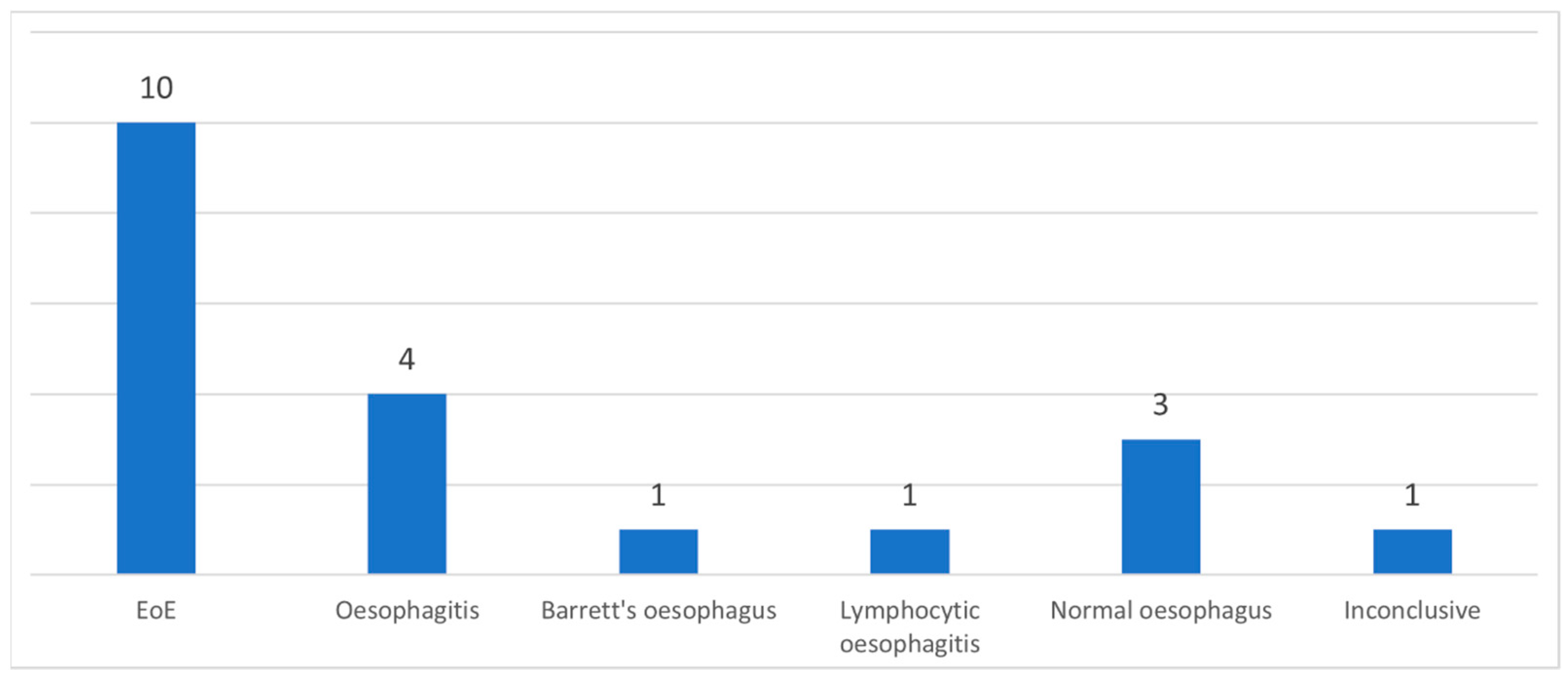
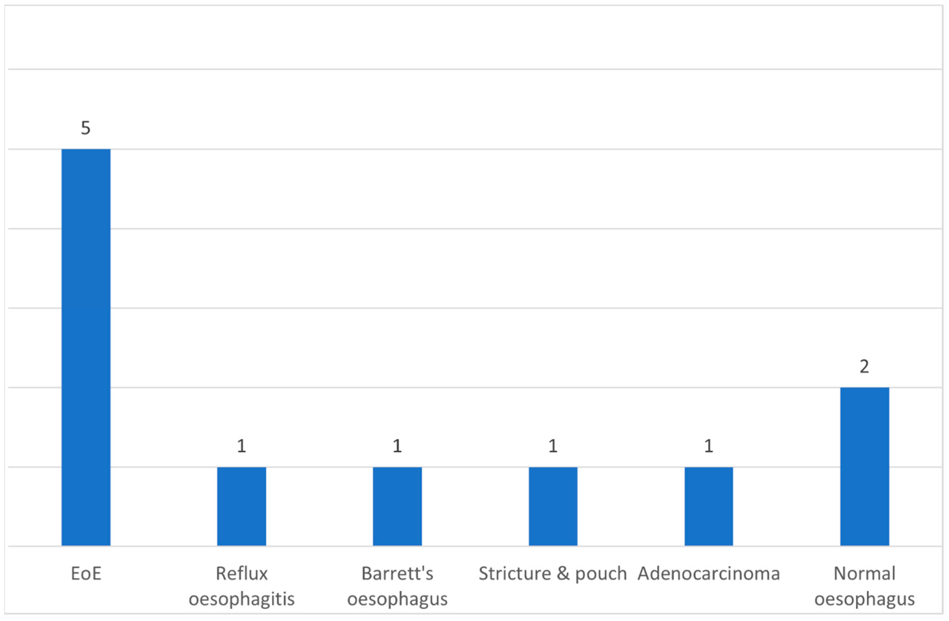
3. Results
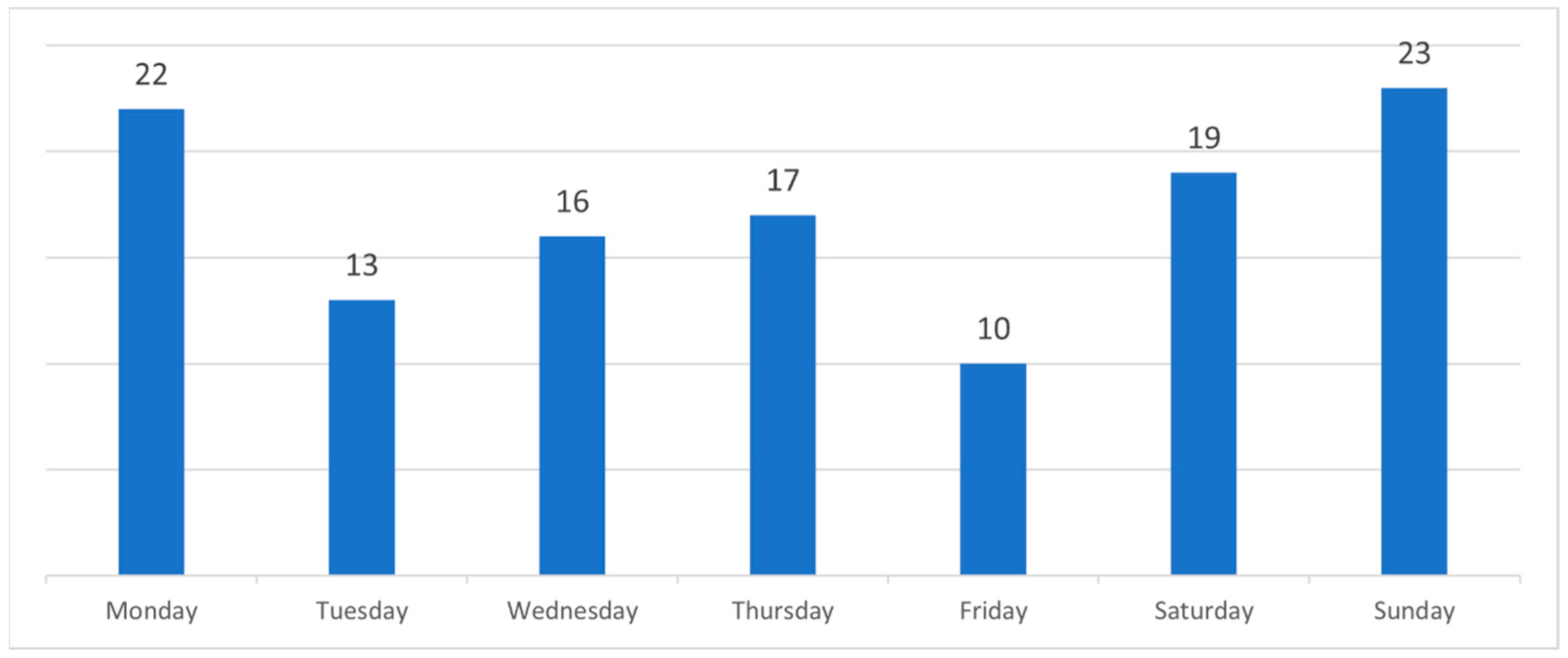
3.1. Demographics
3.2. Interventions
3.3. Follow-Up
4. Discussion
5. Conclusions
Author Contributions
Funding
Institutional Review Board Statement
Informed Consent Statement
Data Availability Statement
Conflicts of Interest
Abbreviations
| ASGEE | American Society for Gastrointestinal Endoscopy |
| ENT | Ear, nose, and throat |
| EoE | Eosinophilic oesophagitis |
| ESGE | European Society of Gastrointestinal Endoscopy |
| FBO | Food bolus obstruction |
| GI | Gastroenterologists |
| GP | General Practitioners |
| Hpf | High power field |
| OGD | Oesophagogastroduodenoscopy |
| RO | Rigid oesophagoscopy |
| TNO | Transnasal oesophagoscopy |
| UK | United Kingdom |
References
- Hardman, J.; Sharma, N.; Smith, J.; Nankivell, P. Conservative management of oesophageal soft food bolus impaction. Cochrane Database Syst. Rev. 2020, 5, Cd007352. [Google Scholar] [CrossRef] [PubMed]
- Birk, M.; Bauerfeind, P.; Deprez, P.H.; Häfner, M.; Hartmann, D.; Hassan, C.; Hucl, T.; Lesur, G.; Aabakken, L.; Meining, A. Removal of foreign bodies in the upper gastrointestinal tract in adults: European Society of Gastrointestinal Endoscopy (ESGE) Clinical Guideline. Endoscopy 2016, 48, 489–496. [Google Scholar] [CrossRef] [PubMed]
- Ntuli, Y.; Bough, I.; Wilson, M. Recognising eosinophilic oesophagitis as a cause of food bolus obstruction. Frontline Gastroenterol. 2020, 11, 11–15. [Google Scholar] [CrossRef] [PubMed]
- Stubington, T.J.; Kamani, T. Food bolus and oesophageal foreign body: A summary of the evidence and proposed management process. Eur. Arch. Otorhinolaryngol. 2021, 278, 3613–3623. [Google Scholar] [CrossRef] [PubMed]
- Attwood, S.E. Overview of eosinophilic oesophagitis. Br. J. Hosp. Med. 2019, 80, 132–138. [Google Scholar] [CrossRef] [PubMed]
- Lucendo, A.J.; Molina-Infante, J.; Arias, Á.; von Arnim, U.; Bredenoord, A.J.; Bussmann, C.; Dias, J.A.; Bove, M.; González-Cervera, J.; Larsson, H.; et al. Guidelines on eosinophilic esophagitis: Evidence-based statements and recommendations for diagnosis and management in children and adults. United Eur. Gastroenterol. J. 2017, 5, 335–358. [Google Scholar] [CrossRef] [PubMed]
- Feraco, A.; Armani, A.; Amoah, I.; Guseva, E.; Camajani, E.; Gorini, S.; Strollo, R.; Padua, E.; Caprio, M.; Lombardo, M. Assessing gender differences in food preferences and physical activity: A population-based survey. Front. Nutr. 2024, 11, 1348456. [Google Scholar] [CrossRef] [PubMed]
- Lombardo, M.; Feraco, A.; Camajani, E.; Gorini, S.; Strollo, R.; Armani, A.; Padua, E.; Caprio, M. Effects of Different Nutritional Patterns and Physical Activity on Body Composition: A Gender and Age Group Comparative Study. Foods 2024, 13, 529. [Google Scholar] [CrossRef] [PubMed]
- Philpott, H.L.; Nandurkar, S.; Thien, F.; Bloom, S.; Lin, E.; Goldberg, R.; Boyapati, R.; Finch, A.; Royce, S.G.; Gibson, P.R. Seasonal recurrence of food bolus obstruction in eosinophilic esophagitis. Intern. Med. J. 2015, 45, 939–943. [Google Scholar] [CrossRef] [PubMed]
- Tsikoudas, A.; Kochillas, X.; Kelleher, R.J.; Mills, R. The management of acute oesophageal obstruction from a food bolus. Can we be more conservative? Eur. Arch. Otorhinolaryngol. 2005, 262, 528–530. [Google Scholar] [CrossRef] [PubMed]
- Price, T.; Jones, S.E.; Montgomery, P.Q. Is current UK management of oesophageal food bolus obstruction evidence based? An e-mail survey and literature review. Eur. Arch. Otorhinolaryngol. 2007, 264, 329–335. [Google Scholar] [CrossRef] [PubMed]
- Dhar, A.; Haboubi, H.N.; Attwood, S.E.; Auth, M.K.H.; Dunn, J.M.; Sweis, R.; Morris, D.; Epstein, J.; Novelli, M.R.; Hunter, H.; et al. British Society of Gastroenterology (BSG) and British Society of Paediatric Gastroenterology, Hepatology and Nutrition (BSPGHAN) joint consensus guidelines on the diagnosis and management of eosinophilic oesophagitis in children and adults. Gut 2022, 71, 1459–1487. [Google Scholar] [CrossRef] [PubMed]
- Straumann, A.; Bussmann, C.; Zuber, M.; Vannini, S.; Simon, H.-U.; Schoepfer, A. Eosinophilic esophagitis: Analysis of food impaction and perforation in 251 adolescent and adult patients. Clin. Gastroenterol. Hepatol. 2008, 6, 598–600. [Google Scholar] [CrossRef] [PubMed]
- Ferrari, D.; Aiolfi, A.; Bonitta, G.; Riva, C.G.; Rausa, E.; Siboni, S.; Toti, F.; Bonavina, L. Flexible versus rigid endoscopy in the management of esophageal foreign body impaction: Systematic review and meta-analysis. World J. Emerg. Surg. 2018, 13, 42. [Google Scholar] [CrossRef] [PubMed]
- Bahgat, M.; Dawe, N.; Flood, L. Eosinophilic oesophagitis: A systematic review for otolaryngologists. J. Laryngol. Otol. 2015, 129, 1156–1166. [Google Scholar] [CrossRef] [PubMed]
- Limketkai, B.N.; Shah, S.C.; Hirano, I.; Bellaguarda, E.; Colombel, J.-F. Epidemiology and implications of concurrent diagnosis of eosinophilic oesophagitis and IBD based on a prospective population-based analysis. Gut 2019, 68, 2152–2160. [Google Scholar] [CrossRef] [PubMed]

Disclaimer/Publisher’s Note: The statements, opinions and data contained in all publications are solely those of the individual author(s) and contributor(s) and not of MDPI and/or the editor(s). MDPI and/or the editor(s) disclaim responsibility for any injury to people or property resulting from any ideas, methods, instructions or products referred to in the content. |
© 2025 by the authors. Published by MDPI on behalf of the Lithuanian University of Health Sciences. Licensee MDPI, Basel, Switzerland. This article is an open access article distributed under the terms and conditions of the Creative Commons Attribution (CC BY) license (https://creativecommons.org/licenses/by/4.0/).
Share and Cite
Misirovs, R.; Kamusella, A.; Miller, M.; Majumdar, S. Underlying Diagnosis of Food Bolus Obstruction Acute Admissions to Otorhinolaryngology: A Shift to Provide the Best Care as per the Current Guidelines. Medicina 2025, 61, 1047. https://doi.org/10.3390/medicina61061047
Misirovs R, Kamusella A, Miller M, Majumdar S. Underlying Diagnosis of Food Bolus Obstruction Acute Admissions to Otorhinolaryngology: A Shift to Provide the Best Care as per the Current Guidelines. Medicina. 2025; 61(6):1047. https://doi.org/10.3390/medicina61061047
Chicago/Turabian StyleMisirovs, Rasads, Anna Kamusella, Michael Miller, and Samit Majumdar. 2025. "Underlying Diagnosis of Food Bolus Obstruction Acute Admissions to Otorhinolaryngology: A Shift to Provide the Best Care as per the Current Guidelines" Medicina 61, no. 6: 1047. https://doi.org/10.3390/medicina61061047
APA StyleMisirovs, R., Kamusella, A., Miller, M., & Majumdar, S. (2025). Underlying Diagnosis of Food Bolus Obstruction Acute Admissions to Otorhinolaryngology: A Shift to Provide the Best Care as per the Current Guidelines. Medicina, 61(6), 1047. https://doi.org/10.3390/medicina61061047